Title: Divergent Pathogenic Properties of Circulating Coxsackievirus A6 Associated. Running Title: CV-A6 Virus Associated with Divergent Lethality
|
|
|
- Katherine McBride
- 5 years ago
- Views:
Transcription
1 JVI Accepted Manuscript Posted Online 21 March 2018 J. Virol. doi: /jvi Copyright 2018 American Society for Microbiology. All Rights Reserved. 1 2 Title: Divergent Pathogenic Properties of Circulating Coxsackievirus A6 Associated with Emerging HFMD Running Title: CV-A6 Virus Associated with Divergent Lethality Authors and affiliations: Shao-Hua Wang 1, 2, Ao Wang 4, Pan-Pan Liu 1, Wen-Yan Zhang 1, Juan Du 1, Shuang Xu 4, Guan-Chen Liu 1, Bai-Song Zheng 1, Chen Huan 1, Ke Zhao 1#, Xiao-Fang Yu 1,3# 1 Institute of Virology and AIDS Research, First Hospital of Jilin University, Changchun, Jilin, China; 2 Key Laboratory of Zoonosis, Ministry of Education, Institute of Zoonosis, Jilin University, Changchun, Jilin, China; 3 Department of Molecular Microbiology and Immunology, Johns Hopkins Bloomberg School of Public Health, Baltimore, Maryland, USA; 4 Jilin Provincial Center for Diseases Control and Prevention, Changchun, Jilin, China. # Address correspondence to Xiao-Fang Yu, xyu2@jhu.edu, or Ke Zhao, paraake@gmail.com
2 ABSTRACT Coxsackievirus A6 (CV-A6) is an emerging pathogen associated with hand, foot, and mouth disease (HFMD). Its genetic characterization and pathogenic properties are largely unknown. Here, we report 39 circulating CV-A6 strains isolated in 2013 from HFMD patients in Northeast China. Three major clusters of CV-A6 were identified and related to CV-A6 mostly from Shanghai, indicating that domestic CV-A6 strains were responsible for HFMD emerging in Northeast China. Four full-length CV-A6 genomes representing each cluster were sequenced and analyzed further. Bootscanning tests indicated that all four CV-A6-Changchun strains were most likely recombinants between CV-A6 prototype Gdula and prototype CV-A4 or CV-A4-related viruses, while the recombination pattern was related to yet distinct from those strains isolated from other regions of China. Furthermore, different CV-A6 viruses showed different capabilities of viral replication, release, and pathogenesis in a mouse model. Further analyses indicated that viral protein 2C contributed to the diverse pathogenic abilities of CV-A6 by causing autophagy and inducing cell death. To our knowledge, this study is the first to report lethal and non-lethal strains of CV-A6 associated with HFMD. The 2C protein region may play a key role in the pathogenicity of CV-A6 viruses.
3 IMPORTANCE Hand, foot, and mouth disease (HFMD) is a major and persistent threat to infants and children. Besides the most common pathogens such as enterovirus 71 (EV-A71) and coxsackievirus A16 (CV-A16), other enteroviruses are increasingly contributing to HFMD. The present study focused on the recently emerging CV-A6 strain. We found that CV-A6 strains isolated in Changchun city in Northeast China were associated with domestic origins. These Changchun viruses were novel recombinants of the CV-A6 prototype Gdula and CV-A4. Our results imply that measures to control CV-A6 transmission are urgently needed. Further analyses revealed differing pathogenicities in strains isolated in a neonatal mouse model. One of the possible causes has been narrowed down to the viral protein 2C, using phylogenetic studies, viral sequences, and direct tests on cultured human cells. Thus, the viral 2C protein is a promising target for anti-viral drugs to prevent CV-A6-induced tissue damaging.
4 INTRODUCTION Hand, foot, and mouth disease (HFMD) is a common infectious disease among children under 5 years of age (1). Enterovirus 71 (EV-A71) and coxsackievirus A16 (CV-A16), belonging to the Picornaviridae family of the enterovirus subfamily are most frequently associated with HFMD in Asian and Pacific countries such as Singapore (2), Japan (3), Taiwan (4), and mainland China (5-9). In addition to the classic hand, foot, and mouth ulcerations, infection due to these viruses can also cause myocarditis, intractable shock, and poliomyelitis-like paralysis (10, 11). In recent years, HFMD cases not associated with EV-A71 or CV-A16 have also been reported (12-17). In particular, coxsackievirus A6 (CV-A6) has been reported to be the predominant cause of HFMD in China, spreading from south to north (14, 18-26). We have reported that CV-A6-related HFMD accounted for more than 60% of all hospitalized cases in the Jilin Province of Northeast China, in 2013 (27). In the same period, EV-A71- or CV-A16-associated HFMD accounted for only 26% of the cases. Similar to other enteroviruses, CV-A6 has a single-stranded positive RNA genome with a length of ~7,400 nucleotides. The genome contains a single open reading frame that encodes a polyprotein, and is flanked by 5'- and 3'-untranslated regions (UTRs). The encoded polyprotein contains three major regions (P1, P2, and P3).These three regions can self-digest into four separate structural proteins (VP1, VP2, VP3, and VP4 from P1) and seven non-structural proteins (2A, 2B, and 2C from P2; 3A, 3B, 3C, and 3D from P3). Similar to EV-A71 and CV-A16 circulating in China (7-9), partial and full-length genome analyses
5 have revealed novel recombination patterns for these circulating CV-A6 strains. These recombinations occurred between CV-A4 and the CV-A6 prototype Gdula (16, 17). In this study, we obtained partial VP1 sequences of 39 CV-A6 strains isolated in Changchun city from Northeast China in Phylogenetic analyses indicated that these Changchun CV-A6 strains were grouped into three different sub-clusters, but mostly derived from strains previously circulating in Shanghai. Four of these Changchun CV-A6 strains were selected to represent each sub-cluster and tested for pathogenicity in a widely used lethal mouse model. One-step growth assay confirmed that the lethality of these Changchun strains correlated with their potency of viral replication and time of viral release. The lethality of these four strains, along with phylogenetic analyses, divided known CV-A6 strains into lethal and non-lethal groups. Examination of the sequence differences between the lethal and non-lethal strains suggested that the viral 2C protein might contribute to viral pathogenicity. This hypothesis was supported by the fact that exogenous expression of 2C in cultured human cells induced cell death but not through apoptosis. Thus, our study not only provided detailed epidemiological information on CV-A6 circulating in Northeast China but also unveiled the viral protein 2C as a pathogenesis contributor and possible target for anti-cv-a6 drug development.
6 MATERIALS AND METHODS Ethics statement and sample selection. The Ethics Committee at the First Hospital of Jilin University approved this study, and the procedures were carried out in accordance with approved guidelines. Informed consent was obtained from the subjects parents or guardians. Out of 101 stool specimens provided by the Jilin Provincial Center for Disease Control and Prevention (CDC) in 2013 and used for this study, 39 were identified as positive for CV-A6 virus using a fluorescent polymerase chain reaction (PCR) kit (Guangzhou Huayin Medical Technology Inc., Guangdong, China). Partial VP1 sequences were retrieved and phylogenetically analyzed. Four representative Changchun CV-A6 strains were selected for DNA isolation, complete genome sequencing, and further analyses. Reverse transcription PCR (RT-PCR), PCR amplification, and sequencing. Extraction of total RNA from stool specimens was performed using TRIzol reagent (Invitrogen, Carlsbad, CA, USA), according to the manufacturer s instructions. The cdna was generated in a 20-μL reaction volume using 1 pm of each cdna primer (28) (primers AN32, AN33, AN34, and AN35; Table 1) and the EasyScript First-Strand cdna Synthesis SuperMix (TransGen Biotech, Beijing, China), according to the provided instructions. PCR reactions were performed in 100-μL volumes containing 10 μl of cdna, 2.5 U of PrimeSTAR HS DNA polymerase (TaKaRa, Dalian, China), 2 μl of specific forward and reverse primers (10 μm each), 8 μl of deoxynucleotide (dntp) mixture (2.5 mm each), and 10 μl of 5 PrimeSTAR buffer (plus MgCl2). The PCR parameters for all reactions were as follows: cdna was denatured at 94 C for 4 min, amplification was performed for 35 cycles
7 consisting of a denaturing step for 30 s at 94 C, a primer annealing step for 30 s at 49 C to 60 C, and a two-part elongation step for 1 to 2 min at 72 C. Extension was carried out at 72 C for 10 min. The reactions were analyzed by electrophoresis on 1.0% agarose gels. The primers used for CV-A6 amplification and detection are listed in Table 1. The locations of primers were designated according to the CV-A6 prototype Gdula strain (GenBank accession no. AY421764). All amplicons were sequenced bi-directionally. Phylogenetic analyses. To determine the phylogenetic relationship between Changchun CV-A6 strains and strains from other locations, CV-A6 sequences, including the prototype Gdula, were aligned using the MEGA software package (version 5.0.5) (29). These alignments were subsequently adjusted manually. Prototypes from the human enterovirus A (HEV-A) group were included as reference sequences, whereas EV-D68 Fermon and poliovirus Sabin 1 were used as the outgroup (Table 2). Neighbor-joining (NJ) trees were constructed using the Kimura 2-parameter model and 1000 bootstrap replicates. Bootstrap values greater than 70% were considered statistically significant for grouping. Different sequence lengths and reference sequences were used and indicated in the figure legend. Cell, antibodies, and virus strains. Human rhabdomyosarcoma RD (no. CCL-136) cells were obtained from the American Type Culture Collection (Manassas, VA, USA). They were grown in modified Eagle s medium (MEM) supplemented with 10% fetal bovine serum (FBS) and 3% L-glutamine at 37 C under 5% CO2. The representative viruses of Changchun046, Changchun097, Changchun098, and Changchun099 were harvested when the
8 cytopathogenic effect (CPE) reached 90%. Viral titers were determined in RD cells by the microplate CPE method, and calculated using the Reed-Muench method (30). The following antibodies were used to detect protein expression: anti-tubulin from Abcam (Cambridge, MA, USA) and anti-hemagglutinin (HA) Covance (Princeton, NJ, USA). All antibodies were used according to the manufacturers protocols. Neonatal mouse infection test. The Jilin University Office of Laboratory Animal Management approved our animal care and experimentation procedures, and we carried out the experiments in accordance with accepted guidelines. One-day-old specific pathogen-free (SPF) ICR neonatal mice (purchased from the Experimental Animal Center, College of Basic Medicine, Jilin University, China) were used to establish the animal model of viral infection. The neonatal mice were randomly divided into five groups, with three litters per group (n = 8 10 mice/litter), and inoculated intra-cerebrally with the above four CV-A6-Changchun viruses or MEM (10 μl/mouse). For 21 days following inoculation, all newborn mice were monitored daily for clinical symptoms and survival rates. The symptoms were scored as follows: 0, healthy; 1, lethargy and inactivity; 2, wasting; 3, limb-shaking weakness; 4, hind limb paralysis; 5, moribund or dead. The mice in the control group were healthy throughout the experiments. The median lethal dose (LD50) was calculated using the Reed-Muench method (30). Histopathological analyses. Three mice with grade 4 5 symptoms from each of the experimental groups, Changchun046 ( cell culture infectious dose 50 (CCID50)/mL), Changchun097 ( CCID50/mL), Changchun098 ( CCID50/mL), and Changchun099
9 ( CCID50/mL) were subjected to histopathological analyses 5 days post-infection. Three normal mice from the control group were also included as the negative control. After the mice were anesthetized, nine tissue or organ types (heart, liver, spleen, lung, kidney, intestine, brain, spine muscle, and hind limb muscle) were harvested and immersion-fixed with 10% formaldehyde solution for 5 days. The samples were dehydrated through an ethanol gradientclarified through dimethylbenzene, and embedded in paraffin. For hematoxylin and eosin (HE) staining, 4-μm sections of the tissue were obtained. Histopathological analyses of the tissues were performed under a light microscope. Viral loads in tissues of newborn infected mice. Injected intracerebrally with Changchun046, Changchun097, Changchun098, Changchun099, or control medium, three challenged mice from each of the mentioned groups and three negative control mice were used to detect viral loads. All samples (heart, liver, spleen, lung, kidney, intestine, brain, spine muscle, hind limb muscle, and blood) were harvested on days 2, 4, and 6 post-infection. All the collected tissue samples were weighed individually and stored at 80 C for further analysis. The collected tissue samples were disrupted, homogenized in sterile phosphate-buffered saline (PBS) with the freeze-thaw/grinding method, and subsequently centrifuged. The supernatants and the blood were subjected to RNA extraction, and viral loads were determined by quantitative real-time reverse transcription PCR (qrt-pcr), as described previously (31). Viral loads were expressed as log10 copies/mg of tissue or log10 copies/ml of blood.
10 qrt-pcr. For qrt-pcr, the CV-A6-specific primers were designed based on the conserved region of VP1as follows: CV-A6-F, AATGAGGCGAGTGTGGAAC, and CV-A6-R, AGGTTGGACACAAAAGTGAACT. The SYBR Green-based qpcr was carried out on an Mx3005P (Agilent Technologies Stratagene, Santa Clara, CA, USA) using the TransStart Top Green qpcr Super Mix (Transgen Biotech). Each 20-μL reaction mixture contained 10 μl of SYBR Green Super Mix (2 ), 0.5 μl of 10 μm each of CV-A6-F and CV-A6-R primers, 7 μl of double-distilled H2O, and 2 μl of cdna template. The cycling conditions were as follows: 50 C for 2 min, 95 C for 10 min, followed by 45 cycles consisting of 95 C for 30 s and 60 C for 1 min. The copy number of the target cdna in the qrt-pcr was determined by a standard curve of 10-fold serial dilutions of a vector containing full-length genome of Changchun046 (ranging from 10 3 to 10 9 copies). Absolute RNA copy number was calculated by using standard dilution curves of plasmids containing the target sequence. The sensitivity of the assay or limit of detection was determined to be the lowest copy number that was amplified consistently within the linear portion of the standard curve. Recombination determination. To determine possible recombination events, full-length CV-A6-Changchun sequences were aligned with sequences of prototype strains from the HEV-A group and subjected to similarity tests. Reference strains with high similarity (i.e., CV-A6 and CV-A4) were kept for the Bootscanning analyses, with a window of 500 bp and a step of 20. Both tests were performed using the SimPlot software package (version 3.5.1) (32). EV-D68 Fermon and poliovirus Sabin 1 were used as the outgroup in both tests.
11 Protein sequence alignment. The amino acid sequences of the P2 region of the four CV-A6 strains and the other 33 CV-A6 strains were aligned using the DNAMAN software package (Version 6.0, USA). Plasmid construction. The sequence coding for the full-length Changchun046-2C (accession no. KT779410) was constructed with a C-terminal HA tag and inserted between the SalI and BamHI sites of VR1012 (33), resulting in the recombinant vector VR1012-Changchun046-2C-HA. VR1012-Changchun097-2C-HA and VR1012-Changchun098-2C-HA were constructed using a similar strategy based on Changchun097-2C (accession no. KT779411) and Changchun098-2C (accession no. KT779412), respectively, using the XbaI and BamHI sites of VR1012. A start codon and a stop codon were assigned to each 2C coding region during PCR to help initiate and terminate translation. Plasmid pegfp-lc3 was constructed by introducing the LC3 coding frame into the pegfp-c1 (BD Biosciences Clontech, Mountain View, CA, USA) vector with the XhoI and BamHI sites. Transfections were performed using the Lipofectamine 2000 reagent (Invitrogen) according to the manufacturer s instructions. Cell proliferation assay. Cell proliferation was assessed using the TransDetect Cell Counting Kit (CCK) (TransGen Biotech), according to the manufacturer s protocol. RD cells were plated in 24-well plates ( cells/well). After 24 h, the plated cells were transfected with VR1012-Changchun046-2C-HA, VR1012-Changchun097-2C-HA, VR1012-Changchun098-2C-HA, and empty VR1012 plasmid. 2C protein from Changchun099 was not included, because it shared identical primary amino acid sequences
12 with Changchun097. At 48 h post-transfection, the cells were harvested and resuspended in 600 μl MEM. Each sample (100 μl) was re-seeded in a 96-well plate, and 10 μl CCK reagent was added to each well. After incubation at 37 C for another 4 h, the number of cells per well was measured via absorbance at 450 nm. All experiments were performed in triplicate. One-step growth curve of CV-A6 strains in cells. RD cells were seeded in a 96-well plate at a density of cells per well one day prior to the assay. The cells were infected with equal amount of lethal Changchun046 and non-lethal Changchun098viruses (quantified with qrt-pcr). After 6 h of viral absorption at 37 C, the supernatant was removed, and the cells were thoroughly washed two times with MEM to remove unbound virus. Then, 100 μl of the maintenance MEM containing 1% FBS was added into each well, and the plates were incubated at 37 C. During incubation, the virus supernatant was harvested at 8, 10, 12, 14, 16, and 18 h post-absorption, and stored at 80 C. Total viral RNAs were extracted using TRIzol reagent (Invitrogen) according to the manufacturer s instructions and quantified by qrt-pcr using CV-A6-specific primers. Fluorescein isothiocyanate (FITC) Annexin V apoptosis detection assay. The possibility of CV-A6 2C-induced apoptosis was tested using FITC Annexin V Apoptosis Detection kit I (BD Biosciences, San Jose, CA, USA) in RD cells by following the manufacturer s protocol. RD cells were seeded in a 12-well plate and transfected with VR1012-Changchun046-2C-HA, VR1012-Changchun097-2C-HA, VR1012-Changchun098-2C-HA, and their control vector VR1012. Or, RD cells were treated
13 with Staurosporine (STS) (final concentration 4nM; Sigma-Aldrich, St. Louis, MO, USA) was also included as the positive control. At 48 h post-transfection (24 h post STS treatment), cells were harvested, washed twice with cold PBS, and resuspended in 200 μl 1 binding buffer. Cell suspension (100 μl) was transferred to a 5-mL culture tube and incubated with 5 μl of FITC-labeled AnnexinV for 15 min at room temperature (25 C) in the dark. Five hundred microliters of 1 binding buffer was added to each tube, and the cells were analyzed by BD FACSCalibur flow cytometry (BD Biosciences) to detect FITC fluorescence. Autophagy detection using fluorescence microscopy. RD cells were seeded on 24-well culture plates and grown to 60 70% confluence. RD cells were co-transfected with CV-A6 2C and pegfp-lc3 using Lipofectamine2000 (Invitrogen), according to the manufacturer s guidelines. The fluorescence signals were visualized using a fluorescence microscope 48 h post-transfection, (Olympus IX83, Tokyo, Japan). Accession codes. All CV-A6-Changchun sequences obtained during the study have been submitted to GenBank ( under accession numbers KT to KT Statistical analyses. The values for the clinical scores and viral loads were analyzed using a non-parametric one-way ANOVA test. The survival rates were evaluated using a log-rank test. The cell proliferation data were analyzed using a two-tailed, unpaired, Student s t-test. All results were expressed as means ± standard deviation (SD). p <0.05 was considered significant.
14 RESULTS Phylogenetic analyses of modern CV-A6 strains in Northeast China. Partial VP1 sequences were obtained from 39 Changchun HFMD patient samples using CV-A6-specific primers. To determine the phylogenetic relationship between Changchun viruses and other reported CV-A6 strains, various published CV-A6 sequences were retrieved from GenBank, including the prototype Gdula, which was isolated in 1949 (34). An NJ tree was constructed from 1000 bootstrap replicates. Poliovirus 1 (Sabin 1, accession no. V01150) and EV-D68 (Fermon, accession no. AY426531) were used as the outgroup. Grouping of Changchun viruses with known CV-A6 strains was observed (Fig. 1A), confirming the classification of these Changchun strains as CV-A6. However, all modern strains clustered together, and were distant from the prototype Gdula. Phylogenetic analyses also identified three major clusters of CV-A6 as being related mostly to CV-A6 from Shanghai, China (Fig. 1A), indicating that multiple introductions of divergent CV-A6 strains from the same city of China were responsible for the HFMD emerging in Northeast China. Some CV-A6 Changchun strains such as Changchun 046 were more closely related to CV-A6 from Japan, while multiple CV-A6 strains persisted in Changchun for years and caused the HFMD epidemic in To further characterize these new CV-A6 viruses, we determined four full-length CV-A6 viral sequences representing the three different clusters in Fig. 1A, including Changchun046, Changchun097, Changchun098, and Changchun099. To examine the phylogenetic relationship between modern CV-A6 strains and other members of HEV-A groups, sequences of additional 34 full-length CV-A6 strains and prototypic strains representing each member of the HEV-A group were retrieved from GenBank, including the CV-A6 prototype Gdula. A similar strategy successfully separated circulating CV-A16 strains from their prototype G-10, and ultimately led to the discovery that the modern CV-A16 was in fact in a recombination form (7). Unlike CV-A16 (7), the resulting NJ tree indicated that modern CV-A6 was 14
15 evolutionarily close to the CV-A6 prototype Gdula and distinct from other members of the HEV-A group (Fig. 1B). However, the four Changchun sequences were located within different subclusters (Fig. 1C). Changchun046 was closely related to another previously reported Changchun sequence, CV-A6/CC13/57, and both might share a common origin in Japan. Changchun097 and Changchun099 were phylogenetically close to one another but distinct from Changchun098, whereas the latter might share a common ancestor with sequences from Shanghai. Circulating CV-A6 viruses produced dose-dependent morbidity and mortality in vivo. To find the pathogenic mechanism and virulence-determining region of CV-A6 viruses, we used a lethal mouse model. To determine whether CV-A6-Changchun strains were lethal to neonatal mice and to measure the LD50 of these viruses, 1-day-old mice were intracerebrally injected with CV-A6-Changchun viruses. The mice were randomly divided into eight groups and injected intracerebrally (10 μl/mouse) with 10-fold serial dilutions of Changchun046 ( CCID50/mL), Changchun097 ( CCID50/mL), Changchun098 ( CCID50/mL), and Changchun099 ( CCID50/mL), with MEM as a negative control for each virus group. The mice that were infected with Changchun046 at titers of CCID50/mL started to show grade 1 symptoms on day 3 post-infection. The symptoms gradually became more severe, until they reached grade 5 on days 6 and 9, respectively (Fig. 2A). The titers of and CCID50/mL Changchun046 caused death on days 3 and 4, with 100% mortality by days 9 and 10 post-infection (Fig. 2A). The log-rank tests suggested that the survival rates in mice treated with and CCID50/mL of Changchun046 were statistically different from the control group (p<0.001). For Changchun046 at lower titers of to CCID50/mL, the virus caused 50 and 28.6% mortality, respectively (Fig. 2A). As expected, 15
16 the control group did not experience any mortality or symptoms during the 21-day period (Fig. 2A). For Changchun097, the challenged mice began to fall sick on day 2 at titers of CCID50/mL, and all mice in these groups quickly reached grade 5 symptoms before day 6 post-infection (Fig. 2B). One hundred percent of the mice injected with Changchun097 at titers of CCID50/mL died on days 2 3 post-inoculation (Fig. 2B). Surprisingly, even at relatively low titers of CCID50/mL, Changchun097 resulted in 78.6 and 33.3% mice mortality, respectively. In addition, the log-rank tests indicated that the survival rates in new-born mice treated with CCID50/mL ofchangchun097were significantly different from the control group (p<0.001). No clinical symptoms or deaths were observed in the negative control group (Fig. 2B). The mice infected with different titers of Changchun099 started to become sick on days 3 5 post-infection (Fig. 2C, grade 2). After increases in the clinical grade of symptoms, the , , and CCID50/mL groups reached grade 5 symptoms on days 6, 7, and 11, respectively. One hundred percent of the infected mice died on days 7, 8, and 11 at the titers of CCID50/mL, whereas the virus caused 76.5% mortality at the titer of CCID50/mL. The survival rates in all four Changchun099-treated groups were significantly different from those in the control group (p<0.001).the control group did not exhibit any mortality or clinical symptoms (Fig. 2C). The challenged mice administered with , , , and CCID50/mL of Changchun098 began to sicken on days 4, 5, 5, and 5, respectively. These groups had survival rates of 43.8, 90, 92.3, and 93.8%, respectively, with a mean clinical score of grade 3 (Fig. 2D). However, only the CCID50/mLChangchun098-treated mice were different from the control group (p<0.05), according to the log-rank test results. The above results suggest that CV-A6-Changchun strains may be divided into lethal 16
17 strains (Changchun046, Changchun097, and Changchun099) and non-lethal strains (Changchun098), and the measured LD50 of Changchun046, Changchun097, and Changchun099 was , , and CCID50/mouse, respectively. Pathological analyses of CV-A6-Changchun strains in neonatal mice. To understand the pathogenesis of CV-A6-Changchun viruses that might be related to the death of the newborn mice, we performed a series of pathological analyses on the nine tissues. The infected mice that were challenged with CCID50/mL of Changchun046, Changchun097, Changchun098, and Changchun099, and presented grade 4 5 clinical scores were collected for pathological analyses. Hind limb muscle and spine muscle fibers exhibited severe necrosis, including muscle bundle fracture (arrows in black), muscle fiber swelling (arrows in green), nuclear dissolution (arrows in red) and shrinkage(arrows in blue) in Changchun046, Changchun097, and Changchun099-infected mice (Fig. 3B, C, D, G, H, and I) compared with those of the negative control mice (Fig. 3A and F). However, no obvious changes were found in the hind limb muscle and spine muscle fibers of Changchun098-infected mice (Fig. 3E and J). Moreover, no detectable pathological changes were observed in the heart (Fig. 3L to O), lung (Fig. 3Q to T), or intestine (Fig. 3V to Y). No obvious pathological symptoms were found in the brain, liver, spleen, or kidney tissues of any CV-A6-Changchun-infected mice (data not shown). These results indicate that three lethal circulating CV-A6-Changchun strains have strong tropism for the muscle tissues, whereas Changchun098 is an exception. These findings correspond to the results of lethal neonatal mice experiments (Fig. 2). Viral loads in various tissues of neonatal mice infected with CV-A6-Changchun viruses. To further investigate the replication and distribution of CV-A6 viruses in infected mice, we measured the viral loads in ten samples at different time points after inoculation. Viral loads were measured in all samples on days 2, 4, and 6, from mice infected with each of 17
18 the four CV-A6-Changchun strains. As shown in Fig. 4, the viruses were detected in almost all tissues at relatively low copy number for all CV-A6-Changchun strains on day 2 post-inoculation. On day 4 post-infection, the viral loads increased steadily in spine muscle, hind limb muscle, and blood, with the copy numbers reaching copies/mg, copies/mg, and copies/ml for Changchun046, respectively; copies/mg, copies/mg, and copies/ml for Changchun097, respectively; and copies/mg, copies/mg, and copies/ml for Changchun099, respectively. However, the viral loads in Changchun098 infected mice were as low as copies/mg, copies/mg, and copies/ml in the corresponding tissues on day 4. These findings suggested that the virus spread systemically and increased steadily on day 4 post-infection. Six days after inoculation, the viruses were detected in the spine muscle, hind limb muscle, and blood at relatively high levels for Changchun046 (Fig. 4A, copies/mg, copies/mg, and copies/ml, respectively), Changchun097 (Fig. 4B, copies/mg, copies/mg, and copies/ml, respectively), and Changchun099 (Fig. 4C, copies/mg, copies/mg, and copies/ml, respectively). Changchun098 (Fig. 4D, copies/mg, copies/mg, and copies/ml, respectively) had relatively low viral loads in these three tissues. These results indicated that on day 6, the viral loads were 2.4 to 3.2 logs higher in the spine muscle, 2.5 to 4.7 logs higher than that in the hind limb muscle, and 0.5 to 2.9 logs higher than that in the blood of the lethal strains than in the non-lethal Changchun098 strain. In the negative control mice, no viral loads were detected in any of the ten selected samples, indicating the specificity of our results. The above results also suggested that CV-A6 had strong tropism to muscle tissues, which might be a major location for viral duplication. Changchun098 was slightly different than Changchun046, Changchun097, and Changchun099 in viral replication and distribution. These results were also consistent with the survival rates and clinical scores in lethal neonatal 18
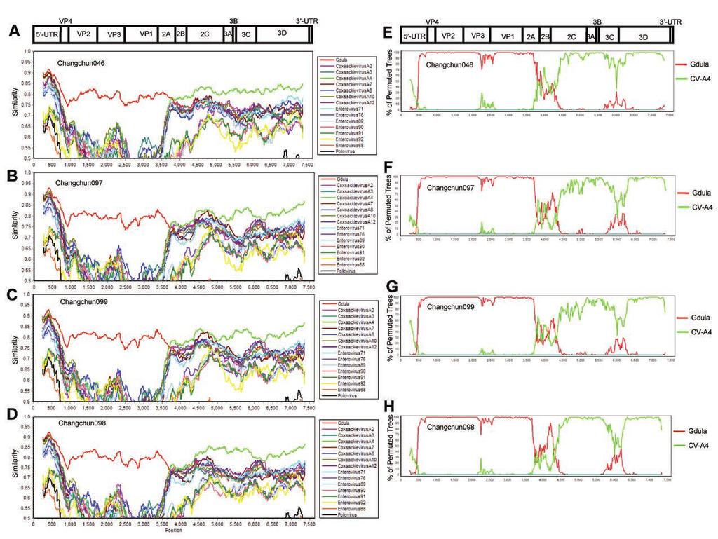
19 mice (Fig. 2), and pathological changes (Fig. 3) after infection. One-step growth curve of CV-A6 in RD cells. To determine whether the lethality of these Changchun CV-A6 strains was due to their potency in viral replication, one-step growth assay with a 2-h interval was performed. Changchun046, as the weakest strain in the lethal group, and Changchun098, as the only non-lethal strain in this study, were tested in RD cells. As shown in Fig. 5, levels of both viruses determined by CV-A6 specific qrt-pcr (data not shown) first increased in the culture medium, indicating viral replication and release, which was then followed by a decrease, most likely due to the viral absorption to RD cells to start the next-round infection. However, even with similar viral dosages to initiate the infection, levels of the lethal strain Changchun046 were constantly higher than those of the non-lethal Changchun098 strain during the replication/release phase, and showed a first peak of viral release at 14 h post-absorption, which was 2 h earlier than that detected with Changchun098. In addition, more Changchun046 viruses were detected than those in Changchun098 at the peak points. These results demonstrated that the viral replication and release of lethal CV-A6 strain was much more potent than that of the non-lethal CV-A6 strain, and this finding was correlated to the lethality of associated CV-A6 strains in the mouse model (Fig. 5 vs. Fig. 2). CV-A6-Changchun strains are products of recombination between Gdula and CV-A4. To reveal possible reasons for the differing pathogenicity observed among CV-A6-Changchun strains, we first checked whether the lethal and non-lethal strains shared different recombination patterns. Similarity tests based on the complete genome were performed, with prototypic strains of members from HEV-A group as reference sequences and each of the four full-length CV-A6-Changchun strains as the query. EV-D68 and poliovirus 1 were used as the outliers. To our surprise, despite their different potency in pathogenesis, all four CV-A6-Changchun strains presented a similar pattern of recombination, which was mostly based on the prototype of CV-A6 and CV-A4 (Fig. 6A to 19
20 D). The recombination was further confirmed by full-length Bootscanning analyses (Fig. 6E to H). In both tests, the P1 region of all four CV-A6-Changchun strains was phylogenetically related to the Gdula prototype (Fig. 6). In addition, CV-A4 was close to the CV-A6-Changchun strains in the P2/P3 regions (Fig. 6). The recombination patterns of all CV-A6-Changchun strains were almost identical throughout the entire genome, with the 2C region (4,105 5,091 bp) of Changchun098 closer to the Gdula strain than that of the other three CV-A6-Changchun strains in both tests (Fig. 6). Indeed, several previous studies have indicated that modern CV-A6 strains were products of recombination between Gdula and other circulating enteroviruses such as CV-A4 (16, 17). The modern CV-A6-Changchun strains are most likely recombinants between Gdula and prototype CV-A4, or an unknown parental virus similar to CV-A4. Phylogenetic analyses of CV-A6-Changchun strains with other CV-A6 strains from China and other countries. To further investigate the cause of CV-A6 lethality, the four Changchun CV-A6 strains were subjected to phylogenetic analyses based on different fragments (i.e., 5'-UTR, P1, P2, and P3). To expand the search, we included 34 additional CV-A6 sequences isolated from other provinces in China and other regions of the world. The prototype CV-A6 Gdula sequence was used as the outgroup. All phylogenetic analyses indicated that the Changchun098 strain was closely related to the PF3/SH/CHN/2013, 5056/SH/CHN/2013, 5039/SH/CHN/2013, and PF1/SH/CHN/2013 strains (Fig. 7A to D), which were all isolated from Shanghai, China. The NJ tree for the P2 sequences best correlated with the lethality of the Changchun CV-A6 strains. The non-lethal Changchun098 was in a separate cluster from the lethal Changchun046, Changchun097, and Changchun099 (Fig. 7C). Similar clustering was not observed for other fragments such as the 5'-UTR (Fig. 7A), P1 (Fig. 7B), and P3 (Fig. 7D). This pattern was consistent with a P2 contribution to the lethality of CV-A6-Changchun strains. 20
21 Viral 2C protein is a contributor to the pathogenicity of CV-A6. To identify residues potentially responsible for the lethality of the virus, protein sequence alignment was performed on the CV-A6 P2 region. As a result, variations between the lethal and non-lethal groups were detected at eight positions (129, 262, 267, 290, 297, 324, 339, and 556), with three of them (324, 339, and 556) being unified within each group (Fig. 8). These three positions were all located on the 2C region of CV-A6, indicating that the 2C protein might be responsible for the viral lethality of CV-A6. To further confirm 2C as a contributor to the pathogenicity of CV-A6, we introduced two more2c protein sequences from CV-A6 strains with reported lethality.weifang/sd/chn/2014/ was effective in killing 5-day-old mice (35), and shared amino acids at positions 324, 339, and 556 with the lethal strains (Fig. 8). TW/2007/00141 had an LD50 of CCID50/mouse (i.e., less lethal than Changchun046) (33) and had two residues (at positions 324 and 339) identical to that of the lethal strains, but one residue (position 556) was identical to that of the non-lethal strains (Fig. 8). This information supported the idea that these three amino acids contribute to the pathogenicity of the 2C protein and the CV-A6 virus. CV-A6 2C protein promotes cell death by inducing autophagy. To determine the contribution of viral 2C protein to the pathogenicity of the CV-A6 virus, we transfected Changchun046-2C, Changchun097-2C, and Changchun098-2C expressing vector into RD cells, and checked for cell survival rate at 48 h post-transfection. Changchun099-2C was excluded, because it shared parts of its amino acid sequence identical to that of Changchun097-2C. As a result, exogenous expression of Changchun046-2C, Changchun097-2C, and Changchun098-2C proteins resulted in 47.57, 68.89, and 27.38% rate of lethality in transfected RD cells, respectively, which were all significantly different from that of the control group (p<0.001, Fig. 9A). Observed cell death rates due to the exogenous 21
22 expression of 2C proteins were correlated to the lethality of associated CV-A6 strains in the mouse model (Fig. 9A vs. Fig. 2). Apoptosis is commonly observed in enterovirus-induced cells death (36-38). To determine whether CV-A6 2C-mediated cell death was due to 2C-induced apoptosis, RD cells were transfected with vectors expressing exogenous 2C proteins or the control vector VR1012, or treated with STS at a final concentration of 4 nm, and tested for Annexin V positivity. As a result, STS induced apoptosis at a rate of 6.42%, which was much higher than that detected in cells transfected with VR1012 (Fig. 9B and C). On the other hand, no apoptosis was observed in the cells expressing the lethal strain-derived Changchun046-2C or Changchun097-2C, or the non-lethal strain-derived Changchun098-2C, compared with cells transfected with VR1012 (Fig. 9B and C). These results indicated that CV-A6 2C protein could not induce apoptotic cell death in RD cells. A previous study has determined that CV-A16 2C induced autophagy in Hela cells by blocking the fusion of autophagosomes with lysosomes and triggering autophagosome accumulation, shown by dot formation of green fluorescent protein (GFP)-tagged LC3 (39). Thus, the CV-A6 2C protein might also induce RD cell death through autophagy. To validate this hypothesis, GFP-LC3 and CV-A6 2Cexpressing vectors were co-transfected into RD cells, and dot formation of GFP-LC3 was determined through fluorescence microscopy at 48 h post-transfection. Rapamycin (also known as sirolimus, Selleck Chemicals, Houston, TX, USA) was included as a positive control to trigger autophagy (36). As shown in Fig. 9D and E, the addition of rapamycin, and not its solvent dimethyl sulfoxide (DMSO), enhanced the dot formation of GFP-LC3, proving the validity of the assay. Comparing cells transfected with the control vector, RD cells expressing CV-A6 2C showed higher numbers of GFP-LC3 dots (Fig. 9D), indicating that autophagy contributed to the cytotoxicity induced by CV-A6 2C. With similar expression levels (Fig. 9F), the lethal strain-derived Changchun046-2C and 22
23 Changchun097-2C induced more GFP-LC3 dot formation than the non-lethal strain-derived Changchun098-2C (Fig. 9D and E). Taken together, our results indicated that CV-A6 2C triggered cell death through autophagy, contributing to the pathogenesis of CV-A6 viruses. 23
24 DISCUSSION Recombination is a common feature of enteroviruses, especially those associated with HFMD. Recently, many studies have reported that multiple HEV-A group viruses, especially non-ev-a71 and non-cv-a16 infections, could co-infect an individual, co-circulate in the same geographical area, and give those viruses the opportunity to undergo recombination (16,20, 35).Our previous studies have also revealed that circulating CV-A16 and EV-A71 were recombinants based on their prototype viruses (7-9). In the present study, we isolated and fully sequenced four CV-A6 strains that were circulating in Changchun city, Northeast China. Although the phylogeny suggested that these four modern CV-A6 strains were closely related to the prototype Gdula, they were in fact recombinants between Gdula (mainly in the P1 region) and CV-A4 (mainly in the P2/P3 regions). A similar recombination pattern was also found in CV-A6 strains isolated in Shanghai in 2013, which shared higher similarity with a recent CV-A4 strain rather than the recent CV-A6 strain in the 2C and 3'-UTR regions, and the recombinant CV-A6 virus led to more generalized rash than that of the non-recombinant CV-A6 virus(16, 40).These findings suggested that co-circulation of CV-A4 and CV-A6 strains in the same geographic region was a relatively common phenomenon in China(20, 41) and would increase the occurrence of recombination between these two serotypes. However, CV-A6 strains isolated from Wenzhou (15) exhibited three different recombination breakpoint patterns, with most strains located at the 3' end of 5'-UTR and 2A regions, whereas some strains showed a close relationship with CV-A2 and CV-A8 viruses, and others were located on the P3 region. Gaunt et al. (17) analyzed CV-A6 strains isolated from Edinburgh in 2008 and 2014, and eight recombinant forms (RF-A H) were circulating worldwide over the past 10 years, with the recombination breakpoints located in the 2A-2C, VP3, and between 5'-UTR and VP1 regions. More recently, it has been reported that recent CV-A6 recombination groups (RF-E, -F, -H, -J, and -K) shared a common 24
25 ancestor (RF-A), and the recombination breakpoints were located between the 2A-2C and 5'-UTR regions (42). Recombination events can play a significant role in the evolution of enterovirus genomes, and breakpoints detected in these studies (2A-2C regions) are well-known recombination hotspots (43). These results, together with our findings, indicated that non-structure regions might potentially contribute to clinical phenotypes and outcomes of CV-A6 infection. Recombination events can provide advantages to a virus, such as hostimmune evasion or enhanced infection. Another feature of enteroviruses is that the transmission of the prototype viruses is difficult to detect. In fact, we are unaware of any recent detections of CV-A6 prototype Gdula or CV-A16 prototype G-10; only a few cases of EV-A71 prototype BrCr have been reported (44). The most plausible explanation to the lost track of prototypic enteroviruses is that recombination is considerably beneficial for viral infection and replication, enabling them to outcompete their prototypes, whose infectivity is comparably inferior. The P1 region in modern enteroviruses is conserved with respect to its homolog in the prototype strains. In other words, no inter-type recombination could be detected in the P1 region coding for all structural proteins (VP1, VP2, VP3, and VP4). Our Bootscanning results also indicated that the recombination events between Gdula and CV-A4 occurred at the 5'-UTR and 2A regions, but not within P1. Our recent study on EV-D68 has indicated that amino acid substitution on VP1 alone affected viral infectivity and contributed to the EV-D68 North America outbreak in 2014 (45). Events such as recombination may introduce dramatic changes to the enteroviral structural proteins, which may lead to failure in virus assembly or impotence in host infection. The four CV-A6-Changchun strains exhibited distinct potencies of lethality in mice. The differences were not due to strain-specific preferences for infecting different tissues or the 25
26 potency of viral replication in vivo. Phylogenetic analyses of the P2 region separated CV-A6 into lethal and non-lethal groups. Furthermore, we identified three unique amino acids in the 2C region that were identical within each group. The 2C protein is the most conserved protein of Picornaviridae viruses. It has been reported that the N-terminal poliovirus 2C protein possesses ATPase and weak GTPase activity (46, 47). The 2C protein of EV-A71 plays an important role in viral replication (47-49), inhibiting activation of the nuclear factor kappa-light-chain-enhancer of activated B cells (NF-κB) pathway (50). In the present study, we also observed that 2C from CV-A6 alone caused cell death through autophagy instead of apoptosis, and we linked this capability to three residues (Fig. 8 and 9). In addition, data from several studies including the present one indicated a close relationship between variations of the viral 2C protein and the lethality of the CV-A6 virus, at least in a mouse model (33, 35). The 2C protein may benefit CV-A6 by causing cell death and promoting the release of the virion-progeny, which would shorten the infection cycle of the virus (Fig. 5). Our observation of 2C-induced autophagy and its association with viral lethality confirms 2C as a potential target for anti-cv-a6 drug development. In conclusion, we obtained four representative CV-A6-Changchun strains from 39 CV-A6-positive samples, and primary phylogenetic analyses indicated that multiple strains from Shanghai contributed most to the CV-A6 circulating in Changchun. The results of animal testing indicated that the Changchun046, Changchun097, and Changchun099 strains were lethal, whereas the Changchun098 strain was non-lethal. The similarity and Bootscanning tests suggested that all four CV-A6-Changchun strains were recombinants of Gdula and prototype CV-A4, or an unknown parental virus similar to CV-A4. These tests failed to reveal a potential reason for distinct lethality. The lethality of these strains determined in vivo correlated with their potency in viral replication and release in vitro. In addition, amino acid variations between the lethal and non-lethal groups were detected at 26
27 several positions; three substitutions in the 2C region were identical within each group and were associated with the capability of inducing autophagy as well as the pathogenicity of the protein. The present report enriches the epidemiological information on CV-A6 in Northeast China, confirming the Gdula/CV-A4 recombination pattern of modern CV-A6 and revealing differing lethality among CV-A6 strains, to which the viral 2C protein may contribute. The viral 2C protein may be a good target for antiviral drug design against CV-A6 infection. Downloaded from on December 30, 2018 by guest 27
28 ACKNOWLEDGMENTS We thank Editage for providing editing services, and Jun-Liang Chang and Yan-Feng Peng for technical assistance FUNDING INFORMATION This work, including the efforts of Xiao-Fang Yu, was funded by the Ministry of Science and Technology of the People s Republic of China (2012CB and 2013ZX ). This work, including the efforts of Shao-Hua Wang, was funded by the Fundamental Research Funds for Jilin University (No. 2014zkf04), and the Sixth Fund for Young Scholars of the First Hospital of Jilin University (No ). This work, including the efforts of Wen-Yan Zhang, was funded by the National Natural Science Foundation of China (No ). This work, including the efforts of Juan Du, was funded by the National Natural Science Foundation of China (No ). This work, including the efforts of Ke Zhao, was funded by the National Natural Science Foundation of China (No ), China Postdoctoral Science Foundation (No. 2017T and No. 2017M610193), and the Norman Bethune Health Science Center of Jilin University (yb201302). The funders had no role in study design, data collection, and interpretation, or the decision to submit the work for publication. 28
29 Figure Legends FIG 1 Phylogenetic status of modern CV-A6 strains. A, Neighbor-joining tree based on a shorter region of the CV-A6 genome (partial VP1, positions , corresponding to the Gdula genome). Only topology is shown for clear demonstration of phylogenetic relationship between strains. B and C, Neighbor-joining tree based on full-length CV-A6 genomes. Prototypes of the HEV-A group, EV-D68 and poliovirus 1 were used as references. The tree indicates that modern CV-A6 strains are phylogenetically close to the prototype Gdula (B), while the subtree indicates that strains from Changchun have different origins (C). All trees were tested with the bootstrap method for 1000 replicates, and values > 70 are shown. indicates CV-A6-Changchun isolates. FIG 2 CV-A6-Changchun virusescause clinical symptoms and mortality in a dose-dependent manner. One-day-old ICR mice (n = 8 10 per litter) were intracerebrally inoculated with 10-fold serial diluted dosages of viruses (10 μl/mouse). Control animals were mock-infected with MEM medium instead of virus (10 μl/mouse). Clinical symptoms and mortality were monitored daily for 21 days post-infection. Neonatal mice challenged with to CCID50/mL of Changchun046 virus (A), to CCID50/mL of Changchun097 virus (B), to CCID50/mL of Changchun099 virus (C), and to CCID50/mL of Changchun098 virus (D), respectively. The log-rank test was used to compare the survival rates of newborn mice between each group and the control group at 21 days post-infection. ***, p < 0.001; *, p < One representative from three independent tests is shown. FIG 3 Pathological analyses of CV-A6 infected newborn mice. One-day-old ICR mice were intracerebrally inoculated with Changchun046 ( CCID50/mL), Changchun097 ( CCID50/mL), Changchun099 ( CCID50/mL), and Changchun098 ( CCID50/mL), or medium (negative control). Infected mice with grade 4 5 exhibited severe hind limb 29
30 paralysis were dissected for HE staining. Representative images from the hind limb muscle, spine muscle, cardiac muscle, and lung and intestine tissues after infection are shown. Hind limb muscle and spine muscle fibers exhibited severe necrosis, including muscle bundle fracture (arrows in black), muscle fiber swelling (arrows in green), nuclear dissolution (arrows in red), and shrinkage (arrows in blue) in Changchun046, Changchun097, and Changchun099 infected mice (B, C, D, G, H, and I). No obvious changes were found in the hind limb muscle and spine muscle fibers of Changchun098 infected mice (E and J), and no detectable pathological changes were observed in the heart (L to O), lung (Q to T), or intestine (V to Y). Magnification, 400 (scale bars, 20 μm). The results are representative of three independent experiments. FIG 4 Kinetics of viral loads levels in various tissues of CV-A6 infected mice at different time points. One-day-old ICR mice were intracerebrally inoculated with Changchun046 ( CCID50/mL, A), Changchun097 ( CCID50/mL, B), Changchun099 ( CCID50/mL, C), and Changchun098 ( CCID50/mL, D), respectively. Viral loads were detected by qrt-pcr at days 2, 4, and 6 post-infection in 10 samples. The results represent the mean virus loads [(log10 copies)/mg of tissue or (log10 copies)/ml of blood] ± SD (3 mice/group, repeated 3 times). The data shown here are representative of three independent experiments. FIG 5 The lethality of CV-A6 correlates with viral replication and release. One-step growth curve of CV-A6 Changchun046 and Changchun098. Based on the genome quantification of qrt-pcr, similar amounts of Changchun046 and Changchun098 viruses were used to infect RD cells. After absorbing the virus for 6 h, the infected cells were washed and replaced by maintain medium, and the levels of released viruses in the medium were determined through qrt-pcr, with a 2-h interval since 8 h post-absorption. The values are shown as means ± SD. 30
31 FIG 6 Similarity tests and Bootscanning analyses of the CV-A6-Changchun strains and the prototype Gdula and CV-A4 on the basis of full-length genomes. The Similarity tests for Changchun046 (A), Changchun097 (C), Changchun099 (E), and Changchun098 (G) were performed with prototypic strains of members from HEV-A group. The test results indicated that modern CV-A6 strains circulating in Changchun were mostly recombinants between CV-A6 and CV-A4, as previously described (16, 17). Thus, for better demonstration, only CV-A4 (accession number AY421762) and CV-A6 Gdula (accession number AY421764) were included as the reference sequences for the Bootscanning tests for Changchun046 (B), Changchun097 (D), Changchun099 (F), and Changchun098 (H). EV-D68 (accession number AY426531) and poliovirus 1 (accession number V01150) were used as the outliers in both Similarity tests and Bootscanning analyses. All tests were performed with full-length viral genomes. A cartoon of the enteroviral genome is located above. FIG 7 Phylogenetic trees construction for 5'-UTR, P1, P2, and P3 regions of CV-A6-Changchun strains with other CV-A6 strains retrieved from the GenBank database. Neighbor-joining trees based on the full-length 5'-UTR (A), P1 (B), P2 (C), and P3 (D) regions were generated by the neighbor-joining method. All trees were tested by bootstrapping for 1,000 replicates, and values > 70 were shown. indicates CV-A6-Changchun isolates. FIG 8 Sequence alignment of the lethal and non-lethal strains of CV-A6. Amino acids sequence alignment of the P2 region ofcv-a6 strains from Changchun and other places of China and worldwide, and specific positions possibly associated with the lethality were indicated, based on the differences between the lethal group (containing Changchun046, Changchun097 and Changchun099) and the non-lethal group (containing Changchun098). 31
32 FIG 9 CV-A6 2C protein induces cell death through autophagy instead of apoptosis in RD cells. (A) Cell lethality caused by different CV-A6 2C protein in RD cells. Changchun046-2C, Changchun097-2C, or Changchun098-2C expressing vector was transfected into RD cells, and the cell proliferation assay was performed at 48 h post-transfection. Western blotting results indicating expression levels of viral 2C proteins for the cell lethality experiment. (B) Representative flow charts for the apoptosis analysis. Changchun046-2C, Changchun097-2C, or Changchun098-2Cexpressing vector was transfected into RD cells. RD cells were harvested at 48 h post-transfection, and cell death was evaluated by flow cytometry. The percentages of apoptotic cells corresponding to the increased FITC fluorescence intensity for each of the experimental conditions are indicated. RD cells transfected with VR1012 or treated with Staurosporine (STS) were used as negative and positive controls, respectively. (C) Bar chart of the apoptosis analysis in RD cells. Western blotting results indicating expression levels of viral 2C proteins for Fig. 9B. (D) Live cell imaging indicating GFP-LC3 aggregation. RD cells were co-transfected with pegfp-lc3 and VR1012 empty vector, Changchun046-2C, Changchun097-2C, or Changchun099-2C. Rapamycin was used as a positive control to induce autophagy. The GFP-LC3 aggregations in the cells were observed by fluorescent microscopy at 48 h post-transfection. Representative images are shown. Magnification, 400 (scale bars, 20 μm). (E) Bar chart indicating average GFP-LC3 dot formation in RD cells expressing CV-A6 2C. Each bar value indicates the average number of dots in ten cells per sample. Rapamycin was used as a positive control. For each sample, number of GFP dots were counted in ten cells. (F) Western blotting results showing 2C protein expression in RD cells from Fig. 9E. The values in Fig. 9A, 9C, and 9E are shown as means ± SD. 32
33 683 TABLE 1 Reverse Transcription, PCR and Sequencing primers used in this study Primer Sequence(5 to 3 ) Location Region Reference AN32 GTYTGCCA a VP1 [28] AN33 GAYTGCCA a VP1 [28] AN34 CCRTCRTA a VP1 [28] AN35 RCTYTGCCA a VP1 [28] 224 GCIATGYTIGGIACICAYRT a VP3 [28] 222 CICCIGGIGGIAYRWACAT a VP1 [28] AN89 CCAGCACTGACAGCAGYNGARAYNGG a VP1 [28] AN88 TACTGGACCACCTGGNGGNAYRWACAT a VP1 [28] FL-F-1C TTAAAACAGCCTGTGGGTTG 1-20 b 5 UTR [19] panev-f CCCCTGAATGCGGCTAATC b 5 UTR [19] panev-r GCTGCTTATGGTGACAATC b 5 UTR [19] 5'UTR-EV-F-FW2 CCCTCTTAGTGGCTGCAATC b VP2 [19] VP1-222-FW2-R TGGGGCAATAGTTAGTGTGA b VP2 [19] 2715-R TTCACCTCCACAACYCCTACYAGC b VP1 [19] 3311-F ATAACTAACACTGCAACCGACC b VP1 [19] 3441-R GCCCARTCATTATGAGTGGC b 2A [19] 4069-R GCMTCCATCYTAGGTATCCCTA b 2B [19] 4613-F GTGGTCACAGTYATGGACGATC b 2C [19] 5477-R GTYGCCGTRCGGAGRATRGGTT b 3B [19] 6150-F GCAGGCRTTGTTCTCYAAGTAT b 3D [19] 5332-F GTTTTCCCAGTCACGACTTYCARGGBGCKTA b 3D [19] 650-R GTCACTGGATGGCCAATCC b 5 UTR This study 5331-F CTCTTTGCTGGGTTCCAAG b 3A This study 7434-R GCTATTCTGGTTATAACAAATTTACC b 3 UTR This study a The locations of all primers are those relative to the genome of PV1 Mahoney (GenBank accession number J02281); b Numbering is based on coxsackievirus A6 strain Gdula (accession No. AY421764); Key to degenerate nucleotides: I = Inosine, K = T+G, M = A+C, R = A+G, W = T+A, Y = C+T, B = C+G+T, V = A+G+C, N = A+T+C+G
34 687 TABLE 2 Reference strains used in this study Strain Type Year Location Length (nt) GenBank accession no. Gdula CV-A USA/ New York 7434 AY Fleetwood CV-A USA/ Delaware 7398 AY Olson CV-A USA/ New York 7395 AY High Point CV-A USA/ North Carolina 7435 AY Swartz CV-A USA/ New York 7400 AY Parker CV-A USA/ New York 7404 AY Donovan CV-A USA/ New York 7396 AY Kowalik CV-A USA/ New York 7409 AY Texas-12 CV-A USA/ Texas 7407 AY G-14 CV-A Republic of South Africa 7415 AY G-10 CV-A Republic of South Africa 7408 U05876 BrCr EV-A71A 1970 USA/ California 7408 U22521 UH1/PM/1997 EV-A71B 1997 MALAYSIA/Kuala Lumpur 7411 AM S10862-SAR-98 EV-A71C 1998 MALAYSIA/KotaSamarahan 7409 DQ FRA EV-A France/Caen 7438 AY BAN EV-A Bangladesh/Bhola 7429 AY BAN EV-A Bangladesh/Comilla 7425 AY BAN EV-A Bangladesh/Barisal 7427 AY RJG7 EV-A92 NA USA 7379 EF Fermon EV-D USA/California 7367 AY Sabin1 Poliovirus USA 7441 V01150 Abbreviations: USA, United States of America; NA, not applicable
35 REFERENCES 1. Bendig JW, Fleming DM Epidemiological, virological, and clinical features of an epidemic of hand, foot, and mouth disease in England and Wales. Commun Dis Rep CDR Rev 6:R Chan KP, Goh KT, Chong CY, Teo ES, Lau G, Ling AE Epidemic hand, foot and mouth disease caused by human enterovirus 71, Singapore. Emerg Infect Dis 9: Shimizu H, Utama A, Onnimala N, Li C, Li-Bi Z, Yu-Jie M, Pongsuwanna Y, Miyamura T Molecular epidemiology of enterovirus 71 infection in the Western Pacific Region. Pediatr Int 46: Ho M, Chen ER, Hsu KH, Twu SJ, Chen KT, Tsai SF, Wang JR, Shih SR An epidemic of enterovirus 71 infection in Taiwan. Taiwan Enterovirus Epidemic Working Group. N Engl J Med 341: Brown BA, Oberste MS, Alexander JP, Jr., Kennett ML, Pallansch MA Molecular epidemiology and evolution of enterovirus 71 strains isolated from 1970 to J Virol 73: Yoke-Fun C, AbuBakar S Phylogenetic evidence for inter-typic recombination in the emergence of human enterovirus 71 subgenotypes. BMC Microbiol 6: Zhao K, Han X, Wang G, Hu W, Zhang W, Yu XF Circulating coxsackievirus A16 identified as recombinant type A human enterovirus, China. Emerg Infect Dis 17: Wang X, Zhu C, Bao W, Zhao K, Niu J, Yu XF, Zhang W Characterization of full-length enterovirus 71 strains from severe and mild disease patients in northeastern China. PLoS One 7:e Wei W, Guo H, Li J, Ren S, Wei Z, Bao W, Hu X, Zhao K, Zhang W, Zhou Y, Sun F, Markham R, Yu XF Circulating HFMD-associated coxsackievirus A16 is genetically and phenotypically distinct from the prototype CV-A16. PLoS One 9:e Howlett SE Coxsackievirus B3-Induced Myocarditis: New Insights Into a Female Advantage. Can J Cardiol 2018: Wieczorek M, Krzysztoszek A Molecular Characterization of Enteroviruses Isolated from Acute Flaccid Paralysis Cases in Poland, Pol J Microbiol 65: Puenpa J, Chieochansin T, Linsuwanon P, Korkong S, Thongkomplew S, Vichaiwattana P, Theamboonlers A, Poovorawan Y Hand, foot, and mouth disease caused by coxsackievirus A6, Thailand, Emerg Infect Dis 19: Montes M, Artieda J, Pineiro LD, Gastesi M, Diez-Nieves I, Cilla G Hand, foot, and mouth disease outbreak and coxsackievirus A6, northern Spain, Emerg Infect Dis Yang F, Yuan J, Wang X, Li J, Du J, Su H, Zhou B, Jin Q Severe hand, foot, and mouth disease and coxsackievirus A6-Shenzhen, China. Clin Infect Dis 59: Guo WP, Lin XD, Chen YP, Liu Q, Wang W, Wang CQ, Li MH, Sun XY, Shi M, Holmes EC, Zhang YZ Fourteen types of co-circulating recombinant enterovirus were associated with hand, foot, and mouth disease in children from 35
36 Wenzhou, China. J Clin Virol 70: Feng X, Guan W, Guo Y, Yu H, Zhang X, Cheng R, Wang Z, Zhang Z, Zhang J, Li H, Zhuang Y, Zhang H, Lu Z, Li M, Yu H, Bao Y, Hu Y, Yao Z A novel recombinant lineage's contribution to the outbreak of coxsackievirus A6-associated hand, foot and mouth disease in Shanghai, China, Sci Rep 5: Gaunt E, Harvala H, Osterback R, Sreenu VB, Thomson E, Waris M, Simmonds P Genetic characterization of human coxsackievirus A6 variants associated with atypical hand, foot and mouth disease: a potential role of recombination in emergence and pathogenicity. J Gen Virol 96: Zhang L, Wang X, Zhang Y, Gong L, Mao H, Feng C, Ojcius DM, Yan J Rapid and sensitive identification of RNA from the emerging pathogen, coxsackievirus A6. Virol J 9: Chen YJ, Chang SC, Tsao KC, Shih SR, Yang SL, Lin TY, Huang YC Comparative genomic analysis of coxsackievirus A6 strains of different clinical disease entities. PLoS One 7:e He YQ, Chen L, Xu WB, Yang H, Wang HZ, Zong WP, Xian HX, Chen HL, Yao XJ, Hu ZL, Luo M, Zhang HL, Ma HW, Cheng JQ, Feng QJ, Zhao DJ Emergence, circulation, and spatiotemporal phylogenetic analysis of coxsackievirus a6- and coxsackievirus a10-associated hand, foot, and mouth disease infections from 2008 to 2012 in Shenzhen, China. J Clin Microbiol 51: Di B, Zhang Y, Xie H, Li X, Chen C, Ding P, He P, Wang D, Geng J, Luo L, Bai Z, Yang Z, Wang M Circulation of Coxsackievirus A6 in hand-foot-mouth disease in Guangzhou, Virol J 11: Lu J, Zeng H, Zheng H, Yi L, Guo X, Liu L, Sun L, Tan X, Li H, Ke C, Lin J Hand, foot and mouth disease in Guangdong, China, in 2013: new trends in the continuing epidemic. Clin Microbiol Infect 20:O442- O Hongyan G, Chengjie M, Qiaozhi Y, Wenhao H, Juan L, Lin P, Yanli X, Hongshan W, Xingwang L Hand, foot and mouth disease caused by coxsackievirus A6, Beijing, Pediatr Infect Dis J 33: Zeng H, Lu J, Zheng H, Yi L, Guo X, Liu L, Rutherford S, Sun L, Tan X, Li H, Ke C, Lin J The Epidemiological Study of Coxsackievirus A6 revealing Hand, Foot and Mouth Disease Epidemic patterns in Guangdong, China. Sci Rep 5: Tan X, Li L, Zhang B, Jorba J, Su X, Ji T, Yang D, Lv L, Li J, Xu W Molecular epidemiology of coxsackievirus A6 associated with outbreaks of hand, foot, and mouth disease in Tianjin, China, in Arch Virol 160: Hu YQ, Xie GC, Li DD, Pang LL, Xie J, Wang P, Chen Y, Yang J, Cheng WX, Zhang Q, Jin Y, Duan ZJ Prevalence of Coxsackievirus A6 and Enterovirus 71 in Hand, Foot and Mouth Disease in Nanjing, China in Pediatr Infect Dis J 34: Han JF, Xu S, Zhang Y, Zhu SY, Wu DL, Yang XD, Liu H, Sun BX, Wu XY, Qin CF Hand, foot, and mouth disease outbreak caused by coxsackievirus A6, China, J Infect 69: Nix WA, Oberste MS, Pallansch MA Sensitive, seminested PCR amplification of VP1 sequences for direct identification of all enterovirus serotypes from original 36
37 clinical specimens. J Clin Microbiol 44: Tamura K, Peterson D, Peterson N, Stecher G, Nei M, Kumar S MEGA5: molecular evolutionary genetics analysis using maximum likelihood, evolutionary distance, and maximum parsimony methods. Mol Biol Evol 28: Reed LJ, Muench H A simple method of estimating 50 percent end-points. Am J Epidemiol 27: Chang J, Li J, Liu X, Liu G, Yang J, Wei W, Zhang W, Yu XF Broad protection with an inactivated vaccine against primary-isolated lethal enterovirus 71 infection in newborn mice. BMC Microbiol 15: Lole KS, Bollinger RC, Paranjape RS, Gadkari D, Kulkarni SS, Novak NG, Ingersoll R, Sheppard HW, Ray SC Full-length human immunodeficiency virus type 1 genomes from subtype C-infected seroconverters in India, with evidence of intersubtype recombination. J Virol 73: Yang L, Mao Q, Li S, Gao F, Zhao H, Liu Y, Wan J, Ye X, Xia N, Cheng T, Liang Z A neonatal mouse model for the evaluation of antibodies and vaccines against coxsackievirus A6. Antiviral Res 134: Sickles GM, Mutterer M, Feorino P, Plager H Recently classified types of Coxsackie virus, group A; behavior in tissue culture. Proc Soc Exp Biol Med 90: Zhang Z, Dong Z, Wei Q, Carr MJ, Li J, Ding S, Tong Y, Li D, Shi W A Neonatal Murine Model of Coxsackievirus A6 Infection for Evaluation of Antiviral and Vaccine Efficacy. J Virol Li X, Zhang J, Chen Z, Yang L, Xing X, Ma X, Yang Z Both PI3K- and mtor-signaling pathways take part in CVB3-induced apoptosis of Hela cells. DNA Cell Biol 32: doi: /dna Xi X, Zhang X, Wang B, Wang T, Wang J, Huang H, Wang J, Jin Q, Zhao Z The interplays between autophagy and apoptosis induced by enterovirus 71. PLoS One 8:e Zhu G, Zheng Y, Zhang L, Shi Y, Li W, Liu Z, Peng B, Yin J, Liu W, He X Coxsackievirus A16 infection triggers apoptosis in RD cells by inducing ER stress. Biochem Biophys Res Commun 441: Shi Y, He X, Zhu G, Tu H, Liu Z, Li W, Han S, Yin J, Peng B, Liu W Coxsackievirus A16 elicits incomplete autophagy involving the mtor and ERK pathways. PLoS One 10:e Feng X, Guan W, Guo Y, Yu H, Zhang X, Cheng R, Wang Z, Zhang Z, Zhang J, Li H, Zhuang Y, Zhang H, Lu Z, Li M, Yu H, Bao Y, Hu Y, Yao Z Genome sequence of a novel recombinant coxsackievirus a6 strain from shanghai, china, Genome Announc Lu QB, Zhang XA, Wo Y, Xu HM, Li XJ, Wang XJ, Ding SJ, Chen XD, He C, Liu LJ, Li H, Yang H, Li TY, Liu W, Cao WC Circulation of Coxsackievirus A10 and A6 in hand-foot-mouth disease in China, PLoS One 7:e Puenpa J, Vongpunsawad S, Osterback R, Waris M, Eriksson E, Albert J, Midgley S, Fischer TK, Eis-Hubinger AM, Cabrerizo M, Gaunt E, Simmonds P, Poovorawan Y Molecular epidemiology and the evolution of human coxsackievirus A6. J Gen Virol 97:
38 Lukashev AN, Lashkevich VA, Ivanova OE, Koroleva GA, Hinkkanen AE, Ilonen J Recombination in circulating Human enterovirus B: independent evolution of structural and non-structural genome regions. J Gen Virol 86: Yang Z, Lu S, Xian J, Ye J, Xiao L, Luo J, Zen K, Liu F Complete genome sequence of a human enterovirus 71 strain isolated in wuhan, china, in Genome Announc Du J, Zheng B, Zheng W, Li P, Kang J, Hou J, Markham R, Zhao K, Yu XF Analysis of Enterovirus 68 Strains from the 2014 North American Outbreak Reveals a New Clade, Indicating Viral Evolution. PLoS One 10:e Mirzayan C, Wimmer E Genetic analysis of an NTP-binding motif in poliovirus polypeptide 2C. Virology 189: Rodriguez PL, Carrasco L Poliovirus protein 2C contains two regions involved in RNA binding activity. J Biol Chem 270: Tang WF, Yang SY, Wu BW, Jheng JR, Chen YL, Shih CH, Lin KH, Lai HC, Tang P, Horng JT Reticulon 3 binds the 2C protein of enterovirus 71 and is required for viral replication. J Biol Chem 282: Wang J, Wu Z, Jin Q COPI is required for enterovirus 71 replication. PLoS One 7:e Zheng Z, Li H, Zhang Z, Meng J, Mao D, Bai B, Lu B, Mao P, Hu Q, Wang H Enterovirus 71 2C protein inhibits TNF-alpha-mediated activation of NF-kappaB by suppressing IkappaB kinase beta phosphorylation. J Immunol 187: Downloaded from on December 30, 2018 by guest 38

39
40

41

42
43
44

45
46

47
Enterovirus 71 Outbreak in P. R. China, 2008
 JCM Accepts, published online ahead of print on 13 May 2009 J. Clin. Microbiol. doi:10.1128/jcm.00563-09 Copyright 2009, American Society for Microbiology and/or the Listed Authors/Institutions. All Rights
Sequence analysis for VP4 of enterovirus 71 isolated in Beijing during 2007 to 2008
 16 2009 3 4 1 Journal of Microbes and Infection, March 2009, Vol. 4, No. 1 2007 2008 71 VP4 1, 2, 2, 2, 1, 2, 2, 2, 1, 2 1., 100730; 2., 100020 : 2007 2008 71 ( EV71), 2007 3 EV71( 1, 2 ) 2008 5 EV71(
Oncolytic Adenovirus Complexes Coated with Lipids and Calcium Phosphate for Cancer Gene Therapy
 Oncolytic Adenovirus Complexes Coated with Lipids and Calcium Phosphate for Cancer Gene Therapy Jianhua Chen, Pei Gao, Sujing Yuan, Rongxin Li, Aimin Ni, Liang Chu, Li Ding, Ying Sun, Xin-Yuan Liu, Yourong
p47 negatively regulates IKK activation by inducing the lysosomal degradation of polyubiquitinated NEMO
 Supplementary Information p47 negatively regulates IKK activation by inducing the lysosomal degradation of polyubiquitinated NEMO Yuri Shibata, Masaaki Oyama, Hiroko Kozuka-Hata, Xiao Han, Yuetsu Tanaka,
RNA extraction, RT-PCR and real-time PCR. Total RNA were extracted using
 Supplementary Information Materials and Methods RNA extraction, RT-PCR and real-time PCR. Total RNA were extracted using Trizol reagent (Invitrogen,Carlsbad, CA) according to the manufacturer's instructions.
Phosphate buffered saline (PBS) for washing the cells TE buffer (nuclease-free) ph 7.5 for use with the PrimePCR Reverse Transcription Control Assay
 for washing the cells TE buffer (nuclease-free) ph 7.5 for use with the PrimePCR Reverse Transcription Control Assay") Catalog # Description 172-5080 SingleShot Cell Lysis Kit, 100 x 50 µl reactions 172-5081 SingleShot Cell Lysis Kit, 500 x 50 µl reactions For research purposes only. Introduction The SingleShot Cell Lysis
Catalog # Description 172-5080 SingleShot Cell Lysis Kit, 100 x 50 µl reactions 172-5081 SingleShot Cell Lysis Kit, 500 x 50 µl reactions For research purposes only. Introduction The SingleShot Cell Lysis
Change of Major Genotype of Enterovirus 71 in Outbreaks of Hand-Foot-and-Mouth Disease in Taiwan between 1998 and 2000
 JOURNAL OF CLINICAL MICROBIOLOGY, Jan. 2002, p. 10 15 Vol. 40, No. 1 0095-1137/02/$04.00 0 DOI: 10.1128/JCM.40.1.10 15.2002 Copyright 2002, American Society for Microbiology. All Rights Reserved. Change
Avian Influenza Virus H7N9. Dr. Di Liu Network Information Center Institute of Microbiology Chinese Academy of Sciences
 Avian Influenza Virus H7N9 Dr. Di Liu Network Information Center Institute of Microbiology Chinese Academy of Sciences Avian Influenza Virus RNA virus, Orthomyxoviruses Influenza A virus Eight Gene segments
Supplemental Materials and Methods Plasmids and viruses Quantitative Reverse Transcription PCR Generation of molecular standard for quantitative PCR
 Supplemental Materials and Methods Plasmids and viruses To generate pseudotyped viruses, the previously described recombinant plasmids pnl4-3-δnef-gfp or pnl4-3-δ6-drgfp and a vector expressing HIV-1 X4
DOI: /ICJ poliovirus. [14] (Hand-Foot-Mouth Disease, HFMD) (Herpangina) 71 [26] [17,18,27] 71 picornaviridae
![DOI: /ICJ poliovirus. [14] (Hand-Foot-Mouth Disease, HFMD) (Herpangina) 71 [26] [17,18,27] 71 picornaviridae](/thumbs/93/112612467.jpg "DOI: /ICJ poliovirus. [14] (Hand-Foot-Mouth Disease, HFMD) (Herpangina) 71 [26] [17,18,27] 71 picornaviridae") 174 DOI: 10.6526/ICJ.2016.404 71 71 [14] (Hand-Foot-Mouth Disease, HFMD) (Herpangina) 71 71 picornaviridae poliovirus / 1990 71 [26] [17,18,27] 71 175 [31] ICR NOD/SCID AG129 hpsgl-1 hscarb2 (MP4) (MP4)
174 DOI: 10.6526/ICJ.2016.404 71 71 [14] (Hand-Foot-Mouth Disease, HFMD) (Herpangina) 71 71 picornaviridae poliovirus / 1990 71 [26] [17,18,27] 71 175 [31] ICR NOD/SCID AG129 hpsgl-1 hscarb2 (MP4) (MP4)
Construction of a hepatocellular carcinoma cell line that stably expresses stathmin with a Ser25 phosphorylation site mutation
 Construction of a hepatocellular carcinoma cell line that stably expresses stathmin with a Ser25 phosphorylation site mutation J. Du 1, Z.H. Tao 2, J. Li 2, Y.K. Liu 3 and L. Gan 2 1 Department of Chemistry,
SURVEILLANCE TECHNICAL
 CHAPTER 5 SURVEILLANCE TECHNICAL ASPECTS 55 Protect - detect - protect Polio eradication strategies can be summed up as protect and detect protect children against polio by vaccinating them, and detect
Test Report. Efficacy of A New JM Nanocomposite Material in Inhibiting Respiratory Syncytial Virus Cellular Infection
 Test Report Efficacy of A New JM Nanocomposite Material in Inhibiting Respiratory Syncytial Virus Cellular Infection Test Reagent New JM Nanocomposite Material Project Commissioner JM Material Technology,
Evidence for Other Non-Poliovirus Enteroviruses Multiplies in L20B Cells. ACCEPTED LUIS SARMIENTO*, PEDRO MÁS, ROSA PALOMERA, LUIS MORIER, MAGILÉ
 CVI Accepts, published online ahead of print on 14 March 2007 Clin. Vaccine Immunol. doi:10.1128/cvi.00017-06 Copyright 2007, American Society for Microbiology and/or the Listed Authors/Institutions. All
Retro-X qrt-pcr Titration Kit User Manual
 Takara Bio USA Retro-X qrt-pcr Titration Kit User Manual Cat. No. 631453 PT3952-1 (030218) 1290 Terra Bella Avenue, Mountain View, CA 94043, USA U.S. Technical Support: techus@takarabio.com United States/Canada
Multi-clonal origin of macrolide-resistant Mycoplasma pneumoniae isolates. determined by multiple-locus variable-number tandem-repeat analysis
 JCM Accepts, published online ahead of print on 30 May 2012 J. Clin. Microbiol. doi:10.1128/jcm.00678-12 Copyright 2012, American Society for Microbiology. All Rights Reserved. 1 2 Multi-clonal origin
Broad protection with an inactivated vaccine against primary-isolated lethal enterovirus 71 infection in newborn mice
 Chang et al. BMC Microbiology (2015) 15:139 DOI 10.1186/s12866-015-0474-9 RESEARCH ARTICLE Open Access Broad protection with an inactivated vaccine against primary-isolated lethal enterovirus 71 infection
Genetic Diversity of Coxsackievirus A16 Associated with Hand, Foot, and Mouth Disease Epidemics in Japan from 1983 to 2003
 JOURNAL OF CLINICAL MICROBIOLOGY, Jan. 2007, p. 112 120 Vol. 45, No. 1 0095-1137/07/$08.00 0 doi:10.1128/jcm.00718-06 Copyright 2007, American Society for Microbiology. All Rights Reserved. Genetic Diversity
CHAPTER 4 RESULTS. showed that all three replicates had similar growth trends (Figure 4.1) (p<0.05; p=0.0000)
 (p<0.05; p=0.0000)") CHAPTER 4 RESULTS 4.1 Growth Characterization of C. vulgaris 4.1.1 Optical Density Growth study of Chlorella vulgaris based on optical density at 620 nm (OD 620 ) showed that all three replicates had similar
CHAPTER 4 RESULTS 4.1 Growth Characterization of C. vulgaris 4.1.1 Optical Density Growth study of Chlorella vulgaris based on optical density at 620 nm (OD 620 ) showed that all three replicates had similar
Nature Medicine: doi: /nm.4322
 1 2 3 4 5 6 7 8 9 10 11 Supplementary Figure 1. Predicted RNA structure of 3 UTR and sequence alignment of deleted nucleotides. (a) Predicted RNA secondary structure of ZIKV 3 UTR. The stem-loop structure
B16-F10 (Mus musculus skin melanoma), NCI-H460 (human non-small cell lung cancer
, NCI-H460 (human non-small cell lung cancer") Electronic Supplementary Material (ESI) for ChemComm. This journal is The Royal Society of Chemistry 2017 Experimental Methods Cell culture B16-F10 (Mus musculus skin melanoma), NCI-H460 (human non-small
Electronic Supplementary Material (ESI) for ChemComm. This journal is The Royal Society of Chemistry 2017 Experimental Methods Cell culture B16-F10 (Mus musculus skin melanoma), NCI-H460 (human non-small
Replication Defective Enterovirus Infections: Implications for Type I Diabetes
 Replication Defective Enterovirus Infections: Implications for Type I Diabetes N. M. Chapman Department of Pathology & Microbiology University of Nebraska Medical Center Enterovirus Genome and 2 Capsid
Picornaviruses. Virion. Genome. Genes and proteins. Viruses and hosts. Diseases. Distinctive characteristics
 Picornaviruses Virion Genome Genes and proteins Viruses and hosts Diseases Distinctive characteristics Virion Naked icosahedral capsid (T=1) Diameter of 30 nm Genome Linear single-stranded RNA, positive
MicroRNA sponges: competitive inhibitors of small RNAs in mammalian cells
 MicroRNA sponges: competitive inhibitors of small RNAs in mammalian cells Margaret S Ebert, Joel R Neilson & Phillip A Sharp Supplementary figures and text: Supplementary Figure 1. Effect of sponges on
A Novel Recombinant Virus Reagent Products for Efficient Preparation Of Hepatitis B Animal Models
 About FivePlus Beijing FivePlus Molecular Medicine Institute was established in 2005. The company has been dedicating itself to continuous innovation of viral vectors. The meaning of FivePlus is based
Instructions for Use. APO-AB Annexin V-Biotin Apoptosis Detection Kit 100 tests
 3URGXFW,QIRUPDWLRQ Sigma TACS Annexin V Apoptosis Detection Kits Instructions for Use APO-AB Annexin V-Biotin Apoptosis Detection Kit 100 tests For Research Use Only. Not for use in diagnostic procedures.
HEK293FT cells were transiently transfected with reporters, N3-ICD construct and
 Supplementary Information Luciferase reporter assay HEK293FT cells were transiently transfected with reporters, N3-ICD construct and increased amounts of wild type or kinase inactive EGFR. Transfections
Evolution of influenza
 Evolution of influenza Today: 1. Global health impact of flu - why should we care? 2. - what are the components of the virus and how do they change? 3. Where does influenza come from? - are there animal
ab Adipogenesis Assay Kit (Cell-Based)
") ab133102 Adipogenesis Assay Kit (Cell-Based) Instructions for Use For the study of induction and inhibition of adipogenesis in adherent cells. This product is for research use only and is not intended
ab133102 Adipogenesis Assay Kit (Cell-Based) Instructions for Use For the study of induction and inhibition of adipogenesis in adherent cells. This product is for research use only and is not intended
Coronaviruses. Virion. Genome. Genes and proteins. Viruses and hosts. Diseases. Distinctive characteristics
 Coronaviruses Virion Genome Genes and proteins Viruses and hosts Diseases Distinctive characteristics Virion Spherical enveloped particles studded with clubbed spikes Diameter 120-160 nm Coiled helical
Supplementary data Supplementary Figure 1 Supplementary Figure 2
 Supplementary data Supplementary Figure 1 SPHK1 sirna increases RANKL-induced osteoclastogenesis in RAW264.7 cell culture. (A) RAW264.7 cells were transfected with oligocassettes containing SPHK1 sirna
The effect of insulin on chemotherapeutic drug sensitivity in human esophageal and lung cancer cells
 The effect of insulin on chemotherapeutic drug sensitivity in human esophageal and lung cancer cells Published in: Natl Med J China, February 10, 2003; Vol 83, No 3, Page 195-197. Authors: JIAO Shun-Chang,
Multi-Parameter Apoptosis Assay Kit
 Multi-Parameter Apoptosis Assay Kit Catalog Number KA1335 5 x 96 assays Version: 05 Intended for research use only www.abnova.com Table of Contents Introduction... 3 Background... 3 Principle of the Assay...
IN VITRO HORMESIS EFFECTS OF SODIUM FLUORIDE ON KIDNEY CELLS OF THREE-DAY-OLD MALE RATS
 292 Tang, An, Du, Zhang, Zhou 292 IN VITRO HORMESIS EFFECTS OF SODIUM FLUORIDE ON KIDNEY CELLS OF THREE-DAY-OLD MALE RATS Qin-qing Tang, a Xiao-jing An, a Jun Du, a Zheng-xiang Zhang, a Xiao-jun Zhou a
The Annexin V Apoptosis Assay
 The Annexin V Apoptosis Assay Development of the Annexin V Apoptosis Assay: 1990 Andree at al. found that a protein, Vascular Anticoagulant α, bound to phospholipid bilayers in a calcium dependent manner.
Figure S1. Schematic presentation of genomic replication of idsiv after transfection and infection. After transfection of idsiv plasmid DNA into 293T
 Figure S1. Schematic presentation of genomic replication of idsiv after transfection and infection. After transfection of idsiv plasmid DNA into 293T cells, the RNA genomes with all modifications are generated
Gladstone Institutes, University of California (UCSF), San Francisco, USA
, San Francisco, USA") Fluorescence-linked Antigen Quantification (FLAQ) Assay for Fast Quantification of HIV-1 p24 Gag Marianne Gesner, Mekhala Maiti, Robert Grant and Marielle Cavrois * Gladstone Institutes, University of
Fluorescence-linked Antigen Quantification (FLAQ) Assay for Fast Quantification of HIV-1 p24 Gag Marianne Gesner, Mekhala Maiti, Robert Grant and Marielle Cavrois * Gladstone Institutes, University of
ADCC Assay Protocol Vikram Srivastava 1, Zheng Yang 1, Ivan Fan Ngai Hung 2, Jianqing Xu 3, Bojian Zheng 3 and Mei- Yun Zhang 3*
 ADCC Assay Protocol Vikram Srivastava 1, Zheng Yang 1, Ivan Fan Ngai Hung 2, Jianqing Xu 3, Bojian Zheng 3 and Mei- Yun Zhang 3* 1 Department of Microbiology, Li Ka Shing Faculty of Medicine, University
Constitutive Reporter Lentiviral Vectors Expressing Fluorescent Proteins
 Constitutive Reporter Lentiviral Vectors Expressing Fluorescent Proteins www.vectalys.com/products/ Constitutive Reporter Lentiviral Vectors Catalog Number referring to this User Manual: 0008VCT; 0009VCT;
Enterovirus D68-US Outbreak 2014 Laboratory Perspective
 Enterovirus D68-US Outbreak 2014 Laboratory Perspective Allan Nix Team Lead Picornavirus Laboratory Polio and Picornavirus Laboratory Branch PAHO / SARInet Webinar November 6, 2014 National Center for
DNA context and promoter activity affect gene expression in lentiviral vectors
 ACTA BIOMED 2008; 79: 192-196 Mattioli 1885 O R I G I N A L A R T I C L E DNA context and promoter activity affect gene expression in lentiviral vectors Gensheng Mao 1, Francesco Marotta 2, Jia Yu 3, Liang
Focus Application. Compound-Induced Cytotoxicity
 xcelligence System Real-Time Cell Analyzer Focus Application Compound-Induced Cytotoxicity For life science research only. Not for use in diagnostic procedures. Featured Study: Using the Time Resolving
Supplementary Fig. 1. Identification of acetylation of K68 of SOD2
 Supplementary Fig. 1. Identification of acetylation of K68 of SOD2 A B H. sapiens 54 KHHAAYVNNLNVTEEKYQEALAK 75 M. musculus 54 KHHAAYVNNLNATEEKYHEALAK 75 X. laevis 55 KHHATYVNNLNITEEKYAEALAK 77 D. rerio
Supporting Information
 Supporting Information Valkenburg et al. 10.1073/pnas.1403684111 SI Materials and Methods ELISA and Microneutralization. Sera were treated with Receptor Destroying Enzyme II (RDE II, Accurate) before ELISA
Norgen s HIV proviral DNA PCR Kit was developed and validated to be used with the following PCR instruments: Qiagen Rotor-Gene Q BioRad icycler
 3430 Schmon Parkway Thorold, ON, Canada L2V 4Y6 Phone: (905) 227-8848 Fax: (905) 227-1061 Email: techsupport@norgenbiotek.com HIV Proviral DNA PCR Kit Product # 33840 Product Insert Background Information
PUMA gene transfection can enhance the sensitivity of epirubicin-induced apoptosis of MCF-7 breast cancer cells
 PUMA gene transfection can enhance the sensitivity of epirubicin-induced apoptosis of MCF-7 breast cancer cells C.-G. Sun 1 *, J. Zhuang 1 *, W.-J. Teng 1, Z. Wang 2 and S.-S. Du 3 1 Department of Oncology,
Norgen s HIV Proviral DNA PCR Kit was developed and validated to be used with the following PCR instruments: Qiagen Rotor-Gene Q BioRad T1000 Cycler
 3430 Schmon Parkway Thorold, ON, Canada L2V 4Y6 Phone: 866-667-4362 (905) 227-8848 Fax: (905) 227-1061 Email: techsupport@norgenbiotek.com HIV Proviral DNA PCR Kit Product# 33840 Product Insert Intended
For in vitro Veterinary Diagnostics only. Kylt Rotavirus A. Real-Time RT-PCR Detection.
 For in vitro Veterinary Diagnostics only. Kylt Rotavirus A Real-Time RT-PCR Detection www.kylt.eu DIRECTION FOR USE Kylt Rotavirus A Real-Time RT-PCR Detection A. General Kylt Rotavirus A products are
Hepatitis B Antiviral Drug Development Multi-Marker Screening Assay
 Hepatitis B Antiviral Drug Development Multi-Marker Screening Assay Background ImQuest BioSciences has developed and qualified a single-plate method to expedite the screening of antiviral agents against
Chronic HIV-1 Infection Frequently Fails to Protect against Superinfection
 Chronic HIV-1 Infection Frequently Fails to Protect against Superinfection Anne Piantadosi 1,2[, Bhavna Chohan 1,2[, Vrasha Chohan 3, R. Scott McClelland 3,4,5, Julie Overbaugh 1,2* 1 Division of Human
Viral Taxonomic Classification
 Viruses Part I Viral Taxonomic Classification Order>> -virales Family>> - viridae Subfamily>> -virinae Genus>> -virus Species Order>> Picornavirales Family>> Picornaviridae Subfamily>> Picornavirinae Genus>>
The humoral immune responses to IBV proteins.
 The humoral immune responses to IBV proteins. E. Dan Heller and Rosa Meir The Hebrew University of Jerusalem, Israel COST FA1207 meeting WG2 + WG3, Budapest, Jan. 2015 1 IBV encodes four major structural
CHARACTERISATION OF INFECTIOUS BURSAL DISEASE VIRUS AND DETERMINATION OF POSSIBLE VACCINE STRAIN(S) IN KENYA
 IN KENYA") CHARACTERISATION OF INFECTIOUS BURSAL DISEASE VIRUS AND DETERMINATION OF POSSIBLE VACCINE STRAIN(S) IN KENYA Investigator: Dr. Mutinda, W.U (BVM, MSc.) Supervisors: Prof. P.N Nyaga, BVM, MSc, PhD Prof.
CHARACTERISATION OF INFECTIOUS BURSAL DISEASE VIRUS AND DETERMINATION OF POSSIBLE VACCINE STRAIN(S) IN KENYA Investigator: Dr. Mutinda, W.U (BVM, MSc.) Supervisors: Prof. P.N Nyaga, BVM, MSc, PhD Prof.
Figure S1 Time-dependent down-modulation of HER3 by EZN No Treatment. EZN-3920, 2 μm. Time, h
 Figure S1 Time-dependent down-modulation of HER3 by EZN-392 HE ER3 mrna A, %Contr rol 12 No Treatment EZN-392, 2 μm 1 8 6 4 2 2 8 24 Time, h Figure S2. Specific target down-modulation by HER3 (EZN-392)
HCC1937 is the HCC1937-pcDNA3 cell line, which was derived from a breast cancer with a mutation
 SUPPLEMENTARY INFORMATION Materials and Methods Human cell lines and culture conditions HCC1937 is the HCC1937-pcDNA3 cell line, which was derived from a breast cancer with a mutation in exon 20 of BRCA1
HIV-1 Virus-like Particle Budding Assay Nathan H Vande Burgt, Luis J Cocka * and Paul Bates
 HIV-1 Virus-like Particle Budding Assay Nathan H Vande Burgt, Luis J Cocka * and Paul Bates Department of Microbiology, Perelman School of Medicine at the University of Pennsylvania, Philadelphia, USA
MATERIALS AND METHODS. Neutralizing antibodies specific to mouse Dll1, Dll4, J1 and J2 were prepared as described. 1,2 All
 MATERIALS AND METHODS Antibodies (Abs), flow cytometry analysis and cell lines Neutralizing antibodies specific to mouse Dll1, Dll4, J1 and J2 were prepared as described. 1,2 All other antibodies used
SUPPLEMENTARY INFORMATION. Supplementary Figures S1-S9. Supplementary Methods
 SUPPLEMENTARY INFORMATION SUMO1 modification of PTEN regulates tumorigenesis by controlling its association with the plasma membrane Jian Huang 1,2#, Jie Yan 1,2#, Jian Zhang 3#, Shiguo Zhu 1, Yanli Wang
Doctoral Degree Program in Marine Biotechnology, College of Marine Sciences, Doctoral Degree Program in Marine Biotechnology, Academia Sinica, Taipei,
 Cyclooxygenase 2 facilitates dengue virus replication and serves as a potential target for developing antiviral agents Chun-Kuang Lin 1,2, Chin-Kai Tseng 3,4, Yu-Hsuan Wu 3,4, Chih-Chuang Liaw 1,5, Chun-
Focus Application. Compound-Induced Cytotoxicity
 xcelligence System Real-Time Cell Analyzer Focus Application Compound-Induced Cytotoxicity Featured Study: Using the Time Resolving Function of the xcelligence System to Optimize Endpoint Viability and
SOME PROPERTIES OF ECHO AND COXSACKIE VIRUSES IN TISSUE CULTURE AND VARIATIONS BY HEAT
 THE KURUME MEDICAL JOURNAL Vol. 9, No. 1, 1962 SOME PROPERTIES OF ECHO AND COXSACKIE VIRUSES IN TISSUE CULTURE AND VARIATIONS BY HEAT SHIGERU YAMAMATO AND MASAHISA SHINGU Department of Microbiology, Kurume
VIRAL TITER COUNTS. The best methods of measuring infectious lentiviral titer
 VIRAL TITER COUNTS The best methods of measuring infectious lentiviral titer FLUORESCENCE CYCLES qpcr of Viral RNA SUMMARY Viral vectors are now routinely used for gene transduction in a wide variety of
Identification of Mutation(s) in. Associated with Neutralization Resistance. Miah Blomquist
 in. Associated with Neutralization Resistance. Miah Blomquist") Identification of Mutation(s) in the HIV 1 gp41 Subunit Associated with Neutralization Resistance Miah Blomquist What is HIV 1? HIV-1 is an epidemic that affects over 34 million people worldwide. HIV-1
Identification of Mutation(s) in the HIV 1 gp41 Subunit Associated with Neutralization Resistance Miah Blomquist What is HIV 1? HIV-1 is an epidemic that affects over 34 million people worldwide. HIV-1
A reverse genetic approach to study the pathogenicity of coxsackievirus A6.
 Biomedical Research 2017; 28 (17): 7619-7625 ISSN 0970-938X www.biomedres.info A reverse genetic approach to study the pathogenicity of coxsackievirus A6. Huang Xu 1*, Peiyan He 2, Dong Han 1 1 Medical
Li et al. Journal of Experimental & Clinical Cancer Research (2018) 37:108
 37:108") Li et al. Journal of Experimental & Clinical Cancer Research (2018) 37:108 https://doi.org/10.1186/s13046-018-0774-7 CORRECTION Correction to: Novel smac mimetic APG- 1387 elicits ovarian cancer cell killing
Li et al. Journal of Experimental & Clinical Cancer Research (2018) 37:108 https://doi.org/10.1186/s13046-018-0774-7 CORRECTION Correction to: Novel smac mimetic APG- 1387 elicits ovarian cancer cell killing
Supplementary Figure 1. SC35M polymerase activity in the presence of Bat or SC35M NP encoded from the phw2000 rescue plasmid.
 1 2 3 4 5 6 7 8 9 10 11 12 13 14 15 16 17 18 19 20 21 22 23 24 25 26 27 Supplementary Figure 1. SC35M polymerase activity in the presence of Bat or SC35M NP encoded from the phw2000 rescue plasmid. HEK293T
Chang Gung University co-commissioned final report. Research Ttitle: Antiviral mechanism study for 254 UVC robot system
 co-commissioned final report Research Ttitle: Antiviral mechanism study for 254 UVC robot system Project/Research Number:: Execution duration: 2014.10.16-2015.01.16 Principle Investigator: Professor Shin-Ru
Regulation of cell signaling cascades by influenza A virus
 The Second International Symposium on Optimization and Systems Biology (OSB 08) Lijiang, China, October 31 November 3, 2008 Copyright 2008 ORSC & APORC, pp. 389 394 Regulation of cell signaling cascades
L6 GLUT4myc Cell Growth Protocol
 L6 GLUT4myc Cell Growth Protocol Background: Parental L6 cells selected for high fusion (2, 3) were stably transfected with a rat GLUT4 cdna carrying a myc epitope (recognized by the commercially available
DATA SHEET. Provided: 500 µl of 5.6 mm Tris HCl, 4.4 mm Tris base, 0.05% sodium azide 0.1 mm EDTA, 5 mg/liter calf thymus DNA.
 Viral Load DNA >> Standard PCR standard 0 Copies Catalog Number: 1122 Lot Number: 150298 Release Category: A Provided: 500 µl of 5.6 mm Tris HCl, 4.4 mm Tris base, 0.05% sodium azide 0.1 mm EDTA, 5 mg/liter
Prevalence and characterization of somatic mutations in Chinese aldosterone-producing adenoma. patients. Supplemental data. First author: Baojun Wang
 Prevalence and characterization of somatic mutations in Chinese aldosterone-producing adenoma patients Supplemental data First author: Baojun Wang Patients and tumor samples A total of 87 patients with
SUPPLEMENTARY INFORMATION. Divergent TLR7/9 signaling and type I interferon production distinguish
 SUPPLEMENTARY INFOATION Divergent TLR7/9 signaling and type I interferon production distinguish pathogenic and non-pathogenic AIDS-virus infections Judith N. Mandl, Ashley P. Barry, Thomas H. Vanderford,
(A) PCR primers (arrows) designed to distinguish wild type (P1+P2), targeted (P1+P2) and excised (P1+P3)14-
 PCR primers (arrows) designed to distinguish wild type (P1+P2), targeted (P1+P2) and excised (P1+P3)14-") 1 Supplemental Figure Legends Figure S1. Mammary tumors of ErbB2 KI mice with 14-3-3σ ablation have elevated ErbB2 transcript levels and cell proliferation (A) PCR primers (arrows) designed to distinguish
1 Supplemental Figure Legends Figure S1. Mammary tumors of ErbB2 KI mice with 14-3-3σ ablation have elevated ErbB2 transcript levels and cell proliferation (A) PCR primers (arrows) designed to distinguish
Lecture 2: Virology. I. Background
 Lecture 2: Virology I. Background A. Properties 1. Simple biological systems a. Aggregates of nucleic acids and protein 2. Non-living a. Cannot reproduce or carry out metabolic activities outside of a
CRISPR/Cas9 cleavage of viral DNA efficiently suppresses hepatitis B virus
 0 0 CRISPR/Cas cleavage of viral DNA efficiently suppresses hepatitis B virus Authors: Vyas Ramanan,, Amir Shlomai,, David B.T. Cox,,,, Robert E. Schwartz,,, Eleftherios Michailidis, Ankit Bhatta, David
Annexin V-APC/7-AAD Apoptosis Kit
 Annexin V-APC/7-AAD Apoptosis Kit Catalog Number KA3808 100 assays Version: 04 Intended for research use only www.abnova.com Table of Contents Introduction... 3 Background... 3 General Information... 4
Existence of reassortant A (H1N2) swine influenza viruses in Saitama Prefecture, Japan
 swine influenza viruses in Saitama Prefecture, Japan") International Congress Series 1263 (2004) 749 753 Existence of reassortant A (H1N2) swine influenza viruses in Saitama Prefecture, Japan Shin ichi Shimada a, *, Takayasu Ohtsuka b, Masayuki Tanaka b, Munehito
International Congress Series 1263 (2004) 749 753 Existence of reassortant A (H1N2) swine influenza viruses in Saitama Prefecture, Japan Shin ichi Shimada a, *, Takayasu Ohtsuka b, Masayuki Tanaka b, Munehito
Reoviruses. Virion. Genome. Genes and proteins. Viruses and hosts. Diseases. Distinctive characteristics
 Reoviruses Virion Genome Genes and proteins Viruses and hosts Diseases Distinctive characteristics Virion Naked icosahedral capsid (T=13), diameter 60-85 nm Capsid consists of two or three concentric protein
INTRABULBAR INOCULATION OF JAPANESE ENCEPHALITIS VIRUS TO MICE
 THE KURUME MEDICAL JOURNAL Vol. 15, No. 1, 1968 INTRABULBAR INOCULATION OF JAPANESE ENCEPHALITIS VIRUS TO MICE TOSHINORI TSUCHIYA Department of Microbiology, and Department of Ophthalmology, Kurume University
Materials and Methods , The two-hybrid principle.
 The enzymatic activity of an unknown protein which cleaves the phosphodiester bond between the tyrosine residue of a viral protein and the 5 terminus of the picornavirus RNA Introduction Every day there
Notch Signaling Pathway Notch CSL Reporter HEK293 Cell line Catalog #: 60652
 Notch Signaling Pathway Notch CSL Reporter HEK293 Cell line Catalog #: 60652 Background The Notch signaling pathway controls cell fate decisions in vertebrate and invertebrate tissues. Notch signaling
An update on the laboratory detection and epidemiology of astrovirus, adenovirus, sapovirus, and enterovirus in gastrointestinal disease
 An update on the laboratory detection and epidemiology of astrovirus, adenovirus, sapovirus, and enterovirus in gastrointestinal disease Christopher McIver, Principal Hospital Scientist, Microbiology Department
Nature Protocols: doi: /nprot Supplementary Figure 1. Fluorescent titration of probe CPDSA.
 Supplementary Figure 1 Fluorescent titration of probe CPDSA. Fluorescent titration of probe CPDSA (10 um) upon addition of GSH in HEPES (10 mm, ph = 7.4) containing 10% DMSO. Each spectrum was recorded
Control and Management of EV 71 Associated HFMD
 Control and Management of EV 71 Associated HFMD Amr Ismail MBBCh, MSc, MD, MACP, FISQua, FABAM, CHPI Consultant Dermatologist Objectives To overview EV 71 associated HFMD outbreaks. To describe prevention
xcelligence Real-Time Cell Analyzers
 xcelligence Real-Time Cell Analyzers Application Note No. 9 A New Way to Monitor Virus-Mediated Cytopathogenicity Introduction One of the most important procedures in virology is the measurement of viral
ab Membrane fluidity kit Instructions for Use For the detection of membrane fluidity in cells
 ab189819 Membrane fluidity kit Instructions for Use For the detection of membrane fluidity in cells This product is for research use only and is not intended for diagnostic use. Version 1 Last Updated
Mesenchymal Stem Cells Reshape and Provoke Proliferation of Articular. State Key Laboratory of Bioreactor Engineering, East China University of
 Mesenchymal Stem Cells Reshape and Provoke Proliferation of Articular Chondrocytes by Paracrine Secretion Lei Xu, Yuxi Wu, Zhimiao Xiong, Yan Zhou, Zhaoyang Ye *, Wen-Song Tan * State Key Laboratory of
Supporting Information
 Supporting Information Harries et al. 1.173/pnas.9923916 A Fig. S1. Disruption of microfilaments within epidermal cells after treatment with 5 M Lat. Images of N. benthamiana cells are from plants expressing
Recommended laboratory tests to identify influenza A/H5 virus in specimens from patients with an influenza-like illness
 World Health Organization Recommended laboratory tests to identify influenza A/H5 virus in specimens from patients with an influenza-like illness General information Highly pathogenic avian influenza (HPAI)
Luminescent platforms for monitoring changes in the solubility of amylin and huntingtin in living cells
 Electronic Supplementary Material (ESI) for Molecular BioSystems. This journal is The Royal Society of Chemistry 2016 Contents Supporting Information Luminescent platforms for monitoring changes in the
Human Rotavirus A. genesig Standard Kit. Non structural protein 5 (NSP5) 150 tests. Primerdesign Ltd. For general laboratory and research use only
 150 tests. Primerdesign Ltd. For general laboratory and research use only") TM Primerdesign Ltd Human Rotavirus A Non structural protein 5 (NSP5) genesig Standard Kit 150 tests For general laboratory and research use only 1 Introduction to Human Rotavirus A Rotavirus is a genus
TM Primerdesign Ltd Human Rotavirus A Non structural protein 5 (NSP5) genesig Standard Kit 150 tests For general laboratory and research use only 1 Introduction to Human Rotavirus A Rotavirus is a genus
B19, see Parvovirus B19 Bone marrow, gene transfer with parvovirus. Erythrovirus, see Parvovirus B19, Simian parvovirus
 ... Subject Index Adeno-associated virus Cap and genome encapsidation 87 DNA integration homologous recombination 90, 91 latency vs replication 77, 78 mechanism 79 requirements 78, 79 site in human genome
Supplemental Figure 1
 Supplemental Figure 1 A S100A4: SFLGKRTDEAAFQKLMSNLDSNRDNEVDFQEYCVFLSCIAMMCNEFFEGFPDK Overlap: SF G DE KLM LD N D VDFQEY VFL I M N FF G PD S100A2: SFVGEKVDEEGLKKLMGSLDENSDQQVDFQEYAVFLALITVMCNDFFQGCPDR
Recombinant Protein Expression Retroviral system
 Recombinant Protein Expression Retroviral system Viruses Contains genome DNA or RNA Genome encased in a protein coat or capsid. Some viruses have membrane covering protein coat enveloped virus Ø Essential
Choosing Between Lentivirus and Adeno-associated Virus For DNA Delivery
 Choosing Between Lentivirus and Adeno-associated Virus For DNA Delivery Presenter: April 12, 2017 Ed Davis, Ph.D. Senior Application Scientist GeneCopoeia, Inc. Outline Introduction to GeneCopoeia Lentiviral
Figure S1. Generation of inducible PTEN deficient mice and the BMMCs (A) B6.129 Pten loxp/loxp mice were mated with B6.
 B6.129 Pten loxp/loxp mice were mated with B6.") Figure S1. Generation of inducible PTEN deficient mice and the BMMCs (A) B6.129 Pten loxp/loxp mice were mated with B6.129-Gt(ROSA)26Sor tm1(cre/ert2)tyj /J mice. To induce deletion of the Pten locus,
Figure S1. Generation of inducible PTEN deficient mice and the BMMCs (A) B6.129 Pten loxp/loxp mice were mated with B6.129-Gt(ROSA)26Sor tm1(cre/ert2)tyj /J mice. To induce deletion of the Pten locus,
Blocking the expression of the hepatitis B virus S gene in hepatocellular carcinoma cell lines with an anti-gene locked nucleic acid in vitro
 Blocking the expression of the hepatitis B virus S gene in hepatocellular carcinoma cell lines with an anti-gene locked nucleic acid in vitro Y.-B. Deng, H.-J. Qin, Y.-H. Luo, Z.-R. Liang and J.-J. Zou
Phylogenetic Analysis of Enterovirus 71 Circulating in Beijing, China from 2007 to 2009
 Phylogenetic Analysis of Enterovirus 71 Circulating in Beijing, China from 2007 to 2009 Junping Zhu 1, Zhen Luo 2, Juan Wang 1, Zigang Xu 2, Hui Chen 1, Dongying Fan 1, Na Gao 1, Guoling Ping 1, Zhen Zhou