Risk for vertebral osteoporosis in postmenopausal women with
|
|
|
- Berniece Ross
- 5 years ago
- Views:
Transcription
1 Risk for vertebral osteoporosis in postmenopausal women with alterations of the mandible Akira Taguchi, DDS, PhD 1 ; Masahiko Ohtsuka, PhD 2 ; Mikio Tsuda, MD, PhD 3 ; Takashi Nakamoto, DDS, PhD 2 ; Ichiro Kodama, MD, PhD 3 ; Koji Inagaki, DDS, PhD 4 ; Toshihide Noguchi, DDS, PhD 4 ; Yoshiki Kudo, MD, PhD 3 ; Yoshikazu Suei, DDS, PhD 1 ; and Keiji Tanimoto, DDS, PhD 2 (1) Department of Oral and Maxillofacial Radiology, Hiroshima University Hospital, Hiroshima, Japan (2) Department of Oral and Maxillofacial Radiology, Division of Medical Intelligence and Informatics, Graduate School of Biomedical Sciences, Hiroshima University, Hiroshima, Japan (3) Department of Obstetrics and Gynecology, Division of Clinical Medical Science, Graduate School of Biomedical Sciences, Hiroshima University, Hiroshima, Japan (4) Department of Periodontology, School of Dentistry, Aichi-Gakuin University, Nagoya, Japan Correspondence to: Akira Taguchi, DDS, PhD Department of Oral and Maxillofacial Radiology, Hiroshima University Hospital, Kasumi, Minami-ku, Hiroshima , Japan Tel: ; Fax: ; akiro@hiroshima-u.ac.jp
2 Abstract Background. Previous studies have suggested that a thin or eroded cortex of the mandible detected on dental panoramic radiographs is associated with low vertebral bone mineral density (BMD) or osteoporosis. However, those studies did not estimate the multivariate-adjusted risk for low vertebral BMD or osteoporosis associated with alterations of the mandible. Methods. BMD of the lumbar vertebrae (L2 L4) was compared among quartiles of cortical width and among three cortical shape categories in 450 postmenopausal women (mean age, 57.2 years), adjusted for potential confounders. The odds ratios for low BMD or osteoporosis according to cortical width and shape were also calculated. Results. Significant associations were found between cortical width and shape and vertebral BMD. The odds ratio for low vertebral BMD associated with the second, third, and lowermost quartiles of cortical width was 1.71 (95% CI, ), 2.30 (95% CI, ), and 5.43 (95% CI, ), respectively, compared to the uppermost quartile. The odds ratios for osteoporosis according to cortical width category were similar to those for low BMD. The odds ratio for low BMD associated with mildly to moderately and severely eroded cortices was 3.85 (95% CI, ) and 7.84 (95% CI, ), respectively, compared to normal cortex. The odds ratio for osteoporosis associated with mildly to moderately and severely eroded cortices was 4.73 (95% CI, ) and (95% CI, ), respectively. Conclusions. Postmenopausal women with alterations of the mandible may have an increased risk for low vertebral BMD or osteoporosis. Keywords: Osteoporosis; Bone; Menopause; Women; Mandible 1
3 Introduction A clinically diagnosed vertebral fracture is a risk factor for subsequent long-term morbidity and mortality in elderly women 1,2. The prevalence of vertebral fractures increases rapidly after the age of 65 years in Japanese and Caucasian women 3. Vertebral fractures are the most common complication of osteoporosis; however, they are frequently asymptomatic and undetected 4,5. Since bone mineral density (BMD) is one of the indicators predicting osteoporotic fractures, postmenopausal women with undetected low skeletal BMD should be referred for BMD testing. However, it is likely that a large segment of postmenopausal women with undetected low BMD will not have BMD testing if they do not have a deep concern of osteoporosis. Postmenopausal women have many opportunities to visit dental clinics for oral care or treatment. A large number of dental panoramic radiographs (approximately 10 million in Japan and 17 million in the United States) are taken annually for the diagnosis and treatment of dental diseases, such as dental caries and periodontal disease 6,7, but not for the diagnosis of non-dental diseases in general dental practice. It would be very beneficial for postmenopausal women with undetected low BMD if the dentists can identify them by incidental finding on dental panoramic radiographs and refer them to medical professionals for BMD testing prior to the incidence of osteoporotic fractures. Some investigators have suggested that a thin or eroded inferior cortex of the mandible detected on dental panoramic radiographs, an indicator of alterations of the mandible, is useful for identifying postmenopausal women with undetected low skeletal BMD or 2
4 osteoporosis However, the possible confounding variables, such as age and body weight, were not adjusted sufficiently in those analyses. It is not clear whether postmenopausal women with alterations of the mandible have an increased risk for vertebral low BMD or osteoporosis after adjusting for potential confounding variables. Therefore, this study estimated the multivariate-adjusted risk for low vertebral BMD or osteoporosis in postmenopausal women with alterations of the mandible. Methods Participants Between 1996 and 2006, 932 women visited our clinic for BMD assessment. Of these, 141 women were patients from the dental clinic in our hospital, 703 were from the gynecology clinic, 62 were from internal medicine, and 26 were from surgery. At the BMD assessment, all the women were asked to give informed consent to a dental panoramic radiographic examination for oral care; 138 women refused the panoramic examination. Of these, 42 were pre- or peri-menopausal women. Of the remaining 752 postmenopausal women who had not menstruated for at least 1 year, 450 postmenopausal women aged years (mean ± SD, 57.2 ± 8.1) with no previous diagnosis of vertebral fractures were recruited for this study. Postmenopausal women whose precise medical history was not confirmed by hospital records or a questionnaire were excluded from this study. Other exclusion criteria were tobacco use (only 6.6%), metabolic bone diseases (hyperparathyroidism, hypoparathyroidism, Paget s disease, osteomalacia, renal osteodystrophy, and osteogenesis imperfecta), cancers with bone metastasis, diabetes, major renal impairment, the use of medications that affect bone metabolism other than 3
5 estrogen, bone destructive lesions in the jawbones (such as malignant tumors or osteomyelitis), non-vertebral osteoporotic fractures, and vertebral osteoporotic fractures that were assessed on X-ray at skeletal BMD assessment semiquantitatively 22. Of the 450 subjects who participated in this study, 105 (23.3%) had used estrogen. One hundred and forty-four subjects had undergone hysterectomy, 37 had undergone unilateral oophorectomy, and 91 had undergone bilateral oophorectomy. The Hiroshima University Institutional Human Subjects Committee approved taking dental panoramic radiographs of the subjects who gave informed consent. BMD assessment and dental panoramic radiography measures BMD at the lumbar vertebrae (L2 L4) was determined using dual-energy X-ray absorptiometry (DXA, DPX-alpha; Lunar, Madison, WI, U.S.A.). Height and weight were measured at the time of DXA measurement. In our clinic, the in vivo short-term precision error of DXA was 1.0% at the lumbar vertebrae. Low BMD was defined as a BMD T score of 1.0 or less. Osteoporosis was defined as a BMD T-score of 2.5 or less according to the World Health Organization (WHO) classification 23. Dental panoramic radiographs were obtained at the time of DXA measurement with a Panoramax (Asahi, Kyoto, Japan) or AZ-3000 (Asahi) instrument. A screen-film system, which consisted of screens of speed group 200 (HG-M; Fuji Photo Film, Tokyo, Japan) and film (UR-2; Fuji Photo Film), was used for the radiographs in 426 subjects. A digital radiography system (FCR 5000; Fuji Photo Film) was used for the radiographs in 24 subjects. The magnification factor was the same for the two panoramic radiography systems. All the dental panoramic radiographs used in this study were satisfactory for the measurements. Two dental panoramic radiographic measures, mandibular inferior 4
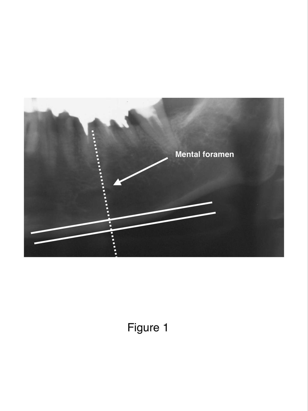
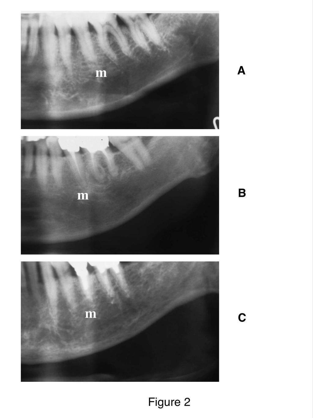
6 cortical width and shape (erosion), were considered indicators of alterations of the mandible and estimated by one oral radiologist (A.T.) with 18 years of clinical experience. The number of teeth remaining was also counted on dental panoramic radiographs excluding the third molar. The mandibular cortical width was measured bilaterally on the radiographs at the site of the mental foramen, as in our previous study 9 (Fig. 1). A line parallel to the long axis of the mandible and tangential to the inferior border of the mandible was drawn. A line perpendicular to this tangent intersecting the inferior border of the mental foramen was constructed, along which the mandibular cortical width was measured using calipers. The mean cortical width on both sides of the mandible was used in this study. The coefficient of variation due to positioning and operator error in the cortical width measure was less than 2%. The intra-observer variation in the cortical width measure was 0.1 mm, which was similar to the interobserver variation 9. Mandibular cortical shape on the dental panoramic radiograph was determined by observing the mandible distally from the mental foramen bilaterally and was categorized into one of three groups according to the method of Klemetti et al. 8 as follows (Fig. 2). Normal cortex: the endosteal margin of the cortex is even and sharp on both sides. Mildly to moderately eroded cortex: the endosteal margin shows semilunar defects (lacunar resorption) or appears to form endosteal cortical residues. Severely eroded cortex: the cortical layer forms heavy endosteal cortical residues and is clearly porous. The overall intra- and interobserver agreement was 92 and 82%, respectively 9. Data analysis 5
7 Pearson s correlation coefficient was calculated to evaluate the associations between cortical width and shape of the mandible, age, and vertebral BMD. Vertebral BMD was also compared among quartiles of cortical width of the mandible and among three categories of cortical shape of the mandible using analysis of covariance, adjusted for age, height, weight, time since menopause, duration of estrogen use (0 for no use), and history of hysterectomy (yes or no) or oophorectomy (yes or no). A logistic regression analysis adjusted for these confounding variables was used to calculate the odds ratios and 95% confidence interval (CI) of low vertebral BMD or osteoporosis according to the quartile of cortical width and cortical shape category. The data were analyzed using the Statistical Package for the Social Sciences (SPSS; version 8.0; SPSS, Chicago, IL, USA). P-values less than 0.05 were considered statistically significant. Results One hundred and eighty (40%) subjects had a normal BMD, 172 (38.2%) subjects had osteopenia (BMD T score of 2.5 to 1.0) at the lumbar vertebrae, and 98 (21.7%) subjects had vertebral osteoporosis (Table 1). Two hundred and fourteen (47.6%) subjects had an eroded cortex of the mandible. When the cortical width of the mandible was divided into quartiles, the ranges of cortical width in the uppermost, second, third, and lowermost quartiles were from 6.92 to 4.66, 4.65 to 4.08, 4.07 to 3.46, and 3.45 to 1.37 mm, respectively. Since a magnification factor was about 1.2, the actual ranges of cortical width in the uppermost, second, third, and lowermost quartiles were considered from 5.77 to 3.88, 3.87 to 3.40, 3.39 to 2.88, and 2.87 to 1.14 mm, respectively. 6
8 Age was significantly correlated with cortical width of the mandible (r = 0.32, P < 0.001) and cortical shape of the mandible (r = 0.48, P < 0.001). Vertebral BMD was also significantly correlated with cortical width of the mandible (r = 0.44, P < 0.001) and cortical shape of the mandible (r = 0.47, P < 0.001). The analysis of covariance adjusted for confounding variables revealed a significant association between the quartile of cortical width of the mandible and vertebral BMD (P < 0.001) (Table 2). Subjects belonging to the lowermost quartile of cortical width had a significantly lower vertebral BMD than those belonging to the uppermost (P < 0.001), second (P < 0.001), or third (P = 0.027) quartiles of cortical width. Furthermore, there was a significant association between the cortical shape of the mandible and vertebral BMD (P < 0.001). Subjects with a severely eroded cortex had a significantly lower vertebral BMD than those with a mildly to moderately eroded cortex (P = 0.001) or normal cortex (P < 0.001). Further adjustment for dentition status (number of teeth remaining) did not change these associations between the quartile of cortical width of the mandible and vertebral BMD or between cortical shape of the mandible and vertebral BMD. The odds ratios of low vertebral BMD associated with the second, third, and lowermost quartiles of cortical width of the mandible were 1.71 (95% CI, ), 2.30 (95% CI, ), and 5.43 (95% CI, ), respectively, compared to the uppermost quartile (Table 3). The odds ratios of vertebral osteoporosis associated with the second, third, and lowermost quartiles of cortical width of the mandible were 0.79 (95% CI, ), 2.95 (95% CI, ), and 6.04 (95% CI, ), respectively, compared to the uppermost quartile. The odds ratios of low vertebral BMD associated with mildly to moderately and severely eroded cortices compared to the normal cortex were 3.85 (95% CI, ) and 7.84 (95% CI, ), 7
9 respectively. The odds ratios of vertebral osteoporosis associated with mildly to moderately and severely eroded cortices compared to the normal cortex were 4.73 (95% CI, ) and (95% CI, ), respectively. The odds ratio of vertebral osteoporosis for 33 subjects with both the lowermost quartile of cortical width and a severely eroded cortex was (95% CI, ) compared to 160 subjects with a normally shaped cortex belonging to both the uppermost and second quartiles of cortical width. Discussion Postmenopausal women with a thinner cortical width had an increased risk for low vertebral BMD or osteoporosis in this study after adjusting for possible confounding variables. In particular, women belonging to the lowermost quartile of cortical width of the mandible had a significantly lower vertebral BMD than those belonging to the other quartiles of cortical width. This implies that a thin cortex determined visually by dentists based on their clinical experience can be used as an indicator for identifying postmenopausal women with a low BMD or osteoporosis. In fact, about 80% of postmenopausal women who were selected visually using a thin cortex alone, as determined by the dentists, had a low skeletal BMD in our recent study 20. Postmenopausal women with an eroded cortex also had an increased risk for low vertebral BMD or osteoporosis after adjusting for confounding variables. Especially, the odds ratio for vertebral osteoporosis was 14 times greater in subjects with a severely eroded cortex compared to those with a normal cortex. Furthermore, the odds ratio for vertebral osteoporosis was 23 times greater in subjects with both a thinner (lowermost 8
10 quartile) and severely eroded cortex compared to those with both a normal and thicker (uppermost and second quartiles) cortex. These women at risk for vertebral osteoporosis should be referred for BMD testing before osteoporotic vertebral fractures occur. Age was significantly correlated with the cortical width of the mandible (r = 0.32, P < 0.001) in this study. Vertebral BMD was also significantly correlated with cortical width of the mandible (r = 0.44, P < 0.001). Devlin and Horner recently reported that subject age was significantly correlated with cortical width of the mandible below the mental foramen (r = 0.26, P = 0.024) in 74 British postmenopausal women aged years (mean age, 62 years) 12. In their study, the cortical width of the mandible was significantly correlated with the BMD T-score recorded at the lumbar vertebrae (L1 L4) (r = 0.52, P < 0.01). Their results were in accord with our results. It is possible that the crude threshold of cortical width for identifying low vertebral BMD or osteoporosis in Japanese postmenopausal women cannot be used directly for British postmenopausal women because of the difference in the size of the mandible. However, the percentage based on the mean cortical width of the mandible in postmenopausal women with a normal vertebral BMD may be used in both Japanese and British women for identifying low vertebral BMD or osteoporosis because a similar correlation was observed between cortical width and vertebral BMD in both races. Devlin and Horner found that the mean cortical width (3.96 mm) in British postmenopausal women with a low BMD (T score of 1.0 or less) was about 84% of the mean cortical width (4.73 mm) in those with a normal BMD 12. In our study, the mean cortical width (3.75 mm) in Japanese postmenopausal women with a low BMD was also about 84% of the mean cortical width (4.45 mm) in those with a normal BMD. Two recent British studies suggest that a diagnostic threshold for cortical width of 3 mm (or less) is the most appropriate threshold for referral for bone 9
11 densitometry in postmenopausal and perimenopausal women 12,13. Considering the use of percent mean cortical width, a diagnostic threshold of 2.8 mm (or less), which was within the lowermost quartile in our study, is suggested as an appropriate threshold for BMD testing in Japanese women. Cortical thinning and erosion of the mandible were regarded as indicators of alterations of the mandible in this study. These changes reflect the increase in widened Haversian canals with resorption surfaces in the cortex 24. In the skeleton, trabecular bone is more sensitive to osteoporotic changes than cortical bone; however, it is not clear whether a similar change is observed in the trabecular part of the mandible. Jonasson et al. reported a significant correlation between the forearm BMD and the coarseness of trabeculation (r = 0.62, P < 0.001) in 80 women with a mean age of 47 years (range, years) 25. White et al. also reported that changes in radiographic trabecular structure, represented by the node-to-terminus strut, in the mandibular incisor region were predictive of hip fracture in 598 elderly women 26. Conversely, Choel et al. indicated that the infra-alveolar trabecular BMD measured using DXA was more sensitive to the local dental status than to the systemic status 27. Klemetti et al. failed to find a significant association between trabecular BMD determined using quantitative computed tomography and skeletal BMD (lumbar spine and femoral neck) determined using DXA in 74 totally or nearly edentulous menopausal women 28. Southard et al. also found no significant association between alveolar trabecular bone architecture in the premolar to molar region of the mandible, represented by fractal dimension, and skeletal BMD (lumbar spine, total hip, and total wrist) determined using DXA in 37 dentate healthy Caucasian women aged years 29. Since the trabecular bone architecture of the mandible is markedly different in the incisor, premolar, and molar regions 30 and is easily 10
12 influenced by dental infections, which contribute to both osteolytic and osteosclerotic changes, we regarded cortical width and the shape of the mandible as indicators of alterations of the mandible in this study. This study has limitations. Our subjects were not healthy volunteers from the community, but were patients who visited our clinic for BMD assessment. Therefore, the subjects are not representative of normal Japanese postmenopausal women. Iki et al. recently reported that in healthy Japanese women aged years, the prevalence of osteoporosis according to the WHO classification was 38.0% at the lumbar vertebrae 31. We cannot compare our subjects aged years with their subjects directly; however, the prevalence of osteoporosis at the lumbar vertebrae (21.7%) was lower than that in their study. The relative younger mean age (57.2 years) might contribute to the lower prevalence of vertebral osteoporosis. These results may limit the interpretation of our findings. Dentition status might influence alterations of the mandible due to the change in masticatory function 32. This might also contribute to vertebral osteoporosis via the change of nutritional intake 33. However, further adjustment for dentition status (number of teeth remaining) did not change the associations among the mandibular cortical parameters and vertebral BMD in this study. Since a number of subjects with tobacco use was small (6.6%) in 752 postmenopausal women who had not menstruated for at least 1 year and the history of their tobacco use varied (current use, former use or no precise history of use), postmenopausal women with tobacco use was excluded from this study. In conclusion, after adjusting for potentially confounding variables, the odds ratio for low vertebral BMD or osteoporosis associated with the lowermost quartile of cortical width of the mandible was 5.43 (95% CI, ) and 6.04 (95% CI, ), respectively, compared to the uppermost quartile. The odds ratio for low vertebral BMD 11
13 or osteoporosis associated with a severely eroded cortex was 7.84 (95% CI, ) and (95% CI, ), respectively, compared to the normal cortex. Postmenopausal women with a thinner or eroded cortex of the mandible detected on dental panoramic radiographs had an increased risk for low vertebral BMD or osteoporosis. Our results suggest that dentists should refer asymptomatic postmenopausal women with alterations of the mandible to medical professionals for bone densitometry. Acknowledgments This study was supported by a Grant-in-Aid for Scientific Research from the Japan Society for the Promotion of Science ( , ). References 1. Kado DM, Browner WS, Palermo L, Nevitt MC, Genant HK, Cummings SR. Vertebral fractures and mortality in older women: a prospective study. Study of Osteoporotic Fractures Research Group. Arch Intern Med 1999;159: Hasserius R, Karlsson MK, Jonsson B, Redlund-Johnell I, Johnell O. Long-term morbidity and mortality after a clinically diagnosed vertebral fracture in the elderly a 12- and 22-year follow-up of 257 patients. Calcif Tissue Int 2005;76: Ross PD, Fujiwara S, Huang C, Davis JW, Epstein RS, Wasnich RD, et al. Vertebral fracture prevalence in women in Hiroshima compared to Caucasians or Japanese in the US. Int J Epidemiol 1995;24: Fechtenbaum J, Cropet C, Kolta S, Verdoncq B, Orcel P, Roux C. Reporting of vertebral fractures on spine X-rays. Osteoporos Int 2005;16:
14 5. Delmas PD, van de Langerijt L, Watts NB, Eastell R, Genant H, Grauer A, et al. Underdiagnosis of vertebral fractures is a worldwide problem: the IMPACT study. J Bone Miner Res 2005;20: Shimano T, Suzuki Y, Sasaki T. Long-term trend of dental radiographic examination in Japan analysis on health insurance data. Dent Radiol 2002;42:9 21 (in Japanese). 7. American Dental Association Survey Center. The 2000 survey of dental practice characteristics of dentists and their patients. Chicago, IL: American Dental Association. 8. Klemetti E, Kolmakov S, Kroger H. Pantomography in assessment of the osteoporosis risk group. Scand J Dent Res 1994;102: Taguchi A, Suei Y, Ohtsuka M, Otani K, Tanimoto K, Ohtaki M. Usefulness of panoramic radiography in the diagnosis of postmenopausal osteoporosis in women. Width and morphology of inferior cortex of the mandible. Dentomaxillofac Radiol 1996;25: Bollen AM, Taguchi A, Hujoel PP, Hollender LG.. Case-control study on self-reported osteoporotic fractures and mandibular cortical bone. Oral Surg Oral Med Oral Pathol Oral Radiol Endod 2000;90: Persson RE, Hollender LG, Powell LV, MacEntee MI, Wyatt CC, Kiyak HA, et al. Assessment of periodontal conditions and systemic disease in older subjects. I. Focus on osteoporosis. J Clin Periodontol 2002;29: Devlin H, Horner K. Mandibular radiomorphometric indices in the diagnosis of reduced skeletal bone mineral density. Osteoporos Int 2002;13: Horner K, Devlin H, Harvey L. Detecting patients with low skeletal bone mass. J Dent 2002;30:
15 14. Drozdzowska B, Pluskiewicz W, Tarnawska B. Panoramic-based mandibular indices in relation to mandibular bone mineral density and skeletal status assessed by dual energy X-ray absorptiometry and quantitative ultrasound. Dentomaxillofac Radiol 2002;31: Nakamoto T, Taguchi A, Ohtsuka M, Suei Y, Fujita M, Tanimoto K, et al. Dental panoramic radiograph as a tool to detect postmenopausal women with low bone mineral density: untrained general dental practitioners diagnostic performance. Osteoporos Int 2003;14: Taguchi A, Sanada M, Krall E, Nakamoto T, Ohtsuka M, Suei Y, et al. Relationship between dental panoramic radiographic findings and biochemical markers of bone turnover. J Bone Miner Res 2003;18: Taguchi A, Suei Y, Sanada M, Ohtsuka M, Nakamoto T, Sumida H, et al. Validation of dental panoramic radiography measures for identifying postmenopausal women with spinal osteoporosis. AJR Am J Roentgenol 2004;183: White SC, Taguchi A, Kao D, Wu S, Service SK, Yoon D, et al. Clinical and panoramic predictors of femur bone mineral density. Osteoporos Int 2005;16: Halling A, Persson GR, Berglund J, Johansson O, Renvert S. Comparison between the Klemetti index and heel DXA BMD measurements in the diagnosis of reduced skeletal bone mineral density in the elderly. Osteoporos Int 2005;16: Lee K, Taguchi A, Ishii K, Suei Y, Fujita M, Nakamoto T, et al. Visual assessment of the mandibular cortex on panoramic radiographs to identify postmenopausal women with low bone mineral densities. Oral Surg Oral Med Oral Pathol Oral Radiol Endod 2005;100: Taguchi A, Tsuda M, Ohtsuka M, Kodama I, Sanada M, Nakamoto T, et al. Use of 14
16 dental panoramic radiographs in identifying younger postmenopausal women with osteoporosis. Osteoporos Int 2006, in press. 22. Genant HK, Wu CY, van Kuijk C, Nevitt MC. Vertebral fracture assessment using a semiquantitative technique. J Bone Miner Res 1993;8: World Health Organization (WHO) Studying Group. Assessment of fracture risk and its application to screening for postmenopausal osteoporosis. WHO Technical Report Series 1994;843: Von Wowern N. Microradiographic and histomorphometric indices of mandibles for diagnosis of osteopenia. Scand J Dent Res 1982;90: Jonasson G, Bankvall G, Kiliaridis S. Estimation of skeletal bone mineral density by means of the trabecular pattern of the alveolar bone, its interdental thickness, and the bone mass of the mandible. Oral Surg Oral Med Oral Pathol Oral Radiol Endod 2001;92: White SC, Atchison KA, Gornbein JA, Nattiv A, Paganini-Hill A, Service SK, et al. Change in mandibular trabecular pattern and hip fracture rate in elderly women. Dentomaxillofac Radiol 2005;34: Choel L, Duboeuf F, Bourgeois D, Briguet A, Lissac M. Trabecular alveolar bone in the human mandible: a dual-energy x-ray absorptiometry study. Oral Surg Oral Med Oral Pathol Oral Radiol Endod 2003;95: Klemetti E, Vainio P, Lassila V, Alhava E. Trabecular bone mineral density of mandible and alveolar height in postmenopausal women. Scand J Dent Res 1993;101: Southard TE, Southard KA, Lee A. Alveolar process fractal dimension and postcranial bone density. Oral Surg Oral Med Oral Pathol Oral Radiol Endod 15
17 2001;91: von Wowern N. Variations in structure within the trabecular bone of the mandible. Scand J Dent Res 1977;85: Iki M, Kagamimori S, Kagawa Y, Matsuzaki T, Yoneshima H, Marumo F. Bone mineral density of the spine, hip and distal forearm in representative samples of the Japanese female population: Japanese Population-Based Osteoporosis (JPOS) Study. Osteoporos Int 2001;12: Elovic RP, Hipp JA, Hayes WC. Maxillary molar extraction causes increased bone loss in the mandible of ovariectomized rats. J Bone Miner Res 1995;10: Sahyoun NR, Lin CL, Krall E. Nutritional status of the older adult is associated with dentition status. J Am Diet Assoc 2003;103:61 6. Figure legends Figure 1. A line parallel to the long axis of the mandible and tangential to the inferior border of the mandible was drawn. A line (dotted line) perpendicular to this tangent intersecting the inferior border of mental foramen was constructed, along which mandibular cortical width (the distance between the two parallel solid lines) was measured. Figure 2. Classification of mandibular inferior cortical shape observed distally from the mental foramen on dental panoramic radiographs: (A) normal cortex, (B) mildly to 16
18 moderately eroded cortex, and (C) severely eroded cortex. A small letter m shows the mental foramen. 17
19
20
21 Table 1. Characteristics of 450 study subjects Mean +/- SD or number of subjects (% subjects) Age (years) /- 8.1 Height (cm) /- 5.7 Weight (kg) /- 7.5 Time since menopause (years) 9.6 +/- 7.6 Mandibular cortical width (mm) 4.0 +/- 1.0 Number of teeth remaining /- 7.3 Mandibular cortical shape Normal cortex 236 (52.4%) Mildly to moderately eroded cortex 175 (38.9%) Severely eroded cortex 39 (8.7%) Diagnosis based on BMD of the lumbar vertebrae Normal 180 (40.0%) Osteopenia 172 (38.2%) Osteoporosis 98 (21.7%) BMD: bone mineral density
22 Table 2. Vertebral bone mineral density according to mandibular cortical width and cortical shape categories Cortical width* Number of subjects Vertebral bone mineral density (g/cm 2 ) P-value Cortical shape* Uppermost quartile / < Second quartile / Third quartile / Lowermost quartile / Normal / < Mildly to moderately eroded / Severely eroded / The results were shown as mean +/- SEM. * Means are significant different from each other after Bonferroni adjustment for multiple comparisons; cortical width (uppermost quartile vs. third quartile: P<0.001, uppermost quartile vs. lowermost quartile: P<0.001, second quartile vs. lowermost quartile: P<0.001, third quartile vs. lowermost quartile: P=0.027) and cortical shape (normal cortex vs. mild to moderately eroded cortex: P<0.001, normal cortex vs. severely eroded cortex: P<0.001, mildly to moderately eroded cortex vs. severely eroded cortex: P=0.001). From the analysis of covariance, adjusted for age, height, weight, time since menopause, duration of estrogen use (0 for none-user), and history of hysterectomy (yes or no) or oophorectomy (yes or no)
23 Table 3. Unadjusted and multivariate-adjusted odds ratio and 95% confidence interval of low vertebral bone mineral density (BMD) or osteoporosis according to mandibular cortical width and shape categories BMD T score of -1.0 or less (Low BMD) BMD T score of -2.5 or less (Osteoporosis) Cortical width Unadjusted Adjusted Unadjusted Adjusted Uppermost quartile Second quartile 1.95 ( ) 1.71 ( ) 1.01 ( ) 0.79 ( ) Third quartile 2.55 ( ) 2.30 ( ) 3.01 ( ) 2.95 ( ) Lowermost quartile 7.86 ( ) 5.43 ( ) 8.70 ( ) 6.04 ( ) Cortical shape Normal Mildly to moderately eroded 4.90 ( ) 3.85 ( ) 6.22 ( ) 4.73 ( ) Severely eroded ( ) 7.84 ( ) ( ) ( ) The multivariate model includes age, height, weight, time since menopause, duration of estrogen use (0 for none-user), and history of hysterectomy (yes or no) or oophorectomy (yes or no).
Diagnostic efficacy of alveolar bone loss of the mandible for identifying
 Diagnostic efficacy of alveolar bone loss of the mandible for identifying postmenopausal women with femoral osteoporosis Kodo Ishii 1, Akira Taguchi 2, Takashi Nakamoto 2, Masahiko Ohtsuka 1, Pipop Sutthiprapaporn
Orthopantomogram as an effective tool for the diagnosis of osteoporosis-a study
 Original Research Article Comparative Study of Mandibular Cortical Index in Orthopantomogram and Bone Mineral Density of Lumbar Vertebrae in Dual Energy X-Ray Absorptiometry in Postmenopausal Females in
Evaluation of Correlation between Width and Morphology of Mandibular Inferior Cortex in Digital Panoramic Radiography and Postmenopausal Osteoporosis
 Iranian Red Crescent Medical Journal ORIGINAL ARTICLE Evaluation of Correlation between Width and Morphology of Mandibular Inferior Cortex in Digital Panoramic Radiography and Postmenopausal Osteoporosis
Panoramic-Based Mandibular Indices and Bone Mineral Density of Femoral Neck and Lumbar Vertebrae in Women
 Original Article Panoramic-Based Mandibular Indices and Bone Mineral Density of Femoral Neck and Lumbar Vertebrae in Women S. Marandi 1, A. Bagherpour 2, M. Imanimoghaddam 3, MR. Hatef 4, AR. Haghighi
The Comparison of Mandibular Radiomorphometric Indices in Panoramic Radiography between Patients with Chronic Periodontitis and Healthy Individuals
 ORIGINAL RESEARCH The Comparison of Mandibular Radiomorphometric 10.5005/jp-journals-10024-1563 Indices in Panoramic Radiography The Comparison of Mandibular Radiomorphometric Indices in Panoramic Radiography
Panoramic radiographs for identifying individuals with undetected osteoporosis
 Japanese Dental Science Review (2009) 45, 109 120 available at www.sciencedirect.com journal homepage: www.elsevier.com/locate/jdsr REVIEW ARTICLE Panoramic radiographs for identifying individuals with
AUTHOR QUERIES DATE 5/22/2013 JOB NAME MAJ ARTICLE THIS QUERY FORM MUST BE RETURNED WITH ALL PROOFS FOR CORRECTIONS
 NUMBER 1 OF 1 AUTHOR QUERIES DATE 5/22/2013 JOB NAME MAJ ARTICLE 201976 QUERIES FOR AUTHORS Mansour et al THIS QUERY FORM MUST BE RETURNED WITH ALL PROOFS FOR CORRECTIONS AU1) Please provide manufacturer
Comparison of Accuracy of Prediction of Osteoporosis in Digital Panoramic Radiography with DEXA Scan in Postmenopausal Women: A Pilot Study
 JIAOMR Comparison of Accuracy of Prediction of Osteoporosis in Digital Panoramic Radiography with Dexa Scan ORIGINAL RESEARCH Comparison of Accuracy of Prediction of Osteoporosis in Digital Panoramic Radiography
The Open Dentistry Journal
 Send Orders for Reprints to reprints@benthamscience.ae 350 The Open Dentistry Journal, 2017, 11, (Suppl-1, M3) 350-359 The Open Dentistry Journal Content list available at: www.benthamopen.com/todentj/
Computer-aided measurement of mandibular cortical width on dental panoramic radiographs for identifying osteoporosis
 Journal of Investigative and Clinical Dentistry (2012), 3, 36 44 ORIGINAL ARTICLE Oral Radiology Computer-aided measurement of mandibular cortical width on dental panoramic radiographs for identifying
Mandibular Cortex Correlates to Alveolar Bone Density in Indonesian Women Aged 40 to 75 Years
 in Indonesian Women Aged 40 to 75 Years Lilies Dwi Sulistyani 1 *, Menik Priaminiarti 2, Elza Ibrahim Auerkari 3, Lindawati Soetanto Kusdhany 4, Benny Sjariefsjah Latief 1 1. Department of Oral and Maxillofacial
Selecting regions of interest on intra oral radiographs. for the prediction of bone mineral density
 Selecting regions of interest on intra oral radiographs for the prediction of bone mineral density W.G.M. Geraets a, J.G.C. Verheij a, P.F. van der Stelt a, K. Horner b, C. Lindh c, K. Nicopoulou-Karayianni
Influence of Age and Gender on Mandible Among South Indian Population
 OnLine Journal of Biological Sciences Original Research Paper Influence of Age and Gender on Mandible Among South Indian Population Kathirvelu Dhandapani and Anburajan Mariamichael Department of Biomedical
MANDIBULAR CORTICALLY THICKNESS AS INDICATOR IN OSTEOPOROSIS SCREENING
 MANDIBULAR CORTICALLY THICKNESS AS INDICATOR IN OSTEOPOROSIS SCREENING Alexandru Georgescu 1, Horia Traian Dumitriu 2 1 Assist. Prof., Faculty of Dental Medicine, Department of Periodontology, University
Mandibular Bone Mineral Density to predict Skeletal Osteoporosis: A Literature Review
 Sweta Pisulkar et al REVIEW ARTICLE 10.5005/jp-journals-10053-0021 Mandibular Bone Mineral Density to predict Skeletal Osteoporosis: A Literature Review 1 Sweta Pisulkar, 2 Gajanan Pisulkar, 3 Ashok Pakhan,
Open Access Evaluation of Radiomorphometric Indices in Panoramic Radiograph A Screening Tool
 Send Orders for Reprints to reprints@benthamscience.ae The Open Dentistry Journal, 2015, 9, (Suppl 2: M9) 303-310 303 Open Access Evaluation of Radiomorphometric Indices in Panoramic Radiograph A Screening
Application of telemedicine to assess mandibular cortical width on panoramic images of dental patients in the Lao People s Democratic Republic
 DOI 10.1007/s11282-015-0198-4 ORIGINAL ARTICLE Application of telemedicine to assess mandibular cortical width on panoramic images of dental patients in the Lao People s Democratic Republic Johnny Sisounthone
Osteoporosis is a silent skeletal disease characterized. Can Dental Students Be Taught to Use Dental Radiographs for Osteoporosis Screening?
 Can Dental Students Be Taught to Use Dental Radiographs for Osteoporosis Screening? Werner Harumiti Shintaku, D.D.S., M.S.; Reyes Enciso, Ph.D., M.S.; John Stansill Covington, D.D.S., M.S., B.S.; Cesar
Panoramic Radiography for Screening Postmenopausal Osteoporosis in India: A Pilot Study
 Panoramic Radiography for Screening Postmenopausal Osteoporosis in India: A Pilot Study Pankaj R Bodade 1, Rajendra N Mody 2 1 MDS, BDS. Associate Professor, Department of Oral Medicine and Radiology,
Computer-aided system for morphometric mandibular index computation (Using dental panoramic radiographs)
") Journal section: Gerodontology Publication Types: Review doi:10.4317/medoral.17637 http://dx.doi.org/doi:10.4317/medoral.17637 Computer-aided system for morphometric mandibular index computation (Using
Journal section: Gerodontology Publication Types: Review doi:10.4317/medoral.17637 http://dx.doi.org/doi:10.4317/medoral.17637 Computer-aided system for morphometric mandibular index computation (Using
Relationship Between Body Mass Index and Local Quality of Mandibular Bone Structure in Elderly Individuals
 Journal of Gerontology: MEDICAL SCIENCES 2002, Vol. 57A, No. 9, M588 M593 Copyright 2002 by The Gerontological Society of America Relationship Between Body Mass Index and Local Quality of Mandibular Bone
Interpreting DEXA Scan and. the New Fracture Risk. Assessment. Algorithm
 Interpreting DEXA Scan and the New Fracture Risk Assessment Algorithm Prof. Samir Elbadawy *Osteoporosis affect 30%-40% of women in western countries and almost 15% of men after the age of 50 years. Osteoporosis
Oral signs and salivary parameters as indicators of possible osteoporosis and osteopenia in postmenopausal women - A study of 45 subjects
 Braz J Oral Sci. January/March 2008 - Vol. 7 - Number 24 Siva Reddy 1 Ramalingam Karthikeyan 2 Herald Justin Sherlin 2 Natesan Anuja 2 Ramani Pratibha 3 Premkumar Priya 3 Thiruvengadam Chandrasekar 4 1
CIC Edizioni Internazionali. Detecting low bone mineral density from dental radiographs: a mini-review. Mini-review
 Detecting low bone mineral density from dental radiographs: a mini-review James Graham Centre for Imaging Science, Institute of Population Health, Faculty of Medicine and Human Sciences, The University
Correlation between Mandibular Radiomorphometric Parameters and Gonial Angle size in Iranian Adults
 Correlation between Mandibular Radiomorphometric Parameters and Gonial Angle size in Iranian Adults Original Article Sanam Mirbeigi 1, Leila Khojastepour 2, Fatameh Ezoddini 3, Yasaman Sabaghzadegan 4
Osteoporosis International. Original Article. Bone Mineral Density and Vertebral Fractures in Men
 Osteoporos Int (1999) 10:265 270 ß 1999 International Osteoporosis Foundation and National Osteoporosis Foundation Osteoporosis International Original Article Bone Mineral Density and Vertebral Fractures
The combination of a histogram-based clustering algorithm and support vector machine for the diagnosis of osteoporosis
 Imaging Science in Dentistry 2013; 43: 153-61 http://dx.doi.org/10.5624/isd.2013.43.3.153 The combination of a histogram-based clustering algorithm and support vector machine for the diagnosis of osteoporosis
COMPUTER TOMOGRAPHY ASSESSMENT OF ALVEOLAR RIDGE MODIFICATIONS IN ELDERLY PATIENTS
 COMPUTER TOMOGRAPHY ASSESSMENT OF ALVEOLAR RIDGE MODIFICATIONS IN ELDERLY PATIENTS Adina Mosnegu, Alexandru Nem Department of Geriatric Stomatology, Dental Medicine Faculty, University of Medicine and
Association between periodontal disease and bone mineral density in postmenopausal women: A cross sectional study
 Journal section: Periodontology Publication Types: Research doi:10.4317/medoral.16.e440 http://dx.doi.org/doi:10.4317/medoral.16.e440 Association between periodontal disease and bone mineral density in
Bone xxx (2011) xxx xxx. Contents lists available at ScienceDirect. Bone. journal homepage:
 xxx xxx. Contents lists available at ScienceDirect. Bone. journal homepage:") BON-09329; No. of pages: 7; 4C: Bone xxx (2011) xxx xxx Contents lists available at ScienceDirect Bone journal homepage: www.elsevier.com/locate/bone A prospective study of mandibular trabecular bone to
BON-09329; No. of pages: 7; 4C: Bone xxx (2011) xxx xxx Contents lists available at ScienceDirect Bone journal homepage: www.elsevier.com/locate/bone A prospective study of mandibular trabecular bone to
OSTEOPOROSIS AND PERIODONTAL DISEASE A REVIEW
 OSTEOPOROSIS AND PERIODONTAL DISEASE A REVIEW Dr. V. GOPINATH 1, Dr.M.N.PRABHU 2. Dr. HEMA SURYAWANSHI 3, 1. Professor, Head of the Department, Department of Periodontology. Chhattisgarh Dental College
Clinical risk factor assessment had better discriminative ability than bone mineral density in identifying subjects with vertebral fracture
 Osteoporos Int (2011) 22:667 674 DOI 10.1007/s00198-010-1260-z ORIGINAL ARTICLE Clinical risk factor assessment had better discriminative ability than bone mineral density in identifying subjects with
Skeletal Manifestations
 Skeletal Manifestations of Metabolic Bone Disease Mishaela R. Rubin, MD February 21, 2008 The Three Ages of Women Gustav Klimt 1905 1 Lecture Outline Osteoporosis epidemiology diagnosis secondary causes
Body Mass Index as Predictor of Bone Mineral Density in Postmenopausal Women in India
 International Journal of Public Health Science (IJPHS) Vol.3, No.4, December 2014, pp. 276 ~ 280 ISSN: 2252-8806 276 Body Mass Index as Predictor of Bone Mineral Density in Postmenopausal Women in India
Comparison of Bone Density of Distal Radius With Hip and Spine Using DXA
 ORIGINAL ARTICLE Comparison of Bone Density of Distal Radius With Hip and Spine Using DXA Leila Amiri 1, Azita Kheiltash 2, Shafieh Movassaghi 1, Maryam Moghaddassi 1, and Leila Seddigh 2 1 Rheumatology
Reporting of Spinal Fractures
 Reporting of Spinal Fractures Poster No.: P-0026 Congress: ESSR 2014 Type: Authors: Keywords: DOI: Scientific Poster H. Al-Chalabi, C. Groves; Bradford/UK Osteoporosis, Structured reporting, Observer performance,
DXA When to order? How to interpret? Dr Nikhil Tandon Department of Endocrinology and Metabolism All India Institute of Medical Sciences New Delhi
 DXA When to order? How to interpret? Dr Nikhil Tandon Department of Endocrinology and Metabolism All India Institute of Medical Sciences New Delhi Clinical Utility of Bone Densitometry Diagnosis (DXA)
Panoramic Radiographic Detection of Systemic Disease By Dr. Allan G. Farman in collaboration with Dr. C. J. Nortjé
 Volume 4, Issue 3 US $6.00 Editor: Allan G. Farman, BDS, PhD (odont.), DSc (odont.), Diplomate of the American Board of Oral and Maxillofacial Radiology, Professor of Radiology and Imaging Sciences, Department
Analysis of Clinical Features of Hip Fracture Patients with or without Prior Osteoporotic Spinal Compression Fractures
 J Bone Metab 2013;20:11-15 http://dx.doi.org/10.11005/jbm.2013.20.1.11 pissn 2287-6375 eissn 2287-7029 Original Article Analysis of Clinical Features of Hip Fracture Patients with or without Prior Osteoporotic
Dr Tuan V NGUYEN. Mapping Translational Research into Individualised Prognosis of Fracture Risk
 Dr Tuan V NGUYEN Bone and Mineral Research Program, Garvan Institute of Medical Research, Sydney NSW Mapping Translational Research into Individualised Prognosis of Fracture Risk From the age of 60, one
Diagnosis of Vertebral Fractures by Vertebral Fracture Assessment
 Journal of Clinical Densitometry, vol. 9, no. 1, 66 71, 2006 Ó Copyright 2006 by The International Society for Clinical Densitometry 1094-6950/06/9:66 71/$32.00 DOI: 10.1016/j.jocd.2005.11.002 Original
Mandibular Inferior Cortex Erosion on Dental Panoramic Radiograph as a Sign of Low Bone Mineral Density in Postmenopausal Women
 THIEME Original Article Mandibular Inferior Cortex Erosion on Dental Panoramic Radiograph as a Sign of Low Bone Mineral Density in Postmenopausal Women Erosão do córtex mandibular inferior em radiografias
Bone Mineral Density and Its Associated Factors in Naresuan University Staff
 Naresuan University Journal 2005; 13(3): 13-18 13 Bone Mineral Density and Its Associated Factors in Naresuan University Staff Supawitoo Sookpeng *, Patsuree Cheebsumon, Malinee Dhanarun, Thanyavee Pengpan
Bone Mass Measurement BONE MASS MEASUREMENT HS-042. Policy Number: HS-042. Original Effective Date: 8/25/2008
 Easy Choice Health Plan, Inc. Harmony Health Plan of Illinois, Inc. Missouri Care, Inc. Ohana Health Plan, a plan offered by WellCare Health Insurance of Arizona, Inc. WellCare Health Insurance of Illinois,
LUMBAR IS IT IMPORTANT? S. Tantawy,, M.D.
 بسم االله الرحمن الرحيم DEXA LATERAL LUMBAR IS IT IMPORTANT? By S. Tantawy,, M.D. Osteopenia,, bone mineral deficiency in the absence of fracture, is an indicator of the bone structural integrity and compared
Are glucocorticoid-induced osteoporosis recommendations sufficient to determine antiosteoporotic treatment for patients with rheumatoid arthritis?
 ORIGINAL ARTICLE Korean J Intern Med 2014;29:509-515 Are glucocorticoid-induced osteoporosis recommendations sufficient to determine antiosteoporotic treatment for patients with rheumatoid arthritis? Joo-Hyun
Building Bone Density-Research Issues
 Building Bone Density-Research Issues Helping to Regain Bone Density QUESTION 1 What are the symptoms of Osteoporosis? Who is at risk? Symptoms Bone Fractures Osteoporosis 1,500,000 fractures a year Kyphosis
Dual-energy Vertebral Assessment
 Dual-energy Vertebral Assessment gehealthcare.com Dual-energy Vertebral Assessment More than 40% of women with normal or osteopenic BMD had a moderate or severe vertebral deformation seen with DVA. Patrick
Prevalence of Osteoporosis p. 262 Consequences of Osteoporosis p. 263 Risk Factors for Osteoporosis p. 264 Attainment of Peak Bone Density p.
 Dedication Preface Acknowledgments Continuing Education An Introduction to Conventions in Densitometry p. 1 Densitometry as a Quantitative Measurement Technique p. 2 Accuracy and Precision p. 2 The Skeleton
Dental Panoramic Radiographic Indices as a Predictor of Osteoporosis in Postmenopausal Saudi Women
 J Bone Metab 2018;25(3):165-173 https://doi.org/10.11005/jbm.2018.25.3.165 pissn 2287-6375 eissn 2287-7029 Original Article Dental Panoramic Radiographic Indices as a Predictor of Osteoporosis in Postmenopausal
International Journal of Health Sciences and Research ISSN:
 International Journal of Health Sciences and Research www.ijhsr.org ISSN: 2249-9571 Original Research Article Osteoporosis- Do We Need to Think Beyond Bone Mineral Density? Dr Preeti Soni 1, Dr Shipra
Prediction of osteoporosis with dental radiographs and age
 (2009) 38, 431 437 2009 The British Institute of Radiology http://dmfr.birjournals.org RESEARCH Prediction of osteoporosis with dental radiographs and age JGC Verheij*,1, WGM Geraets 1, PF van der Stelt
Advanced DXA Using TBS insight
 Advanced DXA Using TBS insight A New Bone Structure Assessment Technique Enhances Identification of Fracture Risk Introduction The World Health Organization defines osteoporosis as asilent disease characterised
Bone Densitometry Radiation dose: what you need to know
 Bone Densitometry Radiation dose: what you need to know John Damilakis, PhD Associate Professor and Chairman University of Crete, Iraklion, Crete, GREECE Estimation of bone status using X-rays Assessment
DEVELOPMENT OF A RISK SCORING SYSTEM TO PREDICT A RISK OF OSTEOPOROTIC VERTEBRAL FRACTURES IN POSTMENOPAUSAL WOMEN
 October 2-4, Liverpool, UK EURO SPINE 2013 DEVELOPMENT OF A RISK SCORING SYSTEM TO PREDICT A RISK OF OSTEOPOROTIC VERTEBRAL FRACTURES IN POSTMENOPAUSAL WOMEN D. Colangelo, L. A. Nasto, M. Mormando, E.
ASJ. How Many High Risk Korean Patients with Osteopenia Could Overlook Treatment Eligibility? Asian Spine Journal. Introduction
 Asian Spine Journal Asian Spine Clinical Journal Study Asian Spine J 2014;8(6):729-734 High http://dx.doi.org/10.4184/asj.2014.8.6.729 risk patients with osteopenia How Many High Risk Korean Patients with
Coincidence of calcified carotid atheromatous plaque, osteoporosis, and periodontal bone loss in dental panoramic radiographs
 Imaging Science in Dentistry 2013; 43: 235-43 http://dx.doi.org/10.5624/isd.2013.43.4.235 Coincidence of calcified carotid atheromatous plaque, osteoporosis, and periodontal bone loss in dental panoramic
Prevalence of vertebral fractures on chest radiographs of elderly African American and Caucasian women
 Osteoporos Int (2011) 22:2365 2371 DOI 10.1007/s00198-010-1452-6 ORIGINAL ARTICLE Prevalence of vertebral fractures on chest radiographs of elderly African American and Caucasian women D. Lansdown & B.
An audit of bone densitometry practice with reference to ISCD, IOF and NOF guidelines
 Osteoporos Int (2006) 17: 1111 1115 DOI 10.1007/s00198-006-0101-6 SHORT COMMUNICATION An audit of bone densitometry practice with reference to ISCD, IOF and NOF guidelines R. Baddoura. H. Awada. J. Okais.
Tooth loss and osteoporosis: the osteodent study
 J Clin Periodontol 2009; 36: 190 197 doi: 10.1111/j.1600-051X.2008.01365.x Tooth loss and osteoporosis: the osteodent study Nicopoulou-Karayianni K, Tzoutzoukos P, Mitsea A, Karayiannis A, Tsiklakis K,
Mædica - a Journal of Clinical Medicine. Interobserver variability of the diagnosis of apical periodontitis on panoramic radiography assessment
 Mædica - a Journal of Clinical Medicine ORIGIN RIGINAL PAPERS Interobserver variability of the diagnosis of apical periodontitis on panoramic radiography assessment Mihaela HEDESIU, MD a ; Andrea SERBANESCU,
Prevalence of Osteoporosis in the Korean Population Based on Korea National Health and Nutrition Examination Survey (KNHANES),
,") Original Article http://dx.doi.org/10.3349/ymj.2014.55.4.1049 pissn: 0513-5796, eissn: 1976-2437 Yonsei Med J 55(4):1049-1057, 2014 Prevalence of Osteoporosis in the n Population Based on National Health
Original Article http://dx.doi.org/10.3349/ymj.2014.55.4.1049 pissn: 0513-5796, eissn: 1976-2437 Yonsei Med J 55(4):1049-1057, 2014 Prevalence of Osteoporosis in the n Population Based on National Health
Challenging the Current Osteoporosis Guidelines. Carolyn J. Crandall, MD, MS Professor of Medicine David Geffen School of Medicine at UCLA
 Challenging the Current Osteoporosis Guidelines Carolyn J. Crandall, MD, MS Professor of Medicine David Geffen School of Medicine at UCLA Whom to screen Which test How to diagnose Whom to treat Benefits
Comparative study of axial and femoral bone mineral density and parameters of mandibular bone quality in patients receiving dental implants
 Osteoporos Int (2007) 18: 703 709 DOI 10.1007/s00198-006-0295-7 ERRATUM M. A. L. Amorim. L. Takayama. V. Jorgetti. R. M. R. Pereira Comparative study of axial and femoral bone mineral density and parameters
Does Salivary Calcium and Phosphate Concentrations Adequately Reflect Bone Mineral Density in Patients with Chronic Periodontitis?
 Global Journal of Health Science; Vol. 8, No. 10; 2016 ISSN 1916-9736 E-ISSN 1916-9744 Published by Canadian Center of Science and Education Does Salivary Calcium and Phosphate Concentrations Adequately
Effect of Precision Error on T-scores and the Diagnostic Classification of Bone Status
 Journal of Clinical Densitometry, vol. 10, no. 3, 239e243, 2007 Ó Copyright 2007 by The International Society for Clinical Densitometry 1094-6950/07/10:239e243/$32.00 DOI: 10.1016/j.jocd.2007.03.002 Original
Objectives. Discuss bone health and the consequences of osteoporosis on patients medical and disability status.
 Objectives Discuss bone health and the consequences of osteoporosis on patients medical and disability status. Discuss the pathophysiology of osteoporosis and major risk factors. Assess the major diagnostic
Efficacy of risedronate in men with primary and secondary osteoporosis: results of a 1-year study
 Rheumatol Int (2006) 26: 427 431 DOI 10.1007/s00296-005-0004-4 ORIGINAL ARTICLE J. D. Ringe Æ H. Faber Æ P. Farahmand Æ A. Dorst Efficacy of risedronate in men with primary and secondary osteoporosis:
Use of DXA / Bone Density in the Care of Your Patients. Brenda Lee Holbert, M.D. Associate Professor Senior Staff Radiologist
 Use of DXA / Bone Density in the Care of Your Patients Brenda Lee Holbert, M.D. Associate Professor Senior Staff Radiologist Important Websites Resources for Clinicians and Patients www.nof.org www.iofbonehealth.org
EVALUATION OF THE CORTICAL BONE THICKNESS IN LUMBAR VERTEBRA USING CT AND RP EXPERIMENTAL TECHNIQUES
 15 th International Conference on Experimental Mechanics PAPER REF: 2711 EVALUATION OF THE CORTICAL BONE THICKNESS IN LUMBAR VERTEBRA USING CT AND RP EXPERIMENTAL TECHNIQUES Elza M.M. Fonseca 1(*), Luisa
The effects of image compression on quantitative measurements of digital panoramic radiographs
 Journal section: Clinical and Experimental Dentistry Publication Types: Research doi:10.4317/medoral.17912 http://dx.doi.org/doi:10.4317/medoral.17912 The effects of image compression on quantitative measurements
Trabecular bone analysis with tomosynthesis in diabetic patients: comparison with CT-based finite-element method
 Trabecular bone analysis with tomosynthesis in diabetic patients: comparison with CT-based finite-element method Poster No.: C-1789 Congress: ECR 2015 Type: Scientific Exhibit Authors: M. Fujii, T. Aoki,
QCT and CT applications in Osteoporosis Imaging
 Q appli in Osteoporosis Imaging Thomas M. Link, MD, PhD Department of Radiology Biomedical Imaging University of California, San Francisco Goals 1. To identify advantages disadvantages of Q compared to
The Influence of Gender on the Cortical Width of the Lower Border of the Mandible and the Mandibular Cortical Index
 Clinical Science Original Article Acta Medica Academica 2018;47(2):149-154 DOI: 10.5644/ama2006-124.226 The Influence of Gender on the Cortical Width of the Lower Border of the Mandible and the Mandibular
Assessment of Alveolar Bone Mineral Density as a Predictor of Lumbar Fracture Probability
 Adv Ther (2013) 30:487 502 DOI 10.1007/s25-013-0028-1 ORIGINAL RESEARCH Assessment of Alveolar Bone Mineral Density as a Predictor of Lumbar Fracture Probability Yoshitomo Takaishi Seizaburo Arita Mitsugi
Clinician s Guide to Prevention and Treatment of Osteoporosis
 Clinician s Guide to Prevention and Treatment of Osteoporosis Published: 15 August 2014 committee of the National Osteoporosis Foundation (NOF) Tipawan khiemsontia,md outline Basic pathophysiology screening
Bone Density Measurement in Women
 Bone Density Measurement in Women Revised 2005 Scope This guideline defines the medical necessity of bone mineral density (BMD) measurement using dualenergy x-ray absorptiometry (DXA or DEXA), and applies
Factors Associated with Treatment Initiation after Osteoporosis Screening
 American Journal of Epidemiology Copyright 2004 by the Johns Hopkins Bloomberg School of Public Health All rights reserved Vol. 160, No. 5 Printed in U.S.A. DOI: 10.1093/aje/kwh245 Factors Associated with
Risk Factors for Postmenopausal Fractures What We Have Learned from The OSTPRE - study
 Risk Factors for Postmenopausal Fractures What We Have Learned from The OSTPRE - study Heikki Kröger Kuopio Musculoskeletal Research Unit, University of Eastern Finland (UEF) Dept. of Orthopaedics, Traumatology
Fragile Bones and how to recognise them. Rod Hughes Consultant physician and rheumatologist St Peter s hospital Chertsey
 Fragile Bones and how to recognise them Rod Hughes Consultant physician and rheumatologist St Peter s hospital Chertsey Osteoporosis Osteoporosis is a skeletal disorder characterised by compromised bone
OSTEOPOROSIS MANAGEMENT AND INVESTIGATION. David A. Hanley, MD, FRCPC
 OSTEOPOROSIS MANAGEMENT AND INVESTIGATION David A. Hanley, MD, FRCPC There is a huge care gap in the management of osteoporosis in this country. As yet unpublished findings from the Canadian Multicentre
Concordance of a Self Assessment Tool and Measurement of Bone Mineral Density in Identifying the Risk of Osteoporosis in Elderly Taiwanese Women
 TZU CHI MED J September 2008 Vol 20 No 3 available at http://ajws.elsevier.com/tcmj Tzu Chi Medical Journal Original Article Concordance of a Self Assessment Tool and Measurement of Bone Mineral Density
Fracture Prediction From Bone Mineral Density in Japanese Men and Women ABSTRACT
 JOURNAL OF BONE AND MINERAL RESEARCH Volume 18, Number 8, 2003 2003 American Society for Bone and Mineral Research Fracture Prediction From Bone Mineral Density in Japanese Men and Women SAEKO FUJIWARA,
Annotations Part III Vertebral Fracture Initiative. International Osteoporosis Foundation March 2011
 Annotations Part III Vertebral Fracture Initiative International Osteoporosis Foundation March 2011 Slide 1-3 Topics to be covered: What is vertebral fracture assessment? How does VFA compare to standard
2013 ISCD Official Positions Adult
 2013 ISCD Official Positions Adult These are the Official Positions of the ISCD as updated in 2013. The Official Positions that are new or revised since 2007 are in bold type. Indications for Bone Mineral
Clinical Study Comparison of QCT and DXA: Osteoporosis Detection Rates in Postmenopausal Women
 International Endocrinology Volume 3, Article ID 895474, 5 pages http://dx.doi.org/.55/3/895474 Clinical Study Comparison of QCT and DXA: Osteoporosis Detection Rates in Postmenopausal Women Na Li, Xin-min
Radiological Morphometric Analysis of the Mandibular Bone Structure after Ovariectomy in Mature Cynomolgus Monkeys
 Oral Science International, May 2005, p.54 63 Copyright 2005, Japanese Stomatology Society. All Rights Reserved. Radiological Morphometric Analysis of the Mandibular Bone Structure after Ovariectomy in
Association of testosterone and bone mineral density with tooth loss in men with chronic periodontitis
 333 Journal of Oral Science, Vol. 53, No. 3, 333-339, 2011 Original Association of testosterone and bone mineral density with tooth loss in men with chronic periodontitis Balendra P. Singh 1), Annu Makker
Visibility of Maxillary and Mandibular Anatomical Landmarks in Digital Panoramic Radiographs: A Retrospective Study
 Visibility of Maxillary and Mandibular Anatomical Landmarks in Digital Panoramic Radiographs: A Retrospective Study Srisha Basappa, Smitha JD, Nishath Khanum*, Santosh Kanwar, Mahesh MS and Archana Patil
Beyond BMD: Bone Quality and Bone Strength
 Beyond BMD: Bone Quality and Bone Strength Consultant / advisor: Amgen, Eli Lilly, Merck Disclosures Mary L. Bouxsein, PhD Beth Israel Deaconess Medical Center Harvard Medical School, Boston, MA mbouxsei@bidmc.harvard.edu
Based on review of available data, the Company may consider the use of denosumab (Prolia) for the
 for the") Applies to all products administered or underwritten by Blue Cross and Blue Shield of Louisiana and its subsidiary, HMO Louisiana, Inc.(collectively referred to as the Company ), unless otherwise provided
Applies to all products administered or underwritten by Blue Cross and Blue Shield of Louisiana and its subsidiary, HMO Louisiana, Inc.(collectively referred to as the Company ), unless otherwise provided
denosumab (Prolia ) Policy # Original Effective Date: 07/21/2011 Current Effective Date: 04/19/2017
 Policy # Original Effective Date: 07/21/2011 Current Effective Date: 04/19/2017") Applies to all products administered or underwritten by Blue Cross and Blue Shield of Louisiana and its subsidiary, HMO Louisiana, Inc.(collectively referred to as the Company ), unless otherwise provided
Applies to all products administered or underwritten by Blue Cross and Blue Shield of Louisiana and its subsidiary, HMO Louisiana, Inc.(collectively referred to as the Company ), unless otherwise provided
Screening points for a peripheral densitometer of the calcaneum for the diagnosis of osteoporosis
 23 Ivorra Cortés J, Román-Ivorra JA, Alegre Sancho JJ, Beltrán Catalán E, Chalmeta Verdejo I, Fernández-Llanio Comella N, Muñoz Gil S Servicio de Reumatología - Hospital Universitario Dr. Peset - Valencia
Oral Alendronate Vs. Three-Monthly Iv Ibandronate In The Treatment Of Postmenopausal Osteoporosis
 Oral Alendronate Vs. Three-Monthly Iv Ibandronate In The Treatment Of Postmenopausal Osteoporosis Miriam Silverberg A. Study Purpose and Rationale More than 70% of fractures in people after the age of
VERTEBRAL FRACTURES ARE THE
 ORIGINAL CONTRIBUTION Long-term Risk of Incident Vertebral Fractures Jane A. Cauley, DrPH Marc C. Hochberg, MD, MPH Li-Yung Lui, MA, MS Lisa Palermo, MS Kristine E. Ensrud, MD, MPH Teresa A. Hillier, MD,
Correlation between Thyroid Function and Bone Mineral Density in Elderly People
 IBBJ Spring 2016, Vol 2, No 2 Original Article Correlation between Thyroid Function and Bone Mineral Density in Elderly People Ali Mirzapour 1, Fatemeh Shahnavazi 2, Ahmad Karkhah 3, Seyed Reza Hosseini
Relationship between Family History of Osteoporotic Fracture and Femur Geometry
 Iranian J Publ Health, 2007, Iranian A supplementary J Publ Health, issue 2007, on Osteoporosis, A supplementary pp.70-74 issue on Osteoporosis, pp.70-74 Relationship between Family History of Osteoporotic
Module 5 - Speaking of Bones Osteoporosis For Health Professionals: Fracture Risk Assessment. William D. Leslie, MD MSc FRCPC
 Module 5 - Speaking of Bones Osteoporosis For Health Professionals: Fracture Risk Assessment William D. Leslie, MD MSc FRCPC Case #1 Age 53: 3 years post-menopause Has always enjoyed excellent health with
Omnisense: At Least As Good As DXA
 Omnisense: At Least As Good As DXA The following document summarizes a series of clinical studies that have been conducted to compare between different qualities of the Sunlight support the claim that
Bone mineral density testing: Is a T score enough to determine the screening interval?
 Interpreting Key Trials CME CREDIT EDUCATIONAL OBJECTIVE: Readers will measure bone mineral density at reasonable intervals in their older postmenopausal patients Krupa B. Doshi, MD, CCD Department of