Temporal miniplates in the frontozygomatic area an anatomical study
|
|
|
- Tyler Norris
- 5 years ago
- Views:
Transcription
1 1 Temporal miniplates in the frontozygomatic area an anatomical study Bruno Ramos Chrcanovic 1* Yves Stenio Lima Cavalcanti 2 Peter Reher 3 1 DDS; Address: Av. Raja Gabaglia, 1000/1209 Gutierrez Belo Horizonte, MG CEP Brazil brunochrcanovic@hotmail.com DDS; yvescavalcanti@hotmail.com 3 DDS, PhD in Oral and Maxillofacial Surgery (University College London - England) dr.peter.reher@gmail.com * Corresponding author ORAL AND MAXILLOFACIAL SURGERY DEPARTMENT, SCHOOL OF DENTISTRY, PONTIFÍCIA UNIVERSIDADE CATÓLICA DE MINAS GERAIS, BELO HORIZONTE, BRAZIL DEPARTMENT OF MORPHOLOGY, INSTITUTE OF BIOLOGICAL SCIENCES, UNIVERSIDADE FEDERAL DE MINAS GERAIS, BELO HORIZONTE, BRAZIL ABSTRACT Purpose The advantages of rigid fixation over wire osteosynthesis are well established for the management of facial trauma. Miniplates in the frontozygomatic area are traditionally applied to the lateral face of the orbital rim, but with some undesirable effects, such as palpability, visibility and risk of penetration into the anterior cranial fossa. The aim of this study was to perform an anatomical study to validate the use of miniplates on the temporal face of the frontozygomatic region. Methods Osseous thickness measurements were performed in 30 skulls, on four points above and four below the suture, at 3 mm intervals, perpendicular to the bone surface. Results There is enough bone thickness to apply the screws, ranging between 4 and 6.5 mm. The first hole over the frontozygomatic suture should receive the smallest screws and the other areas can receive screws up to 6 mm. All drillings are made from the temporal fossa to the orbit, and its contents should therefore be protected during the perforations. At the measured points there is no risk of anterior cranial fossa penetration. Conclusion This study suggests that it is possible to use miniplates at the temporal aspect of the frontozygomatic suture. KEYWORDS Miniplates; rigid fixation; zygomatic bone; traumatology; maxillofacial surgery
2 2 INTRODUCTION The zygomaticomaxillary complex is an essential element of the facial configuration. The zygoma is a diamond-shaped bone located in the middle third of the face, and has relations with the orbit, the maxilla and the temporal fossa. It has lateral, orbital and temporal faces [1]. The four articulations of the zygoma include the frontozygomatic suture (FZS), infraorbital rim, zygomaticomaxillary buttress, and zygomaticotemporal suture. Because of its location, it is subjected to trauma more often than any other element of the face except the nose. Although some injuries will involve an isolated orbital rim or antral wall fracture, most injuries will include the zygomatic bone, and thus the term zygomaticomaxillary [1]. The consequences of such injuries may involve ocular function, orbital shape, facial aesthetics, and mandibular mobility [2]. Trauma of the zygomatic complex constitutes a considerable percentage of all midface fractures and the best treatment time is generally considered to be as early as possible for fractures of the midface [3]. Fractures without displacement do not require surgery. But all fractures requiring surgery should also have some form of fixation [1]. This is particularly true in displaced, unstable cases with wide separation, displacement of the FZS, and rotation [4]. There are many methods to treat zygomatico-orbital fractures. Although simple methods such as elevation with a hook or the temporal approach are often associated with fewer complications, they are generally used in less complex cases. Wire fixation of zygomaticomaxillary fractures was used extensively in the past with satisfactory results, although some rotation or displacement of the fractured ends could not always be avoided, and the inclusion of small but occasionally important fragments could not always be achieved [1]. In the 1970s, introduction of miniplate osteosynthesis for treatment of zygomatic complex fractures revolutionized the treatment [5]. Stability and exactness of the reduction are still debated with regard to the number of plates applied to the facial buttresses (latero- and infraorbital rim, zygomaticomaxillary crest) [6]. Anatomical studies of the zygoma have shown that the FZS has enough bone for miniplate fixation. A sound anatomic knowledge of bony dimensions of this area is very important to avoid the risk of penetrating the orbit and the cranial cavity when drilling [7]. It is also important to know the osseous thicknesses of the region to determine which screw should be used for the plate [8]. On the points above the FZS, there is always a risk of penetrating the orbit, because of the presence of the lachrymal gland fossa, or the anterior cranial fossa. Five millimeters screws have been suggested above the suture, and 7 mm screws below [7, 9]. Although miniplates have been routinely applied to the lateral face of the zygoma, there have been reports of palpability of the plates in this area, suggesting the use of smaller plates [10]. Palpability and visibility of the plates may occur mainly in areas where the skin is thinner, as in the FZS region. With this in mind, the aim of this study was to perform an anatomical study of the FZS region to validate the use of miniplates on its temporal face, evaluating the risks of orbit and cranial fossa penetration, as well as determining the possible screw sizes to be used.
3 3 MATERIAL AND METHODS Thirty skulls were used in this study, obtained from the Department of Morphology (Federal University of Minas Gerais, Brazil). The skulls were well preserved, with no eroded parts, and all measurements were performed on the right side. The measurement points were defined based on the possible uses of 1.5 mm four hole mini plates with or without intermediate segment. The plates were adapted to the temporal face of the FZ suture, and the drilling points marked (Figures 1 and 2). When applying the plate with an intermediate segment, the measurement points were named A, C, F and H, and for the plate without the segment, the measurement points were B, D, E and G (Figures 1 and 2). Therefore a total of 4 measurements above and 4 below the suture were made with a 3 mm distance between each point. The thickness measurements were made with a stainless steel metric dial caliper with 0.1 mm precision, perpendicularly to the bone surface. All thickness measurements were also made perpendicular to the external bone surface, just as in drilling. The possibility of entering the orbit or the cranial fossa was also evaluated. Basic descriptive statistics was employed to analyze the data obtained using standard software (Excel ). The following study was approved by the ethics review committee of the university.
4 4 RESULTS The results show that there is enough bone thickness to apply screws on the temporal face of the FZ region. The mean average bone thickness varied between 4.07 mm (point D) and 6.64 mm (point A) (Figure 3). The lowest measurement was obtained in the area located immediately above the FZS, and the thickness increases gradually when moving upward. Below the suture, the thickness is more constant, although it increases slowly when moving downwards (Figure 3). The data obtained with the depth measurements on points B, D, E, and G used for 1.5 mm miniplates without intermediate segment were statistically analyzed and are shown in Table 1. The first hole over the FZS (point D) showed the smallest mean thickness (4.07 ± 1.49), and the points more distant from the FZ showed the largest mean thickness. The data obtained with the depth measurements on points A, C, F, and H used for 1.5 mm miniplates with intermediate segment are shown in Table 2. The first hole over the FZS (point C) showed the smallest mean thickness (4.56 ± 1.58), and the points more distant from the FZ showed the largest mean thickness. Figures 5A and 5B illustrate a surgical case.
5 5 DISCUSSION The treatment modality in zygomatic complex fractures is still controversial. Some authors still favour percutaneous hook reduction in cases of fresh fractures [11] or consider it in cases of less complex fractures [12], although it is admitted that rigid internal fixation offers better results and the traditional methods have a high rate of malunion [12]. Other authors expose the infraorbital rim routinely and perform multiple osteosynthesis [6] or even expose all or nearly all fracture lines [3]. Gruss et al. [13] see an indication for routine coronal incision even in cases confined to the orbitozygomaticomaxillary region, to restore the zygomatic arch using miniplates and screws. Reasons for a minimizing treatment include the avoidance of multiple surgical approaches, consequent potential infections, additional scars and nerve palsy. In the late 1980s microsystems for internal fixation of maxillofacial fractures were introduced because of a growing demand for smaller systems [14]. The microplates have the advantage that they can anatomically fix small bone pieces [14]. The soft tissue overlying the orbital rim is very thin, thus necessitating a thin plate to prevent visibility, sensibility and palpability. The muscular forces acting on the zygomatic complex are much weaker than those exerted on the mandible. Therefore, the thinner, more adaptable, microplates may be used [15]. But Luhr [14] itself had said that this microsystem was not recommended for osteosynthesis at the FZS because of lack of rigidity. Implantation of biodegradable screws and plates in load-bearing areas such as the zygomaticomaxillary buttress, the infraorbital rim and the FZS seems to be a significant biomechanical challenge to implant stability. Biodegradation is dependent on soft tissue coverage, which may be extremely thin in the periorbital region [16]. Despite the many theoretical benefits that biologic systems may have, the size of the devices, their adaptability, and other clinical handling properties have been found to be inferior to titanium fixation systems [17]. Miniplates offer better stabilization at the fracture site, can be easily adapted, and are placed passively, allowing normal tension and flexion. In selected cases, they may even be placed under local anesthesia, thus reducing the hospitalization time and expense. Miniplates do not allow compression but are rigid due to the increased surface area between the screws and the bone, the increased three-dimensional stability, and the rigidity of the plate itself [18]. The introduction of titanium has contributed to the malleability and biocompatibility of the hardware. Although it is suggested that most zygomatico-orbital fractures can be treated adequately by simple elevation, this is vigorously disputed [19], and some surgeons feel that a single site of fixation is less than ideal [3]. The multiple-site approach is based on the principle of distracting forces generated by the muscles of mastication, particularly the masseter muscle, acting around an axis between the FZS and the lateral buttress. The masseter muscle, however, develops significantly less force in function in patients with zygomatic complex fractures than controls [20], and it is possible that its role has been overemphasized by those proposing extensive osteosynthesis. It is also worthwhile bearing in mind that the frontozygomatic process is part of the buttress system transmitting vertical compressive force from the maxilla, and that this will oppose masseteric distracting forces. It could be demonstrated that a symmetric reconstruction of the malar prominence could be achieved by the FZS fixation [21]. The result remained stable until ultimate bony consolidation, corresponding to experimental findings in human skulls comparing different methods of internal fixation [22] where one miniplate at the FZS line was the minimum requirement for stable fixation. The infra orbital rim is very thin and is usually
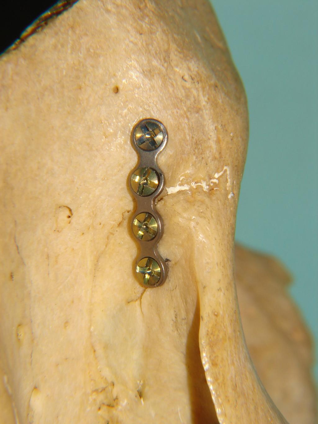
6 6 the least important site for semi-rigid fixation of the zygomatic complex. And routine exposure and reduction of the infraorbital rim and the orbital floor bear the risk of additional trauma to the infraorbital nerve, even if great care is taken. Additional plates at the infraorbital rim or in the region of the zygomaticomaxillary crest are indicated only in cases where the zygomatic bone cannot be reduced by hook reduction and single location fixation. This is by definition necessary only in cases of comminuted fractures [21]. For these reasons, the FZS was the region of our study. But problems such as extrusion, migration, sensibility (particularly in cold weather) and palpability can arise from the use of miniplates in the FZS region because of the thin overlaying skin [23], often leading to a second surgery to remove the miniplates and the screws [24]. The temporal placement suggested here avoids such complications since the miniplates are neither palpable nor visible on this position (Figure 4) and would only be removed in case of infection. When using miniplates without an intermediate segment, the first hole over the FZ suture (point D) should receive the smallest screws and the other areas can receive screws up to 6 mm (Table 1). When using miniplates with an intermediate segment the thinnest area is still immediately above the suture (point C), a result that indicates the use of 5 mm screws. The other areas can receive screws between 6 and 7 mm (Table 2). Therefore, the use of miniplates with an intermediate segment seems to be more appropriate since it avoids drilling at the thinnest area (point D). All measurements showed that the drilling direction is towards the orbit; therefore its contents should be protected during the perforations with an instrument, and depth marks on the drill should be used. At the measured points we did not observe risk of anterior cranial fossa penetration, i.e., there is no risk of anterior cranial fossa penetration when the miniplates are applied to the temporal aspect of the FZ suture. This fact was also observed by Reher and Duarte [9] when internal fixation with miniplates was utilized at the region of the FZS, but at orbital lateral rim. They noted that the lowest point of the anterior cranial fossa that could be reached while drilling perpendicularly to the bone surface was at a mean distance of mm (SD ± 3.49) superior to the FZS. At this point, they observed a mean diploë thickness of 9.18 mm (SD ± 2.51). In 92.06% of the cases (subtracting the standard deviation from the mean value), the measure of 13.5 mm could be used as a reference mark. When one drills above this level, the risk of reaching the cranial cavity using screws bigger than 7.0 mm increases. Considering this, we should not use miniplates with a greater length than 27 mm, because in a miniplate with this maximum length, half of its length (13.5 mm) will be located below the FZS and the other half above, in order to place two screws in each half. The temporal surface of the FZ area on the 30 skulls used in this study was smooth and their shape allowed easy mini plate contouring and adaptation. As shown in tables 1 and 2, the bone thickness in this area is enough to allow screw insertion, of 5 to 6 mm screws. However, care should be taken in elderly patients with thinner bone, since some skulls had a thin bone (the lowest of all measurements was only 1 mm in point D). In these cases plates with an intermediate segment would be indicated, and the smallest screws should usually be applied on the first upper hole (point E). Supraorbital, eyebrow and coronal approaches are usually employed to reach this area, and for the suggested temporal plate placement, the last two seem to be more appropriate, though the coronal approach should be used only in suitable need, i.e., when treatment of associated fractures in the upper third face is
7 7 advisable. The temporalis fascia should be detached from the area and the temporalis muscle retracted posteriorly, exposing the temporal face of the FZ area. Special care should also be taken with regret to drill access and angulation. Drill angulation is forward toward the orbit, and the skin over the temporal fossa as well as the orbit content should be carefully protected. CONCLUSIONS This study suggests that as far as the bone structure is concerned, it is possible to use miniplates at the temporal aspect of the FZ suture. The first hole over the FZ suture should receive the smallest screws and the other areas can receive screws up to 6 mm. All drillings are made from the temporal fossa to the orbit, and its contents should be protected during the perforations. This new position of the mini plates reduces the risk of perforating the anterior cranial fossa, avoids skin palpation of the miniplates, and can be applied using standard approaches, making it an useful option when miniplate fixation in the FZ area is indicated.
8 8 REFERENCES 1. Zachariades N, Mezitis M, Anagnostopoulos PD (1998) Changing trends in the treatment of zigomaticomaxillary complex fractures: a 12 year evaluation of methods used. J Oral Maxillofac Surg 56: Rowe NL, Williams LIJ (1985) Maxillofacial Injuries, Vol. I. Churchill Livingstone, Edinburgh, pp Manson PN, Crawley WA, Yaremchuk MJ, Rochman GM, Hoopes JE, French JH Jr (1985) Midface fractures: advantages of immediate extended open reduction and bone grafting. Plast Reconstr Surg 76: Kaastad E, Freng A (1989) Zygomatico-maxillary fractures. Late results after traction-hook reduction. J Craniomaxillofac Surg 17: Michelet FX, Deymess J, Dessus B (1973) Osteosynthesis with miniaturized screwed plates in maxillofacial surgery. J Maxillofac Surg 1: Vesper M (1996) Der infraorbitale Zugang bei Jochbeinfrakturen. In: Schmelzle R, Bschorer R (eds) Plastische und Wiederherstellungschirurgie ein Jahrbuch. UNI-MED Verlag, Lorsch, pp Zide M J, Wu J (1990) The placement of screws above the zygomaticofrontal suture. J Oral Maxillofac Surg 48: Ewers R (1977) Periorbitale Knochenstrukturen und ihre Bedeutung für die Osteosynthese. Fortschr Kiefer Gesichtschir 22: Reher P, Duarte GC (1994) Miniplates in the frontozygomatic region. An anatomic study. Int J Oral Maxillofac Surg 23: Gruss JS (1996) Discussion: The role of microfixation in malar fractures: a quantitative biophysival study. Plast Reconstr Surg 97: Krumholz K, Niederhagen B, Lepentsiositis J (1991) Zur Therapie isolierter Jochbeinfrakturen. In: Schwenzer N, Pfeifer G (eds) Fortschritte der Kieferund Gesichtschirurgie, Vol. 36. Thieme, Stuttgart, pp O Sullivan ST, Panchal J, O Donoghue JM, Beausang ES, O Shaughnessy M, O Connor TP (1998) Is there still a role for traditional methods in the management of fractures of the zygomatic complex? Injury 29: Gruss JS, Van Wyck L, Phillips JH, Aantonyshyn O (1990) The importance of the zygomatic arch in complex midfacial fracture repair and correction of posttraumatic orbitozygomatic deformities. Plast Reconstr Surg 85: Luhr HG (1988) A microsystem for craniomaxillofacial skeletal fixation. J Craniomaxillofac Surg 16: Fonseca RJ (1998) Discussion: Changing trends in the treatment of zigomaticomaxillary complex fractures: a 12 year evaluation of methods used. J Oral Maxillofac Surg 56: Enislidis G, Lagogiannis G, Wittwer G, Glaser C, Ewers R (2005) Fixation of zygomatic fractures with a biodegradable copolymer osteosynthesis system: short- and long-term results. Int J Oral Maxillofac Surg 34:19-26
9 9 17. Suuronen R, Haers PE, Lindqvist C, Sailer HF (1999) Update on bioresorbable plates in maxillofacial surgery. Facial Plast Surg 15: Berman PD, Jacobs JB (1991) Miniplate fixation of zygomatic fractures. Head Neck 13: Schilli WG (1990) Treatment of zygoma fractures. Oral Maxillofac Surg Clin North Am 2: Dal Santo F, Ellis E 3rd, Throckmorton GS (1992) The effects of zygomatic complex fracture on masseteric muscle force. J Oral Maxillofac Surg 50: Kovács AF, Ghahremani M (2001) Minimization of zygomatic complex fracture treatment. Int J Oral Maxillofac Surg 30: Davidson J, Nickerson D, Nickerson B (1990) Zygomatic fractures: comparison of methods of internal fixation. Plast Reconstr Surg 86: Mitchell DA, MacLeod SP, Bainton R (1995) Multipoint fixation at the frontozygomatic suture with microplates: a technical note. Int J Oral Maxillofac Surg 24: Bhatt V, Langford RJ (2003) Removal of miniplates in maxillofacial surgery: University Hospiral Birrmingham Experience. J Oral Maxillofac Surg 61:
10 10 FIGURE LEGENDS Figure 1. Measurement points in the temporal face of the FZ suture. Points A, C, F and H, were obtained using a miniplate with an intermediate segment; points B, D, E and G using a miniplate without the intermediate segment. Figure 2. Placement of miniplates on the FZ suture. (A) Miniplate without an intermediate segment (points B, D, E and G). (B) Miniplate with an intermediate segment (points A, C, F and H). Figure 3. Mean thicknesses on the selected measurement points (error bars show ± SD). Figure 4. Temporal placement of the miniplates. (A) Lateral view. The bottom screws require more angulation of the screw driver and hand piece toward the temporal fossa. (B) Frontal view. The plates are not palpable at the orbital rims. Figure 5. An illustration of a surgical case. TABLES Table 1 Osseous thicknesses of points used for 1.5 mm miniplates without intermediate segment (n= 30) Mean ± SD Range Mean SD Point B 5.69 ± Point D 4.07 ± Point E 5.21 ± Point G 5.88 ± Table 2 Osseous thicknesses of points used for 1.5 mm miniplates with intermediate segment (n= 30) Mean ± SD Range Mean - SD Point A 6.64 ± Point C 4.56 ± Point F 5.49 ± Point H 6.06 ±
11 A B C D E F G H Orbit Temporal Fossa
12

13
14 (mm) Bone Thickness at the Determined Points A B C D E F G H

15

16

17

18
A New Classification of Zygomatic Fracture Featuring Zygomaticofrontal Suture: Injury Mechanism and a Guide to Treatment
 IBIMA Publishing Plastic Surgery: An International Journal http://www.ibimapublishing.com/journals/psij/psij.html Vol. 2013 (2013), Article ID 383486, 6 pages DOI: 10.5171/2013.383486 Research Article
ZYGOMATIC (MALAR) FRACTURES
 FRACTURES") b854_chapter-12.qxd 1/31/2011 9:40 AM Page 129 ZYGOMATIC (MALAR) FRACTURES CHAPTER 12 Anatomical articulations FZ Fronto-zygomatic ZT Zygomaticotemporal ZMB Zygomatico - maxillary buttress IO Infraorbital
b854_chapter-12.qxd 1/31/2011 9:40 AM Page 129 ZYGOMATIC (MALAR) FRACTURES CHAPTER 12 Anatomical articulations FZ Fronto-zygomatic ZT Zygomaticotemporal ZMB Zygomatico - maxillary buttress IO Infraorbital
MANAGEMENT OF ZYGOMATICO-ORBITAL FRACTURES USING RIGID INTERNAL FIXATION WITH COSMETIC SURGICAL CONSIDERATIONS - CASE REPORT
 MANAGEMENT OF ZYGOMATICO-ORBITAL FRACTURES USING RIGID INTERNAL FIXATION WITH COSMETIC SURGICAL CONSIDERATIONS - CASE REPORT Ong ARM. Management of zygomatico-orbital fracturers using rigid internal fixation
New innovations in craniomaxillofacial fixation: the 2.0 lock system
 LECTURE New innovations in craniomaxillofacial fixation: the 2.0 lock system Brian Alpert, Rolf Gutwald1 and Rainer Schmelzeisen1 Departments of Oral & Maxillofacial Surgery and Surgical & Hospital Dentistry,
Technique Guide. Titanium Wire with Barb and Needle. Surgical Technique Guide for Canthal Tendon Prodecures.
 Technique Guide Titanium Wire with Barb and Needle. Surgical Technique Guide for Canthal Tendon Prodecures. Indications/Features Indications The Synthes Titanium Wire with Barb and straight Needle is
Titanium Wire with Barb and Needle. Surgical Technique Guide for Canthal Tendon Procedures.
 Titanium Wire with Barb and Needle. Surgical Technique Guide for Canthal Tendon Procedures. Technique Guide This publication is not intended for distribution in the USA. Instruments and implants approved
CLINICAL STUDY. Surgical Approaches and Fixation Patterns in Zygomatic Complex Fractures
 CLINICAL STUDY Surgical Approaches and Fixation Patterns in Zygomatic Complex Fractures Sergio Olate, MS, Sergio Monteiro Lima Jr, DDS, Renato Sawazaki, PhD, Roger Willian Fernandes Moreira, PhD, and Márcio
Maxillary and Periorbital Fractures January 2004
 TITLE: Maxillary and Periorbital Fractures SOURCE: Grand Rounds Presentation, UTMB, Dept. of Otolaryngology DATE: January 7, 2004 RESIDENT PHYSICIAN: Gordon Shields, MD FACULTY ADVISOR: Francis B. Quinn,
Technique Guide. Midface Distractor System. For the temporary stabilization and gradual lengthening of the cranial or midfacial bones.
 Technique Guide Midface Distractor System. For the temporary stabilization and gradual lengthening of the cranial or midfacial bones. Table of Contents Introduction Midface Distractor System 2 Indications
Titanium Wire With Barb and Needle
 For Canthal Tendon Procedures Titanium Wire With Barb and Needle Surgical Technique Table of Contents Introduction Titanium Wire With Barb and Needle 2 Indications 2 Surgical Technique Preoperative Planning
RapidSorb Resorbable Tacks. Resorbable Fixation System.
 RapidSorb Resorbable Tacks. Resorbable Fixation System. Fast Safe Resorbable Drill Press Fixed Table of Contents Introduction Overview 2 Indications and Contraindications 4 RapidSorb 5 Surgical Technique
Downloaded from Medico Research Chronicles Assault injury to the face with an axe- A rare case report.
 ISSN No. 2394-3971 Case Report ASSAULT INJURY TO THE FACE WITH AN AXE- A RARE CASE REPORT Dr Sandhya K 1, Dr Bobby John 2, Dr Shobitha G 3 1 Senior resident, Department of Oral and Maxillofacial Surgery,
TRAUMA TO THE FACE AND MOUTH
 Dr.Yahya A. Ali 3/10/2012 F.I.C.M.S TRAUMA TO THE FACE AND MOUTH Bailey & Love s 25 th edition Injuries to the orofacial region are common, but the majority are relatively minor in nature. A few are major
CT of Maxillofacial Injuries
 CT of Maxillofacial Injuries Stuart E. Mirvis, M.D., FACR Department of Radiology University of Maryland School of Medicine Viking 1 1976 MGS 2001 Technology changes the diagnosis Technologic Evolution
Use of Intraoperative Computed Tomography for Revisional Procedures in Patients with Complex Maxillofacial Trauma
 Use of Intraoperative Computed Tomography for Revisional Procedures in Patients with Complex Maxillofacial Trauma The Harvard community has made this article openly available. Please share how this access
Three Dimensional Titanium Mini Plates in Management of Mandibular Fractures
 Biomedical & Pharmacology Journal Vol. 7(1), 241-246 (2014) Three Dimensional Titanium Mini Plates in Management of Mandibular Fractures R. BALAKRISHNAN, VIJAY EBENEZER and ABU DAKIR Department of Oral
SOFT TISSUE SUPPORT IS AN
 ORIGINAL ARTICLE Reconstructive Application of the Endotine Suspension Devices James H. Boehmler IV, MD; Benjamin L. Judson, MD; Steven P. Davison, MD, DDS Objective: To illustrate the potential reconstructive
ORIGINAL ARTICLE. Facial Fracture Classification According to Skeletal Support Mechanisms
 ORIGINAL ARTICLE Facial Fracture Classification According to Skeletal Support Mechanisms Terry L. Donat, MD; Carmen Endress, MD; Robert H. Mathog, MD Objective: To construct, propose, and evaluate the
Core Curriculum Syllabus Emergencies in Otolaryngology-Head and Neck Surgery FACIAL FRACTURES
 Core Curriculum Syllabus Emergencies in Otolaryngology-Head and Neck Surgery A. General Considerations FACIAL FRACTURES Look for other fractures like skull and/or cervical spine fractures Test function
AcUMEDr. LoCKING CLAVICLE PLATE SYSTEM
 AcUMEDr LoCKING CLAVICLE PLATE SYSTEM LoCKING CLAVICLE PLATE SYSTEM Since 1988 Acumed has been designing solutions to the demanding situations facing orthopedic surgeons, hospitals and their patients.
Surgical technique. IMF Screw Set. For temporary, peri opera tive stabilisation of the occlusion in adults.
 Surgical technique IMF Screw Set. For temporary, peri opera tive stabilisation of the occlusion in adults. Table of contents Features and benefits 2 Indications and contraindications 3 Surgical technique
The diagnostic value of Computed Tomography in evaluation of maxillofacial Trauma
 The diagnostic value of Computed Tomography in evaluation of maxillofacial Trauma Qais H. Muassa FICMS College of Dentistry, Babylon University Ibrahim S. Gataa, BDS, FICMS College of Dentistry, Sulaimania
DISTRACTION PRODUCT OVERVIEW. For a wide variety of facial applications
 DISTRACTION PRODUCT OVERVIEW For a wide variety of facial applications DISTRACTION PRODUCT OVERVIEW. STRONG, MODULAR, VERSATILE CRANIOFACIAL DISTRACTION External Midface Distractor Distraction of the maxilla,
Implant Preparation. Page 1
 Why SU-POR? SU-POR Surgical Implants are manufactured from a linear high-density polyethylene. SU-POR Surgical Implants allow for tissue ingrowth because of the interconnecting open pore structure. The
Maxillofacial Injuries Practical Tips
 Saturday, October 29, 2016 Maxillofacial Injuries Practical Tips Suyash Mohan MD, PDCC THE ROOTS OF PENN RADIOLOGY RADIOLOGICAL Assistant Professor of Radiology Assistant Professor of Neurosurgery Neuroradiology
Bones of the skull & face
 Bones of the skull & face Cranium= brain case or helmet Copyright The McGraw-Hill Companies, Inc. Permission required for reproduction or display. The cranium is composed of eight bones : frontal Occipital
Craniomaxillofacial Research
 Journal of Craniomaxillofacial Research Vol. 2, No. (3-4) Application of endoscope and conventional techniques in management of Orbital Floor and Infra-orbital Rim Fracture Reduction Gholamreza Shirani
Orbital Plating System OPS 1.5
 PRODUCT INFORMATION Orbital Plating System OPS 1.5 MODUS Midface 2 Orbital Plating System OPS 1.5 At a glance Orbital Plating System OPS 1.5 INTRODUCTION Fractures of the orbit occur in about 50% of all
SYNPOR POROUS POLYETHYLENE IMPLANTS. For craniofacial and orbital augmentation and reconstruction
 SYNPOR POROUS POLYETHYLENE IMPLANTS For craniofacial and orbital augmentation and reconstruction SURGICAL TECHNIQUE TABLE OF CONTENTS INTRODUCTION SYNPOR Porous Polyethylene Implants 2 Indications and
ISOLATED ZYGOMATIC BONE FRACTURE; MANAGEMENT BY THREE POINT FIXATION
 The Professional Medical Journal 1. BDS, FCPS 2. BDS, FCPS 3. BDS, MSc Community Dentistry 4. BDS, MSc (Trainee) 5. MBBS, FRCS Associate Professor General Surgery LUMHS, Correspondence Address: Dr. Suneel
Technique Guide. Compact 2.0 LOCK Mandible. The locking system for the mandible.
 Technique Guide Compact 2.0 LOCK Mandible. The locking system for the mandible. Table of Contents Introduction Compact 2.0 LOCK Mandible 2 AO Principles 4 Indications and Contraindications 5 Surgical
MEDPOR. Plastic surgery
 MEDPOR Plastic surgery MEDPOR biomaterial MEDPOR has been a trusted name in the industry since 1985, with hundreds of thousands of procedures performed, and hundreds of published clinical reports in reconstructive,
Imaging Orbit/Periorbital Injury
 Imaging Orbit/Periorbital Injury 9 th Nordic Trauma Radiology Course 2016 Stuart E. Mirvis, M.D., FACR Department of Radiology University of Maryland School of Medicine Fireworks Topics to Cover Struts
Biodegradable plates and screws in oral and maxillofacial surgery Buijs, Gerrit Jacob
 University of Groningen Biodegradable plates and screws in oral and maxillofacial surgery Buijs, Gerrit Jacob IMPORTANT NOTE: You are advised to consult the publisher's version (publisher's PDF) if you
Open reduction; plate fixation 1 Principles
 Executive Editor: Peter Trafton Authors: Martin Jaeger, Frankie Leung, Wilson Li Proximal humerus 11-A2 Open reduction, plate fixation Search search... Shortcuts All Preparations All Approaches All Reductions
LCP Superior Clavicle Plate. The anatomically precontoured fixation system with angular stability for clavicle shaft and lateral clavicle.
 Technique Guide LCP Superior Clavicle Plate. The anatomically precontoured fixation system with angular stability for clavicle shaft and lateral clavicle. Table of Contents Introduction LCP Superior Clavicle
Zürich Pediatric Maxillary Distractor
 Distraction Osteogenesis Zürich Pediatric Maxillary Distractor AN INTRAORAL, UNI-DIRECTIONAL DEVICE FOR THE DISTRACTION OF THE MAXILLA Indications & Advantages Zürich Pediatric Maxillary Distractor Developed
Prophylactic Midface Lift in Midfacial Trauma
 Rapid Communication 347 Ryan Brown, MD 1 Kirk Lozada, MD 2 Sameep Kadakia, MD 2 Eli Gordin, MD 3 Yadranko Ducic, MD 4 1 Department of Otolaryngology, Kaiser Permanente, Denver, Colorado 2 Department of
ORIGINAL ARTICLE. A Novel Technique for Malar Eminence Evaluation Using 3-Dimensional Computed Tomography
 ORIGINAL ARTICLE A Novel Technique for Malar Eminence Evaluation Using 3-Dimensional Computed Tomography Sami P. Moubayed, MD; Frederick Duong, MD; Christian Ahmarani, MD, FRCSC; Akram Rahal, MD, FRCSC
THE USE OF TEMPORARY ANCHORAGE DEVICES FOR MOLAR INTRUSION & TREATMENT OF ANTERIOR OPEN BITE By Eduardo Nicolaievsky D.D.S.
 THE USE OF TEMPORARY ANCHORAGE DEVICES FOR MOLAR INTRUSION & TREATMENT OF ANTERIOR OPEN BITE By Eduardo Nicolaievsky D.D.S. Skeletal anchorage, the concept of using the facial skeleton to control tooth
Technique Guide. IMF Screw Set. For intermaxillary fixation.
 Technique Guide IMF Screw Set. For intermaxillary fixation. Table of Contents Introduction IMF Screw Set 2 Indications and Contraindications 3 Surgical Technique Preparation 4 Insert IMF Screw 6 Insert
Clinical Study Open Reduction of Subcondylar Fractures Using a New Retractor
 Plastic Surgery International Volume 2011, Article ID 421245, 5 pages doi:10.1155/2011/421245 Clinical Study Open Reduction of Subcondylar Fractures Using a New Retractor Akira Sugamata, 1 Naoki Yoshizawa,
3.5 mm Clavicle Hook Plates
 A Single Solution for Lateral Clavicle Fractures and Acromioclavicular Joint Dislocations 3.5 mm Clavicle Hook Plates Surgical Technique Discontinued December 2017 DSUS/TRM/1016/1126(1) Table of Contents
Thickened and thinner parts of the skull = important base for understanding of the functional structure of the skull - the transmission of masticatory
 Functional structure of the skull and Fractures of the skull Thickened and thinner parts of the skull = important base for understanding of the functional structure of the skull - the transmission of masticatory
VA-LCP Anterior Clavicle Plate. The anatomically precontoured fixation system with angular stability for clavicle shaft and lateral clavicle.
 Technique Guide VA-LCP Anterior Clavicle Plate. The anatomically precontoured fixation system with angular stability for clavicle shaft and lateral clavicle. Table of Contents Introduction VA-LCP Anterior
CRANIAL RECONSTRUCTION SOLUTIONS
 CRANIAL RECONSTRUCTION SOLUTIONS CRANIAL RECONSTRUCTION SOLUTIONS Your partner of CHOiCE at depuy Synthes CMF, we are dedicated to providing solutions for your individual patient needs. We do this through
Temporal fossa Infratemporal fossa Pterygopalatine fossa Terminal branches of external carotid artery Pterygoid venous plexus
 Outline of content Temporal fossa Infratemporal fossa Pterygopalatine fossa Terminal branches of external carotid artery Pterygoid venous plexus Boundary Content Communication Mandibular division of trigeminal
Five-Year Experience with the Transoral Endoscopically Assisted Treatment of Displaced Condylar Mandible Fractures
 Five-Year Experience with the Transoral Endoscopically Assisted Treatment of Displaced Condylar Mandible Fractures Ralf Schön, M.D., D.M.D., Otto Fakler, M.D., D.M.D., Nils-Claudius Gellrich, M.D., D.M.D.,
Low Profile Neuro Plating System. Surgical Technique
 Low Profile Neuro Plating System Surgical Technique TABLE OF CONTENTS INTRODUCTION Low Profile Neuro Plating System 2 SURGICAL TECHNIQUE Technique 5 PRODUCT INFORMATION Low Profile Neuro Plates 10 Low
DOWNLOAD OR READ : RIGID FIXATION FOR MAXILLOFACIAL SURGERY PDF EBOOK EPUB MOBI
 DOWNLOAD OR READ : RIGID FIXATION FOR MAXILLOFACIAL SURGERY PDF EBOOK EPUB MOBI Page 1 Page 2 rigid fixation for maxillofacial surgery rigid fixation for maxillofacial pdf rigid fixation for maxillofacial
MAXILLOFACIAL TRAUMATOLOGY Department of Maxillofacial Surgery Semmelweis University, Budapest. Dr. Huszár Tamás
 MAXILLOFACIAL TRAUMATOLOGY Department of Maxillofacial Surgery Semmelweis University, Budapest Dr. Huszár Tamás Maxillofacial injuries isolated maxillofacial injury multiple injuries polytrauma (injury
Mandible External Fixator II. Provides treatment for fractures of the maxillofacial area.
 Mandible External Fixator II. Provides treatment for fractures of the maxillofacial area. Technique Guide This publication is not intended for distribution in the USA. Instruments and implants approved
Tikrit University collage of dentistry Dr.Ban I.S. head & neck anatomy 2 nd y. Lec [5] / Temporal fossa :
![Tikrit University collage of dentistry Dr.Ban I.S. head & neck anatomy 2 nd y. Lec [5] / Temporal fossa :](/thumbs/88/115294566.jpg "Tikrit University collage of dentistry Dr.Ban I.S. head & neck anatomy 2 nd y. Lec [5] / Temporal fossa :") Lec [5] / Temporal fossa : Borders of the Temporal Fossa: Superior: Superior temporal line. Inferior: gap between zygomatic arch and infratemporal crest of sphenoid bone. Anterior: Frontal process of the
Lec [5] / Temporal fossa : Borders of the Temporal Fossa: Superior: Superior temporal line. Inferior: gap between zygomatic arch and infratemporal crest of sphenoid bone. Anterior: Frontal process of the
The Skull and Temporomandibular joint II Prof. Abdulameer Al-Nuaimi. E. mail:
 The Skull and Temporomandibular joint II Prof. Abdulameer Al-Nuaimi E-mail: a.al-nuaimi@sheffield.ac.uk E. mail: abdulameerh@yahoo.com Temporal fossa The temporal fossa is a depression on the temporal
Subciliary versus Subtarsal Approaches to Orbitozygomatic Fractures
 CME Subciliary versus Subtarsal Approaches to Orbitozygomatic Fractures Rod J. Rohrich, M.D., Jeffrey E. Janis, M.D., and William P. Adams, Jr., M.D. Dallas, Texas Learning Objectives: After studying this
MEDPOR. Oral maxillofacial surgery
 MEDPOR Oral maxillofacial surgery MEDPOR biomaterial MEDPOR has been a trusted name in the industry since 1985, with hundreds of thousands of procedures performed, and hundreds of published clinical reports
(Jurnal Plastik Rekonstruksi 2017; 1:82-87)
") (Jurnal Plastik Rekonstruksi 2017; 1:82-87) RECONSTRUCTIVE TETRAPOD FRACTURE: SURGICAL ANATOMY REVISITED AS A GUIDE FOR 3D REDUCTION USING CARROLL GIRARD T-BAR SCREW Prasetyanugraheni Kreshanti 1 *, Livia
(Jurnal Plastik Rekonstruksi 2017; 1:82-87) RECONSTRUCTIVE TETRAPOD FRACTURE: SURGICAL ANATOMY REVISITED AS A GUIDE FOR 3D REDUCTION USING CARROLL GIRARD T-BAR SCREW Prasetyanugraheni Kreshanti 1 *, Livia
Anatomy and Physiology. Bones, Sutures, Teeth, Processes and Foramina of the Human Skull
 Anatomy and Physiology Chapter 6 DRO Bones, Sutures, Teeth, Processes and Foramina of the Human Skull Name: Period: Bones of the Human Skull Bones of the Cranium: Frontal bone: forms the forehead and the
What is Hemifacial Microsomia? By Pravin K. Patel, MD and Bruce S. Bauer, MD Children s Memorial Hospital, Chicago, IL
 What is Hemifacial Microsomia? By Pravin K. Patel, MD and Bruce S. Bauer, MD Children s Memorial Hospital, Chicago, IL 773-880-4094 Early in the child s embryonic development the structures destined to
low ProfIle neuro PlaTIng system
 low ProfIle neuro PlaTIng system surgical TeChnIque Table of Contents Introduction Low Profile Neuro Cranial Plating System 2 Surgical Technique Technique 5 Product Information Low Profile Neuro Plates
Technique Guide. ARCH Laminoplasty System. Dedicated System for Open-door Laminoplasty.
 Technique Guide ARCH Laminoplasty System. Dedicated System for Open-door Laminoplasty. Table of Contents Introduction Overview 2 AO ASIF Principles 4 Indications and Contraindications 5 Product Information
Diagnosis of Midface Fractures with CT: What the Surgeon Needs to Know 1
 Note: This copy is for your personal non-commercial use only. To order presentation-ready copies for distribution to your colleagues or clients, contact us at www.rsna.org/rsnarights. EDUCATION EXHIBIT
Technique Guide. SynPOR Porous Polyethylene Implants. For craniofacial and orbital augmentation and reconstruction.
 Technique Guide SynPOR Porous Polyethylene Implants. For craniofacial and orbital augmentation and reconstruction. Table of Contents Introduction SynPOR Porous Polyethylene Implants 2 Indications and Contraindications
Bones Ethmoid bone Inferior nasal concha Lacrimal bone Maxilla Nasal bone Palatine bone Vomer Zygomatic bone Mandible
 splanchnocranium - Consists of part of skull that is derived from branchial arches - The facial bones are the bones of the anterior and lower human skull Bones Ethmoid bone Inferior nasal concha Lacrimal
McHenry Western Lake County EMS System Paramedic, EMT-B and PHRN Optional Continuing Education 2019 #1 Facial Trauma
 McHenry Western Lake County EMS System Paramedic, EMT-B and PHRN Optional Continuing Education 2019 #1 Facial Trauma The face is vital to human appearance and function. Facial injuries can impair a patient
Oral Surgery Dr. Labeed Sami جامعة تكريت كلية طب االسنان املرحلة اخلامسة م.د. لبيد سامي حسن
 جامعة تكريت كلية طب االسنان جراحة الفم مادة املرحلة اخلامسة م.د. لبيد سامي حسن 6102-6102 1 5 th stage Fracture zygomatic complex As the zygomatic bone is closely associated with the maxilla, frontal and
CT of Maxillofacial Fracture Patterns. CT of Maxillofacial Fracture Patterns
 CT of Maxillofacial Fracture Patterns CT of Maxillofacial Fracture Patterns Stuart E. Mirvis, M.D., FACR Department of Radiology University of Maryland School of Medicine Viking 1 1976 MGS 2001 Technology
The treatment of malocclusion after open reduction of maxillofacial fracture: a report of three cases
 CASE REPORT http://dx.doi.org/10.5125/jkaoms..40.2.91 pissn 2234-7550 eissn 2234-5930 The treatment of malocclusion after open reduction of maxillofacial fracture: a report of three cases Sung-Suk Lee,
Technique Guide. LCP Proximal Femoral Hook Plate 4.5/5.0. Part of the LCP Periarticular Plating System.
 Technique Guide LCP Proximal Femoral Hook Plate 4.5/5.0. Part of the LCP Periarticular Plating System. Table of Contents Introduction Features and Benefits 2 AO ASIF Principles 4 Indications 5 Surgical
Current concepts in midface fracture management
 REVIEW C URRENT OPINION Current concepts in midface fracture management AQ1 Alf L. Nastri and Ben Gurney AQ4 Purpose of review Management of midface trauma is complex and challenging and requires a clear
3. The Jaw and Related Structures
 Overview and objectives of this dissection 3. The Jaw and Related Structures The goal of this dissection is to observe the muscles of jaw raising. You will also have the opportunity to observe several
MEDICAL CODING FOR FACIAL INJURIES & RECONSTRUCTION
 MEDICAL CODING FOR FACIAL INJURIES & RECONSTRUCTION Tirbod Fattahi, MD, DDS, FACS Chief & Associate Professor Division of Oral & Maxillofacial Surgery University of Florida Health Science Center, Jacksonville
Pediatric Craniofacial Injuries: Concept of Treatment
 Med. J. Cairo Univ., Vol. 83, No. 1, March: 217-224, 201 5 www.medicaljournalofcairouniversity.net Pediatric Craniofacial Injuries: Concept of Treatment FAWZY T. AL-SAYED, Ph.D.* and MOHAMAD A. SHOEIB,
cally, a distinct superior crease of the forehead marks this spot. The hairline and
 4 Forehead The anatomical boundaries of the forehead unit are the natural hairline (in patients without alopecia), the zygomatic arch, the lower border of the eyebrows, and the nasal root (Fig. 4.1). The
Clinical Note Clinical Outcome of 285 Medpor Grafts used for Craniofacial Reconstruction PATIENTS AND METHODS
 Clinical Note Clinical Outcome of 285 Medpor Grafts used for Craniofacial Reconstruction Roberto Cenzi, MD,* Antonio Farina, MD, y Luca Zuccarino, MD, z Francesco Carinci, MD Ferrara, Italy Porous polyethylene
Elbow Hinge Fixator. Guided Flexion/Extension for Unstable Elbow Fractures.
 Elbow Hinge Fixator. Guided Flexion/Extension for Unstable Elbow Fractures. Surgical Technique MR Safe Radiolucent Table of Contents System Description 3 Indications and Contraindications 4 Fixation Components
MEDPOR. Oculoplastic surgery
 MEDPOR Oculoplastic surgery MEDPOR biomaterial MEDPOR has been a trusted name in the industry since 1985, with hundreds of thousands of procedures performed, and hundreds of published clinical reports
Measurement of the Maxilla and Zygoma as an Aid in Installing Zygomatic Implants
 J Oral Maxillofac Surg 59:1193-1198, 2001 Measurement of the Maxilla and Zygoma as an Aid in Installing Zygomatic Implants Yuki Uchida, DDS, PhD,* Masaaki Goto, DDS, PhD, Takeshi Katsuki DDS, PhD, and
LCP Medial Distal Tibia Plate, without Tab. The Low Profile Anatomic Fixation System with Angular Stability and Optimal Screw Orientation.
 LCP Medial Distal Tibia Plate, without Tab. The Low Profile Anatomic Fixation System with Angular Stability and Optimal Screw Orientation. Technique Guide LCP Small Fragment System Table of Contents Introduction
Contemporary Implant Dentistry
 Contemporary Implant Dentistry C H A P T ER 1 4 O F C O N T E M P OR A R Y O R A L A N D M A X I L L OFA C IA L S U R G E RY B Y : D R A R A S H K H O J A S T EH Dental implant is suitable for: completely
CHAPTER. 1. Uncontrolled systemic disease 2. Retrognathic jaw relationship
 CHAPTER 7 Immediate Implant Supported Restoration of the Edentulous Arch Stephen G. Alfano and Robert M. Laughlin Department of Oral and Maxillofacial Surgery, Naval Medical Center San Diego, San Diego,
Chapter 23: Maxillofacial Trauma. Robert B. Stanley, Jr.
 Chapter 23: Maxillofacial Trauma Robert B. Stanley, Jr. Traditionally, fractures of the facial skeleton have been evaluated and treated in a segmentalized fashion, even if complex injuries were obvious
Post-operative stability of the maxilla treated with Le Fort I and horseshoe osteotomies in bimaxillary surgery
 European Journal of Orthodontics 24 (2002) 471 476 2002 European Orthodontic Society Post-operative stability of the maxilla treated with Le Fort I and horseshoe osteotomies in bimaxillary surgery Kiyoshi
Open and Endoscopic Forehead Lift. Plastic Surgery. For All Brow and Forehead Lift Procedures. Revolutionizing. Soft-Tissue Fixation
 Plastic Surgery Open and Endoscopic Forehead Lift For All Brow and Forehead Lift Procedures Revolutionizing Soft-Tissue Fixation DESIGNED FOR SIMPLICITY AND PREDICTABILITY The versatile design can be applied
Humerus shaft - Reduction & Fixation - Compression plate - AO Surgery Reference. Compression plating
 Humerus shaft 12-A3 ORIF 1. Principles Compression plating Authors Compression plate Compression plating provides fixation with absolute stability for two-part fracture patterns, where the bone fragments
MatrixNEURO Cranial Plating System
 The Next Generation Cranial Plating System MatrixNEURO Cranial Plating System Surgical Technique TABLE OF CONTENTS INTRODUCTION MatrixNEURO Cranial Plating System 2 MatrixNEURO Reconstruction Mesh 6 MatrixNEURO
Dr. Sami Zaqout, IUG Medical School
 The skull The skull is composed of several separate bones united at immobile joints called sutures. Exceptions? Frontal bone Occipital bone Vault Cranium Sphenoid bone Zygomatic bones Base Ethmoid bone
Two Hundred Ninety-Four Consecutive Facial Fractures in an Urban Trauma Center: Lessons Learned
 CME Two Hundred Ninety-Four Consecutive Facial Fractures in an Urban Trauma Center: Lessons Learned Patrick Kelley, M.D., Marcus Crawford, M.D., Stephen Higuera, M.D., and Larry H. Hollier, M.D. Houston,
LCP Superior Clavicle Plate. The anatomically precontoured fixation system with angular stability for clavicle shaft and lateral clavicle.
 LCP Superior Clavicle Plate. The anatomically precontoured fixation system with angular stability for clavicle shaft and lateral clavicle. Surgical Technique This publication is not intended for distribution
Comparative Evaluation of Single Point Fixation at Zygomatic Buttress and Fronto Zygomatic Rim in Zygomatic Complex Fractures -A Prospective Study
 Comparative Evaluation of Single Point Fixation at Zygomatic Buttress and Fronto Zygomatic Rim in Zygomatic Complex Fractures -A Prospective Study *Prathibha Sridhar 1, Shubha Sandeep 2, Kavitha Prasad
Face. Definition: The area between the two ears and from the chin to the eye brows. The muscles of the face
 Face Definition: The area between the two ears and from the chin to the eye brows. The muscles of the face The muscle of facial expression (include the muscle of the face and the scalp). All are derived
Surgical Technique. Calcaneal Locking Plate
 Surgical Technique Calcaneal Locking Plate PERI-LOC Locked Plating System Calcaneal Locking Plate Surgical TechniqueCatalog Infor Table of Contents Introduction...2 Indications...3 Plate Features...3 Patient
Wrist Fixation System
 Wrist Fixation System Anatomy / Fracture Implant EXTRA & SIMPLE ARTICULAR Volar Radius Volar Fixed Angle Plate Volar Bearing Plate Radial Peg Plate Volar Hook Plate Volar Buttress Pin Volar Shear Plate
LCP Medial Proximal Tibial Plate 3.5. Part of the Synthes small fragment Locking Compression Plate (LCP) system.
 system.") LCP Medial Proximal Tibial Plate 3.5. Part of the Synthes small fragment Locking Compression Plate (LCP) system. Technique Guide This publication is not intended for distribution in the USA. Instruments
LCP Medial Proximal Tibial Plate 3.5. Part of the Synthes small fragment Locking Compression Plate (LCP) system. Technique Guide This publication is not intended for distribution in the USA. Instruments
MAXILLOFACIAL TRAUMA. The on-call maxillofacial surgeons can be contacted through the switchboard at the Southern General Hospital
 MAXILLOFACIAL TRAUMA The on-call maxillofacial surgeons can be contacted through the switchboard at the Southern General Hospital Mandibular Injuries Mechanism of injury Assault, falls, RTA-Direct trauma
Departement of Stomatology, The Second Hospital of Lanzhou University, 82 Cuiyingmwen, Chengguan District, Lanzhou City, Gansu Province, China
 European Review for Medical and Pharmacological Sciences Comparative evaluation of 2.0 mm locking plate system vs 2.0 mm non-locking plate system for mandibular angle fracture fixation: a prospective randomized
Osseointegrated dental implant treatment generally
 Placement of Dental Implants Without Flap Surgery: A Clinical Report Bader H. Al-Ansari, BDS, MScD*/Robert R. Morris, DMD** Traditionally, the procedure of implant placement requires a surgical periosteal
2.0 mm Mandible Locking Plate System
 Advanced Plating System for Trauma, Microvascular Reconstruction, and Orthognathic Surgery 2.0 mm Mandible Locking Plate System Surgical Technique TABLE OF CONTENTS INTRODUCTION 2.0 mm Mandible Locking