Supplementary information Novel VCP modulators mi2gate major pathologies of rd10, a mouse model of re2ni2s pigmentosa
|
|
|
- Amelia Sims
- 5 years ago
- Views:
Transcription
1 Supplementary information Novel VCP modulators mi2gate major pathologies of rd1, a mouse model of re2ni2s pigmentosa Hanako Ohashi Ikeda, Norio Sasaoka, Masaaki Koike, Noriko Nakano, Yuki Muraoka, Yoshinobu Toda, Tomohiro Fuchigami, Toshiyuki Shudo, Ayana Iwata, Seiji Hori, Nagahisa Yoshimura & Akira Kakizuka
2 12! Relative ATPase activity (%)! 1! 8! 6! 4! 2!! um!.1um!.1um! 1uM!.1uM!.1uM! 1uM!.1uM!.1uM! 1uM! 1uM! KUS121! KUS187! DBeQ! DMSO! KUS121! KUS187! DBeQ! E. coli! (μm)
3 Luciferase activity/cell ATP/ADP ratio µ AU/ 1 6 cells Supplementary Fig.2 a 2 b c 2 2. *** * ** ** *** *** 1 8 * HG DMSO K69 K94 HG DMSO K69 K94 K121 K187 HG DMSO K94 K121 K187
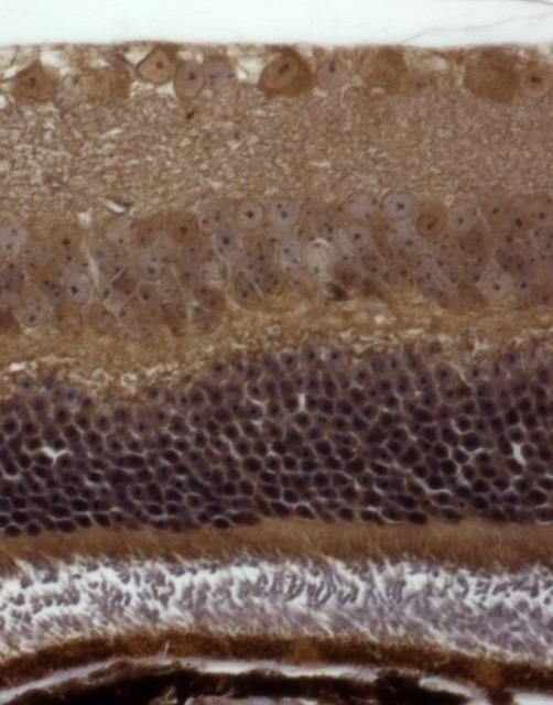
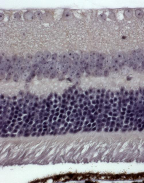
4 Supplementary Fig.3 a b GCL IPL INL OPL ONL IS OS
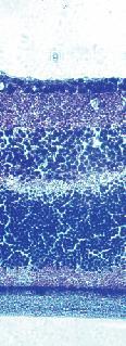
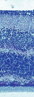
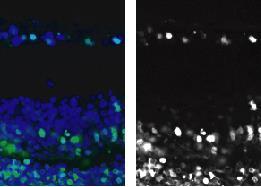
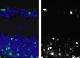
5 Supplementary Fig.4 a control K121 RNFL GCL IPL INL b TUNEL TOTO control TUNEL K121 GCL INL
 P21 a RNFL IPL")


6 a-wave amplitude (µv) latency of a-wave (ms) P21 a RNFL IPL INL OPL ONL IS/OS RPE WT rd1 control KUS121 KUS187 ONL Supplementary Fig.5 b b-wave latency amplitude a-wave 1 µv 2 msec c 2 2 * ** d *** *** C C C
7 b-wave amplitude (µv) a-wave amplitude (µv) b-wave amplitude (µv) Supplementary Fig.6 a 8 6 * 4 2 P25 P29 P33 age (days) b 3 c 生食 C KUS121 K121 生食 C KUS121 K121

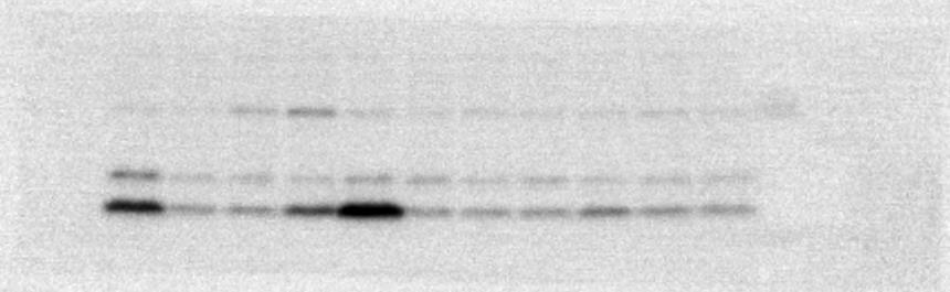
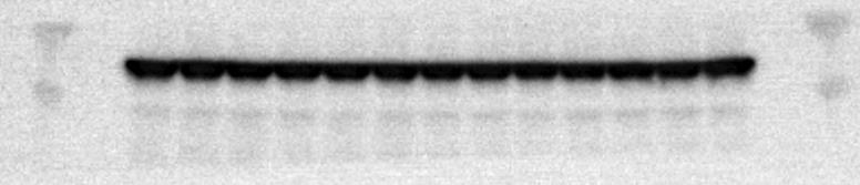
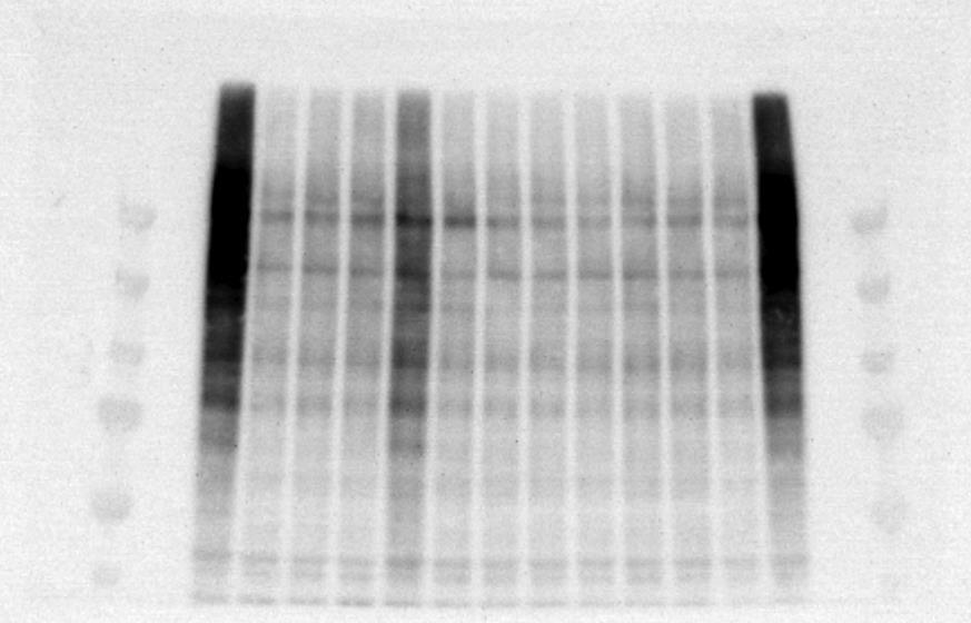
8 Supplementary Fig.7 (KDa) 2 Ubiquitin CHOP 25 Fig.1c LC3 actin 15 37


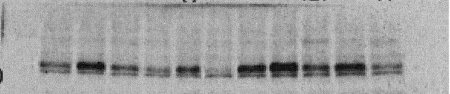
9 Supplementary Fig.8 CHOP 25 P-Akt Akt GRP VCP 1 actin Fig.2f

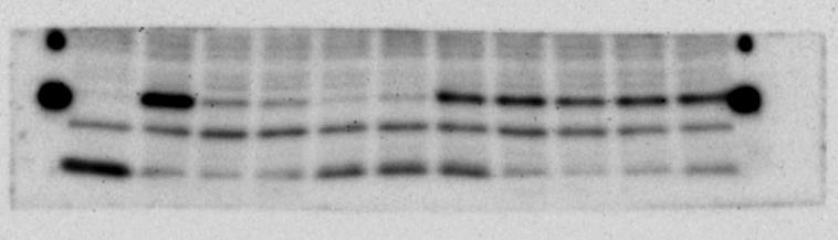

10 Supplementary Fig.9 CHOP 25 actin CHOP 25 Fig.4c actin


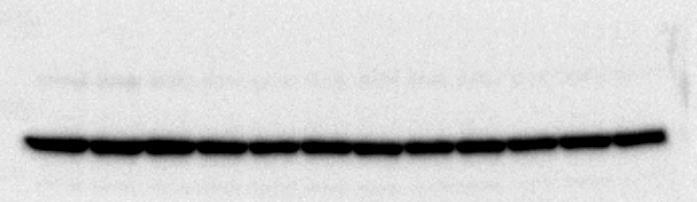
11 WT rd1 C Supplementary Fig.1 CHOP 25 VCP actin Fig.5i
12 Supplementary Figure Legends Supplementary Figure 1 Inhibitory effects of KUS121, KUS187, and DBeQ on the ATPase activity of recombinant VCP purified from E. coli. Supplementary Figure 2 (a) HeLa cells were cultured in medium with low glucose (.25 g/l) for 2 hours, with or without KUSs, and ATP levels were measured with luciferase assays. *** P <.5 vs. DMSO control (Dunnett s test, n=3). HG: medium with 4.5 g/l glucose. Error bars indicate SD. (b) HeLa cells were cultured in medium with low glucose (.2 g/l) for 2 hours, with DMSO or KUSs, and ATP and ADP levels were measured by HPLC. The respective ratios of ATP/ADP are shown. * P <.5, ** P <.1 vs. DMSO control (Dunnett s test, n=3). Error bars indicate SD. (c) HeLa cells were cultured in medium without glucose for 6 hours, with ( µm each KUS, as indicated) or without KUSs, and acetyl-coa levels were measured by HPLC. Supplementary Figure 3 Immunohistochemical analyses of retinal neuronal cells. Retinal sections of 5-month-old C57BL/6 mice were stained with (b) or without (a) an anti-vcp antibody 12, developed with diaminobenzidine and counter-stained with hematoxylin (a, b). Note that all retinal neuronal cells highly expressed VCP. GCL: ganglion cell layer, IPL: inner plexiform layer, INL: inner nuclear layer, OPL: outer plexiform layer, ONL: outer nuclear layer, IS: inner segment, OS: outer segment. Supplementary Figure 4 KUS121 protects retinal neuronal cells from NMDA insult. Retinas from adult C57BL/6 mice were cultured on porous membrane with 2 µm of NMDA with or without KUS121 ( µm) for 4 days. (a) HE-stained retinal sections. In the retina cultured with KUS121, the inner nuclear layer (INL), inner plexiform layer (IPL), ganglion cell layer (GCL), and retinal nerve fiber layer (RNFL) were thicker than that of the retina without KUS121. (b) TUNEL staining of the retinal sections. More TUNEL-positive apoptotic cells (green) were observed in the retina without KUS than with KUS121. Supplementary Figure 5 (a) Cross sectional (vertical) images obtained by spectral-domain optical coherence tomography (SD-OCT) in 21-day-old C57BL/6 wild-type (WT) and rd1 mice with or without KUSs. Vertical bars in the images indicate the thickness of outer nuclear layer. RNFL: retinal nerve fiber layer, IPL: inner plexiform layer, INL: inner nuclear layer, OPL: outer plexiform layer, ONL: outer nuclear layer, IS: inner segment, OS: outer segment, IS/OS: junction line between IS and OS. Scale bar (shown by white color), 1 µm. (b-d) Dark-adapted electroretinogram was performed on 21-day-old C57BL/6 wild-type and rd1 mice. (b) Representative electroretinogram records are shown. Double-headed arrows indicate amplitudes of a- and b-wave, and latency of a-wave. (c) The mean amplitude of the a-wave (measured at 25 msec), which is derived from photoreceptors, in KUS-treated mice was larger than that in control rd1 mice. * P =.45 and.6 in KUS121 and KUS187 vs. control, respectively (Dunnett s test). Error bars indicate SD. (d) Latency of a-wave in electroretinogram. In KUS-treated mice, mean latencies were delayed, compared with those in the control mice. *** P <.1, vs. control (Dunnett s test). Error bars indicate SD.
13 Supplementary Figure 6 (a) Time-dependent changes of b-wave amplitude in dark-adapted electroretinograms in non-treated C57BL/6 wild-type mice. b-wave amplitudes remained unchanged or slightly increased at the ages of 25 to 33 days. * P =.4. (b, c) a-wave (b) and b-wave (c) amplitude of adult C57BL/6 wild-type mice administered saline or KUS121 for 7 days. There were no significant differences in the amplitudes of a-and b-waves between mice administered saline or KUS121 (a-wave amplitude: 23.5 ± 41.6 vs ± 3.2, P =.56, t-test; b-wave amplitude: ± 92.9 vs ± 52.6, P =.86, t-test). Error bars indicate SD. Supplementary Figure 7 Complete scans of the different blots presented in Figure 1c. Lane 1, 1µM MG132; lane 2, control; lane 3, 5µM DBeQ; lane 4, 1µM DBeQ; lane 5, 15µM DBeQ; lane 6: KUS11; lane 7, KUS31; lane 8, KUS69; lane 9, KUS 94; lane 1, KUS121; lane 11, KUS187. Supplementary Figure 8 Complete scans of the different blots presented in Figure 2f. Lane 1, Tm(-); lane 2, control; lane 3, KUS69; lane 4, KUS94; lane 5, KUS121; lane 6, KUS187. Supplementary Figure 9 Complete scans of the different blots presented in Figure 4c. Lane 1, Tm(-); lane 2, control; lane 3, KUS69; lane 4, KUS94; lane 5, KUS121; lane 6, KUS187; lane 7, Tm(-); lane 8, control; lane 9,.1mM ATP; lane 1,.3mM ATP; lane 11, 1mM ATP; lane 12, 3mM methylpyruvate; lane 13, 1mM methylpyruvate. Supplementary Figure 1 Complete scans of the different blots presented in Figure 5i. 2
Measuring of the fovea and foveola using line scans and 3D Macular scans obtained with spectral domain optical coherent tomography.
 Measuring of the fovea and foveola using line scans and 3D Macular scans obtained with spectral domain optical coherent tomography. Vakhrameeva O.A. 1, Moiseenko G.A. 1, Maltsev D.S. 2, Sukhinin M.V. 2,
Ganglion cell analysis by optical coherence tomography (OCT) Jonathan A. Micieli, MD Valérie Biousse, MD
 Jonathan A. Micieli, MD Valérie Biousse, MD") Ganglion cell analysis by optical coherence tomography (OCT) Jonathan A. Micieli, MD Valérie Biousse, MD Figure 1. Normal OCT of the macula (cross section through the line indicated on the fundus photo)
Ganglion cell analysis by optical coherence tomography (OCT) Jonathan A. Micieli, MD Valérie Biousse, MD Figure 1. Normal OCT of the macula (cross section through the line indicated on the fundus photo)
OCT Image Analysis System for Grading and Diagnosis of Retinal Diseases and its Integration in i-hospital
 Progress Report for1 st Quarter, May-July 2017 OCT Image Analysis System for Grading and Diagnosis of Retinal Diseases and its Integration in i-hospital Milestone 1: Designing Annotation tool extraction
Rescue of mutant rhodopsin traffic by metformin-induced AMPK activation accelerates photoreceptor degeneration Athanasiou et al
 Supplementary Material Rescue of mutant rhodopsin traffic by metformin-induced AMPK activation accelerates photoreceptor degeneration Athanasiou et al Supplementary Figure 1. AICAR improves P23H rod opsin
Non-arteritic anterior ischemic optic neuropathy (NAION) with segmental optic disc edema. Jonathan A. Micieli, MD Valérie Biousse, MD
 with segmental optic disc edema. Jonathan A. Micieli, MD Valérie Biousse, MD") Non-arteritic anterior ischemic optic neuropathy (NAION) with segmental optic disc edema Jonathan A. Micieli, MD Valérie Biousse, MD A 75 year old white woman lost vision in the inferior part of her visual
Non-arteritic anterior ischemic optic neuropathy (NAION) with segmental optic disc edema Jonathan A. Micieli, MD Valérie Biousse, MD A 75 year old white woman lost vision in the inferior part of her visual
Concerted action of CB1 cannabinoid receptor and Deleted in Colorectal Cancer (DCC) in axon guidance
 in axon guidance") Anteneh Argaw et al. Supplemental Information and RGC axon guidance 1 Supplemental Information Concerted action of CB1 cannabinoid receptor and Deleted in Colorectal Cancer () in axon guidance Abbreviated
Anteneh Argaw et al. Supplemental Information and RGC axon guidance 1 Supplemental Information Concerted action of CB1 cannabinoid receptor and Deleted in Colorectal Cancer () in axon guidance Abbreviated
Pearls, Pitfalls and Advances in Neuro-Ophthalmology
 Pearls, Pitfalls and Advances in Neuro-Ophthalmology Nancy J. Newman, MD Emory University Atlanta, GA Consultant for Gensight Biologics, Santhera Data Safety Monitoring Board for Quark AION Study Medical-legal
Supplementary Information. Staged decline of neuronal function in vivo in an animal model of Alzheimer s Disease. Supplementary Figures S1-10
 Supplementary Information Staged decline of neuronal function in vivo in an animal model of Alzheimer s Disease Christine Grienberger 1 *, Nathalie L. Rochefort 1 *, Helmuth Adelsberger 1, Horst A. Henning
Method for comparing visual field defects to local RNFL and RGC damage seen on frequency domain OCT in patients with glaucoma.
 Method for comparing visual field defects to local RNFL and RGC damage seen on frequency domain OCT in patients with glaucoma. Donald C. Hood 1,2,* and Ali S. Raza 1 1 Department of Psychology, Columbia
Optical Coherence Tomography: Pearls for the Anterior Segment Surgeon Basic Science Michael Stewart, M.D.
 Optical Coherence Tomography: Pearls for the Anterior Segment Surgeon Basic Science Michael Stewart, M.D. Disclosure OCT Optical Coherence Tomography No relevant financial relationships I will refer to
Supplementary Figure 1. AdipoR1 silencing and overexpression controls. (a) Representative blots (upper and lower panels) showing the AdipoR1 protein
 Representative blots (upper and lower panels) showing the AdipoR1 protein") Supplementary Figure 1. AdipoR1 silencing and overexpression controls. (a) Representative blots (upper and lower panels) showing the AdipoR1 protein content relative to GAPDH in two independent experiments.
Supplementary Figure 1. AdipoR1 silencing and overexpression controls. (a) Representative blots (upper and lower panels) showing the AdipoR1 protein content relative to GAPDH in two independent experiments.
OCT Angiography. Financial Disclosures: Pre-Test: Which one is Correct?
 OCT Angiography Brandon Lujan, MD Medical Director, Casey Reading Center Assistant Professor of Ophthalmology Financial Disclosures: Genentech (Consultant, Grant support, Educational training) UC Berkeley
Soluble Lutein (Lutemax2020 ) Prevents Retinal Damage in Streptozotocin (STZ)- induced Diabetic Rats
 Prevents Retinal Damage in Streptozotocin (STZ)- induced Diabetic Rats") Soluble Lutein (Lutemax2020 ) Prevents Retinal Damage in Streptozotocin (STZ)- induced Diabetic Rats Vijaya Juturu, Ph.D., F.A.C.N. OmniActive Health Technologies, Inc. Morristown, NJ Acknowledgements
Soluble Lutein (Lutemax2020 ) Prevents Retinal Damage in Streptozotocin (STZ)- induced Diabetic Rats Vijaya Juturu, Ph.D., F.A.C.N. OmniActive Health Technologies, Inc. Morristown, NJ Acknowledgements
!! INL!!!! ONL!! IS/OS!!
 SUPPLEMENTARY FIGURES GCL INL ONL IS/OS RPE Choroid GR MR A B C HSD2 Cc RPE CV D Cc RPE CV E RPE Cc CV F RPE Cc CV Figure S1 Figure S1: Immunohistochemistry eidence for glucocorticoid receptor (GR), mineralocorticoid
The Quick Guide to OCT Mastery 50 Real Cases with Expert Analysis
 OPTICAL COHERENCE TOMOGRAPHY The Quick Guide to OCT Mastery 50 Real Cases with Expert Analysis VOL 1 Sanjay Sharma, MD, FRCS, MSc (Epid), MBA Ophthalmologist, Epidemiologist Queen s University, Canada
Retinitis pigmentosa (RP) primarily affects the photoreceptor/pigment
 primarily affects the photoreceptor/pigment") Thickness of Receptor and Post-receptor Retinal Layers in Patients with Retinitis Pigmentosa Measured with Frequency-Domain Optical Coherence Tomography Donald C. Hood, 1,2 Christine E. Lin, 1 Margot A.
Thickness of Receptor and Post-receptor Retinal Layers in Patients with Retinitis Pigmentosa Measured with Frequency-Domain Optical Coherence Tomography Donald C. Hood, 1,2 Christine E. Lin, 1 Margot A.
Brain Advance Access published October 17, doi: /brain/awr264 Brain 2011: Page 1 of 13 1
 Brain Advance Access published October 17, 2011 doi:10.1093/brain/awr264 Brain 2011: Page 1 of 13 1 BRAIN A JOURNAL OF NEUROLOGY Optical coherence tomography segmentation reveals ganglion cell layer pathology
Autoimmune retinopathy associated with colonic adeno. The original publication is available at Instructions for use
 Title Autoimmune retinopathy associated with colonic adeno Author(s)Saito, Wataru; Kase, Satoru; Ohguro, Hiroshi; Ishida CitationGraefe's Archive for Clinical and Experimental Ophth Issue Date 2013-05
Programmed necrosis, not apoptosis, is a key mediator of cell loss and DAMP-mediated inflammation in dsrna-induced retinal degeneration
 Programmed necrosis, not apoptosis, is a key mediator of cell loss and DAMP-mediated inflammation in dsrna-induced retinal degeneration The Harvard community has made this article openly available. Please
SUPPLEMENTARY INFORMATION
 DOI: 10.1038/ncb3461 In the format provided by the authors and unedited. Supplementary Figure 1 (associated to Figure 1). Cpeb4 gene-targeted mice develop liver steatosis. a, Immunoblot displaying CPEB4
Il contributo dell'angio-oct: valutazione integrata della componente nervosa e vascolare della malattia glaucomatosa
 SIMPOSIO G.O.A.L. - LE NUOVE FRONTIERE DIAGNOSTICHE E LE LINEE DI INDIRIZZO AMBULATORIALI DEL GLAUCOMA Coordinatore e moderatore: D. Mazzacane Presidente: L. Rossetti Il contributo dell'angio-oct: valutazione
TGF-β Signaling Regulates Neuronal C1q Expression and Developmental Synaptic Refinement
 Supplementary Information Title: TGF-β Signaling Regulates Neuronal C1q Expression and Developmental Synaptic Refinement Authors: Allison R. Bialas and Beth Stevens Supplemental Figure 1. In vitro characterization
Parallel pathways in the retina
 Retinal origins of parallel pathways in the primate visual system Wednesday, September 23, 2015 Sherry, 2002 1 Parallel pathways in the retina Several different images of the outside world are sent simultaneously
NIH Public Access Author Manuscript Retin Cases Brief Rep. Author manuscript; available in PMC 2012 January 1.
 NIH Public Access Author Manuscript Published in final edited form as: Retin Cases Brief Rep. 2011 ; 5(1): 46 48. doi:10.1097/icb.0b013e3181cafc49. Intact Retinal Tissue and Retinal Pigment Epithelium
Retinitis pigmentosa (RP) is an inherited retinal disease that
 is an inherited retinal disease that") Retina Significant Relationship of Visual Field Sensitivity in Central 108 to Thickness of Retinal Layers in Retinitis Pigmentosa Akira Sayo, Shinji Ueno, Taro Kominami, Satoshi Okado, Daiki Inooka, Shiori
Retina Significant Relationship of Visual Field Sensitivity in Central 108 to Thickness of Retinal Layers in Retinitis Pigmentosa Akira Sayo, Shinji Ueno, Taro Kominami, Satoshi Okado, Daiki Inooka, Shiori
EFFECT OF OPTIC DISK FOVEA DISTANCE ON MEASUREMENTS OF INDIVIDUAL MACULAR INTRARETINAL LAYERS IN NORMAL SUBJECTS
 EFFECT OF OPTIC DISK FOVEA DISTANCE ON MEASUREMENTS OF INDIVIDUAL MACULAR INTRARETINAL LAYERS IN NORMAL SUBJECTS KUNLIANG QIU, MD,* BINYAO CHEN, MD,* HAOYU CHEN, MD,* ENTING GAO, PHD, JIANLING YANG, MD,*
Quantitative Analysis of Major Histocompatibility Complex Class II-Positive Cells in Posterior Segment of Royal College of Surgeons Rat Eyes
 Quantitative Analysis of Major Histocompatibility Complex Class II-Positive Cells in Posterior Segment of Royal College of Surgeons Rat Eyes Keiko Akaishi, Sei-ichi Ishiguro, Yusuf Kemal Durlu and Makoto
Photoreceptor Proliferation and Dysregulation of Cell Cycle Genes in Early Onset Inherited Retinal Degenerations
 University of Pennsylvania ScholarlyCommons Departmental Papers (Vet) School of Veterinary Medicine 3-11-216 Photoreceptor Proliferation and Dysregulation of Cell Cycle Genes in Early Onset Inherited Retinal
Longitudinal Changes of Retinal Thicknesses in Branch Retinal Artery Occlusion: Spectral-Domain Optical Coherence Tomography Study METHODS.
 Retina Longitudinal Changes of Retinal Thicknesses in Branch Retinal Artery Occlusion: Spectral-Domain Optical Coherence Tomography Study Min-Su Kim, 1 Kyeung-Min Kim, 1 Hyung-Bin Lim, 1,2 Young-Joon Jo,
Clinical Study X-Linked Retinoschisis in Juveniles: Follow-Up by Optical Coherence Tomography
 Hindawi BioMed Research International Volume 2017, Article ID 1704623, 5 pages https://doi.org/10.1155/2017/1704623 Clinical Study X-Linked Retinoschisis in Juveniles: Follow-Up by Optical Coherence Tomography
Advances in assessing and managing vision impairment
 Advances in assessing and managing vision impairment John Grigg Associate Professor and Head Discipline of Ophthalmology Consultant Ophthalmologist Sydney Eye Hospital and The Children s Hospital at Westmead
293T cells were transfected with indicated expression vectors and the whole-cell extracts were subjected
 SUPPLEMENTARY INFORMATION Supplementary Figure 1. Formation of a complex between Slo1 and CRL4A CRBN E3 ligase. (a) HEK 293T cells were transfected with indicated expression vectors and the whole-cell
Author(s) Sekiya, Takuro; Yoshimura, Nagahisa. Citation Japanese journal of ophthalmology (
 Sekiya, Takuro; Yoshimura, Nagahisa. Citation Japanese journal of ophthalmology (") Title Concentric division of 10 visual f pigmentosa. Author(s) Ogino, Ken; Otani, Atsushi; Oishi, Sekiya, Takuro; Yoshimura, Nagahisa Citation Japanese journal of ophthalmology ( Issue Date 2013-05 URL
Title Concentric division of 10 visual f pigmentosa. Author(s) Ogino, Ken; Otani, Atsushi; Oishi, Sekiya, Takuro; Yoshimura, Nagahisa Citation Japanese journal of ophthalmology ( Issue Date 2013-05 URL
The ON and OFF Channels
 The visual and oculomotor systems Peter H. Schiller, year 2006 The ON and OFF Channels Questions: 1. How are the ON and OFF channels created for the cones? 2. How are the ON and OFF channels created for
History/principles of the OCT What does the normal retinal OCT look like Vitreal disorders Retinal/RPE disorders Choroidal disorders
 Nathan Lighthizer, O.D., F.A.A.O. Assistant Professor Assistant Dean for Clinical Care Director of Continuing Education Chief of Specialty Care Clinics Chief of Electrodiagnostics Clinic Oklahoma College
Ganglion cell complex scan in the early prediction of glaucoma
 Original article in the early prediction of glaucoma Ganekal S Nayana Super Specialty Eye Hospital and Research Center, Davangere, Karnataka, India Abstract Objective: To compare the macular ganglion cell
Correlation of Optical Coherence Tomography and Retinal Histology in Normal and Pro23His Retinal Degeneration Pig
 Article https://doi.org/10.1167/tvst.7.6.18 Correlation of Optical Coherence Tomography and Retinal Histology in Normal and Pro23His Retinal Degeneration Pig Justine Cheng 1, Elliott H. Sohn 1, Chunhua
Ji Soo Shin, Young Hoon Lee. Department of Ophthalmology, Konyang University College of Medicine, Daejeon, Korea
 pissn: 1011-8942 eissn: 2092-9382 Korean J Ophthalmol 2017;31(6):497-507 https://doi.org/10.3341/kjo.2016.0108 Original Article Changes in Macular Retinal Layers and Peripapillary Nerve Fiber Layer Thickness
In Vivo Foveal Development Using Optical Coherence Tomography
 Retina In Vivo Foveal Development Using Optical Coherence Tomography Helena Lee, Ravi Purohit, Aarti Patel, Eleni Papageorgiou, Viral Sheth, Gail Maconachie, Anastasia Pilat, Rebecca J. McLean, Frank A.
8/6/17. Disclosures Aerie Pharmaceuticals Alcon BioTissue Diopsys Optovue Shire
 Nathan Lighthizer, O.D., F.A.A.O. Associate Professor Assistant Dean for Clinical Care Director of Continuing Education Chief of Specialty Care Clinics Oklahoma College of Optometry Tahlequah, OK lighthiz@nsuok.edu
Regulation of retinalaldehydes isomerization in the chicken inner retina.
 Regulation of retinalaldehydes isomerization in the chicken inner retina. Name of fellow: Nicolas Diaz Laboratory USA: Dr. Andrew Tsin, Department of Biology, University of Texas at San Antonio Laboratory
PREPARED FOR: U.S. Army Medical Research and Materiel Command Fort Detrick, Maryland
 AD Award Number: W81XWH-07-1-0 TITLE: PRINCIPAL INVESTIGATOR: CONTRACTING ORGANIZATION: University of REPORT DATE: TYPE OF REPORT: PREPARED FOR: U.S. Army Medical Research and Materiel Command Fort Detrick,
Research Article Impact of B-Scan Averaging on Spectralis Optical Coherence Tomography Image Quality before and after Cataract Surgery
 Hindawi Ophthalmology Volume 17, Article ID 814847, 8 pages https://doi.org/1.1155/17/814847 Research Article Impact of B-Scan Averaging on Spectralis Optical Coherence Tomography Image Quality before
MATERIALS AND METHODS
 Noninvasive Imaging by Optical Coherence Tomography to Monitor Retinal Degeneration in the Mouse Qiuhong Li, 1 Adrian M. Timmers, 1 Kirk Hunter, 2 Carlos Gonzalez-Pola, 1 Alfred S. Lewin, 3 David H. Reitze,
OCT Angiography The Next Frontier
 Choroid Retina avascular 5/13/2017 OCT Angiography The Next Frontier Pierce Kenworthy OD, FAAO June 9, 2017 OCT Angiography (OCTA) 2016 Non-invasive, motion contrast imaging Represents erythrocyte movement
NIH Public Access Author Manuscript Arch Ophthalmol. Author manuscript; available in PMC 2010 November 18.
 NIH Public Access Author Manuscript Published in final edited form as: Arch Ophthalmol. 2009 July ; 127(7): 875 881. doi:10.1001/archophthalmol.2009.145. Measurement of Local Retinal Ganglion Cell Layer
Advances in OCT Murray Fingeret, OD
 Disclosures Advances in OCT Murray Fingeret, OD Consultant Alcon, Allergan, Bausch & Lomb, Carl Zeiss Meditec, Diopsys, Heidelberg Engineering, Reichert, Topcon Currently Approved OCT Devices OCT Devices
Cirrus TM HD-OCT. Details defi ne your decisions
 Cirrus TM HD-OCT Details defi ne your decisions 2 With high-defi nition OCT Carl Zeiss Meditec takes you beyond standard spectral domain Built on 10 years experience at the vanguard of innovation, Carl
OCT in the Diagnosis and Follow-up of Glaucoma
 OCT in the Diagnosis and Follow-up of Glaucoma Karim A Raafat MD. Professor Of Ophthalmology Cairo University Hmmmm! Do I have Glaucoma or not?! 1 Visual Function 100% - N Gl Structure : - 5000 axon /
Eye Movements, Strabismus, Amblyopia and Neuro-Ophthalmology
 Eye Movements, Strabismus, Amblyopia and Neuro-Ophthalmology Retinal Ganglion Cell Layer Thinning Within One Month of Presentation for Non-Arteritic Anterior Ischemic Optic Neuropathy Mark J. Kupersmith,
Research Article Repeatability of Perimacular Ganglion Cell Complex Analysis with Spectral-Domain Optical Coherence Tomography
 Ophthalmology Volume 2015, Article ID 605940, 5 pages http://dx.doi.org/10.1155/2015/605940 Research Article Repeatability of Perimacular Ganglion Cell Complex Analysis with Spectral-Domain Optical Coherence
Sequential non-arteritic anterior ischemic optic neuropathy (NAION) Jonathan A. Micieli, MD Valérie Biousse, MD
 Jonathan A. Micieli, MD Valérie Biousse, MD") Sequential non-arteritic anterior ischemic optic neuropathy (NAION) Jonathan A. Micieli, MD Valérie Biousse, MD A 68 year old white woman had a new onset of floaters in her right eye and was found to have
Sequential non-arteritic anterior ischemic optic neuropathy (NAION) Jonathan A. Micieli, MD Valérie Biousse, MD A 68 year old white woman had a new onset of floaters in her right eye and was found to have
Retinal Layer Location of Increased Retinal Thickness in Eyes with Subclinical and Clinical Macular Edema in Diabetes Type 2
 Original Paper Received: July 18, 2015 Accepted: July 19, 2015 Published online: August 21, 2015 Retinal Layer Location of Increased Retinal Thickness in Eyes with Subclinical and Clinical Macular Edema
Incidental branch retinal artery occlusion on optical coherence tomography angiography presenting as segmental optic atrophy in a child: a case report
 DOI 10.1186/s12886-017-0653-6 CASE REPORT Incidental branch retinal artery occlusion on optical coherence tomography angiography presenting as segmental optic atrophy in a child: a case report Ji Hyung
Supporting Information
 Supporting Information Early detection of amyloidopathy in Alzheimer's mice by hyperspectral endoscopy. Swati S. More 1, James Beach 2, Robert Vince 1* 1 Center for Drug Design, Academic Health Center,
Novel Role for the Innate Immune Receptor Toll-Like Receptor 4 (TLR4) in the Regulation of the Wnt Signaling Pathway and Photoreceptor Apoptosis
 in the Regulation of the Wnt Signaling Pathway and Photoreceptor Apoptosis") Novel Role for the Innate Immune Receptor Toll-Like Receptor 4 (TLR4) in the Regulation of the Wnt Signaling Pathway and Photoreceptor Apoptosis Hyun Yi 1, Amit K. Patel 1, Chhinder P. Sodhi 2, David J.
Novel Role for the Innate Immune Receptor Toll-Like Receptor 4 (TLR4) in the Regulation of the Wnt Signaling Pathway and Photoreceptor Apoptosis Hyun Yi 1, Amit K. Patel 1, Chhinder P. Sodhi 2, David J.
Cirrus TM HD-OCT. Details define your decisions
 Cirrus TM HD-OCT Details define your decisions 2 With high-definition OCT Carl Zeiss Meditec takes you beyond standard spectral domain Built on 10 years experience at the vanguard of innovation, Carl Zeiss
Supplementary Figure 1. ETBF activate Stat3 in B6 and Min mice colons
 Supplementary Figure 1 ETBF activate Stat3 in B6 and Min mice colons a pstat3 controls Pos Neg ETBF 1 2 3 4 b pstat1 pstat2 pstat3 pstat4 pstat5 pstat6 Actin Figure Legend: (a) ETBF induce predominantly
Supplementary Figure 1 Expression of Crb3 in mouse sciatic nerve: biochemical analysis (a) Schematic of Crb3 isoforms, ERLI and CLPI, indicating the
 Schematic of Crb3 isoforms, ERLI and CLPI, indicating the") Supplementary Figure 1 Expression of Crb3 in mouse sciatic nerve: biochemical analysis (a) Schematic of Crb3 isoforms, ERLI and CLPI, indicating the location of the transmembrane (TM), FRM binding (FB)
Supplementary Figure 1 Expression of Crb3 in mouse sciatic nerve: biochemical analysis (a) Schematic of Crb3 isoforms, ERLI and CLPI, indicating the location of the transmembrane (TM), FRM binding (FB)
Supplementary Figure S I: Effects of D4F on body weight and serum lipids in apoe -/- mice.
 Supplementary Figures: Supplementary Figure S I: Effects of D4F on body weight and serum lipids in apoe -/- mice. Male apoe -/- mice were fed a high-fat diet for 8 weeks, and given PBS (model group) or
Supplemental Figure 1. Western blot analysis indicated that MIF was detected in the fractions of
 Supplemental Figure Legends Supplemental Figure 1. Western blot analysis indicated that was detected in the fractions of plasma membrane and cytosol but not in nuclear fraction isolated from Pkd1 null
Comparison of retinal thickness measurements of normal eyes between topcon algorithm and a graph based algorithm
 University of Iowa Iowa Research Online Proceedings of the Ophthalmic Medical Image Analysis International Workshop 2014 Proceedings Sep 14th, 2014 Comparison of retinal thickness measurements of normal
Use of the Free Electron Laser for the Noninvasive Determination of Retinal Oxyhemoglobin Saturation by Near Infrared Reflectance Spectrophotometry
 Use of the Free Electron Laser for the Noninvasive Determination of Retinal Oxyhemoglobin Saturation by Near Infrared Reflectance Spectrophotometry Ref: Eye, M.C. Escher, 1946 Ref: Eye, M.C. Escher, 1946
Supplementary Figure 1
 Supplementary Figure 1 AAV-GFP injection in the MEC of the mouse brain C57Bl/6 mice at 4 months of age were injected with AAV-GFP into the MEC and sacrificed at 7 days post injection (dpi). (a) Brains
Introduction to Physiological Psychology
 Introduction to Physiological Psychology Vision ksweeney@cogsci.ucsd.edu cogsci.ucsd.edu/~ksweeney/psy260.html This class n Sensation vs. Perception n How light is translated into what we see n Structure
THE STRUCTURE-FUNCTION JUNCTION
 THE STRUCTURE-FUNCTION JUNCTION Craig Thomas, O.D. 3900 West Wheatland Road Dallas, Texas 75237 972-780-7199 thpckc@yahoo.com Paul M. Karpecki, O.D., FAAO 120 N Eagle Creek Drive # 431 Lexington, KY 40509
Outer Retinal Dysfunction in the Absence of Structural Abnormalities in Multiple Sclerosis
 Zurich Open Repository and Archive University of Zurich Main Library Strickhofstrasse 39 CH-8057 Zurich www.zora.uzh.ch Year: 2018 Outer Retinal Dysfunction in the Absence of Structural Abnormalities in
Extending Injury- and Disease-Tolerant Phenotypes by Repetitive Conditioning
 Extending Injury- and Disease-Tolerant Phenotypes by Repetitive Conditioning Promoting Long-Lasting Protection in the CNS Jeff Gidday PhD Neurosurgery Ophthalmology & Visual Sciences Cell Biology & Physiology
Primary blast injury-induced lesions in the retina of adult rats
 Zou et al. Journal of Neuroinflammation 2013, 10:79 JOURNAL OF NEUROINFLAMMATION RESEARCH Open Access Primary blast injury-induced lesions in the retina of adult rats Ying-Ying Zou 1,2, Enci Mary Kan 3,
M-Type Pyruvate Kinase Isoforms and Lactate Dehydrogenase A in the Mammalian Retina: Metabolic Implications
 Retinal Cell Biology M-Type Pyruvate Kinase Isoforms and Lactate Dehydrogenase A in the Mammalian Retina: Metabolic Implications Robert J. Casson, 1,2 John P. M. Wood, 1,2 Guoge Han, 1,2 Thaksaon Kittipassorn,
Targeting neuronal gap junctions in mouse retina offers neuroprotection in glaucoma
 Targeting neuronal gap junctions in mouse retina offers neuroprotection in glaucoma Abram Akopian, Sandeep Kumar, Hariharasubramanian Ramakrishnan, Kaushambi Roy, Suresh Viswanathan, and Stewart A. Bloomfield
Flore De Bats, 1 Benjamin Wolff, 2,3 Martine Mauget-Faÿsse, 2 Claire Scemama, 2 and Laurent Kodjikian Introduction
 Case Reports in Medicine Volume 2013, Article ID 260237, 7 pages http://dx.doi.org/10.1155/2013/260237 Case Report B-Scan and En-Face Spectral-Domain Optical Coherence Tomography Imaging for the Diagnosis
Nature Structural & Molecular Biology: doi: /nsmb Supplementary Figure 1. Generation and validation of mtef4-knockout mice.
 Supplementary Figure 1 Generation and validation of mtef4-knockout mice. (a) Alignment of EF4 (E. coli) with mouse, yeast and human EF4. (b) Domain structures of mouse mtef4 compared to those of EF4 (E.
Ganglion Cell-Inner Plexiform Layer Assesment with Cirrus Hd 5000 in Early Glaucoma
 AASCIT Journal of Medicine 2017; 3(6): 40-45 http://www.aascit.org/journal/medicine ISSN: 2381-1420 (Print); ISSN: 2381-1447 (Online) Ganglion Cell-Inner Plexiform Layer Assesment with Cirrus Hd 5000 in
Foundations. 1. Introduction 2. Gross Anatomy of the Eye 3. Simple Anatomy of the Retina
 Foundations 2. Gross Anatomy of the Eye 3. Simple Anatomy of the Retina Overview Central and peripheral retina compared Muller Glial Cells Foveal Structure Macula Lutea Blood supply to the retina Degenerative
Supplementary Figure 1. Ent inhibits LPO activity in a dose- and time-dependent fashion.
 Supplementary Information Supplementary Figure 1. Ent inhibits LPO activity in a dose- and time-dependent fashion. Various concentrations of Ent, DHBA or ABAH were pre-incubated for 10 min with LPO (50
Development of retinal synaptic arrays in the inner plexiform layer of dark-reared mice
 /. Embryo/, exp. Morph. Vol. 54, pp. 219-227, 1979 219 Printed in Great Britain Company of Biologists Limited 1977 Development of retinal synaptic arrays in the inner plexiform layer of dark-reared mice
THE EYE: RETINA AND GLOBE
 Neuroanatomy Suzanne Stensaas February 24, 2011, 10:00-12:00 p.m. Reading: Waxman Ch. 15. Your histology and gross anatomy books should be useful. Reading: Histology of the Eye from any histology book
Fundus Autofluorescence. Jonathan A. Micieli, MD Valérie Biousse, MD
 Fundus Autofluorescence Jonathan A. Micieli, MD Valérie Biousse, MD The retinal pigment epithelium (RPE) has many important functions including phagocytosis of the photoreceptor outer segments Cone Rod
marker. DAPI labels nuclei. Flies were 20 days old. Scale bar is 5 µm. Ctrl is
 Supplementary Figure 1. (a) Nos is detected in glial cells in both control and GFAP R79H transgenic flies (arrows), but not in deletion mutant Nos Δ15 animals. Repo is a glial cell marker. DAPI labels
Nature Immunology: doi: /ni.3866
 Nature Immunology: doi:10.1038/ni.3866 Supplementary Figure 1 The effect of TIPE2 on chemotaxis. a, The expression of TIPE2 in dhl-60c, dhl-60t, TIPE2-expressing and 15/16Q-expressing dhl-60t neutrophils
SEGMENTATION OF MACULAR LAYERS IN OCT DATA OF TOPOLOGICALLY DISRUPTED MACULA
 SEGMENTATION OF MACULAR LAYERS IN OCT DATA OF TOPOLOGICALLY DISRUPTED MACULA Athira S C 1, Reena M Roy 2 1 P G Scholar, L.B.S Institute of Technology for women Poojappura Trivandrum, 2 Assistant Professor,
 Title Optical Coherence Tomography Angiog Retinal Blood Flow in Eyes with Ret Sugahara, Masako; Miyata, Manabu; I Norimoto; Morooka, Satoshi; Ogino, Author(s) Hirashima, Takako; Yoshikawa, Munem Muraoka,
Spectral domain optical coherence tomography detects
 Spectral domain optical coherence tomography detects early stages of chloroquine retinopathy similar to multifocal electroretinography, fundus autofluorescence and near-infrared autofluorescence Simone
The role of optical coherence tomography in neuro-ophthalmology
 Review Article Page 1 of 18 The role of optical coherence tomography in neuro-ophthalmology John J. Chen 1,2, Fiona Costello 3,4 1 Department of Ophthalmology, 2 Department of Neurology, Mayo Clinic, Rochester,
Supplementary Figure 1. EC-specific Deletion of Snail1 Does Not Affect EC Apoptosis. (a,b) Cryo-sections of WT (a) and Snail1 LOF (b) embryos at
 Cryo-sections of WT (a) and Snail1 LOF (b) embryos at") Supplementary Figure 1. EC-specific Deletion of Snail1 Does Not Affect EC Apoptosis. (a,b) Cryo-sections of WT (a) and Snail1 LOF (b) embryos at E10.5 were double-stained for TUNEL (red) and PECAM-1 (green).
Supplementary Figure 1. EC-specific Deletion of Snail1 Does Not Affect EC Apoptosis. (a,b) Cryo-sections of WT (a) and Snail1 LOF (b) embryos at E10.5 were double-stained for TUNEL (red) and PECAM-1 (green).
Journal of Advances in Molecular Biology, Vol. 1, No. 1, June
 Journal of Advances in Molecular Biology, Vol. 1, No. 1, June 2017 https://dx.doi.org/10.22606/jamb.2017.11003 33 Tryptophan Hydroxylase and Serotonin Receptors 5-HT1A, 5-HT2A, 5-HT3A, 5-HT4, 5-HT5A, 5-HT6
Analysis of Peripapillary Atrophy Using Spectral Domain Optical Coherence Tomography
 Analysis of Peripapillary Atrophy Using Spectral Domain Optical Coherence Tomography The MIT Faculty has made this article openly available. Please share how this access benefits you. Your story matters.
Visual Physiology. Perception and Attention. Graham Hole. Problems confronting the visual system: Solutions: The primary visual pathways: The eye:
 Problems confronting the visual system: Visual Physiology image contains a huge amount of information which must be processed quickly. image is dim, blurry and distorted. Light levels vary enormously.
Apoptosis in Retinal Ganglion Cell Decrease in Human Glaucomatous Eyes
 ELSEVIER Apoptosis in Retinal Ganglion Cell Decrease in Human Glaucomatous Eyes Shigekuni Okisaka, Akira Murakami, Atsushi Mizukawa and Junji Ito Department of Ophthalmology, National Defense Medical College,
Seiji T. Takagi, Yoshiyuki Kita, Asuka Takeyama, and Goji Tomita. 1. Introduction. 2. Subjects and Methods
 Ophthalmology Volume 2011, Article ID 914250, 5 pages doi:10.1155/2011/914250 Clinical Study Macular Retinal Ganglion Cell Complex Thickness and Its Relationship to the Optic Nerve Head Topography in Glaucomatous
Supplementary Table 1. Metabolic parameters in GFP and OGT-treated mice
 Supplementary Table 1. Metabolic parameters in GFP and OGT-treated mice Fasted Refed GFP OGT GFP OGT Liver G6P (mmol/g) 0.03±0.01 0.04±0.02 0.60±0.04 0.42±0.10 A TGs (mg/g of liver) 20.08±5.17 16.29±0.8
Visualize. Analyze. Personalize. OCT + OCTA
 Visualize. Analyze. Personalize. OCT + OCTA A New Approach to Protecting Vision AngioVue OCT Angiography brings valuable new information to clinical practice. Non-invasive visualization of retinal vasculature.
How to Be Efficient and Effective. Disclosure. Topics CASE CM. Case JF 2007 OHTN / POAG? How to Be Efficient and Effective with. with New Technology
 How to Be Efficient and Effective with Disclosure COPE Course ID: 40750 GL Michael Chaglasian has the following disclosures: 1. Advisory Board: Allergan, Inc., Alcon Labs, B+L Carl Zeiss Meditec 2. Research:
Electrodiagnostics Alphabet Soup
 Nathan Lighthizer, O.D., F.A.A.O Assistant Professor, NSUOCO Chief of Specialty Care Clinics Chief of Electrodiagnostics Clinic What is electrodiagnostics testing? Visual Pathway Basic Understanding VEP
Although glaucoma is classified as an optic nerve disease,
 Glaucoma Macular Ganglion Cell Inner Plexiform Layer: Automated Detection and Thickness Reproducibility with Spectral Domain Optical Coherence Tomography in Glaucoma Jean-Claude Mwanza, 1 Jonathan D. Oakley,
Microglia in mouse retina contralateral to experimental glaucoma exhibit multiple signs of activation in all retinal layers
 Rojas et al. Journal of Neuroinflammation 2014, 11:133 JOURNAL OF NEUROINFLAMMATION RESEARCH Open Access Microglia in mouse retina contralateral to experimental glaucoma exhibit multiple signs of activation
Overview. Macular OCT Artifact Study
 Imaging Artifacts Sarah Moyer, CRA, OCT-C Director, Ophthalmic Imaging Kittner Eye Center University of North Carolina Chapel Hill, NC Disclose financial interest now Overview Sarah s Thoughts on Artifacts
Department of Ophthalmology, Beijing Friendship Hospital, Capital Medical University, PR China. Abstract
 Biomedical Research 2017; 28 (14): 6314-6318 ISSN 0970-938X www.biomedres.info Surgical model for ocular ischemic syndrome in mice. Yu Ling, Zhiyong Fu, Yanling Wang * Department of Ophthalmology, Beijing
VISUAL EVOKED POTENTIAL PATTERN ELECTRORETINOGRAM ASSESSMENT OF NEURO VISUAL FUNCTION. Lee Shettle, D.O. Lee Shettle Eye & Hearing
 VISUAL EVOKED POTENTIAL PATTERN ELECTRORETINOGRAM ASSESSMENT OF NEURO VISUAL FUNCTION Lee Shettle, D.O. Lee Shettle Eye & Hearing Overview of VEP / PERG Why should I Use this Test? How does it work? Clinical