Pearls, Pitfalls and Advances in Neuro-Ophthalmology
|
|
|
- Sharon Bailey
- 5 years ago
- Views:
Transcription


1 Pearls, Pitfalls and Advances in Neuro-Ophthalmology Nancy J. Newman, MD Emory University Atlanta, GA Consultant for Gensight Biologics, Santhera Data Safety Monitoring Board for Quark AION Study Medical-legal consultant (IIH and peri-op vision loss) Where? What? Now What? 1


 Same vascular")

 Same vascular territory as")


2 CRAO BRAO Vascular TMVL Acute retinal ischemia Different visual outcomes Same systemic implications Study MRI Results Correlation Boston 2012 Korea 2014 Germany 2015 DWI+ in 31/129 (24%) Same vascular territory as visual loss in 28/31 Small, multiple infarctions DWI+ in 8/33 (24.2%) Same vascular territory as visual loss in 8/8 Small, multiple infarctions DWI+ in 49/213 (23%) Same vascular territory as visual loss in 55% Small, multiple infarctions Neuro sx+ Permanent VL > TMVL Identified cause Embolic cause Neuro sx+ CRAO > BRAO Identified cause Embolic cause Neuro sx+ Identified cause Embolic cause Am J Ophthalmol 2014 TIA + = STROKE 2

")


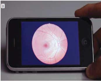

3 (The Neurologist 2012;18: ) Non-mydriatic fundus cameras Easy for non-ophthalmic trained individuals to use No pupillary dilation Able to take quality photographs of the posterior pole Reveals unrecognized findings in ED (Bruce et al. NEJM 2011; 364:387-9) Arch Ophthalmol 2012; 130:


4 Optic Neuropathy Classic Features Decreased visual acuity Abnormal visual field Relative afferent pupillary defect Can see through to the nerve Swollen or pale optic nerve Optic Neuropathy Disc Alternatives Inflammatory Vascular Causes Compressive/Infiltrative Toxic/Nutritional Hereditary Traumatic Optic Neuropathy Elevated intracranial pressure Elevated intraocular pressure 4


 Idiopathic intracranial hypertension")

5 Inflammatory Vascular Causes Compressive/Infiltrative Toxic/Nutritional Hereditary Traumatic Optic Neuropathy Elevated intracranial pressure Elevated intraocular pressure Optic Neuropathy Papilledema Disc swelling from intracranial pressure Any age Painless Bilateral Spares visual acuity Constriction of visual field Papilledema Causes Intracranial mass lesions Hydrocephalus Meningeal processes Cerebral venous thrombosis Idiopathic (pseudotumor cerebri) Idiopathic intracranial hypertension Papilledema Headaches No localizing neurologic symptoms/signs except for VIth No intracranial process, no venous sinus thrombosis Normal CSF contents CSF opening pressure 25cm H2O 5


6 IIH imaging Elevated ICP measured in the lateral decubitus position: neonates: >76 mm H2O, age 1 18 years: >280 mm H2O Normal CSF composition except in neonates who may have up to 19 WBC/mm3 if 0 28 days and up to 9 WBC/mm3 if between 29 and 56 days old; the protein may be as high as 150 mg/dl IIH: Poor visual prognosis Not just a diagnosis of exclusion New diagnostic criteria Papilledema Measure of intracranial pressure Neuroimaging findings Patient s characteristics Black race. Neurology 2008; 70: Male. Neurology 2009; 72:304-9 Severe obesity. J Neuro-Ophthalmol 2013; 33: 4-8 Anemia / sleep apnea syndrome / HTN Rapid onset (fulminant IIH). Neurology 2007; 68:


7 IIH is everywhere there are obese people 7


8 ScientificWorldJournal. 2015; 2015: Clinical course of idiopathic intracranial hypertension with transverse sinus stenosis Neurology 2013;80: TS stenosis After stenting Inflammatory Vascular Causes Compressive/Infiltrative Toxic/Nutritional Hereditary Traumatic Optic Neuropathy Elevated intracranial pressure Elevated intraocular pressure 8


9 Optic Neuropathy Typical Optic Neuritis Inflammation of the optic nerve F:M 3:1 Age: Pain on eye movement Normal or swollen disc Spontaneous improvement Associated with multiple sclerosis ONTT No difference in visual acuity between steroid and placebo groups at 6 months. I.V. steroids may accelerate recovery by 2 to 3 weeks. P.O. steroids doubled the risk of recurrence in either eye. ONTT: MRI predicts the risk of MS Total 50% at 15 yrs 0 Years lesion 72% No lesion 25% (NEJM 326:581, 1992) 9
 Hemorrhage, disk or peripapillary (16 patients) Macular Exudates (8")

:34-43. doi:10.1001/jamaneurol.2013.")


, the segmentation software identifies the outer boundaries of the macular RNFL, IPL, and")
10 Clinical Features of Optic Neuritis with Low Risk of CDMS in Patients with No Brain MRI Lesions No cases of CDMS have developed when any one of the following clinical features* was present: Severe Disc Swelling (21 patients) Hemorrhage, disk or peripapillary (16 patients) Macular Exudates (8 patients) Painless (19 patients) No Light Perception (7 patients) OCT: Retinal Nerve Fiber Layer (RNFL) Thickness Correlates with axonal loss Correlates with visual dysfunction Correlates with: Brain atrophy in MS Disability Quality of life From: Relationships Between Retinal Axonal and Neuronal Measures and Global Central Nervous System Pathology in Multiple Sclerosis JAMA Neurol. 2013;70(1): doi: /jamaneurol Cellular composition of the retinal layers : ILM: inner limiting membrane RNFL: retinal nerve fiber layer GCL: ganglion cell layer IPL: inner plexiform layer INL: inner nuclear layer OPL: outer plexiform layer ONL: outer nuclear layer ELM: external limiting membrane IPS: inner photoreceptor segments OPS: outer photoreceptor segments PR: photoreceptors RPE: retinal pigment epithelium -Healthy subject. 3-dimensional macular volume cube generated by Cirrus HD-OCT from the macular region -The individual layers of the retina are readily discernible, except for GCL and IPL, which are difficult to distinguish. -During the segmentation process (performed in 3-dimension), the segmentation software identifies the outer boundaries of the macular RNFL, IPL, and OPL, as well as the inner boundary of the RPE, which is identified by the conventional Cirrus HD-OCT algorithm. The identification of these boundaries facilitates OCT segmentation, enabling determination of the thicknesses of the macular RNFL, GCL + IPL, the INL + OPL, and the ONL including the inner and outer photoreceptor segments 10
;")


 at 3-4 months of therapy")
 Inflammatory")

11 Fingolimod and Macular Edema 1. Incidence of macular edema is low (~1%); (uveitis, DM increase risk). Ophthalmology 2013; 120: Screening evaluation for uveitis, macular or retinal vascular disease prior to starting, or within the first few weeks of starting fingolimod 3. Re-evaluation (complete eye exam +/- macular OCT) at 3-4 months of therapy (most reported cases of macular edema occurred within 3-4 months) Inflammatory Vascular Causes Compressive/Infiltrative Toxic/Nutritional Hereditary Traumatic Optic Neuropathy Elevated intracranial pressure Elevated intraocular pressure 11


12 Optic Neuritis and NMO Abs Severe Bilateral Bilateral Poor recovery Severe Recurrent Poor recovery Recurrent Optic Neuritis and NMO Abs 12
Ganglion cell analysis by optical coherence tomography (OCT) Jonathan A. Micieli, MD Valérie Biousse, MD
 Jonathan A. Micieli, MD Valérie Biousse, MD") Ganglion cell analysis by optical coherence tomography (OCT) Jonathan A. Micieli, MD Valérie Biousse, MD Figure 1. Normal OCT of the macula (cross section through the line indicated on the fundus photo)
Ganglion cell analysis by optical coherence tomography (OCT) Jonathan A. Micieli, MD Valérie Biousse, MD Figure 1. Normal OCT of the macula (cross section through the line indicated on the fundus photo)
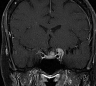
Typical idiopathic intracranial hypertension Optic nerve appearance and brain MRI findings. Jonathan A. Micieli, MD Valérie Biousse, MD
 Typical idiopathic intracranial hypertension Optic nerve appearance and brain MRI findings Jonathan A. Micieli, MD Valérie Biousse, MD A 24 year old African American woman is referred for bilateral optic
Neuro-Ocular Grand Rounds Anthony B. Litwak,OD, FAAO VA Medical Center Baltimore, Maryland
 Neuro-Ocular Grand Rounds Anthony B. Litwak,OD, FAAO VA Medical Center Baltimore, Maryland Dr. Litwak is on the speaker and advisory boards for Alcon and Zeiss Meditek COMMON OPTIC NEUROPATHIES THAT CAN
Optic Nerve Disorders: Structure and Function and Causes
 Optic Nerve Disorders: Structure and Function and Causes Using Visual Fields, OCT and B-scan Ultrasound to Diagnose and Follow Optic Nerve Visual Losses Ohio Ophthalmological Society and Ophthalmic Tech
Neuro-Ocular Grand Rounds
 Neuro-Ocular Grand Rounds Anthony B. Litwak,OD, FAAO VA Medical Center Baltimore, Maryland Dr. Litwak is on the speaker and advisory boards for Alcon and Zeiss Meditek COMMON OPTIC NEUROPATHIES THAT CAN
12/2/16. Ways to differentiate:
 Nate Lighthizer, O.D., F.A.A.O. Assistant Dean for Clinical Care Services Director of CE Chief of Specialty Care Clinics Chief of Electrodiagnostics Clinic Oklahoma College of Optometry lighthiz@nsuok.edu
Index. Note: Page numbers of article titles are in boldface type.
 Index Note: Page numbers of article titles are in boldface type. A Acetazolamide, in idiopathic intracranial hypertension, 49 52, 60 Angiography, computed tomography, in cranial nerve palsy, 103 107 digital
THE SWOLLEN DISC. Valerie Biousse, MD Emory University School of Medicine Atlanta, GA
 THE SWOLLEN DISC Valerie Biousse, MD Emory University School of Medicine Atlanta, GA Updated from: Neuro-Ophthalmology Illustrated. Biousse V, Newman NJ. Thieme, New-York,NY. 2 nd Ed, 2016. Edema of the
OCT Image Analysis System for Grading and Diagnosis of Retinal Diseases and its Integration in i-hospital
 Progress Report for1 st Quarter, May-July 2017 OCT Image Analysis System for Grading and Diagnosis of Retinal Diseases and its Integration in i-hospital Milestone 1: Designing Annotation tool extraction
The Case: A 64 yo man with chronic back pain has elective multilevel lumbar spinal surgery
 The Case: A 64 yo man with chronic back pain has elective multilevel lumbar spinal surgery The Case: Upon awakening from anesthesia, he is blind in both eyes After Non-Ocular Surgeries Nancy J. Newman,
Papilledema. Golnaz Javey, M.D. and Jeffrey J. Zuravleff, M.D.
 Papilledema Golnaz Javey, M.D. and Jeffrey J. Zuravleff, M.D. Papilledema specifically refers to optic nerve head swelling secondary to increased intracranial pressure (IICP). Optic nerve swelling from
OPTIC NERVE SWELLING IN CHILDHOOD
 OPTIC NERVE SWELLING IN CHILDHOOD Melissa W. Ko, MD, FAAN One of the main findings on a pediatric neurologic examination that can instill fear and lead to an urgent referral to neuro-ophthalmology is the
Non-arteritic anterior ischemic optic neuropathy (NAION) with segmental optic disc edema. Jonathan A. Micieli, MD Valérie Biousse, MD
 with segmental optic disc edema. Jonathan A. Micieli, MD Valérie Biousse, MD") Non-arteritic anterior ischemic optic neuropathy (NAION) with segmental optic disc edema Jonathan A. Micieli, MD Valérie Biousse, MD A 75 year old white woman lost vision in the inferior part of her visual
Non-arteritic anterior ischemic optic neuropathy (NAION) with segmental optic disc edema Jonathan A. Micieli, MD Valérie Biousse, MD A 75 year old white woman lost vision in the inferior part of her visual
Intracranial hypertension and headache. Daniel Tibussek, MD
 Intracranial hypertension and headache. Daniel Tibussek, MD none Disclosures Overview Case Clinical presentation of pediatric PTC Nomenclature, Definition What is intracranial hypertension? Diagnostic
Sequential non-arteritic anterior ischemic optic neuropathy (NAION) Jonathan A. Micieli, MD Valérie Biousse, MD
 Jonathan A. Micieli, MD Valérie Biousse, MD") Sequential non-arteritic anterior ischemic optic neuropathy (NAION) Jonathan A. Micieli, MD Valérie Biousse, MD A 68 year old white woman had a new onset of floaters in her right eye and was found to have
Sequential non-arteritic anterior ischemic optic neuropathy (NAION) Jonathan A. Micieli, MD Valérie Biousse, MD A 68 year old white woman had a new onset of floaters in her right eye and was found to have
Fundus Autofluorescence. Jonathan A. Micieli, MD Valérie Biousse, MD
 Fundus Autofluorescence Jonathan A. Micieli, MD Valérie Biousse, MD The retinal pigment epithelium (RPE) has many important functions including phagocytosis of the photoreceptor outer segments Cone Rod
I have nothing to disclose but I
 OPTIC NEUROPATHIES Robert L. Tomsak MD PhD Professor of Ophthalmology and Neurology Wayne State t University it Sh School of Mdii Medicine I have nothing to disclose but I wish I did. dd Road map for this
Brain Advance Access published October 17, doi: /brain/awr264 Brain 2011: Page 1 of 13 1
 Brain Advance Access published October 17, 2011 doi:10.1093/brain/awr264 Brain 2011: Page 1 of 13 1 BRAIN A JOURNAL OF NEUROLOGY Optical coherence tomography segmentation reveals ganglion cell layer pathology
Role Of Various Factors In The Treatment Of Optic Neuritis----A Study Abstract Aim: Materials & Methods Discussion: Conclusion: Key words
 IOSR Journal of Dental and Medical Sciences (IOSR-JDMS) e-issn: 2279-0853, p-issn: 2279-0861.Volume 15, Issue 9 Ver. X (September). 2016), PP 51-57 www.iosrjournals.org Role Of Various Factors In The Treatment
Learn Connect Succeed. JCAHPO Regional Meetings 2015
 Learn Connect Succeed JCAHPO Regional Meetings 2015 OPTIC NEUROPATHY AS EASY AS 1,2,3,4 OPTIC NERVE ANATOMY M. Tariq Bhatti, MD Departments of Ophthalmology and Neurology Duke Eye Center and Duke University
3/16/2018. Optic Nerve Examination. Hassan Eisa Swify FRCS Ed (Ophthalmology) Air Force Hospital
 Air Force Hospital") Optic Nerve Examination Hassan Eisa Swify FRCS Ed (Ophthalmology) Air Force Hospital 1 Examination Structure ( optic disc) Function Examination of the optic disc The only cranial nerve (brain tract) which
Optic Nerve Examination Hassan Eisa Swify FRCS Ed (Ophthalmology) Air Force Hospital 1 Examination Structure ( optic disc) Function Examination of the optic disc The only cranial nerve (brain tract) which
Slide 4. Slide 5. Slide 6
 Slide 1 Slide 4 Demographics El Paso Eye Care Border Healthcare-Based Grand Rounds Derek N. Cunningham, O.D. 80-90% Mexican-Americans Diabetes Hypertension Hyperlipidemia Obesity 70% uninsured High poverty
Prevalence of venous sinus stenosis in Pseudotumor cerebri(ptc) using digital subtraction angiography (DSA)
 using digital subtraction angiography (DSA)") Prevalence of venous sinus stenosis in Pseudotumor cerebri(ptc) using digital subtraction angiography (DSA) Dr.Mohamed hamdy ibrahim MBBC,MSc,MD, PhD Neurology Degree Kings lake university (USA). Fellow
Prevalence of venous sinus stenosis in Pseudotumor cerebri(ptc) using digital subtraction angiography (DSA) Dr.Mohamed hamdy ibrahim MBBC,MSc,MD, PhD Neurology Degree Kings lake university (USA). Fellow
LECTURE # 7 EYECARE REVIEW: PART III
 LECTURE # 7 EYECARE REVIEW: PART III HOW TO TRIAGE EYE EMERGENCIES STEVE BUTZON, O.D. EYECARE REVIEW: HOW TO TRIAGE EYE EMERGENCIES FOR PRIMARY CARE PHYSICIANS Steve Butzon, O.D. Member Director IDOC President
Eye Movements, Strabismus, Amblyopia and Neuro-Ophthalmology
 Eye Movements, Strabismus, Amblyopia and Neuro-Ophthalmology Retinal Ganglion Cell Layer Thinning Within One Month of Presentation for Non-Arteritic Anterior Ischemic Optic Neuropathy Mark J. Kupersmith,
Idiopathic Intracranial Hypertension
 Idiopathic Intracranial Hypertension Dr. Mar'n Su+onBrown MD. FRCPC Neuro-Ophthalmology, Neurology Div of Neurology, Island Health Clinical Assistant Professor, Div of Neurology, UBC Stroke Rapid Assessment
Supplementary Online Content
 Supplementary Online Content Park KH, Kim YK, Woo SJ, et al. Iatrogenic occlusion of the ophthalmic artery after cosmetic facial filler injections: a national survey by the Korean Retina Society. JAMA
Case #1: 68 M with floaters OS
 Case #1: 68 M with floaters OS Point-of-Care Ocular Sonography for the Emergency Department Nate Teismann MD Dept of Emergency Medicine, UCSF Topics in EM 2012 Acute onset of dark spots in L eye 2 days
Neuro-ophthalmologyophthalmology. Marek Michalec, MD.
 Neuro-ophthalmologyophthalmology Marek Michalec, MD. Neuro-ophthalmology Study integrating ophthalmology and neurology Disorders affecting parts of CNS devoted to vision or eye: Afferent system (visual
Idiopathic Intracranial Hypertension (Pseudotumor Cerebri) David I. Kaufman, D.O. Michigan State University Department of Neurology and Ophthalmology
 David I. Kaufman, D.O. Michigan State University Department of Neurology and Ophthalmology") Idiopathic Intracranial Hypertension (Pseudotumor Cerebri) David I. Kaufman, D.O. Michigan State University Department of Neurology and Ophthalmology 26 year old 5 3, 300 pound female with papilledema,
Idiopathic Intracranial Hypertension (Pseudotumor Cerebri) David I. Kaufman, D.O. Michigan State University Department of Neurology and Ophthalmology 26 year old 5 3, 300 pound female with papilledema,
THE EYE: RETINA AND GLOBE
 Neuroanatomy Suzanne Stensaas February 24, 2011, 10:00-12:00 p.m. Reading: Waxman Ch. 15. Your histology and gross anatomy books should be useful. Reading: Histology of the Eye from any histology book
The Prevalence of diabetic optic neuropathy in type 2 diabetes mellitus
 The Prevalence of diabetic optic neuropathy in type 2 diabetes mellitus Received: 25/4/2016 Accepted: 8/12/2016 Introduction Diabetic papillopathy is an atypical form of non-arteritic anterior ischemic
Optical Coherence Tomography: Pearls for the Anterior Segment Surgeon Basic Science Michael Stewart, M.D.
 Optical Coherence Tomography: Pearls for the Anterior Segment Surgeon Basic Science Michael Stewart, M.D. Disclosure OCT Optical Coherence Tomography No relevant financial relationships I will refer to
The role of optical coherence tomography in neuro-ophthalmology
 Review Article Page 1 of 18 The role of optical coherence tomography in neuro-ophthalmology John J. Chen 1,2, Fiona Costello 3,4 1 Department of Ophthalmology, 2 Department of Neurology, Mayo Clinic, Rochester,
CHAPTER 13 CLINICAL CASES INTRODUCTION
 2 CHAPTER 3 CLINICAL CASES INTRODUCTION The previous chapters of this book have systematically presented various aspects of visual field testing and is now put into a clinical context. In this chapter,
BMB Disclosures. Papilledema can be a. Neurological Emergency, Causing Preventable Blindness
 Reasonable Doubt: Can High Intracranial Pressure Occur Without Papilledema? 15 February 2013 Jonathan C. Horton hortonj@vision.ucsf.edu http://www.ucsf.edu/hortonlab BMB Disclosures Financial Disclosures
The Quick Guide to OCT Mastery 50 Real Cases with Expert Analysis
 OPTICAL COHERENCE TOMOGRAPHY The Quick Guide to OCT Mastery 50 Real Cases with Expert Analysis VOL 1 Sanjay Sharma, MD, FRCS, MSc (Epid), MBA Ophthalmologist, Epidemiologist Queen s University, Canada
Cases of visual impairment caused by cerebral venous sinus occlusion-induced intracranial hypertension in the absence of headache
 Zhao et al. BMC Neurology (2018) 18:159 https://doi.org/10.1186/s12883-018-1156-7 CASE REPORT Open Access Cases of visual impairment caused by cerebral venous sinus occlusion-induced intracranial hypertension
Fatigue And Beyond How Vision Captures Disease in MS
 Fatigue And Beyond How Vision Captures Disease in MS Salim Chahin, MD Fellow Multiple Sclerosis University of Pennsylvania Outline Introduction. Visual function testing disease outcomes. Optical Coherence
Anterior Ischemic Optic Neuropathy (AION)
") Anterior Ischemic Optic Neuropathy (AION) Your doctor thinks you have suffered an episode of anterior ischemic optic neuropathy (AION). This is the most common cause of sudden decreased vision in patients
Anterior Ischemic Optic Neuropathy (AION) Your doctor thinks you have suffered an episode of anterior ischemic optic neuropathy (AION). This is the most common cause of sudden decreased vision in patients
OCT Interpretation in Retinal Disease
 OCT Interpretation in Retinal Disease Jay M. Haynie, OD, FAAO Financial Disclosure I have received honoraria or am on the advisory board for the following companies: Carl Zeiss Meditec Advanced Ocular
OPTIC NEUROPATHIES Optic Neuritis vs AION. Jacqueline M.S. Winterkorn, Ph.D., M.D.
 OPTIC NEUROPATHIES Optic Neuritis vs AION Jacqueline M.S. Winterkorn, Ph.D., M.D. OPTIC NEUROPATHIES Inflammatory Optic Neuritis Ischemic Optic Neuropathy Compressive Optic Neuropathy Traumatic Optic
Shared embryology Eye and brain develop from neuro-ectoderm
 The Patient with Visual Loss: Localization of Neuropathologic Disease and Select Diseases of Neuropathologic Interest Steven A. Kane, M.D., Ph.D. The Edward S. Harkness Eye Institute Shared embryology
Measuring of the fovea and foveola using line scans and 3D Macular scans obtained with spectral domain optical coherent tomography.
 Measuring of the fovea and foveola using line scans and 3D Macular scans obtained with spectral domain optical coherent tomography. Vakhrameeva O.A. 1, Moiseenko G.A. 1, Maltsev D.S. 2, Sukhinin M.V. 2,
Unexplained visual loss in seven easy steps
 Unexplained visual loss in seven easy steps Andrew G. Lee, MD Chair Ophthalmology, Houston Methodist Hospital, Professor, Weill Cornell MC; Adjunct Professor, Baylor COM, U Iowa, UTMB Galveston, UT MD
Longitudinal Changes of Retinal Thicknesses in Branch Retinal Artery Occlusion: Spectral-Domain Optical Coherence Tomography Study METHODS.
 Retina Longitudinal Changes of Retinal Thicknesses in Branch Retinal Artery Occlusion: Spectral-Domain Optical Coherence Tomography Study Min-Su Kim, 1 Kyeung-Min Kim, 1 Hyung-Bin Lim, 1,2 Young-Joon Jo,
Analysis of Fundus Photography and Fluorescein Angiography in Nonarteritic Anterior Ischemic Optic Neuropathy and Optic Neuritis
 pissn: 1011-8942 eissn: 2092-9382 Korean J Ophthalmol 2016;30(4):289-294 http://dx.doi.org/10.3341/kjo.2016.30.4.289 Original Article Analysis of Fundus Photography and Fluorescein Angiography in Nonarteritic
Misdiagnosed Vogt-Koyanagi-Harada (VKH) disease and atypical central serous chorioretinopathy (CSC)
 disease and atypical central serous chorioretinopathy (CSC)") HPTER 12 Misdiagnosed Vogt-Koyanagi-Harada (VKH) disease and atypical central serous chorioretinopathy (S) linical Features VKH disease is a bilateral granulomatous panuveitis often associated with exudative
HPTER 12 Misdiagnosed Vogt-Koyanagi-Harada (VKH) disease and atypical central serous chorioretinopathy (S) linical Features VKH disease is a bilateral granulomatous panuveitis often associated with exudative
NIH Public Access Author Manuscript Retin Cases Brief Rep. Author manuscript; available in PMC 2012 January 1.
 NIH Public Access Author Manuscript Published in final edited form as: Retin Cases Brief Rep. 2011 ; 5(1): 46 48. doi:10.1097/icb.0b013e3181cafc49. Intact Retinal Tissue and Retinal Pigment Epithelium
Retro-bulbar visual anatomy Optic nerves carry. Normal left ocular fundus. Retinal nerve fiber layer anatomy
 The Patient with Visual Loss: Localization of Neuropathologic Disease and Select Diseases of Neuropathologic Interest Steven A. Kane, M.D., Ph.D. The Edward S. Harkness Eye Institute Shared embryology
Moncef Khairallah, MD
 Moncef Khairallah, MD Department of Ophthalmology, Fattouma Bourguiba University Hospital Faculty of Medicine, University of Monastir Monastir, Tunisia INTRODUCTION IU: anatomic form of uveitis involving
This is the author's manuscript of the article published in final edited form as:
 Nonmydriatic Fundus Photography: A Practical Review for the Neurologist Devin D. Mackay, M.D.; Beau B. Bruce, M.D., Ph.D. From the Departments of Neurology, Ophthalmology, and Neurosurgery (DDM), Indiana
Ji Soo Shin, Young Hoon Lee. Department of Ophthalmology, Konyang University College of Medicine, Daejeon, Korea
 pissn: 1011-8942 eissn: 2092-9382 Korean J Ophthalmol 2017;31(6):497-507 https://doi.org/10.3341/kjo.2016.0108 Original Article Changes in Macular Retinal Layers and Peripapillary Nerve Fiber Layer Thickness
Khalil Zahra, M.D Neuro-interventional radiology
 Khalil Zahra, M.D Neuro-interventional radiology 1 Disclosure None 2 Outline Etiology and pathogensis Imaging techniques and Features Literature review Treatment modalities Endovascular techniques Long
Eye and b develop fr neuro-ect. Their func. to disease. Blood ocular/bra. The eye is The eye is window in
 The Patient wit th Visual Loss: Localization of Neuropathologic Disease and Select Diseases of Neuropathologic Interest Steven A. Kan e, MD M.D., PhD Ph.D. The Edward S. Harkness Eye Institute The eye
OBSTRUCTIVE sleep apnea
 CLINICAL SCIENCES Papilledema and Obstructive Sleep Apnea Syndrome Valerie A. Purvin, MD; Aki Kawasaki, MD; Robert D. Yee, MD Objectives: To characterize the pathogenesis and clinical features of optic
Ganglion cell complex scan in the early prediction of glaucoma
 Original article in the early prediction of glaucoma Ganekal S Nayana Super Specialty Eye Hospital and Research Center, Davangere, Karnataka, India Abstract Objective: To compare the macular ganglion cell
Speaker Disclosure Statement. " Dr. Tim Maillet and Dr. Vladimir Kozousek have no conflicts of interest to disclose.
 Speaker Disclosure Statement Dr. Tim Maillet and Dr. Vladimir Kozousek have no conflicts of interest to disclose. Diabetes Morbidity Diabetes doubles the risk of stroke. Diabetes quadruples the risk of
What Is O.C.T. and Why Should I Give A Rip? OCT & Me How Optical Coherence Tomography Changed the Life of a Small Town Optometrist 5/19/2014
 OCT & Me How Optical Coherence Tomography Changed the Life of a Small Town Optometrist Email: myoder@wcoil.com Mark A. Yoder, O.D. 107 N. Main Street PO Box 123 Bluffton, OH 45817 @yoderod 115.02 Histoplasma
WORKSHOP B Ophthalmic Imaging: All Hands on Tech! COPE Course PS
 WORKSHOP B Ophthalmic Imaging: All Hands on Tech! COPE Course 44334-PS Ophthalmic Imaging: All Hands on Tech! Southern College of Optometry April 17, 2015 COPE #44334-PS Faculty Dr. Michael Gerstner Dr.
SOUTH-EAST EUROPEAN JOURNAL of OPHTHALMOLOGY 2015; 1 (1) 34 40
 34 40") Review article SOUTH-EAST EUROPEAN JOURNAL of OPHTHALMOLOGY 2015; 1 (1) 34 40 Retinal nerve fiber layer versus peripapillary capillary density assessment A powerful tool for detecting optic nerve head
Review article SOUTH-EAST EUROPEAN JOURNAL of OPHTHALMOLOGY 2015; 1 (1) 34 40 Retinal nerve fiber layer versus peripapillary capillary density assessment A powerful tool for detecting optic nerve head
An Organized Approach to the Patient with Papilledema and IIH
 An Organized Approach to the Patient with Papilledema and IIH Leonard V. Messner, OD, FAAO James L. Fanelli, OD, FAAO Please silence all mobile devices and remove items from chairs so others can sit. Unauthorized
Local Intra-arterial Fibrinolysis in Treatment of Incomplete Ophthalmic Artery Occlusion A Case Report
 CASE REPORT Local Intra-arterial Fibrinolysis in Treatment of Incomplete Ophthalmic Artery Occlusion A Case Report Shih-Ting Fang, Pao-Sheng Yen 1, Chien-Chung Chen, Yuan-Chieh Lee Department of Ophthalmology,
A Case of Carotid-Cavernous Fistula
 A Case of Carotid-Cavernous Fistula By : Mohamed Elkhawaga 2 nd Year Resident of Ophthalmology Alexandria University A 19 year old male patient came to our outpatient clinic, complaining of : -Severe conjunctival
Cirrus TM HD-OCT. Details defi ne your decisions
 Cirrus TM HD-OCT Details defi ne your decisions 2 With high-defi nition OCT Carl Zeiss Meditec takes you beyond standard spectral domain Built on 10 years experience at the vanguard of innovation, Carl
Case Report Optic Disk Pit with Sudden Central Visual Field Scotoma
 Case Reports in Ophthalmological Medicine Volume 2016, Article ID 1423481, 4 pages http://dx.doi.org/10.1155/2016/1423481 Case Report Optic Disk Pit with Sudden Central Visual Field Scotoma Nikol Panou
11/10/2017. Headache and Increased Pressure: A tale of 2 cases. Kathleen Digre MD University of Utah TWO CASES. 23 yo medical practice manager
 Headache and Increased Pressure: A tale of 2 cases Kathleen Digre MD University of Utah TWO CASES 23 yo medical practice manager September 2016 began developing intense frontal headaches first intermittent
3/16/2018. Optic nerve axons of retinal ganglion cells. 1.2 million nerve fibers. ON sheath: continuous with the meninges dura arachnoid and pia mater
 Optic nerve axons of retinal ganglion cells 1.2 million nerve fibers. ON sheath: continuous with the meninges dura arachnoid and pia mater 1 1.Visual Acuity 2.Color Vision 3.Pupil 4.Contrast sensitivity
Clinically Significant Macular Edema (CSME)
") Clinically Significant Macular Edema (CSME) 1 Clinically Significant Macular Edema (CSME) Sadrina T. Shaw OMT I Student July 26, 2014 Advisor: Dr. Uwaydat Clinically Significant Macular Edema (CSME) 2
Clinically Significant Macular Edema (CSME) 1 Clinically Significant Macular Edema (CSME) Sadrina T. Shaw OMT I Student July 26, 2014 Advisor: Dr. Uwaydat Clinically Significant Macular Edema (CSME) 2
Rare Presentation of Ocular Toxoplasmosis
 Case Report Rare Presentation of Ocular Toxoplasmosis Rakhshandeh Alipanahi MD From Department of Ophthalmology, Nikookari Eye Hospital, Tabriz University of Medical Sciences, Tabriz, Iran. Correspondence:
OCT Interpretation. Financial Disclosure. Jay M. Haynie, OD, FAAO. OCT Image Layers 7/21/2014
 OCT Interpretation Jay M. Haynie, OD, FAAO Financial Disclosure I have received honoraria or am on the advisory board for the following companies: Olympia Tacoma Renton Kennewick - Washington Carl Zeiss
Supplementary information Novel VCP modulators mi2gate major pathologies of rd10, a mouse model of re2ni2s pigmentosa
 Supplementary information Novel VCP modulators mi2gate major pathologies of rd1, a mouse model of re2ni2s pigmentosa Hanako Ohashi Ikeda, Norio Sasaoka, Masaaki Koike, Noriko Nakano, Yuki Muraoka, Yoshinobu
Method for comparing visual field defects to local RNFL and RGC damage seen on frequency domain OCT in patients with glaucoma.
 Method for comparing visual field defects to local RNFL and RGC damage seen on frequency domain OCT in patients with glaucoma. Donald C. Hood 1,2,* and Ali S. Raza 1 1 Department of Psychology, Columbia
53 year old woman attends your practice for routine exam. She has no past medical history or family history of note.
 Case 1 Normal Tension Glaucoma 53 year old woman attends your practice for routine exam. She has no past medical history or family history of note. Table 1. Right Eye Left Eye Visual acuity 6/6 6/6 Ishihara
Reports. Macular Thickness as a Potential Biomarker of Mild Alzheimer s Disease
 Reports Macular Thickness as a Potential Biomarker of Mild Alzheimer s Disease Although several postmortem findings in the retina of patients with Alzheimer s disease (AD) are available, 1 new biomarkers
Principle of OCT. Reading Between the Lines: OCT Interpretation. Initial Concept. Advantage: High Resolution Cross Section Images
 Principle of OCT Reading Between the Lines: OCT Interpretation Mohammad Rafieetary, OD, FAAO mrafieetary@charlesretina.com Introduction Optical Biopsy Morphologic Evaluation of Live Tissue Measurements
EFFECT OF OPTIC DISK FOVEA DISTANCE ON MEASUREMENTS OF INDIVIDUAL MACULAR INTRARETINAL LAYERS IN NORMAL SUBJECTS
 EFFECT OF OPTIC DISK FOVEA DISTANCE ON MEASUREMENTS OF INDIVIDUAL MACULAR INTRARETINAL LAYERS IN NORMAL SUBJECTS KUNLIANG QIU, MD,* BINYAO CHEN, MD,* HAOYU CHEN, MD,* ENTING GAO, PHD, JIANLING YANG, MD,*
OPTIC DISC PIT Pathogenesis and Management OPTIC DISC PIT
 OPTIC DISC PIT Pathogenesis and Management Abdel-Latif Siam Ain Shams University Cairo Egypt OPTIC DISC PIT Congenital pit is an atypical coloboma usually located on the temporal edge of the disc, associated
History/principles of the OCT What does the normal retinal OCT look like Vitreal disorders Retinal/RPE disorders Choroidal disorders
 Nathan Lighthizer, O.D., F.A.A.O. Assistant Professor Assistant Dean for Clinical Care Director of Continuing Education Chief of Specialty Care Clinics Chief of Electrodiagnostics Clinic Oklahoma College
Ethambutol (EMB), the first-line drug used to treat mycobacterium
, the first-line drug used to treat mycobacterium") Eye Movements, Strabismus, Amblyopia and Neuro-Ophthalmology Ganglion Cell Layer and Inner Plexiform Layer as Predictors of Vision Recovery in Ethambutol-Induced Optic Neuropathy: A Longitudinal OCT Analysis
Eye Movements, Strabismus, Amblyopia and Neuro-Ophthalmology Ganglion Cell Layer and Inner Plexiform Layer as Predictors of Vision Recovery in Ethambutol-Induced Optic Neuropathy: A Longitudinal OCT Analysis
8/6/17. Disclosures Aerie Pharmaceuticals Alcon BioTissue Diopsys Optovue Shire
 Nathan Lighthizer, O.D., F.A.A.O. Associate Professor Assistant Dean for Clinical Care Director of Continuing Education Chief of Specialty Care Clinics Oklahoma College of Optometry Tahlequah, OK lighthiz@nsuok.edu
Evolving glaucoma management True diagnostic integration for the preservation of vision
 Evolving glaucoma management True diagnostic integration for the preservation of vision // GLAUCOMA MANAGEMENT MADE BY ZEISS The moment you are certain it is glaucoma. This is the moment we work for. There
Neuropathy (NAION) and Avastin. Clinical Assembly of the AOCOO-HNS Foundation May 9, 2013
 and Avastin. Clinical Assembly of the AOCOO-HNS Foundation May 9, 2013") Non Arteritic Ischemic Optic Neuropathy (NAION) and Avastin Shalom Kelman, MD Clinical Assembly of the AOCOO-HNS Foundation May 9, 2013 Anterior Ischemic Optic Neuropathy Acute, painless, visual loss,
Non Arteritic Ischemic Optic Neuropathy (NAION) and Avastin Shalom Kelman, MD Clinical Assembly of the AOCOO-HNS Foundation May 9, 2013 Anterior Ischemic Optic Neuropathy Acute, painless, visual loss,
Manifestations of central retinal artery occlusion revealed by fundus fluorescein angiography are associated with the degree of visual loss
 2420 Manifestations of central retinal artery occlusion revealed by fundus fluorescein angiography are associated with the degree of visual loss HONGXIA GONG 1, QIUYING SONG 2 and LANHUI WANG 3 1 Department
An A to Z guide on Epiretinal Membranes (ERMs) Paris Tranos PhD,ICO,FRCS OPHTHALMICA Vitreoretinal & Uveitis Department
 Paris Tranos PhD,ICO,FRCS OPHTHALMICA Vitreoretinal & Uveitis Department") An A to Z guide on Epiretinal Membranes (ERMs) Paris Tranos PhD,ICO,FRCS OPHTHALMICA Vitreoretinal & Uveitis Department Types of ERM Natural history OCT prognostic factors ERM with co-existing pathology
An A to Z guide on Epiretinal Membranes (ERMs) Paris Tranos PhD,ICO,FRCS OPHTHALMICA Vitreoretinal & Uveitis Department Types of ERM Natural history OCT prognostic factors ERM with co-existing pathology
PRIMUS 200 from ZEISS The essential OCT
 EN 00_00I The contents of the brochure may differ from the current status of approval of the product in your country. Please contact your regional representative for more information. Subject to change
Benign Multiple Sclerosis is Associated with Reduced Thinning of the Retinal Nerve Fiber and Ganglion Cell Layers in Non-Optic-Neuritis Eyes
 JCN Open Access pissn 1738-6586 / eissn 2005-5013 / J Clin Neurol 2015;11(3):241-247 / http://dx.doi.org/10.3988/jcn.2015.11.3.241 ORIGINAL ARTICLE Benign Multiple Sclerosis is Associated with Reduced
Learn Connect Succeed. JCAHPO Regional Meetings 2017
 Learn Connect Succeed JCAHPO Regional Meetings 2017 NO FINANCIAL DISCLOSURES Technician s Role in Neuro-Ophthalmology Workup Beth Koch COT, ROUB Cleveland 9/16/2017 What Tests Are You Expected To Perform?
Cirrus TM HD-OCT. Details define your decisions
 Cirrus TM HD-OCT Details define your decisions 2 With high-definition OCT Carl Zeiss Meditec takes you beyond standard spectral domain Built on 10 years experience at the vanguard of innovation, Carl Zeiss
Is it Papilloedema? John Ross Ainsworth Orthoptic staff Birmingham Children s Hospital Birmingham and Midland Eye Centre University of Birmingham
 Is it Papilloedema? John Ross Ainsworth Orthoptic staff Birmingham Children s Hospital Birmingham and Midland Eye Centre University of Birmingham Aims Children/young people A bit about hypoplasia / NFL
Optic Disc Evaluation: Is the Optic Disc Glaucomatous and Has it Progressed?
 Optic Disc Evaluation: Is the Optic Disc Glaucomatous and Has it Progressed? Jody Piltz-Seymour, M.D. Clinical Professor Perelman School of Medicine University of Pennsylvania Wills Glaucoma Service Valley
OCT in the Diagnosis and Follow-up of Glaucoma
 OCT in the Diagnosis and Follow-up of Glaucoma Karim A Raafat MD. Professor Of Ophthalmology Cairo University Hmmmm! Do I have Glaucoma or not?! 1 Visual Function 100% - N Gl Structure : - 5000 axon /
Diabetic Retinopathy. Barry Emara MD FRCS(C) Giovanni Caboto Club October 3, 2012
 Giovanni Caboto Club October 3, 2012") Diabetic Retinopathy Barry Emara MD FRCS(C) Giovanni Caboto Club October 3, 2012 Outline Statistics Anatomy Categories Assessment Management Risk factors What do you need to do? Objectives Summarize the
Diabetic Retinopathy Barry Emara MD FRCS(C) Giovanni Caboto Club October 3, 2012 Outline Statistics Anatomy Categories Assessment Management Risk factors What do you need to do? Objectives Summarize the
Grand Rounds. Eddie Apenbrinck M.D. University of Louisville School of Medicine Department of Ophthalmology & Visual Sciences 6/20/2014
 Grand Rounds Eddie Apenbrinck M.D. University of Louisville School of Medicine Department of Ophthalmology & Visual Sciences 6/20/2014 Subjective CC: sudden painless loss of vision OD HPI: 75 year old
Optical Coherence Tomograpic Features in Idiopathic Retinitis, Vasculitis, Aneurysms and Neuroretinitis (IRVAN)
") Columbia International Publishing Journal of Ophthalmic Research (2014) Research Article Optical Coherence Tomograpic Features in Idiopathic Retinitis, Vasculitis, Aneurysms and Neuroretinitis (IRVAN)
Columbia International Publishing Journal of Ophthalmic Research (2014) Research Article Optical Coherence Tomograpic Features in Idiopathic Retinitis, Vasculitis, Aneurysms and Neuroretinitis (IRVAN)
ISCHEMIC OPTIC neuropathy (ION)
") OBSERVATION Ischemic Optic Neuropathy Associated With Internal Carotid Artery Dissection Valérie Biousse, MD; Monique Schaison, MD; Pierre-Jean Touboul, MD; Jacques D Anglejan-Chatillon, MD; Marie-Germaine
OBSERVATION Ischemic Optic Neuropathy Associated With Internal Carotid Artery Dissection Valérie Biousse, MD; Monique Schaison, MD; Pierre-Jean Touboul, MD; Jacques D Anglejan-Chatillon, MD; Marie-Germaine
OCULAR HEMORRHAGES. ROSCOE J. KENNEDY, M.D. Department of Ophthalmology
 OCULAR HEMORRHAGES ROSCOE J. KENNEDY, M.D. Department of Ophthalmology Ocular hemorrhages are important not only because they produce visual loss but also because they usually indicate a disorder elsewhere
Icd 10 elevated intracranial pressure 1960s 8mm stag films Dealerworld login Text twist games lol How to get past the FRB on zte Z828 Sitemap
 Icd 10 elevated intracranial pressure 1960s 8mm stag films Dealerworld login Text twist games lol How to get past the FRB on zte Z828 Sitemap Click to share on Facebook (Opens in new window) Click to share
VISUAL EVOKED POTENTIAL PATTERN ELECTRORETINOGRAM ASSESSMENT OF NEURO VISUAL FUNCTION. Lee Shettle, D.O. Lee Shettle Eye & Hearing
 VISUAL EVOKED POTENTIAL PATTERN ELECTRORETINOGRAM ASSESSMENT OF NEURO VISUAL FUNCTION Lee Shettle, D.O. Lee Shettle Eye & Hearing Overview of VEP / PERG Why should I Use this Test? How does it work? Clinical