@HUHEYE in Haiti 2018
|
|
|
- Maurice Owens
- 5 years ago
- Views:
Transcription
1 @HUHEYE in Haiti 2018 Eye Evaluation and Common Eye Diseases Leslie S. Jones, MD Associate Professor and Interim Chair Residency Program Director Glaucoma Services Director Department of Ophthalmology Howard University
2 Blindness Legal blindness is a VA in the better eye worse than or equal to 20/200 or VF < 20 degrees in diameter Vision impairment is defined as having 20/40 or worse vision in the better eye even with eye glasses Preventable Blindness Largely a Public Health solution Vitamin A Deficiency Trachoma Onchocerciasis Treatable Blindness Largely an Ophthalmologic solution Cataract Diabetic Retinopathy Glaucoma Congenital/Heritable Disorders Surgically Amenable Refractive Errors
3 Vision Problems in the U.S. US Census ,386,252 : 11,798,940 Black & 8,884,286 Hispanic Visual Impairment : 3,406,280 Blindness: 1,046,920 D 7,095 D 2,608 M 56,701 M 18,385 V 73,714 V 23,594 AMD: 1,651,335 Cataract: 20,476,040 D 2,731 D 38,335 M 27,370 M 356,447 V 35,657 V 470,160 Diabetic Retinopathy : 5,353,233 Glaucoma: 2,227,485 D 11,508 D 7,324 M 99,193 M 45,748 V 131,032 V56,296 Vision Problems in the U.S. - Prevalence of Adult Vision Impairment and Age Related Eye Disease In America NEI & Prevent Blindness America
4 Eye Disease Prevalence and Projections (Number of Adults 40 Years and Older in The U.S.) Estimates Projections (In Millions) (In Millions) Age related Macular Degeneration ( with Associated vision loss) Glaucoma Diabetic Retinopathy Cataract *Another 7.3 million people are at substantial risk for vision loss from AMD Source: NEI

5 Haiti Lascahobas Friends of the Children of Lascahobas Saint-Marc Hospitals for Humanity Fort-Liberte HU/NOAH NY
:536-43.")
6 Eye Disease Prevalence in Retrospective ,702 patients, 49% female, 6 months to 92 years Refractive error 53% 981 (26%) Cataract Lascahobas Haiti 323 Cataract surgeries 706 (19%) Glaucoma Duong HV, Westfield KC, Jones LS, Mitchell J, Carr T. A survey of ocular diseases in an isolated rural Haitian community: a retrospective evaluation. J Natl Med Assoc Nov-Dec;104(11-12): PubMed PMID:

7 Normal Eye
8 Eight Part Exam 1. External Exam 2. Visual Acuity 3. Confrontational Visual Fields 4. Extraocular Motility 5. Pupils 6. Anterior Segment 7. Intraocular Pressure 8. Dilated Fundus Exam
 Fraction (distance at which patient can read letters/distance at which normal eye can read letters) With correction (cc) Without correction (sc) Pinhole* Distance (V) Near (N) Hold")
9 Eight Part Exam I. External Exam Eyeballs Size Setting in the orbit Symmetry and alignment Eyelids Thickness, uniformity, lesions Location of lid to cornea and pupil Margins and lashes II. Visual Acuity (VA) Fraction (distance at which patient can read letters/distance at which normal eye can read letters) With correction (cc) Without correction (sc) Pinhole* Distance (V) Near (N) Hold near card inches from patient
10 Eight Part Exam 1. External Exam 2. Visual Acuity 3. Confrontational Visual Fields 4. Extraocular Motility 5. Pupils 6. Anterior Segment 7. Intraocular Pressure 8. Dilated Fundus Exam
11 Eight Part Exam III. Confrontational Visual Fields (CVFs) Only portion with left eye written first Documented from patient s perspective Examiner positioned in front of patient at equal eye level comparing their left eye to patient s right and vice versa One quadrant at a time Ask patient to count fingers
12 Eight Part Exam 1. External Exam 2. Visual Acuity 3. Confrontational Visual Fields 4. Extraocular Motility 5. Pupils 6. Anterior Segment 7. Intraocular Pressure 8. Dilated Fundus Exam
 Versions Ductions Nine cardinal fields of gaze Esodeviation and exodeviation Hirschberg light reflex test")
13 Eight Part Exam IV. Extraocular Motility (EOM) Versions Ductions Nine cardinal fields of gaze Esodeviation and exodeviation Hirschberg light reflex test V. Pupils Size Shape Reactivity Pay attention to symmetry >2mm requires further work-up Afferent pupillary defect
14

15
16 Eight Part Exam 1. External Exam 2. Visual Acuity 3. Confrontational Visual Fields 4. Extraocular Motility 5. Pupils 6. Anterior Segment 7. Intraocular Pressure 8. Dilated Fundus Exam

17 Not necessary for pupil to be dilated Position patient in slit lamp Dim background lights Hold lens in one hand of the examiner in front of the patient s eye. Gradually pull the lens towards you until the lens is filled with the retinal image


18 Slit-lamp Biomicroscope
, Meibomian glands, unusual coloration of skin or lashes, anything notable on lid")
19 Lids and lashes Comment on eyelid position (ex. Ptosis), Meibomian glands, unusual coloration of skin or lashes, anything notable on lid eversion

20 Conjunctiva Comment on pigment, vessels (episcleral, scleral), lesions, post-surgical changes (blebs, tubes, thinning)

21 Cornea Comment on clarity, location of lesions (corneal layer and clock hour), edema or thinning, vascularization/pannus
 http://www.oculist.net/downaton502/prof/ebook/duanes/pages/v6/ch024/006f.")
22 Anterior Chamber Comment on depth, presence of cells (WBC, pigment, RBC) or protein/flare, postsurgical changes (tubes, shunts, IOLs)

23 Iris Comment on reactivity, pupil shape, nodules/lesions, atrophy/illumination defects, post-surgical changes

24 Lens Comment on clarity, color, post-surgical changes, capsular changes (pigment, PCO)
25 Eight Part Exam 1. External Exam 2. Visual Acuity 3. Confrontational Visual Fields 4. Extraocular Motility 5. Pupils 6. Anterior Segment 7. Intraocular Pressure 8. Dilated Fundus Exam

26 Eight Part Exam VII. Intraocular Pressure (IOP) Goldmann applanation Applanator tip 3.06 mm in diameter Scleral rigidity is force directed away from globe, tear film surface tension is force directed toward globe, so amount of force applied to produce circular area of flattening 3.06mm in diameter will cancel out aforementioned forces


27 icare Tonometer/Tonopen
28 Eight Part Exam VII. Intraocular Pressure (IOP) Mean IOP approximately 16mmHg Non-Gaussian distribution with a skew toward higher pressures Colton T, Ederer F. The distribution of intraocular pressures in the general population Nov-Dec; 25(3):123-9
29 Eight Part Exam 1. External Exam 2. Visual Acuity 3. Confrontational Visual Fields 4. Extraocular Motility 5. Pupils 6. Anterior Segment 7. Intraocular Pressure 8. Dilated Fundus Exam

30 Indirect Ophthalmoscopy Pupil should be fully dilated Position patient, laying down or reclining Dim background lights Ensure that the patient s and the observer s eye are aligned Hold lens in one hand of the examiner in front of the patient s eye. Gradually pull the lens towards you until the whole lens is filled with the retinal image Examination


31 Direct Ophthalmoscopy

32 Retina 9 layers of neurosensory retina Macula Ganglion cell layer is more than 1 cell thick Peripheral Retina Extends from the macula to ora serrata

33 Normal Fundus
34 Documentation
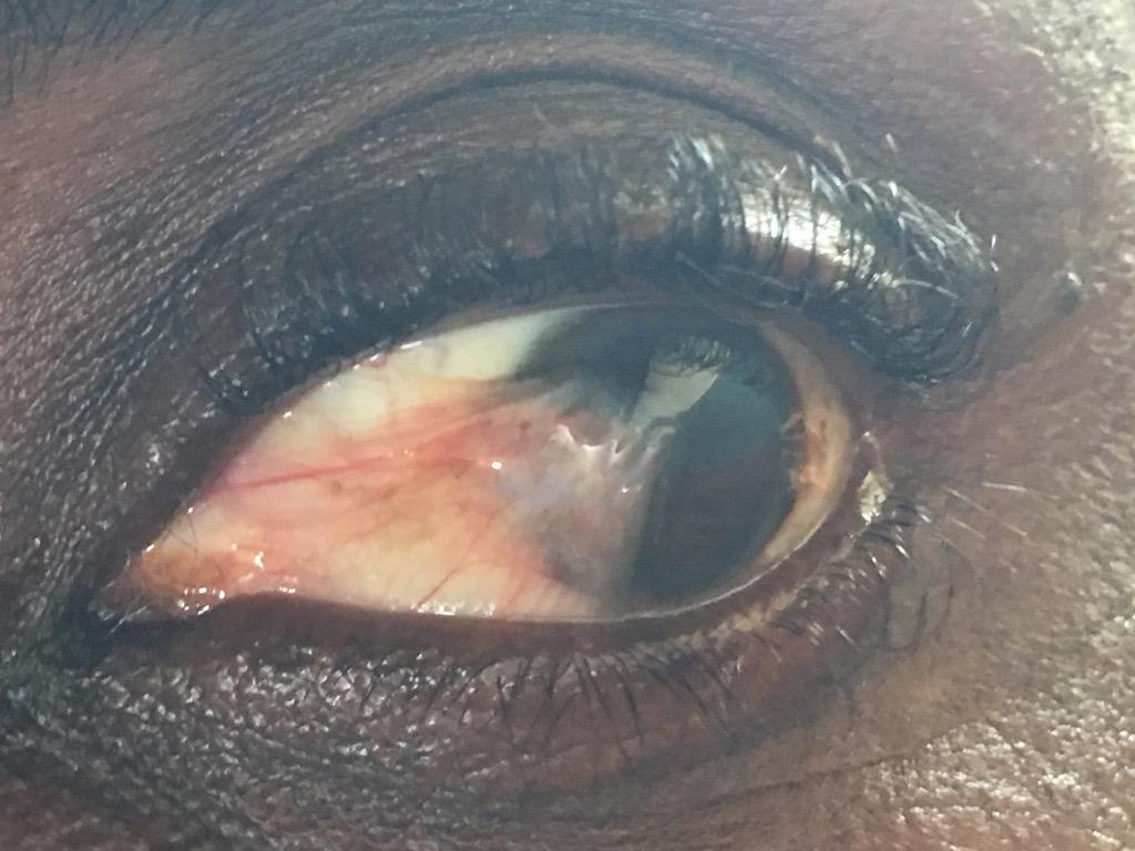

35 Pinguecula/Pterygium

36 Describe this photo
37 Cataract The lens is transparent, biconvex and responsible for diopters of convergent refractive power Cataract is a clouding of the eye s naturally clear lens Age-related cataract is the main cause of blindness and visual impairment throughout the world Cataracts cause blindness in more than 15,000,000 people worldwide and 40,000,000 by the year 2025


38 Cataract

39 Vision with a Cataract
40 Cataract Etiology Aging changes Drug-induced lens changes Trauma Metabolic cataract Nutritional Uveitis Exfoliation Cataract and skin disease
41 Signs and Symptoms Decreased visual Acuity Glare Myopic shift Monocular diplopia Diagnosis History and Physical Eight part eye examination Glare testing Contrast Sensitivity Visual Field Testing Cataract

42 Describe this photo
43 Glaucoma Glaucoma refers to a group of diseases optic neuropathy with associated visual field loss elevated intraocular pressure (IOP) is one of the primary risk factors Three factors determine the IOP: The Rate of aqueous humor production by the ciliary body Resistance to aqueous outflow across the trabecular meshwork - Schlemm s canal system The level of episcleral venous pressure Commonly accepted range for normal IOP is mmhg In most cases of > IOP is > resistance to aqueous humor outflow
44 Glaucoma Between 80,000 and 116,000 Americans are legally blind secondary to glaucoma with each year an additional 5,500 pts become legally blind Approximately 3 million Americans 40 yrs or older have POAG and other forms of glaucoma or related conditions add to these totals 50% don t know they have the disease Glaucoma is the most frequent cause of blindness in Black Americans Classifications Open Angle glaucoma Angle- closure glaucoma Combined-mechanism glaucoma Childhood glaucoma
45 Glaucoma Signs and Symptoms Blurred vision Halos Loss of vision and visual field Diagnosis History and physical Eight part eye examination Gonioscopy Visual field Ultrasonography pachymetry HRT/OCT

46 Aqueous Outflow Channels

47 Histology of Aqueous Outflow Channels

48 Normal Optic Nerve Appearance of nerve tissue ON color Disc Margin Cup to disc ratio

49 Glaucoma
50 Glaucomatous Optic Nerve
51 Glaucoma Visual Field Loss Patterns of Glaucomatous Nerve Loss Paracentral scotoma Arcuate or Bjerrum scotoma Nasal step Temporal wedge
52 Humphrey Visual Fields
53 Treatment Medical Adrengic agonists improves aqueous outflow Alpha 2 adrengic agonists reduces aqueous secretion Parasympathomimetic (miotic) agents improves aqueous outflow Prostaglandin analogs Increases uveoscleral outflow Carbonic anhydrase inhibitors- reduces aquious secretion Hyperosmotic agents reduces aqueous volume Surgical Laser Iriditomy Laser Trabeculoplasty Incisional Trabeculectomy Full thickness


54 Medications


55 Laser Iridotomy/Trabeculoplasty


56 Filtration Surgery
57 Abbreviations you may encounter DES CAM PCO NS CS PSC CEIOL TRAB Dry eye syndrome Complexion associated melanosis Posterior capsular opacification Nuclear sclerosis Cortical spoking Posterior subcapsular cataract Cataract extraction with intraocular lens placement Trabeculectomy
58 Contact Information Leslie S. Jones, MD Associate Professor and Chair Department of Ophthalmology Howard University Department Office (202)
02/03/2014. Average Length: 23mm (Infant ~16mm) Approximately the size of a quarter Volume: ~5mL
 Approximately the size of a quarter Volume: ~5mL") Identify the anatomy of the eye. Explain the basic physiology of the parts of the eye. Briefly discuss various surgeries related to different parts of the anatomy. Average Length: 23mm (Infant ~16mm) Approximately
Identify the anatomy of the eye. Explain the basic physiology of the parts of the eye. Briefly discuss various surgeries related to different parts of the anatomy. Average Length: 23mm (Infant ~16mm) Approximately
Glaucoma Clinical Update. Barry Emara MD FRCS(C) Giovanni Caboto Club October 3, 2012
 Giovanni Caboto Club October 3, 2012") Glaucoma Clinical Update Barry Emara MD FRCS(C) Giovanni Caboto Club October 3, 2012 Objectives Understand the different categories of glaucoma Recognize the symptoms and signs of open angle and angle-closure
Glaucoma Clinical Update Barry Emara MD FRCS(C) Giovanni Caboto Club October 3, 2012 Objectives Understand the different categories of glaucoma Recognize the symptoms and signs of open angle and angle-closure
GLAUCOMA. An Overview
 GLAUCOMA An Overview Compiled by Campbell M Gold (2004) CMG Archives http://campbellmgold.com --()-- IMPORTANT The health information contained herein is not meant as a substitute for advice from your
Collaboration in the care of glaucoma patients and glaucoma suspects. Barry Emara MD FRCS(C) Nico Ristorante November 29, 2012
 Nico Ristorante November 29, 2012") Collaboration in the care of glaucoma patients and glaucoma suspects Barry Emara MD FRCS(C) Nico Ristorante November 29, 2012 Goals of Collaboration Patient-centred and evidence based approach Timely access
Collaboration in the care of glaucoma patients and glaucoma suspects Barry Emara MD FRCS(C) Nico Ristorante November 29, 2012 Goals of Collaboration Patient-centred and evidence based approach Timely access
Written by Administrator Wednesday, 13 January :27 - Last Updated Thursday, 21 January :34
 angle closure glaucoma A type of glaucoma caused by a sudden and severe rise in eye pressure. Occurs when the pupil enlarges too much or too quickly, and the outer edge of the iris blocks the eye s drainage
ASSESSING THE EYES. Structures. Eyelids Extraocularmuscles Eyelashes Lacrimal glands: Lacrimal ducts Cornea Conjunctiva Sclera Pupils Iris.
 ASSESSING THE EYES Structures External Eyelids Extraocularmuscles Eyelashes Lacrimal glands: Lacrimal ducts Cornea Conjunctiva Sclera Pupils Iris 1 2 Structures Internal Optic disc Physiological cup Retinal
Glaucoma. Glaucoma. Optic Disc Cupping
 Glaucoma What is Glaucoma? Bruce James A group of diseases in which damage to the optic nerve occurs as a result of intraocualar pressure being above the physiological norm for that eye Stoke Mandeville
Frequently Asked Questions about General Ophthalmology:
 1. Normal Eye Structure The eye is a slightly asymmetrical globe, about an inch in diameter. The parts of the eye include: Cornea (a clear dome over the iris), Iris (the pigmented part); Pupil (the black
Glaucoma. How is Glaucoma Diagnosed? Glaucoma Testing
 Glaucoma How is Glaucoma Diagnosed? Glaucoma Testing There is no single test for glaucoma. The diagnosis is made by evaluating the patient from a number of perspectives, using specialized instruments.
WORLD GLAUCOMA WEEK What is Glaucoma? Can I develop glaucoma if I have increased eye pressure?
 WORLD GLAUCOMA WEEK What is Glaucoma? Glaucoma is a group of diseases that damage the optic nerve and can result in gradual vision loss and blindness. However, with early detection and treatment, you can
OCCLUSIVE VASCULAR DISORDERS OF THE RETINA
 OCCLUSIVE VASCULAR DISORDERS OF THE RETINA Learning outcomes By the end of this lecture the students would be able to Classify occlusive vascular disorders (OVD) of the retina. Correlate the clinical features
Management of Angle Closure Glaucoma Hospital Authority Convention 18 May 2015
 Management of Angle Closure Glaucoma Hospital Authority Convention 18 May 2015 Jimmy Lai Clinical Professor Department of Ophthalmology The University of Hong Kong 1 Primary Angle Closure Glaucoma PACG
Scrub In. What is the function of vitreous humor? What does the pupil do when exposed to bright light? a. Maintain eye shape and provide color vision
 Scrub In What is the function of vitreous humor? a. Maintain eye shape and provide color vision b. Maintain eye shape and refract light rays c. Provide night vision and color vision d. Provide night vision
A LITTLE ANATOMY. three layers of eye: 1. outer: corneosclera. 2. middle - uvea. anterior - iris,ciliary body. posterior - choroid
 GLAUCOMA A LITTLE ANATOMY three layers of eye: 1. outer: corneosclera 2. middle - uvea anterior - iris,ciliary body posterior - choroid connection at the pars plana between post and ant uvea 3. retina
NEPTUNE RED BANK BRICK
 NEPTUNE RED BANK BRICK Diabetes & The Eye Diabetics are more likely to develop Cataracts at a younger age. Diabetics are twice as likely to develop Glaucoma when compared to non-diabetics. The primary
THE CHRONIC GLAUCOMAS
 THE CHRONIC GLAUCOMAS WHAT IS GLAUCOMA? People with glaucoma have lost some of their field of all round vision. It is often the edge or periphery that is lost. That is why the condition can be missed until
VN 122 MODULE F EYE AND VISION DISORDERS OCULAR HISTORY 1. PATIENT PERCEPTION OF PROBLEM 2. DECREASED VISION? 3. BLURRED, DOUBLE, DISTORTED?
 VN 122 MODULE F EYE AND VISION DISORDERS OCULAR HISTORY 1. PATIENT PERCEPTION OF PROBLEM 2. DECREASED VISION? 3. BLURRED, DOUBLE, DISTORTED? 4. PAIN? QUALITY OF PAIN? 5. ITCHING, DRY? 6. BOTH EYES? 7.
NEW YORK UNIVERSITY SCHOOL OF MEDICINE DEPARTMENT OF OPHTHALMOLOGY EDUCATIONAL OBJECTIVES AND GOALS
 NEW YORK UNIVERSITY SCHOOL OF MEDICINE DEPARTMENT OF OPHTHALMOLOGY EDUCATIONAL OBJECTIVES AND GOALS Revision Date: 6/30/06 Distribution Date: 7/6/06 The Department of Ophthalmology at the NYU Medical Center
Glaucoma. Cornea. Iris
 Glaucoma Introduction Glaucoma is a group of eye diseases that can lead to blindness if not treated. Openangle glaucoma, the most common form of glaucoma, affects about 3 million Americans. Half of those
Goals. Glaucoma PARA PEARL TO DO. Vision Loss with Glaucoma
 Glaucoma Janet R. Fett, OD Drs. Kincaid, Fett and Tharp So Sioux City, NE eyewear21@hotmail.com Goals Understand Glaucoma Disease process Understand how your data (objective and subjective) assists in
Ocular Pathology I 6234_16385 Rm HBSB 203-E 1:00-3:00pm. Tonya G. Ketcham, OD, PhD , RM 2113
 Ocular Pathology I 6234_16385 Rm HBSB 203-E 1:00-3:00pm Tonya G. Ketcham, OD, PhD tketcham@optometry.uh.edu 3-1799, RM 2113 Course Syllabus Course Description To describe normal anomalies and pathologic
Determining the Diagnosis Complete guide to ICD-10-CM coding conventions and guidelines
 ICD-10 ESSENTIALS 2019 Determining the Diagnosis Complete guide to ICD-10-CM coding conventions and guidelines Power up your coding optum360coding.com Contents List of Case Studies... iii List of Figures...
SUPPLEMENTARY INFORMATION
 SUPPLEMENTARY INFORMATION Contents METHODS... 2 Inclusion and exclusion criteria... 2 Supplementary table S1... 2 Assessment of abnormal ocular signs and symptoms... 3 Supplementary table S2... 3 Ocular
You can see vivid colours again after cataract management at Sankar Foundation Eye Hospital
 The Department of Cataract in our Sankar Foundation is equipped with state-of-the-art operation theatres, surgical microscope phacoemulsification machine and microsurgical instruments. And also the department
Glaucoma. What is glaucoma? Eye Words to Know. What causes glaucoma?
 2014 2015 Glaucoma What is glaucoma? Glaucoma is a disease that damages your eye s optic nerve. It usually happens when fluid builds up in the front part of your eye. That extra fluid increases the pressure
Reducing vision loss in chronic eye disease
 THEME vision at risk Anthony Fong MBBS, BOptom, is Clinical Fellow, City Eye Centre, Brisbane, Queensland. antfong@ hotmail.com Graham Lee MD, MBBS, FRANZCO, is Associate Professor, The University of Queensland.
53 year old woman attends your practice for routine exam. She has no past medical history or family history of note.
 Case 1 Normal Tension Glaucoma 53 year old woman attends your practice for routine exam. She has no past medical history or family history of note. Table 1. Right Eye Left Eye Visual acuity 6/6 6/6 Ishihara
OCT : retinal layers. Extraocular muscles. History. Central vs Peripheral vision. History: Temporal course. Optical Coherence Tomography (OCT)
") Optical Coherence Tomography (OCT) OCT : retinal layers 7 Central vs Peripheral vision Extraocular muscles RPE E Peripheral Vision: Rods (95 million) 30% Ganglion cells Central Vision: Cones (5 million)
Optical Coherence Tomography (OCT) OCT : retinal layers 7 Central vs Peripheral vision Extraocular muscles RPE E Peripheral Vision: Rods (95 million) 30% Ganglion cells Central Vision: Cones (5 million)
Ophthalmology. Ophthalmology Services
 Ophthalmology Ophthalmology Services The Ophthalmology service offers the latest and most comprehensive eye care for patients. With a dedicated team of eye surgeons and consultants, we treat vision problems
Divakar Gupta Glaucoma Fellow, Duke Eye Center 5/14/16
 Divakar Gupta Glaucoma Fellow, Duke Eye Center 5/14/16 Pathophysiology of glaucoma Consider risk factors of glaucoma Understand the side effects of glaucoma medications Diagnostic testing Leading cause
UC SF. g h. Eye Trauma. Martha Neighbor, MD Emergency Services San Francisco General Hospital University of California
 UC SF Eye Trauma sf g h Martha Neighbor, MD Emergency Services San Francisco General Hospital University of California Goals Recognize vision threatening eye emergencies Treat them when we can Know when
Intro to Glaucoma/2006
 Intro to Glaucoma/2006 Managing Patients with Glaucoma is Exciting Interesting Challenging But can often be frustrating! Clinical Challenges To identify patients with risk factors for possible glaucoma.
Around The Globe in 60 Minutes
 Around The Globe in 60 Minutes Around the GLOBE in Sixty Minutes Basic Ocular Anatomy, Examination, and Diagnostic Techniques Introduction Focusing on canine and feline ocular anatomy and basic examination
Glaucoma What You Should Know
 Glaucoma What You Should Know U.S. DEPARTMENT OF HEALTH AND HUMAN SERVICES National Institutes of Health National Eye Institute The National Eye Institute (NEI) conducts and supports research that leads
Assisting in Ophthalmology. Copyright 2011, 2007, 2003, 1999 by Saunders, an imprint of Elsevier Inc. All rights reserved.
 Assisting in Ophthalmology Learning Objectives Define, spell, and pronounce the terms listed in the vocabulary. Apply critical thinking skills in performing patient assessment and care. Explain the differences
Downloaded from:
 Philippin, H; Shah, P; Burton, M (2012) The next step: Detailed assessment of an adult glaucoma patient. Community eye health / International Centre for Eye Health, 25 (79-80). pp. 50-53. ISSN 0953-6833
Syllabus-Ophthalmology Rotation Course: Objectives & Goals LOYOLA UNIVERSITY CHICAGO STRITCH SCHOOL OF MEDICINE
 Syllabus-Ophthalmology Rotation Course: Objectives & Goals LOYOLA UNIVERSITY CHICAGO STRITCH SCHOOL OF MEDICINE Department of Ophthalmology Course Objectives: By Core Competencies GENERAL INFORMATION:
Technicians & Nurses Program
 ASCRS ASOA Symposium & Congress Technicians & Nurses Program May 6-10, 2016 New Orleans I have none Sunday, May 8, 2016, 10:30-11:30am Amy Jost, BS, COMT, CCRC Cincinnati Eye Institute Identify the tech
The second most common causes of blindness worldwide. ( after cataract) The commonest cause of irreversible blindness in the world Estimated that 3%
 The commonest cause of irreversible blindness in the world Estimated that 3%") The second most common causes of blindness worldwide. ( after cataract) The commonest cause of irreversible blindness in the world Estimated that 3% of our population age > 40 have glaucoma In the past:
The second most common causes of blindness worldwide. ( after cataract) The commonest cause of irreversible blindness in the world Estimated that 3% of our population age > 40 have glaucoma In the past:
1/25/2019 OCT & OCTA RETINAL IMAGING: HOW TO PREVENT RAGING GLAUCOMA! THE ORIGINAL RAGING GLAUCOMA OCT RETINAL IMAGING OPTIC NERVE HEAD EXAMINATION
 OCT & OCTA RETINAL IMAGING: HOW TO PREVENT RAGING GLAUCOMA! Craig Thomas, O.D. 3900 West Wheatland Road Dallas, Texas 75237 972-780-7199 thpckc@yahoo.com THE ORIGINAL RAGING GLAUCOMA 47-year-old Black
Understanding. Glaucoma. National Glaucoma Research
 Understanding National Research Understanding What is? is not just one disease, but a group of eye diseases that damage the optic nerve the bundle of nerve fibers that carries information from the eye
Vision loss in elderly. Erica Weir, April 2015
 Vision loss in elderly Erica Weir, April 2015 1 Burden Enter nursing homes 3 years earlier Twice the risk of falling 4x the risk of hip fracture Independent risk factor for delirium What are the leading
The Orbit. The Orbit OCULAR ANATOMY AND DISSECTION 9/25/2014. The eye is a 23 mm organ...how difficult can this be? Openings in the orbit
 The eye is a 23 mm organ...how difficult can this be? OCULAR ANATOMY AND DISSECTION JEFFREY M. GAMBLE, OD COLUMBIA EYE CONSULTANTS OPTOMETRY & UNIVERSITY OF MISSOURI DEPARTMENT OF OPHTHALMOLOGY CLINICAL
JINNAH SINDH MEDICAL UNIVERSITY STUDY GUIDE- OPHTHALMOLOGY YEAR 4,
 INTRODUCTION Pakistan, the 7th most populous country in the world, has an urban population of 38.8% and rural dwellers of 61.2%. The country has faced challenges with vision impairment and blindness as
Vision loss in elderly
 Vision loss in elderly Erica Weir, February 2016 1 Burden Enter nursing homes 3 years earlier Twice the risk of falling 4x the risk of hip fracture Independent risk factor for delirium What are the leading
GLAUCOMA SUMMARY BENCHMARKS FOR PREFERRED PRACTICE PATTERN GUIDELINES
 SUMMARY BENCHMARKS FOR PREFERRED PRACTICE PATTERN GUIDELINES Introduction These are summary benchmarks for the Academy s Preferred Practice Pattern (PPP) guidelines. The Preferred Practice Pattern series
THE EYE: RETINA AND GLOBE
 Neuroanatomy Suzanne Stensaas February 24, 2011, 10:00-12:00 p.m. Reading: Waxman Ch. 15. Your histology and gross anatomy books should be useful. Reading: Histology of the Eye from any histology book
Cairo University Faculty of Medicine. Course Specifications Course title: Ophthalmology (Code): OPH-409. Department of Ophthalmology
: OPH-409. Department of Ophthalmology") Cairo University Faculty of Medicine Department of Ophthalmology Course Specifications Course title: Ophthalmology (Code): OPH-409 Department of Ophthalmology Fourth academic year of M.B.B.Ch. program
Cairo University Faculty of Medicine Department of Ophthalmology Course Specifications Course title: Ophthalmology (Code): OPH-409 Department of Ophthalmology Fourth academic year of M.B.B.Ch. program
CLASS-y Laser Treats Glaucoma
 Article # 404 Comments About the Author Released: Author: Category: March 12th, 2014 Issue #0314 Ehud Assia Feature S S S S S CLASS-y Laser Treats Glaucoma Transforming complex, invasive and risky glaucoma
Mild NPDR. Moderate NPDR. Severe NPDR
 Diabetic retinopathy Diabetic retinopathy is the most common cause of blindness in adults aged 35-65 years-old. Hyperglycaemia is thought to cause increased retinal blood flow and abnormal metabolism in
The Anterior Segment & Glaucoma Visual Recognition & Interpretation of Clinical Signs
 The Anterior Segment & Glaucoma Visual Recognition & Interpretation of Clinical Signs Quiz created by Jane Macnaughton MCOptom & Peter Chapman BSc MCOptom FBDO CET Accreditation C19095 2 CET Points (General)
Training Checking Vision Tonometry
 Training 101-2 Checking Vision Tonometry Checking Vision The classic example of an eye chart is the Snellen eye chart, in general they show 11 rows of capital letters. The top row contains one letter (usually
4/22/16. Eye. External Anatomy of Eye. Accessory Structures. Bio 40B Dr. Kandula
 Eye Bio 40B Dr. Kandula External Anatomy of Eye Accessory Structures l Eyebrows l Levator Palpebrae Superioris - opens eye l Eyelashes l Ciliary glands modified sweat glands l Small sebaceous glands l
Recurrent intraocular hemorrhage secondary to cataract wound neovascularization (Swan Syndrome)
") Recurrent intraocular hemorrhage secondary to cataract wound neovascularization (Swan Syndrome) John J. Chen MD, PhD; Young H. Kwon MD, PhD August 6, 2012 Chief complaint: Recurrent vitreous hemorrhage,
Recurrent intraocular hemorrhage secondary to cataract wound neovascularization (Swan Syndrome) John J. Chen MD, PhD; Young H. Kwon MD, PhD August 6, 2012 Chief complaint: Recurrent vitreous hemorrhage,
Test Bank for Medical Surgical Nursing An Integrated Approach 3rd Edition by White
 Test Bank for Medical Surgical Nursing An Integrated Approach 3rd Edition by White Link full download : http://testbankair.com/download/test-bank-for-medical-surgical-nursing-anintegrated-approach-3rd-edition-by-white/
THE CHRONIC GLAUCOMAS
 THE CHRONIC GLAUCOMAS WHAT IS GLAUCOMA People with glaucoma have lost some of their field of all round vision. It is often the edge or periphery that is lost. That is why the condition can be missed until
Glaucoma. Glaucoma. Glaucoma. Trevor Arnold, MS, DVM, DACVO
 Glaucoma Trevor Arnold, MS, DVM, DACVO Glaucoma Physiology of Aqueous Humor Produced in the ciliary body Flows out the iridocorneal angle and ciliary cleft High intraocular pressures are caused by a decreased
Save time at your check-in and register online before your appointment! It s as easy as 1-2-3
 Save time at your check-in and register online before your appointment! It s as easy as 1-2-3 1. Go online to www.blackhillseyes.com 2. Click this logo on our home page for the link to register: 3. Set-up
Eye and Ocular Adnexa, Auditory Systems
 Eye and Ocular Adnexa, Auditory Systems CPT copyright 2011 American Medical Association. All rights reserved. Fee schedules, relative value units, conversion factors and/or related components are not assigned
Vision I. Steven McLoon Department of Neuroscience University of Minnesota
 Vision I Steven McLoon Department of Neuroscience University of Minnesota 1 Eye Cornea Sclera Conjunctiva 2 Eye The conjunctiva lines the inner surface of the eyelids and outer surface of the sclera. 3
3/16/2018. Ultrasound Biomicroscopy in Glaucoma By Ahmed Salah Abdel Rehim. Prof. of Ophthalmology Al-Azhar University
 Ultrasound Biomicroscopy in Glaucoma By Ahmed Salah Abdel Rehim Prof. of Ophthalmology Al-Azhar University 1 Ultrasound biomicroscopy (UBM) is a recent technique to visualize anterior segment with the
Ophthalmology. Glaucoma
 Ophthalmology Glaucoma The Ophthalmology service offers the latest and most comprehensive eye care for patients. With a dedicated team of eye surgeons and consultants, we treat vision problems ranging
Chronicity. Narrow Minded. Course Outline. Acute angle closure. Subacute angle closure. Classification of Angle Closure 5/19/2014
 Chronicity Narrow Minded The management of narrow angles in the optometric practice Acute Subacute Chronic Aaron McNulty, OD, FAAO Course Outline Classification of Angle Closure Evaluation of narrow angles
Ocular Anatomy for the Paraoptometric
 Ocular Anatomy for the Paraoptometric Minnesota Optometric Association Paraoptometric CE Friday September 30, 2016 Lindsay A. Sicks, OD, FAAO Assistant Professor, Illinois College of Optometry lsicks@ico.edu
Diabetic Retinopathy Screening Program in the Cree Region of James Bay of Quebec
 RUIS McGILL VIRTUAL HEALTH AND SOCIAL SERVICES CENTRE (CvSSS) SIMPLIFYING TELEHEALTH! Diabetic Retinopathy Screening Program in the Cree Region of James Bay of Quebec Nurse and Imager Training Prepared
Cataract Surgery Co-Management
 Cataract Surgery Co-Management Phacoemulsification, Clear-Lens Extraction, and LensX INCLUSION CRITERIA: Significant visual complaints (decreased VA, increased glare, decreased Activities of Daily Living
9/25/2017 CASE. 67 years old On 2 topical meds since 3 years. Rx: +3.0 RE LE
 CASE 67 years old On 2 topical meds since 3 years Rx: +3.0 /-0.5@65 RE +2.5/-0.5@115 LE IOP : 17 RE 19 LE CD: 0.5 RE 0.6 LE 1 67 years old On 2 topical meds since 3 years Rx: +3.0 /-0.5@65 RE +2.5/-0.5@115
Year 2 MBChB Clinical Skills Session Ophthalmoscopy. Reviewed & ratified by: Mr M Batterbury Consultant Ophthalmologist
 Year 2 MBChB Clinical Skills Session Ophthalmoscopy Reviewed & ratified by: o Mr M Batterbury Consultant Ophthalmologist Learning objectives o To understand the anatomy and physiology of the external and
relative s privacy, do not identify your relative by full name in any assignment.
 Overview Do you or a family member have glaucoma? Do you wonder what this diagnosis means? Glaucoma affects tens of millions of people worldwide. Despite its prevalence, many people lack accurate information
LECTURE # 7 EYECARE REVIEW: PART III
 LECTURE # 7 EYECARE REVIEW: PART III HOW TO TRIAGE EYE EMERGENCIES STEVE BUTZON, O.D. EYECARE REVIEW: HOW TO TRIAGE EYE EMERGENCIES FOR PRIMARY CARE PHYSICIANS Steve Butzon, O.D. Member Director IDOC President
Special Senses: The Eye
 Unit 4 Special Senses: The Eye ESSENTIALS OF HUMAN ANATOMY & PHYSIOLOGY The Senses General senses of touch Temperature Pressure Pain Special senses Smell Taste Sight Hearing Equilibrium The Eye and Vision
Understanding Angle Closure
 Case Understanding Angle Closure Dominick L. Opitz, OD, FAAO Associate Professor Illinois College of Optometry 56 year old Caucasian Male Primary Eye Exam BCVA: 20/25 OD with+1.25 DS 20/25 OS with +1.75
PREVALENCE OF GLAUCOMA AMONG FISHERMEN COMMUNITY OF MUNDRA TALUKA OF KUTCH DISTRICT- A CROSS- SECTIONAL STUDY
 ORIGINAL RESEARCH PREVALENCE OF GLAUCOMA AMONG FISHERMEN COMMUNITY OF MUNDRA TALUKA OF KUTCH DISTRICT- A CROSS- SECTIONAL STUDY Sanjay Upadhyay 1, Jayantilal Shah 2 1 Assistant Professor, 2 Associate Professor,
GENERAL INFORMATION GLAUCOMA GLAUCOMA
 GENERAL INFORMATION GLAUCOMA GLAUCOMA WHAT IS GLAUCOMA? Glaucoma is commonly known as the sneak thief of sight because it can cause irreversible vision loss without any obvious symptoms. The term glaucoma
Corporate Medical Policy
 Corporate Medical Policy Optical Coherence Tomography (OCT) Anterior Segment of the Eye File Name: Origination: Last CAP Review: Next CAP Review: Last Review: optical_coherence_tomography_(oct)_anterior_segment_of_the_eye
CHAPTER 13 CLINICAL CASES INTRODUCTION
 2 CHAPTER 3 CLINICAL CASES INTRODUCTION The previous chapters of this book have systematically presented various aspects of visual field testing and is now put into a clinical context. In this chapter,
PedsCases Podcast Scripts
 PedsCases Podcast Scripts This is a text version of a podcast from Pedscases.com on Approach to Childhood Glaucoma. These podcasts are designed to give medical students an overview of key topics in pediatrics.
Speaker Disclosure Statement. " Dr. Tim Maillet and Dr. Vladimir Kozousek have no conflicts of interest to disclose.
 Speaker Disclosure Statement Dr. Tim Maillet and Dr. Vladimir Kozousek have no conflicts of interest to disclose. Diabetes Morbidity Diabetes doubles the risk of stroke. Diabetes quadruples the risk of
CHARTING THE NEW COURSE FOR MIGS
 CHARTING THE NEW COURSE FOR MIGS SEE WHAT S ON THE HORIZON CyPass Micro-Stent the next wave in micro-invasive glaucoma surgery. MICRO-INVASIVE GLAUCOMA SURGERY (MIGS) OFFERS A REVOLUTIONARY APPROACH TO
GLAUCOMA (2006) PHILIPPINE GLAUCOMA SOCIETY
 PHILIPPINE GLAUCOMA SOCIETY") GLAUCOMA (2006) PHILIPPINE GLAUCOMA SOCIETY CPM 9 TH EDITION Philippine Glaucoma Society (PGS) Correspondence to: Eye Referral Center, T. M. Kalaw Street, Ermita, Manila Telephone: 524-7119/525-9360 Mobile:
GLAUCOMA (2006) PHILIPPINE GLAUCOMA SOCIETY CPM 9 TH EDITION Philippine Glaucoma Society (PGS) Correspondence to: Eye Referral Center, T. M. Kalaw Street, Ermita, Manila Telephone: 524-7119/525-9360 Mobile:
Optometric Postoperative Cataract Surgery Management
 Financial Disclosures Optometric Postoperative Cataract Surgery Management David Dinh, OD Oak Cliff Eye Clinic Dallas Eye Consultants March 10, 2015 Comanagement Joint cooperation between two or more specialists
11/30/2009. Glaukosis: ancient greek term meaning sparkling or shining appearance of pupil
 Normal Ocular Anatomy Glaucoma Dr Sunil Deokule, MD Asst. Prof and Glaucoma Specialist University of Kentucky Definition Glaukosis: ancient greek term meaning sparkling or shining appearance of pupil Optic
Eye Fluids. Dr. Mohamed Saad Daoud
 Eye Fluids 1 Reference Books: Text Book of Medical physiology (Guyton and Hall) Eleventh edition 2 Fluid System of the Eye (Intraocular Fluid) The eye is filled with intraocular fluid, which maintains
Ophthalmic Trauma Update
 Ophthalmic Trauma Update Richard S. Davidson, M.D. Professor of Ophthalmology Vice Chair for Quality and Clinical Affairs UCHealth Eye Center University of Colorado School of Medicine August 5, 2017 Financial
Secondary Glaucomas. Mr Nick Strouthidis MBBS MD PhD FRCS FRCOphth FRANZCO Consultant Ophthalmologist, Glaucoma Service, Moorfields Eye Hospital
 Secondary Glaucomas Mr Nick Strouthidis MBBS MD PhD FRCS FRCOphth FRANZCO Consultant Ophthalmologist, Glaucoma Service, Moorfields Eye Hospital Introduction: What is glaucoma? Glaucoma is the name given
_ Assessment of the anterior chamber. Review of anatomy of the angle
 Assessment of the anterior chamber Dr Simon Barnard PhD BSc FCOptom FAAO DCLP Department of Optometry & Visual Science City University London, UK Review of anatomy of the angle Figure 1. Anatomical section
Efficacy of latanoprost in management of chronic angle closure glaucoma. Kumar S 1, Malik A 2 Singh M 3, Sood S 4. Abstract
 Original article Efficacy of latanoprost in management of chronic angle closure glaucoma Kumar S 1, Malik A 2 Singh M 3, Sood S 4 1 Associate Professor, 2 Assistant Professor, 4 Professor, Department of
Chapter 7, Section 1 Review Questions. Directions: Place the letter of the best definition next to each key term. Name PER Date
 Name PER Date Chapter 7, Section 1 Review Questions Directions: Place the letter of the best definition next to each key term. A. the middle layer of the wall of the eye B. the structure between the choroid
XUE HUI Department of Histology& Embryology, Basic Medicine College of Jilin University
 SENSE ORGAN XUE HUI Department of Histology& Embryology, Basic Medicine College of Jilin University EYE fibrous globe lens photosensitive cells a system of cells and nerves concentric layers the sclera
The Special Senses: Part A
 PowerPoint Lecture Slides prepared by Janice Meeking, Mount Royal College CHAPTER 15 The Special Senses: Part A Warm Up What is the function of the eyeball? List any structures of the eyeball that you
Cairo University Faculty of Medicine. Course Specifications Course title: Ophthalmology (Code): OPH-411. Department of Ophthalmology
: OPH-411. Department of Ophthalmology") Course Specifications Course title: Ophthalmology (Code): OPH-411 Fourth academic year of M.B.B.Ch. program Date of specification approval: 2016 A) Basic Information: Allocated marks: 250 marks Course
Course Specifications Course title: Ophthalmology (Code): OPH-411 Fourth academic year of M.B.B.Ch. program Date of specification approval: 2016 A) Basic Information: Allocated marks: 250 marks Course
Identify the choice that best completes the statement or answers the question.
 Chapter 5. The Eye Multiple Choice Identify the choice that best completes the statement or answers the question. 1. The most common type of eye disorder is: A. Refractive errors B. Macular conditions
 measure of your overall performance. An isolated glucose test is helpful to let you know what your sugar level is at one moment, but it doesn t tell you whether or not your diabetes is under adequate control
Advanced Examination of the Retina: Scleral Indentation & Retinal 3-Mirror
 Advanced Examination of the Retina: Scleral Indentation & Retinal 3-Mirror Meredith Whiteside, OD, FAAO Nimesh Patel, OD, FAAO John Shan, OD, FAAO Please silence all mobile devices. Unauthorized recording
DIRECT REFERRAL OF CATARACT PATIENTS COMMUNITY OPTOMETRIST PROTOCOL AND GUIDELINES
 DIRECT REFERRAL OF CATARACT PATIENTS COMMUNITY OPTOMETRIST PROTOCOL AND GUIDELINES October 2013 Preoperative assessment History Symptoms/social history: is the patient visually affected and do they want
Cases CFEH. CFEH Facebook Case #4
 CFEH Cases CFEH Facebook Case #4 A 42 year old female has noticed a floater in her left eye for many years but no flashes. She also reports hazy vision in this eye that has been present all her life. She
Clinical Approach To Refractive Errors. Dr. Faizur Rahman Associate Professor Peshawar Medical College
 Clinical Approach To Refractive Errors Dr. Faizur Rahman Associate Professor Peshawar Medical College Learning objectives By the end of this lecture the students would be able to; Correlate optics with
WGA. The Global Glaucoma Network
 The Global Glaucoma Network Fort Lauderdale April 30, 2005 Indications for Surgery 1. The decision for surgery should consider the risk/benefit ratio. Note: Although a lower IOP is generally considered