Course # Getting to Know Your OCT
|
|
|
- Silvester Foster
- 5 years ago
- Views:
Transcription
1 Course # 140 Getting to Know Your OCT

2 Course Title: Lecturer: Getting to Know Your OCT Brad Sutton, OD, FAAO IU School of Optometry Financial Disclosures No financial disclosures Optical Coherence Tomography-OCT Has changed the way the eye is examined & treated. Has revolutionized the diagnosis & therapy of eye disease. Allows for earlier and more sensitive diagnosis. Allows for better understanding of disease mechanisms OCT 1991 James Fujimoto at MIT Original research instrument 400 A-scans / second Current SD-OCT: around 27,000 to 50,000 A-scans / sec Current Swept Source-OCT up to 249,000 A-scans / sec. Commercial units about 100,000 per second OCT angiography So how did we get here? OCT First commercial OCT company in 1992: Advanced Ophthalmic Devices Sold on August 27 th of 1993 to Humphrey- Now Carl Zeiss Meditech First commercial OCT from them in 1996 OCT By 2008, Zeiss had sold 10,000 OCT s Now, there are 8 companies that manufacture OCT s commercially 1
 OptoVue (I Vue and RTVue): $40,000 - $60,000 Heidelberg Spectralis: $80,000: FAF Topcon: has fundus camera, OPTOS Some have no normative data in vivo histology Working mechanism: similar to B scan")
3 Different Companies and Prices OCT Stratus: $53,000 (lower refurbished) Cirrus: $70-75,000 ( new unit with SD- OCT, fundus camera, and FAF?$) OptoVue (I Vue and RTVue): $40,000 - $60,000 Heidelberg Spectralis: $80,000: FAF Topcon: has fundus camera, OPTOS Some have no normative data in vivo histology Working mechanism: similar to B scan (optical vs. acoustic reflectivity) but uses infrared light Resolution: < 10 microns (Stratus OCT III), 3-5 microns with SD and SS technology Different optical reflectivity in various tissue structures: false color map. Often best to view in black and white for fine detail Courtesy Dan Hammer, PSI Image quality Poor signal strength equates to unreliable readings, only use 7 and above Images and reliability can be negatively impacted by media opacities, high myopia, patient movement, highly abnormal disc sizes, and segmentation errors (very important!) Beware red disease, split NFL bundles for example Average RNFL loss of about 1 micron / year Importance of normative database Cirrus normative database Typically take demographic factors in to account, but not refractive error. This can be very important with high myopes, who will have thinner NFL than their counterparts with equal demographics Composition of normative database also very important 284 individuals Age 18 to 84 Refractive error to -12:00 43% Caucasian 24% Asian 18% African American 12% Hispanic 1% Indian Small amount of others combined 2
.")
 Larger scan area (6mm X 6mm).")


4 Spectral Domain OCT Cirrus OCT Easier to use. Faster (1/75 the time it takes TD). Greater resolution (3-5 Microns vs.10) Larger scan area (6mm X 6mm). Can create three dimensional cubed images. Precise registration (can follow the exact retinal area each visit). 3 D retinal layer segmentation maps. Focus range of +/- 20 diopters: Loaded at original patient entry EDI Cube ERM (different patients) with Stratus and Cirrus OCT AMD LSLO fundus image with overlay of retinal thickness map High Myopia with ICSC 3D layer segmentation maps provide detailed visualization of histology and pathology Precise location of raster lines indicated on LSLO fundus image 3D retinal thickness map 3D segmentation of RPE layer 3D segmentation of ILM and RPE layers Courtesy of Bascom Palmer Eye Institute, Miami, Florida Courtesy of Bascom Palmer Eye Institute, Miami, Florida 3


 as SD OCT Allows for wide")


5 High Myopia with ICSC LSLO fundus image with overlay of retinal thickness map 3D layer segmentation maps provide detailed visualization of histology and pathology 3D retinal thickness map 3D segmentation of RPE layer 3D segmentation of ILM and RPE layers Courtesy of Bascom Palmer Eye Institute, Miami, Florida AMD FAF PIL Very important! Line seen at junction of inner and outer segments of the photoreceptors Extremely useful for evaluating disease state and visual potential Ophthalmology calls it the ellipsoid line or ellipsoid zone Swept Source OCT Adaptive Optics (Images courtesy of Dr. Steve Burns) Twice as fast (twice as many A-scans / second) as SD OCT Allows for wide field imaging (12mm vs. 6-9 mm). Easily gets ONH and macula in the same scan Longer wavelength of light, so can image much more effectively through media opacities, and penetrates much better in to the choroid (2.6 mm depth vs. 2.3mm) 4


6 Quantifying edema : BRVO S/P Intravitreal Kenalog Injection x 2 mos 20/20- Macular edema returns Central Retinal Vein Occlusion Another Injection 20/20 20/70 20/25 Pars planitis with chronic CME 20/80 S/P 2 Intravitreal Steroid Injections 20/30 Diamox! 5


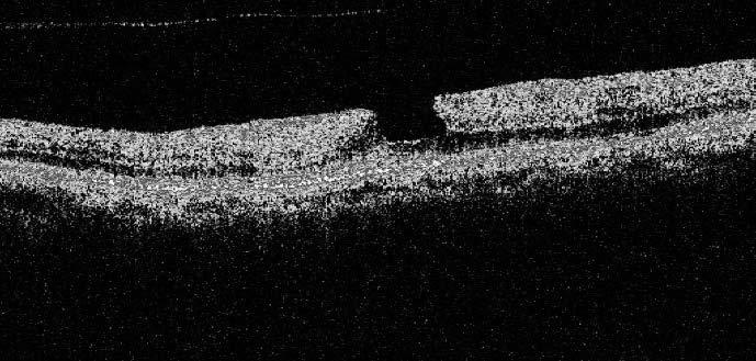
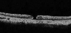
7 What else is going on? Good eye? RP WITH CME RP WITH CME OCT Post-op CME Vitreoretinal Interface Disorders Idiopathic Epiretinal Membrane Vitreomacular Traction Syndrome Idiopathic Macular Hole Full thickness Macular Hole Macular hole sizes Small <= 250 microns Medium microns Large > 400 microns Horizontal diameter at narrowest point 6


8 New grading system ERM VMA with no change in foveal contour: Stage 0 VMT with disruption of foveal contour: Stage 1 VMT with small or medium FT hole: Stage 2 VMT with medium or large full thickness hole: Stage 3 Any full thickness hole without VMT: stage 4 Lamellar hole Psuedohole from ERM Membranous growth of glial cells on retina surface Can be asymptomatic or very bothersome Metamorphopsia is common More common after PVD Tractional macular holes, cysts, CME, neurosensory RD s; retinal and choroidal folds, etc. Rerm ERM ERM + Cystoid Edema ERM with Macular Edema ERM Cysts ERM lemon drops ERM Neurosensory RD 7


9 ILM fracture ERM with psuedoholes 20/30 Lemon drops ERM with lamellar hole ERM OS Full Thickness Macular Hole ERM Macular Holes: 70 Y/O female Full thickness macular hole & ERM- OS BCVA 20/400 Pseudohole 20/25- OD 8
10 Pseudohole Macular hole repair Posterior hyaloid Photoreceptors 20/25! VMTS 3yrs later 20/20- VFTS with early hole Foveal detachment 9

11 VMTS & ERM Posterior hyaloid ERM VMA on an ERM VFTS spontaneous resolution after 3 months Jetrea / Ocriplasmin Effective at breaking VMA about 26% of the time in clinical trials Costs $3000-$4000 per injection Reports of decreased visual function after injection, usually not permanent 10


12 Jetrea Jetrea Factors that increase success in real world settings (Ophthalmology Times on-line) to about 50%... VMA < 1500 microns in diameter Age < 65 Full thickness macular hole present Phakic eye No ERM If PIL line disrupted at one week after injection (recovers by one month), then 75% chance of success This PIL disruption may correlate with reports of temporary reduction in visual function VFTS VFTS Full thickness hole OD, VMTS OS 11


13 Anti-VMA! Macular Hole Formation Attached Operculum Macular Hole Posterior Hyaloid Operculum Cystic spaces OS Stage 4 Macular Hole Large Stage 4 Hole Macular Hole 12


14 Central Serous Retinopathy CSR with PED ICSC with LEMON DROPS Old ICSC ICSC has abnormally thick choroid on SD OCT EDI: Normal is 250 microns. drops of water on a windshield SD-OCT 13

15 SD-OCT SD-OCT Macular Degeneration Drusen- Dry AMD Drusenoid PED- Soft Drusen 14


16 SD PED PED VA= 20/70 PED Subretinal fluid.or is there something else? Same concept CNVM Miscellaneous Retinal Conditions S/P Avastin injection- What s new? 15


17 Chloroquine maculopathy Chloroquine OCT Plaquenil toxicity: Flying Saucer Sign Medullated Nerve Fibers? Retinal Detachment Bullous RD Macula off 16


18 S/P RD surgery 6 Mos. 20/40 Macula off RD PDR with traction RD Development of Foveal Retinoschisis Foveal Retinoschisis Another Patient Foveal Retinoschisis OS OD 17


19 hypotony Solar Maculopathy Choroidal Folds Focal defect in PR outer segments and RPE Solar Maculopathy Solar maculopathy Solar maculopathy Courtesy Dr. Jerome Sherman 18
20 Adult Vitelliform Also Adult Vitelliform Best s dystrophy Cone dystrophy Cone dystrophy OCT IJXT with ILM drape 20/40 ILM drape 19


21 ORT s ORT s Optic nerve head and glaucoma Use of GCC analysis with glaucoma 20


22 GCC analysis VF OU GCC and VF Loss comparison Stroke and GCC loss Stroke and GCC loss # 2 Stroke and GCC loss #2 21


23 Optic Nerve Pit Optic Nerve Pit Pit? ONH Colobomas ONH Coloboma OCT Papilledema- IIH 25Y/O female Dg: IIH Diamox Tx 22


24 Resolving 23 y/o male- IIH Resolution ONH Drusen ONH DRUSEN SD-OCT ONH DRUSEN SD OCT Improved with EDI 23


25 Color SD-OCT Anterior Segment OCT Visante image Many units available with anterior segment capability New cap available for Cirrus 4000 and 5000 that allows Visante style imaging 24


26 Angle:Optovue Cells in A/C Plateau Iris Wound leak with choroidals Wound leak post repair 25

27 Scleral lens Scleral lens Scleral lens landing zone Two for the price of one! THE END! 26
OCT Interpretation. Financial Disclosure. Jay M. Haynie, OD, FAAO. OCT Image Layers 7/21/2014
 OCT Interpretation Jay M. Haynie, OD, FAAO Financial Disclosure I have received honoraria or am on the advisory board for the following companies: Olympia Tacoma Renton Kennewick - Washington Carl Zeiss
Mark Dunbar: Disclosure
 Important Things to Understand About OCT Mark T. Dunbar, O.D., F.A.A.O. Bascom Palmer Eye Institute University of Miami, School of Medicine Mark Dunbar: Disclosure Optometry Advisory Board for: Allergan
8/6/17. Disclosures Aerie Pharmaceuticals Alcon BioTissue Diopsys Optovue Shire
 Nathan Lighthizer, O.D., F.A.A.O. Associate Professor Assistant Dean for Clinical Care Director of Continuing Education Chief of Specialty Care Clinics Oklahoma College of Optometry Tahlequah, OK lighthiz@nsuok.edu
OCT Interpretation in Retinal Disease
 OCT Interpretation in Retinal Disease Jay M. Haynie, OD, FAAO Financial Disclosure I have received honoraria or am on the advisory board for the following companies: Carl Zeiss Meditec Advanced Ocular
History/principles of the OCT What does the normal retinal OCT look like Vitreal disorders Retinal/RPE disorders Choroidal disorders
 Nathan Lighthizer, O.D., F.A.A.O. Assistant Professor Assistant Dean for Clinical Care Director of Continuing Education Chief of Specialty Care Clinics Chief of Electrodiagnostics Clinic Oklahoma College
Optical Coherence Tomography in Diabetic Retinopathy. Mrs Samantha Mann Consultant Ophthalmologist Clinical Lead of SEL-DESP
 Optical Coherence Tomography in Diabetic Retinopathy Mrs Samantha Mann Consultant Ophthalmologist Clinical Lead of SEL-DESP Content OCT imaging Retinal layers OCT features in Diabetes Some NON DR features
Optical Coherence Tomography (OCT)
") Understanding and Interpreting OCT Mark Dunbar: Disclosure The Swiss Army Pocket Knife of Eye Care Mark T. Dunbar, O.D., F.A.A.O. Bascom Palmer Eye Institute University of Miami, School of Medicine Consultant
Understanding and Interpreting OCT Mark Dunbar: Disclosure The Swiss Army Pocket Knife of Eye Care Mark T. Dunbar, O.D., F.A.A.O. Bascom Palmer Eye Institute University of Miami, School of Medicine Consultant
Cirrus TM HD-OCT. Details defi ne your decisions
 Cirrus TM HD-OCT Details defi ne your decisions 2 With high-defi nition OCT Carl Zeiss Meditec takes you beyond standard spectral domain Built on 10 years experience at the vanguard of innovation, Carl
Optical Coherence Tomography: Pearls for the Anterior Segment Surgeon Basic Science Michael Stewart, M.D.
 Optical Coherence Tomography: Pearls for the Anterior Segment Surgeon Basic Science Michael Stewart, M.D. Disclosure OCT Optical Coherence Tomography No relevant financial relationships I will refer to
What Is O.C.T. and Why Should I Give A Rip? OCT & Me How Optical Coherence Tomography Changed the Life of a Small Town Optometrist 5/19/2014
 OCT & Me How Optical Coherence Tomography Changed the Life of a Small Town Optometrist Email: myoder@wcoil.com Mark A. Yoder, O.D. 107 N. Main Street PO Box 123 Bluffton, OH 45817 @yoderod 115.02 Histoplasma
Cirrus TM HD-OCT. Details define your decisions
 Cirrus TM HD-OCT Details define your decisions 2 With high-definition OCT Carl Zeiss Meditec takes you beyond standard spectral domain Built on 10 years experience at the vanguard of innovation, Carl Zeiss
EPIRETINAL MEMBRANE & VITREOMACULAR TRACTION
 EPIRETINAL MEMBRANE & VITREOMACULAR TRACTION Management of ERM and VMT K.V.Chalam,MD,PhD,MBA,FACS Professor and Director of Retina Loma Linda Eye Institute Los Angeles, USA REVIEW ANATOMY The vitreous
Often asymptomatic but can cause a reduction in BCVA and distortion of vision.
 Christopher Wolfe, OD, FAAO, Dipl. ABO Epiretinal Membrane (ERM) and Vitreomacular Traction (VMT) Epiretinal membrane (macular pucker, cellophane maculopathy, premacular fibrosis) consists of a layer of
OPTIC DISC PIT Pathogenesis and Management OPTIC DISC PIT
 OPTIC DISC PIT Pathogenesis and Management Abdel-Latif Siam Ain Shams University Cairo Egypt OPTIC DISC PIT Congenital pit is an atypical coloboma usually located on the temporal edge of the disc, associated
Managing the Vitreomacular Interface
 Managing the Vitreomacular Interface A Guide to VMA, VMT, Holes and ERM Anna K. Bedwell, OD, FAAO Indiana University School of Optometry Please silence all mobile devices and remove items from chairs so
R&M Solutions
 Mohamed Hosny El-Bradey, MD., Assistant Professor of Ophthalmology, Tanta University. Wael El Haig, MD., Professor of Ophthalmology. Zagazeeg University. 1 Myopic CNV is considered the most common vision
Advances in OCT Murray Fingeret, OD
 Disclosures Advances in OCT Murray Fingeret, OD Consultant Alcon, Allergan, Bausch & Lomb, Carl Zeiss Meditec, Diopsys, Heidelberg Engineering, Reichert, Topcon Currently Approved OCT Devices OCT Devices
Posterior Segment Update
 Posterior Segment Update Featured Speaker: Dr. Kyle Cheatham, FAAO, DIP ABO DISCLOSURE STATEMENT We have no direct financial or proprietary interest in any companies, products or services mentioned in
Incorporating OCT Angiography Into Patient Care
 Incorporating OCT Angiography Into Patient Care Beth A. Steele, OD, FAAO OCT A: Introduction Isolates microvascular circulation from OCT image data Axial resolution = 5 microns (i.e. fine capillaries visible)
The Quick Guide to OCT Mastery 50 Real Cases with Expert Analysis
 OPTICAL COHERENCE TOMOGRAPHY The Quick Guide to OCT Mastery 50 Real Cases with Expert Analysis VOL 1 Sanjay Sharma, MD, FRCS, MSc (Epid), MBA Ophthalmologist, Epidemiologist Queen s University, Canada
Ophthalmic Imager Role
 MASTER OCT Ophthalmic Photographers Society October 18, 2014 Chicago, IL James B Soque, CRA COA Pamela A Weber, MD Island Retina Shirley, New York Commack, New York Financial Disclosure Genentech Ophthotech
3/6/2014. Hoda MH Mostafa MD Associate Professor of Ophthalmology Cairo University. The author has no proprietary interest. Today s Objectives
 Hoda MH Mostafa MD Associate Professor of Ophthalmology Cairo University The author has no proprietary interest Today s Objectives Identify the CLINICAL SCENARIOS IN MACULAR EDEMA where OCT plays a MAJOR
11/29/2016 MACULAR MALADIES: TYPICAL & ATYPICAL CASES
 MACULAR MALADIES: TYPICAL & ATYPICAL CASES Dawn Pewitt, OD, FAAO Triad Eye Institute, Grove, OK Dpewitt@triadeye.com Disclosure Statement: No financial disclosures COPE 51218-PS Please silence all mobile
Re(nal and OCT Grand Rounds
 Op#cal Coherence Tomography Op(cal: Light- based Re(nal and OCT Grand Rounds Steven Ferrucci, OD, FAAO Chief, Optometry Sepulveda VA Professor, SCCO/MBKU Coherence: property of light waves in which the
VITREOMACULAR UPDATE FOR THE PRIMARY CARE OD
 VITREOMACULAR UPDATE FOR THE PRIMARY CARE OD VITREOMACULAR UPDATE FOR THE PRIMARY CARE OD 1 2 DISCLOSURE STATEMENT I have received lecture honoraria from TearScience. I have no direct financial or proprietary
Optical Coherence Tomograpic Features in Idiopathic Retinitis, Vasculitis, Aneurysms and Neuroretinitis (IRVAN)
") Columbia International Publishing Journal of Ophthalmic Research (2014) Research Article Optical Coherence Tomograpic Features in Idiopathic Retinitis, Vasculitis, Aneurysms and Neuroretinitis (IRVAN)
Columbia International Publishing Journal of Ophthalmic Research (2014) Research Article Optical Coherence Tomograpic Features in Idiopathic Retinitis, Vasculitis, Aneurysms and Neuroretinitis (IRVAN)
OCT Angiography The Next Frontier
 Choroid Retina avascular 5/13/2017 OCT Angiography The Next Frontier Pierce Kenworthy OD, FAAO June 9, 2017 OCT Angiography (OCTA) 2016 Non-invasive, motion contrast imaging Represents erythrocyte movement
Vitreomacular interface disorders. Ghanbari MD 1393:10:25
 Vitreomacular interface disorders Ghanbari MD 1393:10:25 Human vitreous after dissection of the sclera, choroid, and retina. Lamellar structure of the posterior vitreous cortex (PVC) in the monkey. V =
Learn Connect Succeed. JCAHPO Regional Meetings 2016
 Learn Connect Succeed JCAHPO Regional Meetings 2016 pearls and pitfalls of ophthalmic imaging JCHAPO 2016 Conference Vikas Chopra, M.D. Medical Director, UCLA Doheny Eye Centers Pasadena Principal Investigator,
The Future of Retinal Imaging Has Arrived!
 Financial Disclosure The Future of Retinal Imaging Has Arrived! Joseph J. Pizzimenti, OD, FAAO Carlo Pelino, OD, FAAO! Pizzimenti:! Honoraria! Kemin! Nicox! Review of Optometry! Optometric Management!
The Foundation WHAT IS THE RETINA? continued next page. RETINA HEALTH SERIES Facts from the ASRS
 The Foundation American Society of Retina Specialists Committed to improving the quality of life of all people with retinal disease. Vitreomacular Traction Syndrome The vitreous humor is a transparent,
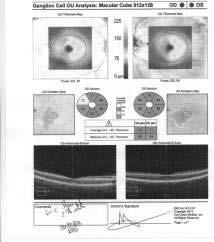
Ganglion cell analysis by optical coherence tomography (OCT) Jonathan A. Micieli, MD Valérie Biousse, MD
 Jonathan A. Micieli, MD Valérie Biousse, MD") Ganglion cell analysis by optical coherence tomography (OCT) Jonathan A. Micieli, MD Valérie Biousse, MD Figure 1. Normal OCT of the macula (cross section through the line indicated on the fundus photo)
Ganglion cell analysis by optical coherence tomography (OCT) Jonathan A. Micieli, MD Valérie Biousse, MD Figure 1. Normal OCT of the macula (cross section through the line indicated on the fundus photo)
Learn Connect Succeed. JCAHPO Regional Meetings 2017
 Learn Connect Succeed JCAHPO Regional Meetings 2017 How Retinal Imaging Guides Treatment Odette Margit Houghton MD Question 1 Which OCT has the highest resolution? A: Swept source OCT B: Spectral domain
OCT Assessment of the Vitreoretinal Relationship in CSME
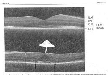
 December 2007 Sonia Rani John et al. - IFIS 375 ORIGINAL ARTICLE OCT Assessment of the Vitreoretinal Relationship in CSME Dr. Manoj S. DNB FRCS, Dr. Unnikrishnan Nair MS DO FRCS, Dr. Gargi Sathish MS Introduction
Fundus Autofluorescence. Jonathan A. Micieli, MD Valérie Biousse, MD
 Fundus Autofluorescence Jonathan A. Micieli, MD Valérie Biousse, MD The retinal pigment epithelium (RPE) has many important functions including phagocytosis of the photoreceptor outer segments Cone Rod
Early diagnosis and treatment of VMT with single Intravitreal Injection of Pharmacologic Vitreolysis. Stratos Gotzaridis MD Athens
 Early diagnosis and treatment of VMT with single Intravitreal Injection of Pharmacologic Vitreolysis Stratos Gotzaridis MD Athens The Vitreous Body Gel composed of 98-99% water 1% macromolecules Glycoproteins
Title: OCT Analysis Workshop: Interpretation of OCT printouts
 Title: OCT Analysis Workshop: Interpretation of OCT printouts Authors: David Yang, OD, FAAO Staff Optometrist, VA Palo Alto Health Care System Associate Clinical Professor, UC Berkeley School of Optometry
Disclosures. Definitions. Goals. Imaging and glaucoma 3/22/2016
 Pinakin Davey OD, PhD, FAAO Professor and Director of Research Disclosures Principal investigator for ivue OCT trial Principal investigator Topcon FDA trials for Maestro and OCT 2000 Consultant for Topcon
Audit of Macular Hole Surgery, Visual Outcome Prediction on OCT Appearance of Macular Hole
 International Journal of Ophthalmology & Visual Science 2017; 2(4): 93-97 http://www.sciencepublishinggroup.com/j/ijovs doi: 10.11648/j.ijovs.20170204.13 Audit of Macular Hole Surgery, Visual Outcome Prediction
Diagnosis in AMD. Managing your AMD Patients
 Managing your AMD Patients Robert W. Dunphy, O.D., F.A.A.O. Diagnosis in AMD Have suspicion Identify relative risk Conduct surveillance Biometry Utilize technology to facilitate detection of change / stability
Principle of OCT. Reading Between the Lines: OCT Interpretation. Initial Concept. Advantage: High Resolution Cross Section Images
 Principle of OCT Reading Between the Lines: OCT Interpretation Mohammad Rafieetary, OD, FAAO mrafieetary@charlesretina.com Introduction Optical Biopsy Morphologic Evaluation of Live Tissue Measurements
VMA at the macula resulting in VMT
 Ocriplasmina for pharmacologic treatment in VMT Teresio Avitabile 1 Introduction PVD is a normal, physiologic process that occurs with aging; however, in some cases, PVD is incomplete Incomplete PVD localized
Optical Coherence Tomography (OCT) in Uveitis Piergiorgio Neri, BMedSc, MD, PhD Head Ocular Immunology Unit
 in Uveitis Piergiorgio Neri, BMedSc, MD, PhD Head Ocular Immunology Unit") The Eye Clinic Polytechnic University of Marche Head: Prof Alfonso Giovannini November, 1991 Optical Coherence Tomography (OCT) in Uveitis Piergiorgio Neri, BMedSc, MD, PhD Head Ocular Immunology Unit
The Eye Clinic Polytechnic University of Marche Head: Prof Alfonso Giovannini November, 1991 Optical Coherence Tomography (OCT) in Uveitis Piergiorgio Neri, BMedSc, MD, PhD Head Ocular Immunology Unit
Applying structure-function to solve clinical cases
 Applying structure-function to solve clinical cases Professor Michael Kalloniatis Centre for Eye Health, and, School of Optometry and Vision Science Acknowledgements Some material prepared by Nayuta Yoshioka
When optical coherence tomography (OCT)
") Macular Imaging: SD-OCT in nterior Segment Surgical Practice Many pathologic processes of the macula can be visualized or quantified only with this modality. y Steven G. Safran, MD When optical coherence
Macular Imaging: SD-OCT in nterior Segment Surgical Practice Many pathologic processes of the macula can be visualized or quantified only with this modality. y Steven G. Safran, MD When optical coherence
University Hospital Basel. Optical Coherence Tomography Emerging Role in the Assessment of MS PD Dr. Konstantin Gugleta
 University Hospital Basel Optical Coherence Tomography Emerging Role in the Assessment of MS PD Dr. Konstantin Gugleta 15th State of the Art SMSS, Lucerne January 2013 Retinal Nerve Fiber Layer 1.200.000
Objective Assessment of Macula and Optic Nerve
 Objective Assessment of Macula and Optic Nerve Jerry Sherman Disclosure: Dr. Sherman has lectured and received honorarium from Carl Zeiss Meditec, Topcon, Optovue, Optos, and PHP, Diopsys, Eye Solutions,Quantel,
PRIMUS 200 from ZEISS The essential OCT
 PRIMUS 200 from ZEISS The essential OCT Seeing beyond the surface. ZEISS PRIMUS 200 // INNOVATION MADE BY ZEISS Clear Visualization. Advanced Technology. Reliability. Essential elements of your first OCT.
An A to Z guide on Epiretinal Membranes (ERMs) Paris Tranos PhD,ICO,FRCS OPHTHALMICA Vitreoretinal & Uveitis Department
 Paris Tranos PhD,ICO,FRCS OPHTHALMICA Vitreoretinal & Uveitis Department") An A to Z guide on Epiretinal Membranes (ERMs) Paris Tranos PhD,ICO,FRCS OPHTHALMICA Vitreoretinal & Uveitis Department Types of ERM Natural history OCT prognostic factors ERM with co-existing pathology
An A to Z guide on Epiretinal Membranes (ERMs) Paris Tranos PhD,ICO,FRCS OPHTHALMICA Vitreoretinal & Uveitis Department Types of ERM Natural history OCT prognostic factors ERM with co-existing pathology
Dehiscence of detached internal limiting membrane in eyes with myopic traction maculopathy with spontaneous resolution
 Hirota et al. BMC Ophthalmology 2014, 14:39 RESEARCH ARTICLE Open Access Dehiscence of detached internal limiting membrane in eyes with myopic traction maculopathy with spontaneous resolution Kazunari
How to Be Efficient and Effective. Disclosure. Topics CASE CM. Case JF 2007 OHTN / POAG? How to Be Efficient and Effective with. with New Technology
 How to Be Efficient and Effective with Disclosure COPE Course ID: 40750 GL Michael Chaglasian has the following disclosures: 1. Advisory Board: Allergan, Inc., Alcon Labs, B+L Carl Zeiss Meditec 2. Research:
Case report 12/10/2014. Delphine Lam ; Dr Mayer Srour Service d ophtalmologie Professeur E.Souied Université Paris Est
 Case report 12/10/2014 Delphine Lam ; Dr Mayer Srour Service d ophtalmologie Professeur E.Souied Medical history Man, 75 years old Complaint: Vision loss in left eye in June 2014 Past ophthalmologic history:
PRIMUS 200 from ZEISS The essential OCT
 EN 00_00I The contents of the brochure may differ from the current status of approval of the product in your country. Please contact your regional representative for more information. Subject to change
Yasser R. Serag, MD Tamer Wasfi, MD El- Saied El-Dessoukey, MD Magdi S. Moussa, MD Anselm Kampik, MD
 Microperimetric Evaluation of Brilliant Blue G- assisted Internal Limiting Membrane Peeling By Yasser R. Serag, MD Tamer Wasfi, MD El- Saied El-Dessoukey, MD Magdi S. Moussa, MD Anselm Kampik, MD The internal
OCT Evaluation of the Retina Alison Bozung, OD, FAAO
 Alison Bozung, OD, FAAO Rob Wooldridge, OD, FAAO Bozung: No relevant financial relationships with commercial interests Wooldridge: Speakers Bureau/honoraria from Aerie, Alcon, Allergan, Bausch & Lomb,
ATLAS OF OCT. Retinal Anatomy in Health & Pathology by Neal A. Adams, MD. Provided to you by:
 ATLAS OF OCT Retinal Anatomy in Health & Pathology by Neal A. Adams, MD Provided to you by: Atlas of OCT The OCT Atlas is written by Neal A. Adams, MD, and produced by Heidelberg Engineering, Inc. to help
The College of Optometrists - Learning outcomes for the Professional Certificate in Medical Retina
 Learning outcomes for the Professional Certificate in Medical Retina, incorporating diabetic retinopathy screening and age related macular degeneration The professional certificate is a prerequisite to
PREDICTIVE FACTORS OF VISUAL OUTCOME FOR VITREOMACULAR TRACTION SYNDROME AFTER VITRECTOMY
 PREDICTIVE FACTORS OF VISUAL OUTCOME FOR VITREOMACULAR TRACTION SYNDROME AFTER VITRECTOMY Downloaded from https://journals.lww.com/retinajournal by mv7bzw+nz2blpko//cqyhwu2mokppdiwuep6ir1molueskh0dp9rbmb7dum5a2/cp6zifirtq3zbawzt+95f/m61fycawpqbpe8y2wuyzwnns2gw3+gmrxei6x11wu+s
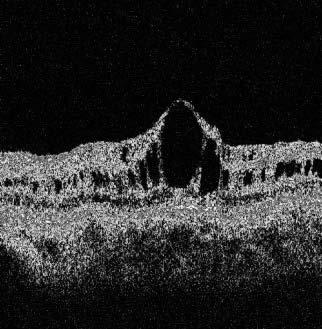
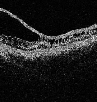
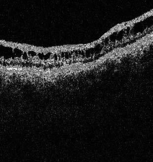
DOME SHAPED MACULOPATHY. Ιωάννης Ν. Βαγγελόπουλος Χειρ. Οφθαλμίατρος - Βόλος
 DOME SHAPED MACULOPATHY Ιωάννης Ν. Βαγγελόπουλος Χειρ. Οφθαλμίατρος - Βόλος DOME SHAPED MACULOPATHY-DEFINITIONS The entity Dome Shaped Macula ( DSM ) was first described by Gaucher and associates in 2008
Re)nal and OCT Grand Rounds. What's new in OCT? Principles of AngioVue OCTA. Vascular Imaging No Referral Needed 3/9/18. Spectral Domain: Many Op3ons
nal and OCT Grand Rounds. What's new in OCT? Principles of AngioVue OCTA. Vascular Imaging No Referral Needed 3/9/18. Spectral Domain: Many Op3ons") Spectral Domain: Many Op3ons Re)nal and OCT Grand Rounds Steven Ferrucci, OD, FAAO Chief, Optometry Sepulveda VA Professor, SCCO/MBKU Ease of use Customer support Integra)on of other technology FAF Color
Spectral Domain: Many Op3ons Re)nal and OCT Grand Rounds Steven Ferrucci, OD, FAAO Chief, Optometry Sepulveda VA Professor, SCCO/MBKU Ease of use Customer support Integra)on of other technology FAF Color
OCT in Diabetic Macular Edema and its Correlation with Flourescein Angiography
 Uvea OCT in Diabetic Macular Edema and its Correlation with Flourescein Angiography Kirti Jaisingh MS Kirti Jaisingh MS, Yashpal Goel* MS, Kshitij Aditya** DO * Guru Nanak Eye Centre, New Delhi ** Baba
OCT Angiography in Primary Eye Care
 OCT Angiography in Primary Eye Care An Image Interpretation Primer Julie Rodman, OD, MS, FAAO and Nadia Waheed, MD, MPH Table of Contents Diabetic Retinopathy 3-6 Choroidal Neovascularization 7-9 Central
SOCT Copernicus REVO. * - Currently import and overlay are avaibale in manual mode only
 SOCT Copernicus REVO Easy Operation (Full auto & Auto mode) Auto alignment (Z-position, C-gate, Focus, Tomogram) Voice guide (support patient through examination) Powerful analysis tools Enhanced tomograms
IN NICU OCT UTILIZES A CONCEPT KNOWN AS INTERFEROMETRY APPLICATIONS FOR OCT THE PRIMARY USE IN THE EYE - RETINA
 2016 25 YEARS OF OPTICAL COHERENCE TOMOGRAPHY OPTICAL COHERENCE TOMOGRAPHY IN NICU Marcin Stopa, MD, PhD, FEBO Department of Ophthalmology, Chair of Ophthalmology and Optometry. Poznan University of Medical
Steven Ferrucci, OD. FAAO; Jeffry Gerson, OD, FAAO; Robert Prouty, OD, FAAO; Leo semes OD, FAAO
 PARDON THE OBJECTION: RETINA Steven Ferrucci, OD. FAAO; Jeffry Gerson, OD, FAAO; Robert Prouty, OD, FAAO; Leo semes OD, FAAO 1. Introductions/Disclosures (Ferrucci) 2. The genetics of AMD (Gerson) a. Background
AperTO - Archivio Istituzionale Open Access dell'università di Torino
 AperTO - Archivio Istituzionale Open Access dell'università di Torino Artifacts in automatic retinal segmentation using different optical coherence tomography instruments. This is the author's manuscript
Optical coherence tomography of the vitreoretinal interface in macular hole formation
 1092 St Thomas s Hospital, London V Tanner D S Chauhan T L Jackson T H Williamson Correspondence to: Mr V Tanner, Royal Berkshire Hospital, London Road, Reading RG1 5AN, UK tannerone@aol.com Accepted for
ZEISS AngioPlex OCT Angiography. Clinical Case Reports
 Clinical Case Reports Proliferative Diabetic Retinopathy (PDR) Case Report 969 PROLIFERATIVE DIABETIC RETINOPATHY 1 1-year-old diabetic female presents for follow-up of proliferative diabetic retinopathy
We are IntechOpen, the world s leading publisher of Open Access books Built by scientists, for scientists. International authors and editors
 We are IntechOpen, the world s leading publisher of Open Access books Built by scientists, for scientists 3,700 108,500 1.7 M Open access books available International authors and editors Downloads Our
Retinal Complications of Obstructive Sleep Apnea A Growing Concern!
 Retinal Complications of Obstructive Sleep Apnea A Growing Concern! Jay M. Haynie, OD, FAAO Financial Disclosure I have received honoraria or am on the advisory board for the following companies: Carl
Structural examina.on: Imaging
 ManaMa: Glaucoma Structural examina.on: Imaging Luís Abegão Pinto, MD, PhD Department of Ophthalmology CHLC Lisbon Faculty of Medicine, Lisbon University 1 11-10- 2013 Structural changes Qualitative changes
Technicians & Nurses Program
 ASCRS ASOA Symposium & Congress Technicians & Nurses Program April 17-21, 2015 San Diego, California Optical Coherence Tomography: Essentials in Anterior and Posterior Segment Imaging Michael Stewart,
Outline. Outline. Vitreous Development & Anatomy OPT - 243
 2010 OPT - 243 Vitreous Disorders & Vitreoretinal Disorders of the Posterior Pole I Leo Semes, OD, FAAO 100% 0% 0% 0% 0% Which of these gives the best resolution for studying vitreoretinal disorders of
Goals of this Course. Imaging Technologies. Imaging Technologies. The Future of Retinal Imaging Has Arrived! Richard Spaide, MD NY Retina Consultants
 The Future of Retinal Imaging Has Arrived! William Jones, OD Steven Ferrucci, OD Joseph J. Pizzimenti, OD (moderator) Optometric Retina Society Goals of this Course! To provide an overview of PS Imaging!
evaluation of vitreoretinal adhesions in exudative AMD using optical coherence tomography
 evaluation of vitreoretinal adhesions in exudative AMD using optical coherence tomography Dr. Mahmoud Alaa Abouhusssein, FRCO Lecturer of ophthalmology, Alexandria university Dr. Amir Ramadan Gomaa, MD
Introducing ANGIOVUE ESSENTIAL. Built on the Avanti Widefield OCT Platform. OCT Angiography for Primary Eye Care
 Introducing ANGIOVUE ESSENTIAL Built on the Avanti Widefield OCT Platform OCT Angiography for Primary Eye Care Transform Your View of the Retina OCT Angiography (OCTA) is a quick non-invasive test that
Do You See What I See!!! Shane R. Kannarr, OD
 Do You See What I See!!! Shane R. Kannarr, OD skannarr@kannarreyecare.com Define Specialty Testing Additional Test to: Prove/Disprove Diagnosis To monitor progression of a condition To document a condition
OCT Image Analysis System for Grading and Diagnosis of Retinal Diseases and its Integration in i-hospital
 Progress Report for1 st Quarter, May-July 2017 OCT Image Analysis System for Grading and Diagnosis of Retinal Diseases and its Integration in i-hospital Milestone 1: Designing Annotation tool extraction
Moving forward with a different perspective
 Moving forward with a different perspective The Leader In Vision Diagnostics Offers A New Perspective Marco has served the eyecare community by offering exceptional lane products and automated high tech
ZEISS AngioPlex OCT Angiography Overview ZEISS OCT Angiography
 ZEISS AngioPlex OCT Angiography Overview ZEISS OCT Angiography California, ZEISS AngioPlex Ultra-clear visualization of microvascular blood flow using non-invasive OCT angiography 2 AngioPlex OCT Angiography
FALSE! True or False: Back to Basics. Key to Retinal Assessment: Making Visible what is Invisible Jerome Sherman
 Key to Retinal Assessment: Making Visible what is Invisible Jerome Sherman Disclosure: Dr. Sherman has lectured and received honorarium from Carl Zeiss Meditec, Topcon, Optovue, Optos, Eye Solutions, PHP,
We are IntechOpen, the world s leading publisher of Open Access books Built by scientists, for scientists. International authors and editors
 We are IntechOpen, the world s leading publisher of Open Access books Built by scientists, for scientists 3,900 116,000 120M Open access books available International authors and editors Downloads Our
Mariam Raouf Fadel M.B., B.Ch. M.Sc., Cairo University. A thesis. Submitted by. For partial fulfillment of. MD Degree in Ophthalmology
 Correlation of fundus autofluorescence and spectral domain OCT findings of the macula with visual outcome after successful repair of rhegmatogenous retinal detachment A thesis Submitted by Mariam Raouf
Fundus Autofluorescence and its PRACTICAL applications: Retina Beyond the Color. Start to think about this. Disclosure 5/21/2015
 Fundus Autofluorescence and its PRACTICAL applications: Retina Beyond the Color Jeffry D. Gerson, O.D., F.A.A.O Olathe, KS jgerson@hotmail.com Start to think about this. Disclosure I have worked with/consulted
Diabetic Retinopathy Clinical Research Network
 Diabetic Retinopathy Clinical Research Network Comparison of Time Domain OCT and Spectral Domain OCT Retinal Thickness Measurement in Diabetic Macular Edema Version 1.0 June 16, 2009 comparison of td vs
RETINA 2018 OBJECTIVES OCT VERY USEFUL INFORMATION SAFE AND FRIENDLY 1/11/2018 KELLY MITCHELL
 RETINA 2018 KELLY MITCHELL OBJECTIVES HIGHLIGHT NEW DIAGNOSTIC & TREATMENT OPTIONS REVIEW DIAGNOSTIC KEYS OF SELECT RETINAL DISEASES DISCUSS USE OF IMAGING AND REFERRAL RECOURSES FOR PATIENT BENEFIT OCT
Financial Disclosures
 Financial Disclosures Consultant Genentech, Regeneron, Allergan, Thrombogenics, Optos, and ArcticDx Grant Support Regeneron, Allergan Mathew W. MacCumber, MD, PhD Professor & Assoc. Chair for Research
Re)nal and OCT Grand Rounds
nal and OCT Grand Rounds") Op#cal Coherence Tomography Op)cal: Light- based Re)nal and OCT Grand Rounds Steven Ferrucci, OD, FAAO Chief, Optometry Sepulveda VA Professor, SCCO/MBKU Coherence: property of light waves in which the
Op#cal Coherence Tomography Op)cal: Light- based Re)nal and OCT Grand Rounds Steven Ferrucci, OD, FAAO Chief, Optometry Sepulveda VA Professor, SCCO/MBKU Coherence: property of light waves in which the
Study of clinical significance of optical coherence tomography in diagnosis & management of diabetic macular edema
 Original Research Article Study of clinical significance of optical coherence tomography in diagnosis & management of diabetic macular edema Neha Kantilal Desai 1,*, Somesh Vedprakash Aggarwal 2, Sonali
Choroidal Mapping; a Novel Approach for Evaluating Choroidal Thickness and Volume
 Imaging Technique Choroidal Mapping; a Novel Approach for Evaluating Choroidal Thickness and Volume Jila Noori 1, MD; Mohammad Riazi Esfahani 1,2, MD Fedra Hajizadeh 2, MD; Mohammad-Mehdi Zaferani 1, MD
Vitreomacular Traction: Management
 Miscellaneous Refractive Surgery Vitreomacular Traction: Management Raji K. MS, DNB Raji K. MS, DNB, A.K. Upadhyay MS, S. Waikar MS, DNB, P. Tiwari MBBS Department of Ophthalmology, Command Hospital (WC)
RETINA REVEALED. Dynamic Developments in AMD Diagnosis and Treatment (2014) The Dawn of: Pharmaco-Genetics (aka Nutrigenomics) Jerome Sherman, OD
 The Dawn of: Pharmaco-Genetics (aka Nutrigenomics) Jerome Sherman, OD") RETINA REVEALED Dynamic Developments in AMD Diagnosis and Treatment (2014) The Dawn of: Pharmaco-Genetics (aka Nutrigenomics) Jerome Sherman, OD Disclosures: Jerome Sherman Dr. Sherman has lectured, received
RETINA REVEALED Dynamic Developments in AMD Diagnosis and Treatment (2014) The Dawn of: Pharmaco-Genetics (aka Nutrigenomics) Jerome Sherman, OD Disclosures: Jerome Sherman Dr. Sherman has lectured, received
Case Report Optic Disk Pit with Sudden Central Visual Field Scotoma
 Case Reports in Ophthalmological Medicine Volume 2016, Article ID 1423481, 4 pages http://dx.doi.org/10.1155/2016/1423481 Case Report Optic Disk Pit with Sudden Central Visual Field Scotoma Nikol Panou
Objectives. Unexplained Vision Loss: Where Do I Go From Here. History. History. Drug Induced Vision Loss
 Objectives Unexplained Vision Loss: Where Do I Go From Here Denise Goodwin, OD, FAAO Coordinator, Neuro-ophthalmic Disease Clinic Pacific University College of Optometry goodwin@pacificu.edu Know the importance
PROSPECTIVE THREE-DIMENSIONAL ANALYSIS OF STRUCTURE AND FUNCTION IN VITREOMACULAR ADHESION CURED BY PHARMACOLOGIC VITREOLYSIS
 PROSPECTIVE THREE-DIMENSIONAL ANALYSIS OF STRUCTURE AND FUNCTION IN VITREOMACULAR ADHESION CURED BY PHARMACOLOGIC VITREOLYSIS Kevin R. Tozer, BS,* Wolfgang Fink, PhD, ** Alfredo A. Sadun, MD, PhD, FARVO,
Why Is Imaging Critical in My Uveitis Practice?
 Why Is Imaging Critical in My Uveitis Practice? Dilraj S. Grewal, MD Developed in collaboration Imaging Is the Backbone of Uveitis Workup and Monitoring Treatment Response FP FAF B- scan Multimodal Imaging
CLINICAL COURSE OF VITREOMACULAR ADHESION MANAGED BY INITIAL OBSERVATION
 CLINICAL COURSE OF VITREOMACULAR ADHESION MANAGED BY INITIAL OBSERVATION VISHAK J. JOHN, MD,* HARRY W. FLYNN, JR., MD,* WILLIAM E. SMIDDY, MD,* ADAM CARVER, MD, ROBERT LEONARD, MD, HOMAYOUN TABANDEH, MD,
Swept-Source OCT Angiography: SS OCT Angio TM
 Swept-Source OCT Angiography: SS OCT Angio TM Not available in all countries, please check with your distributor. 2015.09 Swept-Source OCT Angiography: SS OCT Angio TM Introduction Optical coherence tomography
HOCT-1I 1F All-in-One Optical Coherence Tomography with Fundus
 HOCT-1I 1F All-in-One Optical Coherence Tomography with Fundus Specification Type Resolution(in Tissue) A scan Rate Scan Range SD-OCT / Fundus Z :6~7um, XY:20um 68,000 A-scan/sec. [Fundus] X:6-12mm, Y:6-9mm,
Clinical Trial Endpoints for Macular Diseases
 Clinical Trial Endpoints for Macular Diseases Developed in collaboration Learning Objective Upon completion, participants should be able to: Summarize types of biomarkers of progression and treatment response