Mark Dunbar: Disclosure
|
|
|
- Rolf Cameron
- 5 years ago
- Views:
Transcription


1 Important Things to Understand About OCT Mark T. Dunbar, O.D., F.A.A.O. Bascom Palmer Eye Institute University of Miami, School of Medicine Mark Dunbar: Disclosure Optometry Advisory Board for: Allergan Carl Zeiss Meditec Regeneron Mark Dunbar does not own stock in any of the above companies


2 OCT I Stratus SD OCT SD OCT Commercially Available Carl Zeiss: Cirrus OptiVue: Avantis, RTVue, ivue Heidelberg: Spectralis Topcon Optos Made by OPKO SOCT Copernicus Now owned by Cannon SD-OCT Differences Hardware is relatively similar It s all about the software! Device should be easy to use and patient friendly Should be competitively priced 2
 Over 90,000 A scans per second Compares repeat scans acquired at the same position in the retina to look for changes")
3 Advances in SD-OCT Improving software Faster virtual angiography Noise reduction/over sampling technology Wider and deeper scans Greater density in the scans Improvements in 3D imaging Enhanced depth imaging imaging choroid Progression analysis software 2015 OCT Angiography (OCTA) Over 90,000 A scans per second Compares repeat scans acquired at the same position in the retina to look for changes The OCT anatomy of the eye is static The only changes in scans are caused by blood flowing through vasculature Blood flow within the eye is captured using motion contrast. What makes a good scan Signal strength Glaucoma: minimum of 7, 6 can t rely on data Less important in retina Make sure there is no algorithm failure Need a good tear film Have patient blink before the scan Good patient Good OCT operator 3

4 3Types of OCT Scans Retina scan Glaucoma scan Anterior Segment scan The Printout: Retina Cirrus Retina: 2 Important Scans to Acquire Raster Scan Macular Cube 4


5 New HD Raster Scans More beautiful b scans right where you want them with Smart HD Scan workflow HD 1 Line 100x 100x averaged VIVID Image Enhancement Technology Improved vitreous assessment Publication quality image HD 21 Line 21 lines 4/8x averaged Ideal for anti VEGF therapy monitoring HD Radial 12 lines 8x averaged Fovea as common reference point Ideal for macular hole assessment & surgical planning HD Cross 5 horizontal 5 vertical 8x averaged The The Macular Cube Cube 5


6 The Power of the Cube Incredible Scan Density 3 D viewing Advanced segmentation Guided progression analysis Change analysis Drusen and geographic atrophy analysis Ganglion cell analysis ivue Heidelberg Spectralis 6


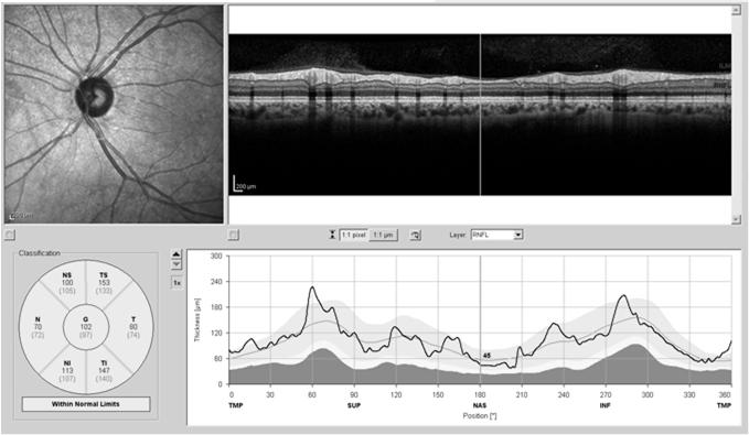
7 Zeiss Cirrus Heidelberg Spectralis OptoVue ivue Main Clinical Utilities of OCT High resolution evaluation of retinal anatomy Diagnosis of conditions difficult to establish with traditional fundus exam Quantitative assessment of retinal and vitreoretinal anatomic alterations Objective means for monitoring disease progression and/or therapeutic response Diagnose and determine progression of glaucoma The Anatomy 7


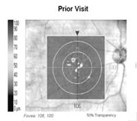
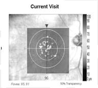
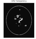
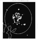
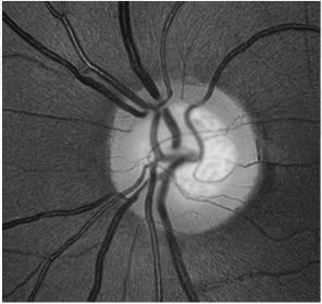
8 Understanding and Interpreting Recognizing Macular Edema Where is the Fluid? Where is the Fluid? 8

9 PED involving fovea RPE Detachment 9


10 Central Serous Chorioretinopathy (CSR) 44 y/o Female: Notes blur in the LE X 1 mo BCVA: 20/25 53 y/o Hispanic Male /20 VA 10

")
11 53 y/o Hispanic Male PED 53 y/o Hispanic Male 8/2012 2/2014 Choroidal Neovascularization (CNV) 11
12 Degenerative Myopia with CNV Early AMD 12


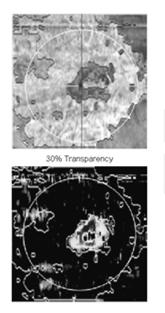
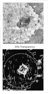
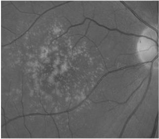
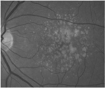
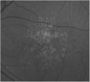
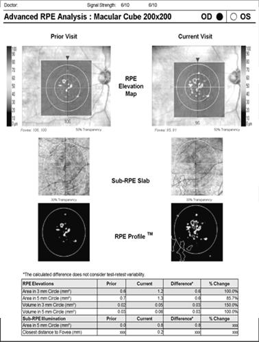
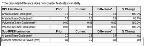
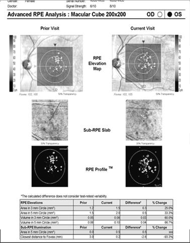
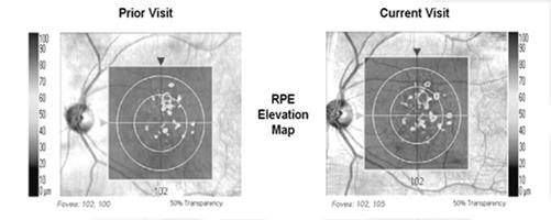
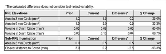
13 RPE Elevations: If the RPE is raised, a new proprietary algorithm for Cirrus maps and measures the area and volume of the elevations. Advanced RPE Analysis Gain new insights on your AMD patients RPE Elevations Sub-RPE Illumination Sub-RPE Illumination. If the RPE is absent or has lost integrity a new proprietary algorithm for Cirrus can map and measure the affected area. 13
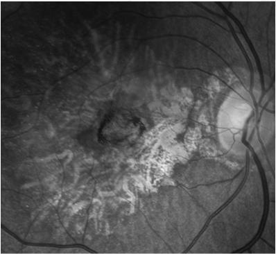
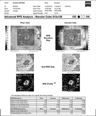
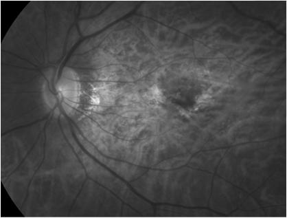
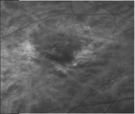
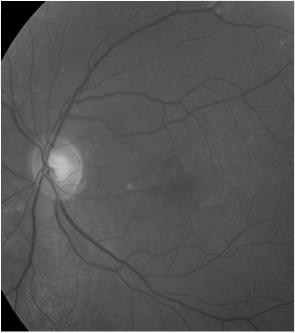
14 Esther: Geographic Atrophy /28/2010 vs. 1/18/ /28/2010 1/18/

15 3/20/10 6/5/2012 3/20/10 6/5/
16 16


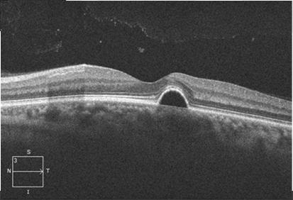
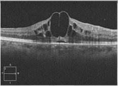
17 Full Thickness Macular Hole 66 yo Female Blurred vision RE X 6 weeks Longstanding blurred vision LE X 2 yrs 17


18 64 y/o White Female Blurred VA X 10 days Seen 2 mo ago: normal exam VA: 20/40 Heidelberg Spectralis 18


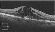
19 When is a hole.a hole? 67 y/o Hispanic Male 20/25 19


20 67 y/o Hispanic Male Told that he had a retinal problem 20


21 Macular Edema 5/7/ /200 6/9/ /25 7/28/ /200 Conditions Affecting IS/OS Junction AKA: PIL, Ellipsoid Line 20/70 20/60 57 y/o Hispanic Male: 21

22 20/70 20/70 20/60 Solar Maculopathy 22

23 SDOCT in Glaucoma 23


24 Traditional Methods of Assessing Glaucoma IOP monitoring Major risk factor Subjective evaluation of the optic nerve Visual field testing The value of the OCT in glaucoma Picking up early change That is difficult to see on clinical exam Before it shows up on the visual field Showing progression Which one has glaucoma? 24


25 At 95% specificity, up to 35% of eyes had abnormal average RNFL thickness 4 years before development of visual field loss and 19% of eyes had abnormal results 8 years before field loss. Conclusions: Assessment of RNFL thickness with OCT was able to detect glaucomatous damage before the appearance of VF defects on SAP. In many subjects, significantly large lead times were seen when applying OCT as an ancillary diagnostic tool. 25


")

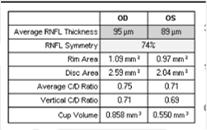
26 Glaucoma Analysis with the RTVue: Nerve Head Map Provides Cup Area Rim Area RNFL Map 16 sector analysis compares sector values to normative database and color codes result based on probability values (p values) TSNIT graph Color shaded regions represent normative database ranges based on p values 26


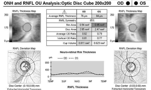
27 RNFL Quadrants and Clock Hours Inter visit Tolerance: Clock Hours: 5 7 microns Quadrant: microns You Need 2 Different Scans for Glaucoma Analysis RNFL Print out With ON data Macular Cube print out Ganglion Cell Analysis 27


28 Macular Cube Average RNFL Thickness 75 microns tipping point Floor Affect in Advanced Glaucoma microns Disc Area Small < 2 mm Medium > 2 mm Large > 3 mm 28
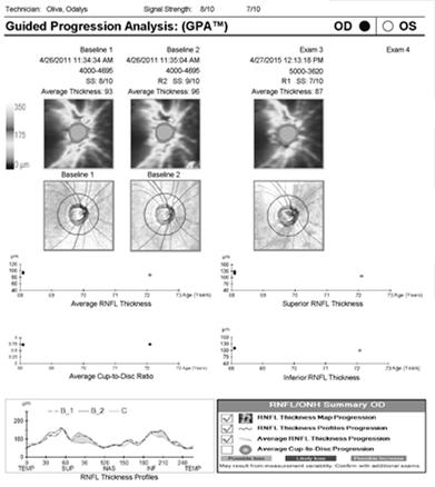
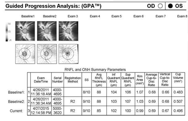
29 Clinical Pearls With Cirrus SDOCT in Glaucoma Do 3 RNFL scans at a time Ensures consistency/reliability On follow up 2 of the scans can be used as the baseline for guided progression analysis GPA 1 st Scan 1 2nd Scan 2 29


30 3rd Scan 3 51 y/o Hispanic Female Reports shadow peripherally in her LE TA: on 3 visits 30
31 31


32 Berta: 65 y/o Hispanic Female Followed for OHTN : TA

33


34 Monday 3/27/15 TA 25/29 December TA 22/22 Inter visit Tolerance: Clock Hours: 5 7μ Quadrant: 10 12μ 34
35 35


36 Tania: 44 y/o Hispanic Female Has been seen several times over the yrs for routine eye care 1998: TA 20/22 09/05: TA 18/20 12/07: 19/20 Tania: 44 y/o Hispanic Female 12/08: TA: 25/21 Pach: 610/620 μ u OCT done 1/5/08 for review 4/20/09: TA 23/24 4/19/10: TA 23/25 10/11/2010: TA 22/ /5/08 36

37 4/20/09 4/20/
38 Tania Ocular HTN No treatment Is there a reason to justify treating her? What is her risk for developing glaucoma? 5 yrs vs. lifetime? Issues Relevant to Tania What is his risk of actually developing glaucoma? From OHTS: Depends mostly on corneal thickness? IOP of mmhg Ave Corneal thickness < 556 µ: 36% Risk Corneal thickness 565 to 588 µ: 13% 38
Optical Coherence Tomography (OCT)
") Understanding and Interpreting OCT Mark Dunbar: Disclosure The Swiss Army Pocket Knife of Eye Care Mark T. Dunbar, O.D., F.A.A.O. Bascom Palmer Eye Institute University of Miami, School of Medicine Consultant
Understanding and Interpreting OCT Mark Dunbar: Disclosure The Swiss Army Pocket Knife of Eye Care Mark T. Dunbar, O.D., F.A.A.O. Bascom Palmer Eye Institute University of Miami, School of Medicine Consultant
Advances in OCT Murray Fingeret, OD
 Disclosures Advances in OCT Murray Fingeret, OD Consultant Alcon, Allergan, Bausch & Lomb, Carl Zeiss Meditec, Diopsys, Heidelberg Engineering, Reichert, Topcon Currently Approved OCT Devices OCT Devices
OCT Interpretation in Retinal Disease
 OCT Interpretation in Retinal Disease Jay M. Haynie, OD, FAAO Financial Disclosure I have received honoraria or am on the advisory board for the following companies: Carl Zeiss Meditec Advanced Ocular
Course # Getting to Know Your OCT
 Course # 140 Getting to Know Your OCT Course Title: Lecturer: Getting to Know Your OCT Brad Sutton, OD, FAAO IU School of Optometry Financial Disclosures No financial disclosures Optical Coherence Tomography-OCT
Cirrus TM HD-OCT. Details defi ne your decisions
 Cirrus TM HD-OCT Details defi ne your decisions 2 With high-defi nition OCT Carl Zeiss Meditec takes you beyond standard spectral domain Built on 10 years experience at the vanguard of innovation, Carl
8/6/17. Disclosures Aerie Pharmaceuticals Alcon BioTissue Diopsys Optovue Shire
 Nathan Lighthizer, O.D., F.A.A.O. Associate Professor Assistant Dean for Clinical Care Director of Continuing Education Chief of Specialty Care Clinics Oklahoma College of Optometry Tahlequah, OK lighthiz@nsuok.edu
How to Be Efficient and Effective. Disclosure. Topics CASE CM. Case JF 2007 OHTN / POAG? How to Be Efficient and Effective with. with New Technology
 How to Be Efficient and Effective with Disclosure COPE Course ID: 40750 GL Michael Chaglasian has the following disclosures: 1. Advisory Board: Allergan, Inc., Alcon Labs, B+L Carl Zeiss Meditec 2. Research:
History/principles of the OCT What does the normal retinal OCT look like Vitreal disorders Retinal/RPE disorders Choroidal disorders
 Nathan Lighthizer, O.D., F.A.A.O. Assistant Professor Assistant Dean for Clinical Care Director of Continuing Education Chief of Specialty Care Clinics Chief of Electrodiagnostics Clinic Oklahoma College
OCT Interpretation. Financial Disclosure. Jay M. Haynie, OD, FAAO. OCT Image Layers 7/21/2014
 OCT Interpretation Jay M. Haynie, OD, FAAO Financial Disclosure I have received honoraria or am on the advisory board for the following companies: Olympia Tacoma Renton Kennewick - Washington Carl Zeiss
Cirrus TM HD-OCT. Details define your decisions
 Cirrus TM HD-OCT Details define your decisions 2 With high-definition OCT Carl Zeiss Meditec takes you beyond standard spectral domain Built on 10 years experience at the vanguard of innovation, Carl Zeiss
Structural examina.on: Imaging
 ManaMa: Glaucoma Structural examina.on: Imaging Luís Abegão Pinto, MD, PhD Department of Ophthalmology CHLC Lisbon Faculty of Medicine, Lisbon University 1 11-10- 2013 Structural changes Qualitative changes
Glaucoma Pearls and Grand Rounds Vision Expo East 2016
 Glaucoma Pearls and Grand Rounds Vision Expo East 2016 Murray Fingeret, Ben Gaddie, Richard Madonna Disclosures Murray Fingeret - Consultant Alcon, Allergan, Bausch & Lomb, Carl Zeiss Meditec, Dyopsys,
Introducing ANGIOVUE ESSENTIAL. Built on the Avanti Widefield OCT Platform. OCT Angiography for Primary Eye Care
 Introducing ANGIOVUE ESSENTIAL Built on the Avanti Widefield OCT Platform OCT Angiography for Primary Eye Care Transform Your View of the Retina OCT Angiography (OCTA) is a quick non-invasive test that
Title: OCT Analysis Workshop: Interpretation of OCT printouts
 Title: OCT Analysis Workshop: Interpretation of OCT printouts Authors: David Yang, OD, FAAO Staff Optometrist, VA Palo Alto Health Care System Associate Clinical Professor, UC Berkeley School of Optometry
Optical Coherence Tomography: Pearls for the Anterior Segment Surgeon Basic Science Michael Stewart, M.D.
 Optical Coherence Tomography: Pearls for the Anterior Segment Surgeon Basic Science Michael Stewart, M.D. Disclosure OCT Optical Coherence Tomography No relevant financial relationships I will refer to
PRIMUS 200 from ZEISS The essential OCT
 PRIMUS 200 from ZEISS The essential OCT Seeing beyond the surface. ZEISS PRIMUS 200 // INNOVATION MADE BY ZEISS Clear Visualization. Advanced Technology. Reliability. Essential elements of your first OCT.
PRIMUS 200 from ZEISS The essential OCT
 EN 00_00I The contents of the brochure may differ from the current status of approval of the product in your country. Please contact your regional representative for more information. Subject to change
Incorporating OCT Angiography Into Patient Care
 Incorporating OCT Angiography Into Patient Care Beth A. Steele, OD, FAAO OCT A: Introduction Isolates microvascular circulation from OCT image data Axial resolution = 5 microns (i.e. fine capillaries visible)
STRUCTURE & FUNCTION An Integrated Approach for the Detection and Follow-up of Glaucoma. Module 3a GDx
 STRUCTURE & FUNCTION An Integrated Approach for the Detection and Follow-up of Glaucoma Module 3a GDx Educational Slide Deck Carl Zeiss Meditec, Inc. November 2005 1 Structure & Function Modules Module
Moving forward with a different perspective
 Moving forward with a different perspective The Leader In Vision Diagnostics Offers A New Perspective Marco has served the eyecare community by offering exceptional lane products and automated high tech
SOCT Copernicus REVO. * - Currently import and overlay are avaibale in manual mode only
 SOCT Copernicus REVO Easy Operation (Full auto & Auto mode) Auto alignment (Z-position, C-gate, Focus, Tomogram) Voice guide (support patient through examination) Powerful analysis tools Enhanced tomograms
OCT Angiography in Primary Eye Care
 OCT Angiography in Primary Eye Care An Image Interpretation Primer Julie Rodman, OD, MS, FAAO and Nadia Waheed, MD, MPH Table of Contents Diabetic Retinopathy 3-6 Choroidal Neovascularization 7-9 Central
What Is O.C.T. and Why Should I Give A Rip? OCT & Me How Optical Coherence Tomography Changed the Life of a Small Town Optometrist 5/19/2014
 OCT & Me How Optical Coherence Tomography Changed the Life of a Small Town Optometrist Email: myoder@wcoil.com Mark A. Yoder, O.D. 107 N. Main Street PO Box 123 Bluffton, OH 45817 @yoderod 115.02 Histoplasma
Evolving glaucoma management True diagnostic integration for the preservation of vision
 Evolving glaucoma management True diagnostic integration for the preservation of vision // GLAUCOMA MANAGEMENT MADE BY ZEISS The moment you are certain it is glaucoma. This is the moment we work for. There
Innovations in Retina:
 Innovations in Retina: Impressions From a Blue Collar Lunch Pail Optometrist Mark T. Dunbar, O.D., F.A.A.O. Bascom Palmer Eye Institute University of Miami, Miller School of Medicine Miami, FL Disclosures
Visualize. Analyze. Personalize. OCT + OCTA
 Visualize. Analyze. Personalize. OCT + OCTA A New Approach to Protecting Vision AngioVue OCT Angiography brings valuable new information to clinical practice. Non-invasive visualization of retinal vasculature.
Do You See What I See!!! Shane R. Kannarr, OD
 Do You See What I See!!! Shane R. Kannarr, OD skannarr@kannarreyecare.com Define Specialty Testing Additional Test to: Prove/Disprove Diagnosis To monitor progression of a condition To document a condition
tracking progression we can better manage our patients. Like any tool, any instrument you ve got to
 EIYESS ALBEINUTI, MD 1 As we know OCT has become very instrumental in taking care of glaucoma patients whether we have the ability to objectively image the RNFL and therefore pickup earlier signs of damage
OCT Angiography The Next Frontier
 Choroid Retina avascular 5/13/2017 OCT Angiography The Next Frontier Pierce Kenworthy OD, FAAO June 9, 2017 OCT Angiography (OCTA) 2016 Non-invasive, motion contrast imaging Represents erythrocyte movement
Financial Disclosure. Maximizing OCT in Diagnosis and Management of Glaucoma. Case 1: Dennis, 65yo WM DIAGNOSIS: CASES 1 2 7/29/2016
 Financial Disclosure Maximizing OCT in Diagnosis and Management of Glaucoma Aerie Pharmaceuticals Alcon Laboratories Allergan Carl Zeiss Meditec Danica J. Marrelli, OD, FAAO AAO Diplomate, Glaucoma University
Visualize. Analyze. Personalize. OCT + OCTA. with
 Visualize. Analyze. Personalize. OCT + OCTA with Avanti Widefield OCT with AngioVue OCTA Imaging Comprehensive Structural and Functional Imaging in a Single Imaging Platform Comprehensive OCT Imaging The
R&M Solutions
 Mohamed Hosny El-Bradey, MD., Assistant Professor of Ophthalmology, Tanta University. Wael El Haig, MD., Professor of Ophthalmology. Zagazeeg University. 1 Myopic CNV is considered the most common vision
Ganglion cell analysis by optical coherence tomography (OCT) Jonathan A. Micieli, MD Valérie Biousse, MD
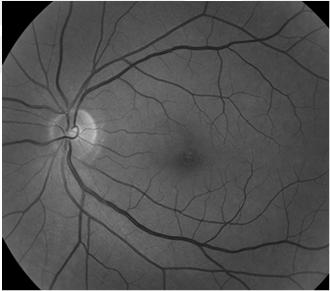
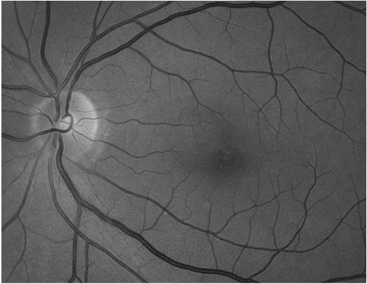
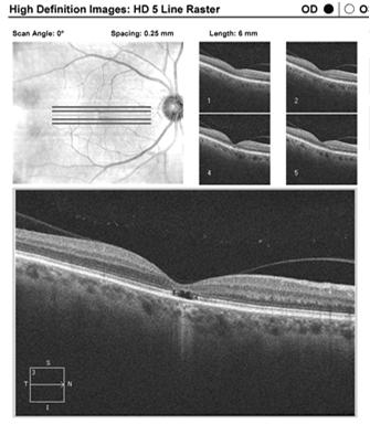
 Jonathan A. Micieli, MD Valérie Biousse, MD") Ganglion cell analysis by optical coherence tomography (OCT) Jonathan A. Micieli, MD Valérie Biousse, MD Figure 1. Normal OCT of the macula (cross section through the line indicated on the fundus photo)
Ganglion cell analysis by optical coherence tomography (OCT) Jonathan A. Micieli, MD Valérie Biousse, MD Figure 1. Normal OCT of the macula (cross section through the line indicated on the fundus photo)
ZEISS AngioPlex OCT Angiography Overview ZEISS OCT Angiography
 ZEISS AngioPlex OCT Angiography Overview ZEISS OCT Angiography California, ZEISS AngioPlex Ultra-clear visualization of microvascular blood flow using non-invasive OCT angiography 2 AngioPlex OCT Angiography
ZEISS AngioPlex OCT Angiography Making the revolutionary, routine.
 ZEISS AngioPlex OCT Angiography Making the revolutionary, routine. The moment that revolutionary insight becomes routine. // OCT ANGIOGRAPHY MADE BY ZEISS CIRRUS with AngioPlex creates a new era in both
Automated Visual Field Analysis for Glaucoma
 Island of Vision Automated Visual Field Analysis for Glaucoma Normal Visual Field Parameters 60 superior 60 nasal 75 inferior 100 temporal Macula the central 13 Fovea the central 3 Visual field is limited
Noel de Jesus Atienza, MD, MSc and Joseph Anthony Tumbocon, MD
 Original Article Philippine Journal of OPHTHALMOLOGY Diagnostic Accuracy of the Optical Coherence Tomography in Assessing Glaucoma Among Filipinos. Part 1: Categorical Outcomes Based on a Normative Database
HOW TO MAKE THE MOST OF A NEW OCT. with Kelly Kerksick, OD
 HOW TO MAKE THE MOST OF A NEW OCT with Kelly Kerksick, OD 3 How to Make the Most of a New OCT Kelly Kerksick, OD, graduated from Southern College of Optometry and immediately started her own private practice
Choroidal Mapping; a Novel Approach for Evaluating Choroidal Thickness and Volume
 Imaging Technique Choroidal Mapping; a Novel Approach for Evaluating Choroidal Thickness and Volume Jila Noori 1, MD; Mohammad Riazi Esfahani 1,2, MD Fedra Hajizadeh 2, MD; Mohammad-Mehdi Zaferani 1, MD
Il contributo dell'angio-oct: valutazione integrata della componente nervosa e vascolare della malattia glaucomatosa
 SIMPOSIO G.O.A.L. - LE NUOVE FRONTIERE DIAGNOSTICHE E LE LINEE DI INDIRIZZO AMBULATORIALI DEL GLAUCOMA Coordinatore e moderatore: D. Mazzacane Presidente: L. Rossetti Il contributo dell'angio-oct: valutazione
OCT in the Diagnosis and Follow-up of Glaucoma
 OCT in the Diagnosis and Follow-up of Glaucoma Karim A Raafat MD. Professor Of Ophthalmology Cairo University Hmmmm! Do I have Glaucoma or not?! 1 Visual Function 100% - N Gl Structure : - 5000 axon /
Optical Coherence Tomography in Diabetic Retinopathy. Mrs Samantha Mann Consultant Ophthalmologist Clinical Lead of SEL-DESP
 Optical Coherence Tomography in Diabetic Retinopathy Mrs Samantha Mann Consultant Ophthalmologist Clinical Lead of SEL-DESP Content OCT imaging Retinal layers OCT features in Diabetes Some NON DR features
C a t a r a c t G l a u c o m a R e t i n a R e f r a c t i v e. The GDxVCC Early answers and ongoing assessment for glaucoma
 C a t a r a c t G l a u c o m a R e t i n a R e f r a c t i v e The GDxVCC Early answers and ongoing assessment for glaucoma The quantifiable approach to quality care Only Humphrey GPA software Early insight
The Role of Phenotype in Selectively Enriching Patients for Clinical Studies
 The Role of Phenotype in Selectively Enriching Patients for Clinical Studies Developing Treatments for Dry Age-Related Macular Degeneration (AMD) Workshop November 15, 2014 National Academy of Sciences
Angio-OCT. Degenerazione Maculare Legata all Eta. Giuseppe Querques
 Angio-OCT Degenerazione Maculare Legata all Eta Giuseppe Querques Department of Ophthalmology, IRCCS Ospedale San Raffaele, University Vita Salute San Raffaele, Milan, Italy Financial Disclosure ADVISORY
Method for comparing visual field defects to local RNFL and RGC damage seen on frequency domain OCT in patients with glaucoma.
 Method for comparing visual field defects to local RNFL and RGC damage seen on frequency domain OCT in patients with glaucoma. Donald C. Hood 1,2,* and Ali S. Raza 1 1 Department of Psychology, Columbia
Comparative evaluation of time domain and spectral domain optical coherence tomography in retinal nerve fiber layer thickness measurements
 Original article Comparative evaluation of time domain and spectral domain optical coherence tomography in retinal nerve fiber layer thickness measurements Dewang Angmo, 1 Shibal Bhartiya, 1 Sanjay K Mishra,
CHAPTER 13 CLINICAL CASES INTRODUCTION
 2 CHAPTER 3 CLINICAL CASES INTRODUCTION The previous chapters of this book have systematically presented various aspects of visual field testing and is now put into a clinical context. In this chapter,
Technicians & Nurses Program
 ASCRS ASOA Symposium & Congress Technicians & Nurses Program April 17-21, 2015 San Diego, California Optical Coherence Tomography: Essentials in Anterior and Posterior Segment Imaging Michael Stewart,
RETINA REVEALED. Dynamic Developments in AMD Diagnosis and Treatment (2014) The Dawn of: Pharmaco-Genetics (aka Nutrigenomics) Jerome Sherman, OD
 The Dawn of: Pharmaco-Genetics (aka Nutrigenomics) Jerome Sherman, OD") RETINA REVEALED Dynamic Developments in AMD Diagnosis and Treatment (2014) The Dawn of: Pharmaco-Genetics (aka Nutrigenomics) Jerome Sherman, OD Disclosures: Jerome Sherman Dr. Sherman has lectured, received
RETINA REVEALED Dynamic Developments in AMD Diagnosis and Treatment (2014) The Dawn of: Pharmaco-Genetics (aka Nutrigenomics) Jerome Sherman, OD Disclosures: Jerome Sherman Dr. Sherman has lectured, received
Widefield Retinal Imaging with Auto Fluorescence Technology in the Optometric Practice
 Widefield Retinal Imaging with Auto Fluorescence Technology in the Optometric Practice This course will define ultra-widefield retinal imaging and autofluorescence for the attendee. Will show how it is
Diagnosis in AMD. Managing your AMD Patients
 Managing your AMD Patients Robert W. Dunphy, O.D., F.A.A.O. Diagnosis in AMD Have suspicion Identify relative risk Conduct surveillance Biometry Utilize technology to facilitate detection of change / stability
AperTO - Archivio Istituzionale Open Access dell'università di Torino
 AperTO - Archivio Istituzionale Open Access dell'università di Torino Artifacts in automatic retinal segmentation using different optical coherence tomography instruments. This is the author's manuscript
PLEX Elite 9000 from ZEISS Swept-Source OCT
 PLEX Elite 9000 from ZEISS Swept-Source OCT Uncovering the undiscovered. ZEISS PLEX Elite 9000 // INNOVATION MADE BY ZEISS 2 Ultra-wide angiography En face montage Image courtesy of Prof. G. Querques,
ZEISS AngioPlex OCT Angiography. Clinical Case Reports
 Clinical Case Reports Proliferative Diabetic Retinopathy (PDR) Case Report 969 PROLIFERATIVE DIABETIC RETINOPATHY 1 1-year-old diabetic female presents for follow-up of proliferative diabetic retinopathy
Translating data and measurements from stratus to cirrus OCT in glaucoma patients and healthy subjects
 Romanian Journal of Ophthalmology, Volume 60, Issue 3, July-September 2016. pp:158-164 GENERAL ARTICLE Translating data and measurements from stratus to cirrus OCT in glaucoma patients and healthy subjects
Optical coherence tomography (OCT) is a relatively new noninvasive. The Use of Optical Coherence Tomography in Neurology DIAGNOSTIC UPDATE
 is a relatively new noninvasive. The Use of Optical Coherence Tomography in Neurology DIAGNOSTIC UPDATE") DIAGNOSTIC UPDATE The Use of Optical Coherence Tomography in Neurology Cédric Lamirel, MD,* Nancy Newman, MD,* Valérie Biousse, MD* Departments of *Ophthalmology, Neurology, and Neurological Surgery, Emory
DIAGNOSTIC UPDATE The Use of Optical Coherence Tomography in Neurology Cédric Lamirel, MD,* Nancy Newman, MD,* Valérie Biousse, MD* Departments of *Ophthalmology, Neurology, and Neurological Surgery, Emory
Optical Coherence Tomography (OCT) in Uveitis Piergiorgio Neri, BMedSc, MD, PhD Head Ocular Immunology Unit
 in Uveitis Piergiorgio Neri, BMedSc, MD, PhD Head Ocular Immunology Unit") The Eye Clinic Polytechnic University of Marche Head: Prof Alfonso Giovannini November, 1991 Optical Coherence Tomography (OCT) in Uveitis Piergiorgio Neri, BMedSc, MD, PhD Head Ocular Immunology Unit
The Eye Clinic Polytechnic University of Marche Head: Prof Alfonso Giovannini November, 1991 Optical Coherence Tomography (OCT) in Uveitis Piergiorgio Neri, BMedSc, MD, PhD Head Ocular Immunology Unit
Disclosures. Definitions. Goals. Imaging and glaucoma 3/22/2016
 Pinakin Davey OD, PhD, FAAO Professor and Director of Research Disclosures Principal investigator for ivue OCT trial Principal investigator Topcon FDA trials for Maestro and OCT 2000 Consultant for Topcon
Ophthalmic Imager Role
 MASTER OCT Ophthalmic Photographers Society October 18, 2014 Chicago, IL James B Soque, CRA COA Pamela A Weber, MD Island Retina Shirley, New York Commack, New York Financial Disclosure Genentech Ophthotech
Structure WGA. Structure WGA. Structural Assessment in Glaucoma. What s New
 New Developments in the Structural and Functional Assessment of the Glaucomas John G. Flanagan PhD, MCOptom, FAAO Professor, School of Optometry, University of Waterloo Professor, Dept of Ophthalmol &
Mark T. Dunbar Disclosures. Hot Topics in Retina. The Challenge. Where is the Fluid? 4/12/2016
 A PANEL DISCUSSION: RAPID-FIRE POSTERIOR SEGMENT UPDATE! Steve Ferrucci, OD Chief, Optometry Sepulveda VA 16111 Plummer St #112e Sepulveda CA, 91343 Mark T. Dunbar, OD Bascom Palmer Eye Institute Miami,
Is this glaucoma? Leo Semes, OD Michael Chaglasian, OD Danica Marrelli, OD. Optometry s Meeting 2015 Seattle, WA
 Is this glaucoma? Leo Semes, OD Michael Chaglasian, OD Danica Marrelli, OD Optometry s Meeting 2015 Seattle, WA Case 1. 54 WM Engineer is referred to UAB Eye Care as a glaucoma suspect. Mild myopic refractive
Reproducibility of Choroidal Thickness Measurements Across Three Spectral Domain Optical Coherence Tomography Systems
 Reproducibility of Choroidal Thickness Measurements Across Three Spectral Domain Optical Coherence Tomography Systems The MIT Faculty has made this article openly available. Please share how this access
OCT Image Analysis System for Grading and Diagnosis of Retinal Diseases and its Integration in i-hospital
 Progress Report for1 st Quarter, May-July 2017 OCT Image Analysis System for Grading and Diagnosis of Retinal Diseases and its Integration in i-hospital Milestone 1: Designing Annotation tool extraction
OtticaFisiopatologica
 Anno quindicesimo dicembre 2010 How to assess the retinal nerve fiber layer thickness Antonio Ferreras Miguel Servet University Hospital, Zaragoza. Aragón Health Sciences Institute University of Zaragoza
Corporate Medical Policy
 Corporate Medical Policy Glaucoma, Evaluation by Ophthalmologic Techniques File Name: Origination: Last CAP Review: Next CAP Review: Last Review: glaucoma_evaluation_by_ophthalmologic_techniques 3/2001
The Measure of Confidence
 Heidelberg_936357.qxd:Layout 1 5/9/08 12:01 PM 12:02 Page 1 (Cyan (Magenta (Yellow (Black (UV Five Powerful Solutions to Fit Your Practice PowerCheck Glaucoma FastCheck+ GPS Software and Retina Edema Index
Fundus Autofluorescence and its PRACTICAL applications: Retina Beyond the Color. Start to think about this. Disclosure 5/21/2015
 Fundus Autofluorescence and its PRACTICAL applications: Retina Beyond the Color Jeffry D. Gerson, O.D., F.A.A.O Olathe, KS jgerson@hotmail.com Start to think about this. Disclosure I have worked with/consulted
Beyond the C/D Ratio: Evaluating a Glaucomatous Optic Nerve. Marcus Gonzales, OD, FAAO Cedar Springs Eye Clinic COPE ID#: GL
 Beyond the C/D Ratio: Evaluating a Glaucomatous Optic Nerve Marcus Gonzales, OD, FAAO Cedar Springs Eye Clinic COPE ID#: 27809-GL Points to Remember Glaucoma affects the ONH in characteristic patterns
Posterior Segment Update
 Posterior Segment Update Featured Speaker: Dr. Kyle Cheatham, FAAO, DIP ABO DISCLOSURE STATEMENT We have no direct financial or proprietary interest in any companies, products or services mentioned in
OCT Angiography. Financial Disclosures: Pre-Test: Which one is Correct?
 OCT Angiography Brandon Lujan, MD Medical Director, Casey Reading Center Assistant Professor of Ophthalmology Financial Disclosures: Genentech (Consultant, Grant support, Educational training) UC Berkeley
Objective Assessment of Macula and Optic Nerve
 Objective Assessment of Macula and Optic Nerve Jerry Sherman Disclosure: Dr. Sherman has lectured and received honorarium from Carl Zeiss Meditec, Topcon, Optovue, Optos, and PHP, Diopsys, Eye Solutions,Quantel,
Early Detection Of Glaucoma Clinical Clues. Points To Live By. Glaucoma Risk Factors 10/3/2014
 Early Detection Of Glaucoma Clinical Clues Eric E. Schmidt, O.D. Omni Eye Specialists Wilmington, NC schmidtyvision@msn.com Points To Live By 25% of G pxs NEVER have IOP >21mm 50% of G pxs have trough
A Review Of Risk Factors. Early Detection Of Glaucoma Clinical Clues. A risk factor analysis is critical. Points To Live By
 A Review Of Risk Factors Early Detection Of Glaucoma Clinical Clues Eric E. Schmidt, O.D. Omni Eye Specialists Wilmington, NC schmidtyvision@msn.com FINDACAR Family history IOP Nearsightedness Diabetes/Vascular
Simply the best OCT & OCTA image quality.
 Avanti Widefield OCT with AngioVue OCT Angiography Simply the best OCT & OCTA image quality. Dear Friends of Optovue, Since introducing Spectral Domain OCT to the ophthalmology market in 2006, Optovue
New Technologies in Glaucoma Management: From ERG to OCT
 What s New and What s Next in Glaucoma New Technologies in Glaucoma Management: From ERG to OCT Ben Gaddie, OD FAAO Murray Fingeret, OD FAAO IOP 24- Hour IOP Role of hysteresis in glaucoma risk Cerebrospinal
University Hospital Basel. Optical Coherence Tomography Emerging Role in the Assessment of MS PD Dr. Konstantin Gugleta
 University Hospital Basel Optical Coherence Tomography Emerging Role in the Assessment of MS PD Dr. Konstantin Gugleta 15th State of the Art SMSS, Lucerne January 2013 Retinal Nerve Fiber Layer 1.200.000
HOCT-1I 1F All-in-One Optical Coherence Tomography with Fundus
 HOCT-1I 1F All-in-One Optical Coherence Tomography with Fundus Specification Type Resolution(in Tissue) A scan Rate Scan Range SD-OCT / Fundus Z :6~7um, XY:20um 68,000 A-scan/sec. [Fundus] X:6-12mm, Y:6-9mm,
3/6/2014. Hoda MH Mostafa MD Associate Professor of Ophthalmology Cairo University. The author has no proprietary interest. Today s Objectives
 Hoda MH Mostafa MD Associate Professor of Ophthalmology Cairo University The author has no proprietary interest Today s Objectives Identify the CLINICAL SCENARIOS IN MACULAR EDEMA where OCT plays a MAJOR
Principle of OCT. Reading Between the Lines: OCT Interpretation. Initial Concept. Advantage: High Resolution Cross Section Images
 Principle of OCT Reading Between the Lines: OCT Interpretation Mohammad Rafieetary, OD, FAAO mrafieetary@charlesretina.com Introduction Optical Biopsy Morphologic Evaluation of Live Tissue Measurements
Optical Coherence Tomograpic Features in Idiopathic Retinitis, Vasculitis, Aneurysms and Neuroretinitis (IRVAN)
") Columbia International Publishing Journal of Ophthalmic Research (2014) Research Article Optical Coherence Tomograpic Features in Idiopathic Retinitis, Vasculitis, Aneurysms and Neuroretinitis (IRVAN)
Columbia International Publishing Journal of Ophthalmic Research (2014) Research Article Optical Coherence Tomograpic Features in Idiopathic Retinitis, Vasculitis, Aneurysms and Neuroretinitis (IRVAN)
A Formula to Predict Spectral Domain Optical Coherence Tomography (OCT) Retinal Nerve Fiber Layer Measurements Based on Time Domain OCT Measurements
 Retinal Nerve Fiber Layer Measurements Based on Time Domain OCT Measurements") pissn: 1011-8942 eissn: 2092-9382 Korean J Ophthalmol 2012;26(5):369-377 http://dx.doi.org/10.3341/kjo.2012.26.5.369 Original Article A Formula to Predict Spectral Domain Optical Coherence Tomography (OCT)
pissn: 1011-8942 eissn: 2092-9382 Korean J Ophthalmol 2012;26(5):369-377 http://dx.doi.org/10.3341/kjo.2012.26.5.369 Original Article A Formula to Predict Spectral Domain Optical Coherence Tomography (OCT)
Fundus Autofluorescence. Jonathan A. Micieli, MD Valérie Biousse, MD
 Fundus Autofluorescence Jonathan A. Micieli, MD Valérie Biousse, MD The retinal pigment epithelium (RPE) has many important functions including phagocytosis of the photoreceptor outer segments Cone Rod
The World s fastest OCT. As simple as pressing. the start button
 The World s fastest OCT As simple as pressing the start button lution continues Optopol engineering team, designers of the first commercially available Spectral Domain OCT in the world, are proud to present
7/25/2018. You talk about glaucoma in cup-to-disc ratios ASSESSING THE OPTIC DISC: IS PHOTOGRAPHY STILL NECESSARY IN THE OCT ERA?
 MAXIMIZING YOUR DIAGNOSTIC TECHNOLOGIES: SOMETHING OLD, NEW, BORROWED AND BLUE Greg Caldwell, OD Joseph Sowka, OD Jessica Steen, OD ASSESSING THE OPTIC DISC: IS PHOTOGRAPHY STILL NECESSARY IN THE OCT ERA?
Maximizing Your Diagnostic Technologies: Something Old, New, Borrowed and Blue Greg Caldwell, OD Joseph Sowka, OD Jessica Steen, OD
 Maximizing Your Diagnostic Technologies: Something Old, New, Borrowed and Blue Greg Caldwell, OD Joseph Sowka, OD Jessica Steen, OD Please silence all mobile devices and remove items from chairs so others
Learn Connect Succeed. JCAHPO Regional Meetings 2016
 Learn Connect Succeed JCAHPO Regional Meetings 2016 pearls and pitfalls of ophthalmic imaging JCHAPO 2016 Conference Vikas Chopra, M.D. Medical Director, UCLA Doheny Eye Centers Pasadena Principal Investigator,
Op#c Nerve Head & Re#nal Imaging
 Op#c Nerve Head & Re#nal Imaging Dr Cesar Carrillo October, 2014 Vien#ane/NOC **Disclaimer** The images contained in this presenta5on are not my own, they can be found on the web OUTLINE Indica5ons Confocal
SOUTH-EAST EUROPEAN JOURNAL of OPHTHALMOLOGY 2015; 1 (1) 34 40
 34 40") Review article SOUTH-EAST EUROPEAN JOURNAL of OPHTHALMOLOGY 2015; 1 (1) 34 40 Retinal nerve fiber layer versus peripapillary capillary density assessment A powerful tool for detecting optic nerve head
Review article SOUTH-EAST EUROPEAN JOURNAL of OPHTHALMOLOGY 2015; 1 (1) 34 40 Retinal nerve fiber layer versus peripapillary capillary density assessment A powerful tool for detecting optic nerve head
Overview. Macular OCT Artifact Study
 Imaging Artifacts Sarah Moyer, CRA, OCT-C Director, Ophthalmic Imaging Kittner Eye Center University of North Carolina Chapel Hill, NC Disclose financial interest now Overview Sarah s Thoughts on Artifacts
1/25/2019 OCT & OCTA RETINAL IMAGING: HOW TO PREVENT RAGING GLAUCOMA! THE ORIGINAL RAGING GLAUCOMA OCT RETINAL IMAGING OPTIC NERVE HEAD EXAMINATION
 OCT & OCTA RETINAL IMAGING: HOW TO PREVENT RAGING GLAUCOMA! Craig Thomas, O.D. 3900 West Wheatland Road Dallas, Texas 75237 972-780-7199 thpckc@yahoo.com THE ORIGINAL RAGING GLAUCOMA 47-year-old Black
Retinal Complications of Obstructive Sleep Apnea A Growing Concern!
 Retinal Complications of Obstructive Sleep Apnea A Growing Concern! Jay M. Haynie, OD, FAAO Financial Disclosure I have received honoraria or am on the advisory board for the following companies: Carl
NIH Public Access Author Manuscript JAMA Ophthalmol. Author manuscript; available in PMC 2013 September 10.
 NIH Public Access Author Manuscript Published in final edited form as: JAMA Ophthalmol. 2013 May ; 131(5): 693 694. doi:10.1001/jamaophthalmol.2013.692. Effect of Intravitreous Anti Vascular Endothelial
The Optic Nerve Head In Glaucoma. Clinical Pearl #1. Characteristics of Normal Disk 9/26/2017. Initial detectable damage Structure vs function
 The Optic Nerve Head In Glaucoma Clinical Pearl #1 Eric E. Schmidt, O.D., F.A.A.O. Omni Eye Specialists Wilmington,NC schmidtyvision@msn.com Glaucoma is an optic neuropathy Initial detectable damage Structure
Non-arteritic anterior ischemic optic neuropathy (NAION) with segmental optic disc edema. Jonathan A. Micieli, MD Valérie Biousse, MD
 with segmental optic disc edema. Jonathan A. Micieli, MD Valérie Biousse, MD") Non-arteritic anterior ischemic optic neuropathy (NAION) with segmental optic disc edema Jonathan A. Micieli, MD Valérie Biousse, MD A 75 year old white woman lost vision in the inferior part of her visual
Non-arteritic anterior ischemic optic neuropathy (NAION) with segmental optic disc edema Jonathan A. Micieli, MD Valérie Biousse, MD A 75 year old white woman lost vision in the inferior part of her visual
New Concepts in Glaucoma Ben Gaddie, OD Moderator Murray Fingeret, OD Louis Pasquale, MD
 New Concepts in Glaucoma Ben Gaddie, OD Moderator Murray Fingeret, OD Louis Pasquale, MD New Concepts in Glaucoma Optical Coherence Tomography: Is it necessary and needed to diagnose and monitor glaucoma?
Peripapillary Retinal Thickness. Maps in the Evaluation of Glaucoma Patients: A Novel Concept.
 Peripapillary Retinal Thickness Maps in the Evaluation of Glaucoma Patients: A Novel Concept The Harvard community has made this article openly available. Please share how this access benefits you. Your
OCT Evaluation of the Retina Alison Bozung, OD, FAAO
 Alison Bozung, OD, FAAO Rob Wooldridge, OD, FAAO Bozung: No relevant financial relationships with commercial interests Wooldridge: Speakers Bureau/honoraria from Aerie, Alcon, Allergan, Bausch & Lomb,
We are IntechOpen, the world s leading publisher of Open Access books Built by scientists, for scientists. International authors and editors
 We are IntechOpen, the world s leading publisher of Open Access books Built by scientists, for scientists 3,700 108,500 1.7 M Open access books available International authors and editors Downloads Our