SPECT and PET Imaging: DaT Scan, Cerebral Blood Flow and Epilepsy
|
|
|
- Darcy Cook
- 5 years ago
- Views:
Transcription


1 SPECT and PET Imaging: DaT Scan, Cerebral Blood Flow and Epilepsy Dana Mathews Ph.D. M.D. Division of Nuclear Medicine Department of Radiology The University of Texas Southwestern Medical Center

2 Financial Disclosures/ Conflicts of Interest/FDA off Label No financial disclosures No COI FDA discussion of drugs not yet approved: F18 AV 133 (VMAT scan)

3 Frederick J. Bonte, M.D
4 SAMs Objectives Individuals attending this session will at the end of the session be able to Describe how DATScan may be used to distinguish etiologies of tremor Restate Uses of SPECT perfusion imaging in cerebrovascular disease and eplilepsy Describe how FDG PET brain imaging may be useful in seizure localization in epilepsy
5 Parkinson s Disease (PD) Neurodegenerative disorder whose symptoms may include, tremor, rigidity, postural instability, abnormal gait and in some cases dementia Often difficult to diagnose in early stages and the tremor can be mistaken for other causes of tremor, most commonly essential tremor (ET). There are high false positive and false negative rates for each of these diagnoses based on clinical diagnosis alone, even among movement disorder experts. PD is treated with a variety of medications whose goal is provide DA. These medications are ineffective for ET and may exacerbate ET. DaTscan can help distinguish between these two diagnoses (often with higher concordance in NM reading experts than clinical experts)
6 Other Disorders in the Parkinsonian Syndromes Progressive supranuclear palsy (PSP) Corticobasal Degeneration (CBD) Multisystem Atrophy (MSA) All these share with idiopathic PD loss of DA neurons but differ in other symptoms and are much less common
7 Drug Induced Parksonian Symptoms Very common cause of misdiagnosis Results from pharmacologic agents that block DA receptors but don t affect DAT Common drugs include neuroleptics (eg prochlorperazine, and metoclopromide), and calcium channel blockers DIP more common in women and elderly and does not clearly relate to dosage or length of usage DATScan will look normal!

8 Cummings et al Brain 2011
9 DaTScan ( I 123 Ioflupane) Images presynaptic dopamine transporters (DAT) whose function is retrieve the neurotransmitter dopamine from the synaptic cleft of dopaminergic neurons In Parkinson s Disease and other Parkinsonian disorders, dopamine is severally depleted, resulting in a lower number of nigrostriatal nerve terminals, with subsequent down regulation in DAT DaTscan is a cocaine analogue with high affinity and high selectivity for DAT with greatest uptake in the caudate and putamen
10 Imaging with I 123 ioflupaine DATScan Referring physician prescribes thyroid blockade solution or tablets Referring physician decides which medications should be held prior to scanning On day of scan, patient takes thyroid blocking medication 1 hour prior to injection Patient injected slowly with 5mCi I 123 ioflupane Hydrate well and continue to do so for next 48 hrs
11 Medications Affecting Imaging Either 100mg potassium iodide or 400 mg potassium chlorateblocks I 123 uptake in thyroid Amoxapine, amphetamine, benztropine, bupropion, buspirone, cocaine, mazindol, methamphetamine, methylphenidate, norephedrine, phentermine, phenylpropanolamine, selegiline, sertraline, citalopram and paroxetine may interfere with imaging (should be discontinued for scan)
12 Imaging with DaTScan After 3 hour uptake, patient images are acquired for approximately 30 minutes 13 cm to no greater than 15 cm distance from patient to collimator Fan beam collimators * 5 five minute dynamic tomos are acquired for 180 degrees of rotation 128x128 matrix
13 Processing Parameters for DATscan Filtered backprogection Butterworth Filter Order 8 cut off 0.46 on Siemens scanner No Masking


14 Interpretation of DaTscan Normal Grade 1 Grade 2 Grade 3
15 DATScan I 123 Ioflupane Not PS PS


16 Asymmetric PD

17 F18 AV 133 VMAT agent for PD
18 Benefits of DaTScan Improves diagnosis in early PD Distinguishes PSD from other sources of tremor Can have a significant impact on patient management ( May change management in approximately 30%) May save money on ineffective medications
19 SPECT Cerebral Perfusion Imaging Vascular Reserve- Allows evaluation of cerebral perfusion in much the same way as cardiac perfusion imaging. By using acetazolamide as a vasodilator, areas of the brain that are already maximally vasodilated may be revealed, yielding information about the impact of arterial stenoses and occlusions Trial Balloon Occlusion- Allows evaluation of the cerebral collateral circulation in response to temporary occlusion of a major cerebral artery Dementia Demonstrates perfusion patterns in demonstration which corresponds to brain areas of hypometabolism seen with PET FDG Epilepsy-Allows evaluation of areas of potential abnormal perfusion associated with seizure foci in focal epilepsy Other uses
20 SPECT Cerebral Perfusion Imaging The CBF tracers commonly used for the purposes discussed in this lecture are Tc 99m ethylene cysteine dimer (ECD) and Tc 99m hexamethylprop yleneamine oxime (HMPAO) Both tracers cross the blood brain barrier rapidly after injection and both are rapidly metabolized into a hydrophilic form that will not cross back through the blood brain barrier Both are stabilized so that they can be injected several hours after preparation. This allows their use for capturing what may be a transient event, such as a temporary arterial occlusion or a seizure Both may be imaged using SPECT or SPECT CT gamma cameras optimized for brain imaging
21 Evaluation of Vascular Reserve Patient injected with 1 g acetazolamide, a vasodilator. Patient then injected with perfusion tracer (usually Tc 99m ECD or HMPAO) Brain SPECT Scan performed. If areas of decreased perfusion seen Baseline scan is performed on a different day. Relative changes in perfusion distribution are indications of areas of reduced reserve Areas of fixed reduction in perfusion usually indication of prior infarct which will not improve with reperfusion efforts.

22


23 Diamox Baseline
24 Trial Balloon Occlusion This refers to the temporary occlusion of a major cerebral artery usually the internal carotid artery. This is done to evaluate collateral blood flow in the event that the artery being evaluated needs temporary or permanent occlusion in order to treat and aneurysm in the brain or a tumor in the neck. Angiographic catheter placed in vessel of interest Balloon inflated, serial neurological exams and injection of tracer, while the balloon inflated. Inflation in the balloon maintained in the artery for approximately 15 minutes or until the patient becomes symptomatic (whew!!!) Balloon deflated SPECT scan performed
25 Complete Circle of Willis which occurs about 50% of the time

26 TBO of right ICA for patient with large thyroid cancer
27 TBO of basilar artery in patient with BA stenosis, baseline normal 2


28 Attempts to dilate that stenosis did not go well 2
29 Patient undergoing left internal carotid TBO. What is the problem? 2
30 What is the arrow pointing to?


31 The arrow is pointing to the PCA which in this case is fetal in origin, meaning it arises from the ICA and not the BA

32 Patient undergoing left internal carotid TBO. What is the problem? 3
33 SPECT CBF and FDG PET SPECT CBF shows perfusion of brain tissue FDG PET shows metabolism of brain tissue Because there is tight coupling in the regulation of both blood flow and metabolism, these two scan types can often show the same patterns of abnormality in a brain area affected by a disease process For example, prior to the widespread availability of FDG PET imaging, SPECT CBF was used to image a whole range of disease processes that are now mainly evaluated by brain FDG PET. These include dementia and epilepsy


34 SPECT CBF in ATD FTD
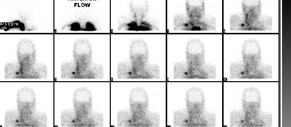
35 Cerebral Perfusion Scanning in Brain Death Brain death refers to cessation of all functions of the brain including the brain stem. Brain death is determined by clinical examination of the patient for evidence of lack of brainstem functions At times, there is desire to have additional confirmatory evidence of brain death. This may include obtaining an electrical encephalogram (EEG) or a scan to confirm lack of intracerebral perfusion When the brain is irreversibly damage through either direct trauma or anoxic injury, swelling occurs which eventually raises intracerebral pressure to the point where arterial brain perfusion no longer occurs
36 Cerebral Perfusion Imaging in Brain Death Can be done using Tc 99m DTPA. Benefits-can be repeated in a short period of time Drawbacks-captures only arterial phase and may be difficult to interpret for less experienced readers Can be done using Tc 99m HMPAO or ECD Benefits-easier to interpret on the delayed images Drawbacks- Not repeatable until about 24 hours

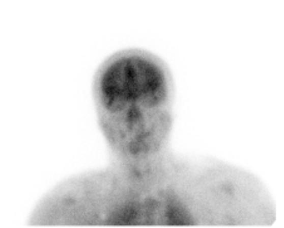
37 Intracerebral perfusion remains

38 Intracerebral perfusion absent
39 SPECT and PET Imaging in Epilepsy About 50/10,000 new cases of epilepsy each year. About 1/3 of these patients are not controlled by antiepileptic drugs (AED). About half of these patients will develop focal epilepsy, most commonly involving the temporal lobes. Some of this patients may benefit significantly from temporal lobectomy MRI is very important in identifying structural lesions which may be epileptogenic. These most commonly are hippocampal sclerosis, but other causes include cortical dysplasia and mass lesions such as tumors, cavernomas, arteriovenous malformations, etc However, about 15-30% of patients with intractable focal epilepsy have no identifiable structural lesions These are the patients where SPECT CBF and FDG PET may be very beneficial in identifying the seizure focus
40 SPECT and PET Imaging in Epilepsy Between seizures, the blood flow and metabolism of the brain area surrounding the seizure focus may be reduced, indication poor function of these areas. During seizures, the blood flow and metabolism in these areas may be increased. However this increase is very transient so for optimal evaluation, the patient has to be injected with tracer during a seizure. When an ictal (during seizure) and interictal (between seizures) scans are compared, the changes in blood flow and metabolism help identify the regions of concern.


41 Patient with left temporal lobe cavernoma and Interictal PET showing hypo metabolism in left temporal lobe

42 SPECT CBF in epilepsy identifying perfusion changes between the right temporal lobe between and during seizures


43 Ictal SPECT Interictal SPECT Interictal PET


44 Other PET Tracers Used in Imaging Epileptogenic Foci

45 A. MRI, B. PET with AMT, C. PET with FDG, D. Intracranial electrode Location, E. EEG in patient with TS
46 CME Questions 1. DATScan imaging is likely to be abnormal in patients with Tremor of Cerebellar origin Drug induced parkinsonian tremor Parkinson s Disease Essential Tremor
47 CME Questions 1. DATScan imaging is likely to be abnormal in patients with Tremor of Cerebellar origin Drug induced parkinsonian tremor Parkinson s Disease Essential Tremor References 1-2
48 CME Questions 2. Syndromes involving loss of CNS dopamine may include a. Parksinson s Disease b. Progressive Supranuclear Palsy c. Multisystem atrophy d. All may result from loss of dopamine
49 CME Questions 2. Syndromes involving loss of CNS dopamine may include a. Parksinson s Disease b. Progressive Supranuclear Palsy c. Multisystem atrophy d. All may result from loss of dopamine References 1-2
50 CME Questions 3. SPECT CBF can be used to evaluate a. Vascular reserve b. Cerebral collateral perfusion c. Epilepsy d. CBF can evaluate all of the above References 3 and 5
51 CME Questions 3. SPECT CBF can be used to evaluate a. Vascular reserve b. Cerebral collateral perfusion c. Epilepsy d. CBF can evaluate all of the above References 1-4
52 CME Questions 4. In evaluating epilepsy, the time when increased CBF at the seizure focus is most likely to be visualized is a. During a seizure b. Just after a seizure c. Between seizures d. Anytime
53 CME Questions 4. In evaluating epilepsy, the time when increased CBF at the seizure focus is most likely to be visualized is a. During a seizure b. Just after a seizure c. Between seizures d. Anytime Reference 4
54 CME Questions 5. Cerebral blood flow imaging of the brain and FDG PET imaging of the brain often show similar findings in dementia and epilepsy because a. They are showing the same process b. There is tight coupling between brain blood flow and brain metabolism c. They both measure brain oxygenation d. They don t have anything to do with each other
55 CME Questions 5. Cerebral blood flow imaging of the brain and FDG PET imaging of the brain often show similar findings in dementia and epilepsy because a. They are showing the same process b. There is tight coupling between brain blood flow and brain metabolism c. They both measure brain oxygenation d. They don t have anything to do with each other Reference 4

56 References Bajaj N, Hauser RA, Grachev ID: Clinical utility of dopamine transporter single photon emission CT (DaT-SPECT) with ( 123 I) ioflupane in diagnosis of parkinsonian syndromes. J Neurolog Neurosurg Psychiatry 2013l84L Tatsch K, Poepperl G: Nigrostriatal dopamine terminal imaging with dopamine transporter SPECT: An update. J Nucl Med 2013; 54: Mathews D, Walker BS, Purdy PD, Batjer H, Allen BC, Eckard DA, Devous MD Sr, Bonte FJ. Brain blood flow SPECT in temporary balloon occlusion of carotid and intracerebral arteries. J Nucl Med Aug;34(8): Duncan JS, Winston GP, Koepp MJ, et al. Brain imaging in the assessment for epilepsy surgery. The Lancet/Neurology. 2016;15: Kumar A, Chugani HT. The role of radionuclide imaging in epilepsy, part 1: Sporadic temporal and extratemporal lobe epilepsy. J Nucl Med 2013; 54: Kumar A, Asano E, Chugani HT. a [11C]-methyl-tryptophan PET for tracer localization of epileptogenic brain regions: clinical studies. Biomark Med 2011;5:
Update on functional brain imaging in Movement Disorders
 Update on functional brain imaging in Movement Disorders Mario Masellis, MSc, MD, FRCPC, PhD Assistant Professor & Clinician-Scientist Sunnybrook Health Sciences Centre University of Toronto 53 rd CNSF
Corporate Medical Policy
 Corporate Medical Policy Dopamine Transporter Imaging with Single Photon Emission File Name: Origination: Last CAP Review: Next CAP Review: Last Review: dopamine_transporter_imaging_with_single_photon_emission_computed_tomography
Brain Perfusion SPECT
 APPROVED BY: Director of Radiology Page 1 of 5 Brain Perfusion SPECT Primary Indications: Brain perfusion SPECT is most commonly performed (1) to aid in identification of the epileptogenic focus in patients
Dose Calibration for DaTscan
 GE Healthcare Dose Calibration for DaTscan Please review the simple steps below to learn how to properly calibrate your dose of DaTscan. 1 2 3 4 5 6 7 8 9 Reading DaTscan in the Dose Calibrator 1. Place
DIFFERENTIAL DIAGNOSIS SARAH MARRINAN
 Parkinson s Academy Registrar Masterclass Sheffield DIFFERENTIAL DIAGNOSIS SARAH MARRINAN 17 th September 2014 Objectives Importance of age in diagnosis Diagnostic challenges Brain Bank criteria Differential
Parkinson s Disease in the Elderly A Physicians perspective. Dr John Coyle
 Parkinson s Disease in the Elderly A Physicians perspective Dr John Coyle Overview Introduction Epidemiology and aetiology Pathogenesis Diagnosis and clinical features Treatment Psychological issues/ non
High Resolution Ictal SPECT: Enhanced Epileptic Source Targeting?
 High Resolution Ictal SPECT: Enhanced Epileptic Source Targeting? Marvin A Rossi MD, PhD RUSH Epilepsy Center Research Lab http://www.synapticom.net Chicago, IL USA Medically-Refractory Epilepsy 500,000-800,000
PET and SPECT in Epilepsy
 PET and SPECT in Epilepsy 12.6.2013 William H Theodore MD Chief, Clinical Epilepsy Section NINDS NIH Bethesda MD American Epilepsy Society Annual Meeting Disclosures Entity DIR NINDS NIH Elsevier Individual
Nuclear neurology. Zámbó Katalin Department of Nuclear Medicine
 Nuclear neurology Zámbó Katalin Department of Nuclear Medicine To refresh your memory Brain has a high rate of oxidative metabolism. It has no reserves either of oxygen or of glucose and has a very limited
Multimodal Imaging in Extratemporal Epilepsy Surgery
 Open Access Case Report DOI: 10.7759/cureus.2338 Multimodal Imaging in Extratemporal Epilepsy Surgery Christian Vollmar 1, Aurelia Peraud 2, Soheyl Noachtar 1 1. Epilepsy Center, Dept. of Neurology, University
GE Healthcare. Protocol Manual. Abnormal Scan. Normal Scan. Period-Shaped. Comma-Shaped
 GE Healthcare Protocol Manual Normal Scan Comma-Shaped Abnormal Scan Period-Shaped 1 Contents Section 1: Introduction... 4 1.1 Scope 1.2 What is DaTscan? Section 2: Image Acquisition, Interpretation, and
Dopamine Transporter Imaging With Single-Photon Emission Computed. Tomography
 Dopamine Transporter Imaging With Single-Photon Emission Computed Tomography Policy Number: 6.01.54 Last Review: 9/2018 Origination: 9/2015 Next Review: 9/2019 Policy Blue Cross and Blue Shield of Kansas
Differential Diagnosis of Hypokinetic Movement Disorders
 Differential Diagnosis of Hypokinetic Movement Disorders Dr Donald Grosset Consultant Neurologist - Honorary Professor Institute of Neurological Sciences - Glasgow University Hypokinetic Parkinson's Disease
GE Healthcare. Protocol Manual. Abnormal Scan. Normal Scan. Period-Shaped. Comma-Shaped
 GE Healthcare Protocol Manual Normal Scan Comma-Shaped Abnormal Scan Period-Shaped Contents Section 1: Introduction... 4 1.1 Scope 1.2 What is DaTscan? Section 2: Image Acquisition, Interpretation, and
Form D1: Clinician Diagnosis
 Initial Visit Packet Form D: Clinician Diagnosis NACC Uniform Data Set (UDS) ADC name: Subject ID: Form date: / / Visit #: Examiner s initials: INSTRUCTIONS: This form is to be completed by the clinician.
Imaging biomarkers for Parkinson s disease
 3 rd Congress of the European Academy of Neurology Amsterdam, The Netherlands, June 24 27, 2017 Teaching Course 6 MDS-ES/EAN: Neuroimaging in movement disorders - Level 2 Imaging biomarkers for Parkinson
Dopamine Transporter Imaging With Single-Photon Emission Computed. Tomography
 Dopamine Transporter Imaging With Single-Photon Emission Computed Tomography Policy Number: 6.01.54 Last Review: 9/2017 Origination: 9/2015 Next Review: 9/2018 Policy Blue Cross and Blue Shield of Kansas
Introduction, use of imaging and current guidelines. John O Brien Professor of Old Age Psychiatry University of Cambridge
 Introduction, use of imaging and current guidelines John O Brien Professor of Old Age Psychiatry University of Cambridge Why do we undertake brain imaging in AD and other dementias? Exclude other causes
Blood Supply. Allen Chung, class of 2013
 Blood Supply Allen Chung, class of 2013 Objectives Understand the importance of the cerebral circulation. Understand stroke and the types of vascular problems that cause it. Understand ischemic penumbra
SPECT Dopamin Transporter lmaging Agent
 SPECT Dopamin Transporter lmaging Agent Product Summary Product Name DATrace-123 Injection Active Ingredient Action Mechanism DATrace-123 is used as a radiopharmaceutical for the SPECT imaging of Dopamine
Round table: Moderator; Fereshteh Sedaghat, MD, PhD Brain Mapping in Dementias and Non-invasive Neurostimulation
 Round table: Moderator; Fereshteh Sedaghat, MD, PhD Brain Mapping in Dementias and Non-invasive Neurostimulation 1. Reflection of Mild Cognitive Impairment (MCI) and Dementias by Molecular Imaging, PET
Parkinson s Disease Initial Clinical and Diagnostic Evaluation. J. Timothy Greenamyre, MD, PhD
 Parkinson s Disease Initial Clinical and Diagnostic Evaluation J. Timothy Greenamyre, MD, PhD Involuntary tremulous motion, with lessened muscular power, in parts not in action and even when supported
Views and Reviews. [ 123 I]FP-CIT (DaTscan) SPECT Brain Imaging in Patients with Suspected Parkinsonian Syndromes ABSTRACT
![Views and Reviews. [ 123 I]FP-CIT (DaTscan) SPECT Brain Imaging in Patients with Suspected Parkinsonian Syndromes ABSTRACT](/thumbs/76/73233066.jpg "Views and Reviews. [ 123 I]FP-CIT (DaTscan) SPECT Brain Imaging in Patients with Suspected Parkinsonian Syndromes ABSTRACT") Views and Reviews [ 123 I]FP-CIT (DaTscan) SPECT Brain Imaging in Patients with Suspected Parkinsonian Syndromes Robert A. Hauser, MD, Donald G. Grosset, MD From the Departments of Neurology, Molecular
Views and Reviews [ 123 I]FP-CIT (DaTscan) SPECT Brain Imaging in Patients with Suspected Parkinsonian Syndromes Robert A. Hauser, MD, Donald G. Grosset, MD From the Departments of Neurology, Molecular
Applicable Neuroradiology
 For the Clinical Neurology Clerkship LSU Medical School New Orleans Amy W Voigt, MD Clerkship Director Introduction The field of Radiology first developed following the discovery of X-Rays by Wilhelm Roentgen
Laura Tormoehlen, M.D. Neurology and EM-Toxicology Indiana University
 Laura Tormoehlen, M.D. Neurology and EM-Toxicology Indiana University Disclosures! No conflicts of interest to disclose Neuroimaging 101! Plain films! Computed tomography " Angiography " Perfusion! Magnetic
Reimbursement for DaTscan
 GE Healthcare Reimbursement for DaTscan DaTscan is a radiopharmaceutical indicated for striatal dopamine transporter visualization using singlephoton emission computed tomography (SPECT) brain imaging
Transcranial sonography in movement disorders
 Transcranial sonography in movement disorders Uwe Walter 1st Residential Training of the European Society of Neurosonology and Cerebral Hemodynamics September 7-12, 2008 Bertinoro, Italy Department of
Overview. Overview. Parkinson s disease. Secondary Parkinsonism. Parkinsonism: Motor symptoms associated with impairment in basal ganglia circuits
 Overview Overview Parkinsonism: Motor symptoms associated with impairment in basal ganglia circuits The differential diagnosis of Parkinson s disease Primary vs. Secondary Parkinsonism Proteinopathies:
Neurodegenerative Disease. April 12, Cunningham. Department of Neurosciences
 Neurodegenerative Disease April 12, 2017 Cunningham Department of Neurosciences NEURODEGENERATIVE DISEASE Any of a group of hereditary and sporadic conditions characterized by progressive dysfunction,
brain MRI for neuropsychiatrists: what do you need to know
 brain MRI for neuropsychiatrists: what do you need to know Christoforos Stoupis, MD, PhD Department of Radiology, Spital Maennedorf, Zurich & Inselspital, University of Bern, Switzerland c.stoupis@spitalmaennedorf.ch
Dementia Update. October 1, 2013 Dylan Wint, M.D. Cleveland Clinic Lou Ruvo Center for Brain Health Las Vegas, Nevada
 Dementia Update October 1, 2013 Dylan Wint, M.D. Cleveland Clinic Lou Ruvo Center for Brain Health Las Vegas, Nevada Outline New concepts in Alzheimer disease Biomarkers and in vivo diagnosis Future trends
Parkinson s Disease: initial diagnosis, initial treatment & non-motor features. J. Timothy Greenamyre, MD, PhD
 Parkinson s Disease: initial diagnosis, initial treatment & non-motor features J. Timothy Greenamyre, MD, PhD Involuntary tremulous motion, with lessened muscular power, in parts not in action and even
Interictal High Frequency Oscillations as Neurophysiologic Biomarkers of Epileptogenicity
 Interictal High Frequency Oscillations as Neurophysiologic Biomarkers of Epileptogenicity December 10, 2013 Joyce Y. Wu, MD Associate Professor Division of Pediatric Neurology David Geffen School of Medicine
Pathogenesis of Degenerative Diseases and Dementias. D r. Ali Eltayb ( U. of Omdurman. I ). M. Path (U. of Alexandria)
. M. Path (U. of Alexandria)") Pathogenesis of Degenerative Diseases and Dementias D r. Ali Eltayb ( U. of Omdurman. I ). M. Path (U. of Alexandria) Dementias Defined: as the development of memory impairment and other cognitive deficits
Pathogenesis of Degenerative Diseases and Dementias D r. Ali Eltayb ( U. of Omdurman. I ). M. Path (U. of Alexandria) Dementias Defined: as the development of memory impairment and other cognitive deficits
Imaging in epilepsy: Ictal perfusion SPECT and SISCOM
 Imaging in epilepsy: Ictal perfusion SPECT and SISCOM Patrick Dupont Laboratory for Cognitive Neurology Laboratory for Epilepsy Research Medical Imaging Research Center KU Leuven, Belgium E-mail: Patrick.Dupont@med.kuleuven.be
CEREBRAL BLOOD FLOW AND METABOLISM
 Supported by: HURO/0901/069/2.3.1 HU-RO-DOCS CEREBRAL BLOOD FLOW AND METABOLISM Part 3 Modern imaging methods SPECT, PET, nmri History of Nuclear Medicine Starts with the invention of the X-ray 1946: radioactive
Molecular Imaging and the Brain
 Molecular imaging technologies are playing an important role in neuroimaging, a branch of medical imaging, by providing a window into the living brain. Where CT and conventional MR imaging provide important
FDG-PET e parkinsonismi
 Parkinsonismi FDG-PET e parkinsonismi Valentina Berti Dipartimento di Scienze Biomediche, Sperimentali e Cliniche Sez. Medicina Nucleare Università degli Studi di Firenze History 140 PubMed: FDG AND parkinsonism
Dopamine Transporter Imaging with Single Photon Emission Computed Tomography
 Last Review Status/Date: September 2014 Page: 1 of 14 Photon Emission Computed Tomography Description Dopamine transporter imaging with single photon emission computed tomography (DAT-SPECT) is being evaluated
Dementia Update. Daniel Drubach, M.D. Division of Behavioral Neurology Department of Neurology Mayo Clinic Rochester, Minnesota
 Dementia Update Daniel Drubach, M.D. Division of Behavioral Neurology Department of Neurology Mayo Clinic Rochester, Minnesota Nothing to disclose Dementia Progressive deterioration in mental function
Neuro Quiz 29 Transcranial Doppler Monitoring
 Verghese Cherian, MD, FFARCSI Penn State Hershey Medical Center, Hershey Quiz Team Shobana Rajan, M.D Suneeta Gollapudy, M.D Angele Marie Theard, M.D Neuro Quiz 29 Transcranial Doppler Monitoring This
Basal ganglia motor circuit
 Parkinson s Disease Basal ganglia motor circuit 1 Direct pathway (gas pedal) 2 Indirect pathway (brake) To release or augment the tonic inhibition of GPi on thalamus Direct pathway There is a tonic inhibition
Physiological Markers of Pharmacoresistant Epilepsy December 2, 2011
 Physiological Markers of Pharmacoresistant Epilepsy December 2, 2011 Jerome Engel, Jr., MD, PhD Director of the Seizure Disorder Center The Jonathan Sinay Distinguished Professor of Neurology, Neurobiology,
! slow, progressive, permanent loss of neurologic function.
 UBC ! slow, progressive, permanent loss of neurologic function.! cause unknown.! sporadic, familial or inherited.! degeneration of specific brain region! clinical syndrome.! pathology: abnormal accumulation
Brain imaging for the diagnosis of people with suspected dementia
 Why do we undertake brain imaging in dementia? Brain imaging for the diagnosis of people with suspected dementia Not just because guidelines tell us to! Exclude other causes for dementia Help confirm diagnosis
Intracranial Studies Of Human Epilepsy In A Surgical Setting
 Intracranial Studies Of Human Epilepsy In A Surgical Setting Department of Neurology David Geffen School of Medicine at UCLA Presentation Goals Epilepsy and seizures Basics of the electroencephalogram
UPSTATE Comprehensive Stroke Center. Neurosurgical Interventions Satish Krishnamurthy MD, MCh
 UPSTATE Comprehensive Stroke Center Neurosurgical Interventions Satish Krishnamurthy MD, MCh Regional cerebral blood flow is important Some essential facts Neurons are obligatory glucose users Under anerobic
Cardiac Imaging Tests
 Cardiac Imaging Tests http://www.medpagetoday.com/upload/2010/11/15/23347.jpg Standard imaging tests include echocardiography, chest x-ray, CT, MRI, and various radionuclide techniques. Standard CT and
Dopamine Transporter Imaging With Single-Photon Emission Computed Tomography
 Dopamine Transporter Imaging With Single-Photon Emission Computed Tomography Applies to all products administered or underwritten by Blue Cross and Blue Shield of Louisiana and its subsidiary, HMO Louisiana,
Neuromodulation in Epilepsy. Gregory C. Mathews, M.D., Ph.D.
 Neuromodulation in Epilepsy Gregory C. Mathews, M.D., Ph.D. Disclosure There are no disclosures to share with regards to this presentation. Epilepsy Basics What is epilepsy? Partial versus generalized
RADIOPHARMACEUTICALS FOR NEUROIMAGING. Prof. Cristina Maria Moriguchi Jeckel Brain Institute of Rio Grande do Sul, PUCRS
 RADIOPHARMACEUTICALS FOR NEUROIMAGING Prof. Cristina Maria Moriguchi Jeckel Brain Institute of Rio Grande do Sul, PUCRS Workshop on Quantitative SPECT and PET Brain Studies BRA6024 PUCRS, Porto Alegre,
ASL Perfusion Imaging: Concepts and Applications
 ASL Perfusion Imaging: Concepts and Applications David C. Alsop, Ph.D. Beth Israel Deaconess Medical Center and Harvard Medical School, Boston USA INTRODUCTION Arterial Spin Labeling (ASL) perfusion imaging
Parkinson Disease. Lorraine Kalia, MD, PhD, FRCPC. Presented by: Ontario s Geriatric Steering Committee
 Parkinson Disease Lorraine Kalia, MD, PhD, FRCPC Key Learnings Parkinson Disease (L. Kalia) Key Learnings Parkinson disease is the most common but not the only cause of parkinsonism Parkinson disease is
PRESURGICAL EVALUATION. ISLAND OF COS Hippocrates: On the Sacred Disease. Disclosure Research-Educational Grants. Patients with seizure disorders
 PRESURGICAL EVALUATION Patients with seizure disorders Gregory D. Cascino, MD Mayo Clinic Disclosure Research-Educational Grants Mayo Foundation Neuro Pace, Inc. American Epilepsy Society American Academy
/ / / / / / Hospital Abstraction: Stroke/TIA. Participant ID: Hospital Code: Multi-Ethnic Study of Atherosclerosis
 Multi-Ethnic Study of Atherosclerosis Participant ID: Hospital Code: Hospital Abstraction: Stroke/TIA History and Hospital Record 1. Was the participant hospitalized as an immediate consequence of this
Epilepsy Surgery, Imaging, and Intraoperative Neuromonitoring: Surgical Perspective
 Epilepsy Surgery, Imaging, and Intraoperative Neuromonitoring: Surgical Perspective AC Duhaime, M.D. Director, Pediatric Neurosurgery, Massachusetts General Hospital Professor, Neurosurgery, Harvard Medical
Assessment of Cerebrovascular Reserve before and after STA-MCA Bypass Surgery by SPECT and SPM Analysis
 Assessment of Cerebrovascular Reserve before and after STA-MCA Bypass Surgery by SPECT and SPM Analysis Joo-Hyun O, MD 1 Kyung-Sool Jang, MD 2 Ie-Ryung Yoo, MD 1 Sung-Hoon Kim, MD 1 Soo-Kyo Chung, MD 1
What if it s not Alzheimer s? Update on Lewy body dementia and frontotemporal dementia
 What if it s not Alzheimer s? Update on Lewy body dementia and frontotemporal dementia Dementia: broad term for any acquired brain condition impairing mental function such that ADLs are impaired. Includes:
Epilepsy. Hyunmi Choi, M.D., M.S. Columbia Comprehensive Epilepsy Center The Neurological Institute. Seizure
 Epilepsy Hyunmi Choi, M.D., M.S. Columbia Comprehensive Epilepsy Center The Neurological Institute Seizure Symptom Transient event Paroxysmal Temporary physiologic dysfunction Caused by self-limited, abnormal,
NEUROLOGY FOR PRIMARY CARE. Sea Island, Georgia The Cloister at Sea Island September 29 October 2, 2016
 NEUROLOGY FOR PRIMARY CARE Sea Island, Georgia The Cloister at Sea Island September 29 October 2, 2016 Thursday, September 29th: 7:30 am 8:00 am Registration and Breakfast 8:00 am 9:00 am Faculty 1 The
Overview Blood supply of the brain What is moyamoya disease? > 1
 Moyamoya Disease Overview Moyamoya disease is caused by blocked arteries at the base of the brain. The name "moyamoya" means "puff of smoke" in Japanese and describes the appearance of tiny vessels that
Case reports functional imaging in epilepsy
 Seizure 2001; 10: 157 161 doi:10.1053/seiz.2001.0552, available online at http://www.idealibrary.com on Case reports functional imaging in epilepsy MARK P. RICHARDSON Medical Research Council Fellow, Institute
MUSCULOSKELETAL AND NEUROLOGICAL DISORDERS
 MUSCULOSKELETAL AND NEUROLOGICAL DISORDERS There are a wide variety of Neurologic and Musculoskeletal disorders which can impact driving safety. Impairment may be the result of altered muscular, skeletal,
SCINTIGRAPHY OF THE CENTRAL NERVOUS SYSTEM Part 1: Introduction and BBB studies
 SCINTIGRAPHY OF THE CENTRAL NERVOUS SYSTEM Part 1: Introduction and BBB studies George N. Sfakianakis MD Professor of Radiology and Pediatrics Director, Division of Nuclear Medicine October 2009 FIRST
Dystonia: Title. A real pain in the neck. in All the Wrong Places
 Focus on CME at the University of Western Ontario Dystonia: Title in All the Wrong Places A real pain in the neck By Mandar Jog, MD, FRCPC and; Mary Jenkins, MD, FRCPC What is dystonia? Dystonia is a neurologic
Parkinsonism or Parkinson s Disease I. Symptoms: Main disorder of movement. Named after, an English physician who described the then known, in 1817.
 Parkinsonism or Parkinson s Disease I. Symptoms: Main disorder of movement. Named after, an English physician who described the then known, in 1817. Four (4) hallmark clinical signs: 1) Tremor: (Note -
PET IMAGING (POSITRON EMISSION TOMOGRAPY) FACT SHEET
 FACT SHEET") Positron Emission Tomography (PET) When calling Anthem (1-800-533-1120) or using the Point of Care authorization system for a Health Service Review, the following clinical information may be needed to
Positron Emission Tomography (PET) When calling Anthem (1-800-533-1120) or using the Point of Care authorization system for a Health Service Review, the following clinical information may be needed to
Comparative Analysis of MR Imaging, Positron Emission Tomography, and Ictal Single-photon Emission CT in Patients with Neocortical Epilepsy
 AJNR Am J Neuroradiol 22:937 946, May 2001 Comparative Analysis of MR Imaging, Positron Emission Tomography, and Ictal Single-photon Emission CT in Patients with Neocortical Epilepsy Sung-Il Hwang, Jae
Il ruolo di nuove tecniche di imaging per la diagnosi precoce di demenza
 Parma, 23 maggio 2017 Il ruolo di nuove tecniche di imaging per la diagnosi precoce di demenza Livia Ruffini SC Medicina Nucleare Azienda Ospedaliero-Universitaria di Parma PET AND SPECT STUDIES OF THE
Facility IBA 18/9 Cyclotron Accelerates p and d. Facility GMP Grade Clean Room Automated Dispenser Synthera Rig FDG 07/11/2014
 Dr Chris Marshall Director Introduction to PET Positron Emission Tomography Positron is anti matter equivalent of the electron Isotopes that are proton rich can decay by emission of a positron Positron
Radionuclides in Medical Imaging. Danielle Wilson
 Radionuclides in Medical Imaging Danielle Wilson Outline Definitions History and development Radionuclide applications & techniques in imaging Conclusion Definition #1 : Radionuclide An unstable nucleus
Creation and validation of an I-123 FP-CIT template for statistical image analysis using high-resolution SPECT for parkinsonian patients
 Ann Nucl Med (2016) 30:477 483 DOI 10.1007/s12149-016-1085-8 ORIGINAL ARTICLE Creation and validation of an I- FP-CIT template for statistical image analysis using high-resolution SPECT for parkinsonian
Neuroimaging for dementia diagnosis. Guidance from the London Dementia Clinical Network
 Neuroimaging for dementia diagnosis Guidance from the London Dementia Clinical Network Authors Dr Stephen Orleans-Foli Consultant Psychiatrist, West London Mental Health NHS Trust Dr Jeremy Isaacs Consultant
SPECT IMAGING AND MAIN MEDICAL APPLICATIONS
 SPECT IMAGING AND MAIN MEDICAL APPLICATIONS Teresa Alonso Ubago Raúl Gijón Villanova María Ramos Ontiveros Medical image and instrumentation. UGR Course 2015-2016 Index 1.Introduction: History 2.What is
Imaging for Epilepsy Diagnosis December 2, 2011
 Imaging for Epilepsy Diagnosis December 2, 2011 Samuel Wiebe, MD University of Calgary Canada American Epilepsy Society Annual Meeting Disclosure University of Calgary Hopewell Professorship of Clinical
version 2.0, approved February 7, 1999 I. Purpose Background Information and Definitions III. Common Indications IV. Procedure
 Society of Nuclear Medicine Procedure Guideline for Brain Perfusion Single Photon Emission Computed Tomography (SPECT) Using Tc-99m Radiopharmaceuticals version 2.0, approved February 7, 1999 A u t h o
TCD AND VASOSPASM SAH
 CURRENT TREATMENT FOR CEREBRAL ANEURYSMS TCD AND VASOSPASM SAH Michigan Sonographers Society 2 Nd Annual Fall Vascular Conference Larry N. Raber RVT-RDMS Clinical Manager General Ultrasound-Neurovascular
responsiveness HMPAO SPECT in Parkinson's disease before and after levodopa: correlation with dopaminergic (SSmTc HMPAO) as a tracer in 21 patients
 as a tracer in 21 patients") 18 1ournal of Neurology, Neurosurgery, and Psychiatry 1994;57:18-185 Department of Neurology H S Markus A J Lees Institute of Nuclear Medicine, University College and Middlesex School of Medicine, London,
18 1ournal of Neurology, Neurosurgery, and Psychiatry 1994;57:18-185 Department of Neurology H S Markus A J Lees Institute of Nuclear Medicine, University College and Middlesex School of Medicine, London,
NEUROLOGY FOR PRIMARY CARE. San Diego, California Hotel del Coronado August 9 12, 2018
 NEUROLOGY FOR PRIMARY CARE San Diego, California Hotel del Coronado August 9 12, 2018 Thursday, August 9th: 7:00 am 7:30 am Registration and Hot Breakfast 7:30 am 8:30 am Faculty 1 The Neurological Exam
CT INTERPRETATION COURSE
 CT INTERPRETATION COURSE Refresher Course ASTRACAT October 2012 Stroke is a Clinical Diagnosis A clinical syndrome characterised by rapidly developing clinical symptoms and/or signs of focal loss of cerebral
PRODUCT MONOGRAPH INCLUDING PATIENT MEDICATION INFORMATION
 PRODUCT MONOGRAPH INCLUDING PATIENT MEDICATION INFORMATION DaTscan Ioflupane ( 123 I) 74 MBq/ml solution for injection Diagnostic Radiopharmaceutical GE Healthcare Canada Inc. Date of Preparation: 2300
Fundamentals of Nuclear Medicine Brain Imaging
 Fundamentals of Nuclear Medicine Brain Imaging Nick Gulliver Chief Clinical Technologist, Department of Nuclear Medicine & PET-CT, King s College Hospital NHS Foundation Trust, London, UK EANM Technologist
Nuclear imaging of the human brain
 Nuclear imaging of the human brain Steven Laureys Coma Science Group Cyclotron Research Centre & Neurology Dept. University of Liège, Belgium Neuroimaging structure function Neuroimaging: Modalities Structural
Brain SPECT Used to Evaluate Vasospasm After Subarachnoid Hemorrhage. Correlation with Angiography and Transcranial Doppler
 CLINICAL NUCLEAR MEDICINE Volume 26, Number 2, pp 125 130 2001, Lippincott Williams & Wilkins Brain SPECT Used to Evaluate Vasospasm After Subarachnoid Hemorrhage Correlation with Angiography and Transcranial
Case Conference: Neuroradiology. Case 1: Tumor Case 1: 22yo F w/ HA and prior Seizures
 Case Conference: Neuroradiology Case 1: 22yo F w/ HA and prior Seizures David E. Rex, MD, PhD Stanford University Hospital Department of Radiology Case 1: Tumor Most likely gangiloglioma, oligodendroglioma,
Dementia. Stephen S. Flitman, MD Medical Director 21st Century Neurology
 Dementia Stephen S. Flitman, MD Medical Director 21st Century Neurology www.neurozone.org Dementia is a syndrome Progressive memory loss, plus Progressive loss of one or more cognitive functions: Language
DEMENTIA 101: WHAT IS HAPPENING IN THE BRAIN? Philip L. Rambo, PhD
 DEMENTIA 101: WHAT IS HAPPENING IN THE BRAIN? Philip L. Rambo, PhD OBJECTIVES Terminology/Dementia Basics Most Common Types Defining features Neuro-anatomical/pathological underpinnings Neuro-cognitive
[(PHY-3a) Initials of MD reviewing films] [(PHY-3b) Initials of 2 nd opinion MD]
![[(PHY-3a) Initials of MD reviewing films] [(PHY-3b) Initials of 2 nd opinion MD]](/thumbs/89/98619893.jpg "[(PHY-3a) Initials of MD reviewing films] [(PHY-3b) Initials of 2 nd opinion MD]") 2015 PHYSICIAN SIGN-OFF (1) STUDY NO (PHY-1) CASE, PER PHYSICIAN REVIEW 1=yes 2=no [strictly meets case definition] (PHY-1a) CASE, IN PHYSICIAN S OPINION 1=yes 2=no (PHY-2) (PHY-3) [based on all available
2015 PHYSICIAN SIGN-OFF (1) STUDY NO (PHY-1) CASE, PER PHYSICIAN REVIEW 1=yes 2=no [strictly meets case definition] (PHY-1a) CASE, IN PHYSICIAN S OPINION 1=yes 2=no (PHY-2) (PHY-3) [based on all available
212 Index C-SB-13,
 Index A Acetylcholinesterase inhibitor, treatment, 15 Age-associated memory impairment (AAMI), 5 Alzheimer s disease (AD), 40, 95 96 apolipoprotein E genotype and risk for, 58 cellular neurodegeneration
Neuropharmacology NOTES
 Neuropharmacology NOTES Contents Topic Page # Lecture 1- Intro to Neurochemical Transmission & Neuromodulation 2 Lecture 2- Serotonin & Noradrenaline 7 Lecture 3- Acetylcholine & Dopamine 14 Lecture 4-
Principles Arteries & Veins of the CNS LO14
 Principles Arteries & Veins of the CNS LO14 14. Identify (on cadaver specimens, models and diagrams) and name the principal arteries and veins of the CNS: Why is it important to understand blood supply
NEUROIMAGING IN PANS/PANDAS
 NEUROIMAGING IN PANS/PANDAS Harry T. Chugani, M.D. Chief, Pediatric Neurology Nemours A.I. dupont Hospital for Children Wilmington, Delaware, USA Professor of Pediatrics and Neurology Thomas Jefferson
Non-Invasive Techniques
 Non-Invasive Techniques Key: Does not hurt the organism Psychology 372 Physiological Psychology Steven E. Meier, Ph.D. Listen to the audio lecture while viewing these slides or view the video presentation
Non-Invasive Techniques
 Many Procedures Non-Invasive Techniques Key: Does not hurt the organism Psychology 372 Physiological Psychology Steven E. Meier, Ph.D. Listen to the audio lecture while viewing these slides or view the
Making Every Little Bit Count: Parkinson s Disease. SHP Neurobiology of Development and Disease
 Making Every Little Bit Count: Parkinson s Disease SHP Neurobiology of Development and Disease Parkinson s Disease Initially described symptomatically by Dr. James Parkinson in 1817 in An Essay on the
Surgery for Medically Refractory Focal Epilepsy
 Surgery for Medically Refractory Focal Epilepsy Seth F Oliveria, MD PhD The Oregon Clinic Neurosurgery Director of Functional Neurosurgery: Providence Brain and Spine Institute Portland, OR Providence
Presurgical Evaluation before Epilepsy Surgery
 Presurgical Evaluation before Epilepsy Surgery Epilepsy Course for Neurology Resident 2015 Kanjana Unnwongse- Wehner, MD Prasat Neurological Epilepsy Center Facts About Epilepsy & Surgery Localization-related
Cerebrovascular Disorders. Blood, Brain, and Energy. Blood Supply to the Brain 2/14/11
 Cerebrovascular Disorders Blood, Brain, and Energy 20% of body s oxygen usage No oxygen/glucose reserves Hypoxia - reduced oxygen Anoxia - Absence of oxygen supply Cell death can occur in as little as
FRONTOTEMPORAL DEGENERATION: OVERVIEW, TRENDS AND DEVELOPMENTS
 FRONTOTEMPORAL DEGENERATION: OVERVIEW, TRENDS AND DEVELOPMENTS Norman L. Foster, M.D. Director, Center for Alzheimer s Care, Imaging and Research Chief, Division of Cognitive Neurology, Department of Neurology
Surgical Treatment for Movement Disorders
 Surgical Treatment for Movement Disorders Seth F Oliveria, MD PhD The Oregon Clinic Neurosurgery Director of Functional Neurosurgery: Providence Brain and Spine Institute Portland, OR Providence St Vincent