Vascular Malformations
|
|
|
- Florence Charles
- 6 years ago
- Views:
Transcription

1 Vascular Malformations LTC Robert Shih Chief of Neuroradiology Walter Reed Medical Center Special thanks to LTC Alice Smith (retired)
2 Disclosures: None. This presentation reflects the personal views of the speaker and not of the US government or Department of Defense.
3 What imaging pattern is associated with higher risk of hemorrhage in a patient with dural arteriovenous fistula (davf)? 1. Pseudosubarachnoid sign 2. Pseudophlebitic pattern 3. Palm tree" appearance 4. Brush stroke" appearance
4 Regarding cavernous malformations, why might intravenous contrast be administered? 1. Evaluate the full extent of the lesion 2. Assess for presence of other cavernous malformations 3. Look for associated developmental venous anomaly 4. Assess arterial and venous supply T1+Gd Cavernous Malformations
5 Which imaging finding would be most characteristic of a capillary telangiectasia? 1. Mild to moderate mass effect 2. Faint stippled appearance on T1 post contrast 3. High signal on T1 weighted imaging 4. Presence of a hemosiderin rim
6 Objectives Describe the typical imaging appearance of the four intraaxial vascular malformations. Understand their clinical significance and the possible treatment options. List a few extraaxial vascular malformations.
7 Vascular Malformations Arteriovenous malformation (AVM) Classic AVM (intraaxial nidus) Pial arteriovenous fistula (pavf) Dural arteriovenous fistula (davf) Developmental venous anomaly (DVA) Cavernous malformation Capillary telangiectasia Shunt Mixed Malformation = Combo No Shunt
8 Teaching Point There are four intra-axial vascular malformations: AVM, cavernous malformation, developmental venous anomaly, and capillary telangiectasia.


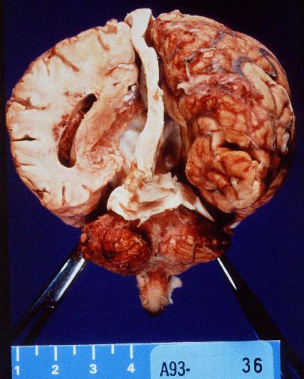
9 Classic AVM 98% solitary Multiple AVMs usually syndromic: Hereditary hemorrhagic telangiectasia (HHT) Cerebrofacial arteriovenous metameric syndrome (CAMS)


10 Hereditary Hemorrhagic Telangiectasia An angiodysplastic disorder with AD inheritance May have multiple pial AVMs or AVFs


11 Intra/extracranial AVMs: CAMS Cerebrofacial Arteriovenous Metameric Syndrome
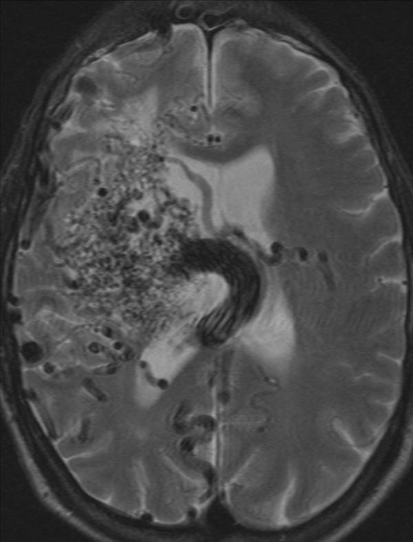
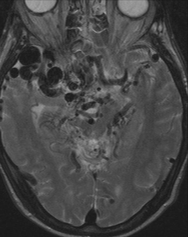
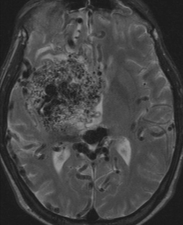

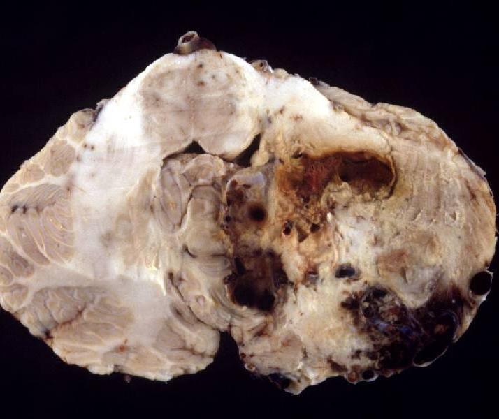
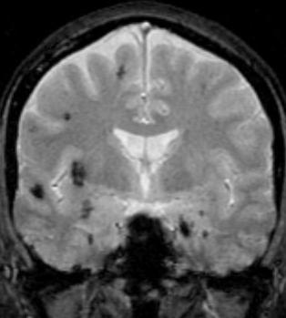
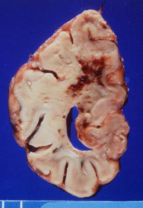
12 T2 AVM: Parenchymal Vascular Nidus


13 AVM: Clinical Significance Peak presentation age: year old Risk of hemorrhage: 2-4%/year ~50% present with symptoms of hemorrhage Seizures, headaches, focal neurologic deficits NECT
 = 1 Medium (3-6 cm) = 2 Large (>6")

14 AVM Grading: Spetzler-Martin Size Small (<3cm) = 1 Medium (3-6 cm) = 2 Large (>6 cm) = 3 Location Noneloquent = 0 Eloquent = 1 Venous drainage Superficial = 0 Deep = 1
")
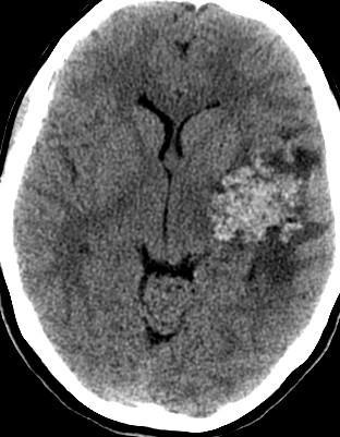
15 AVM Imaging: CT Intraaxial nidus of abnormal vessels Variable Hemorrhage Calcification: 25-30% Enhance post-contrast CTA: Enlarged feeding arteries and draining veins (AV shunting) CECT


16 AVM Imaging: CT NECT NECT Calcification Hemorrhage


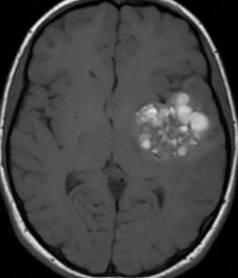
17 AVM Imaging: MRI flow voids T2 FLAIR


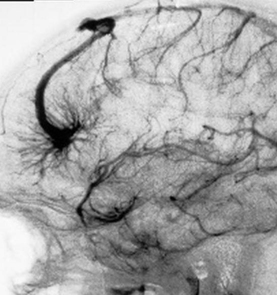
18 AVM: Feeding Arteries & Draining Veins

19 AVM Imaging: Catheter Angiography Must image ICA, ECA, & vertebral circulation 27-32% of AVMs have dual arterial supply

20 AVM: Associated Abnormalities Flow-related aneurysm on feeding artery: 10-15% Intranidal aneurysm: >50% Vascular steal : Ischemia in adjacent brain
 Pedicle aneurysm Intranidal aneurysm")

21 Increased Risk of Hemorrhage NECT Location (deep) Periventricular Basal ganglia Thalamus Arterial (aneurysm) Pedicle aneurysm Intranidal aneurysm Difficult to detect by MR Venous (stenosis or ectasia) Central venous drainage Obstruction of venous outflow Varix or venous pouch Small nidus! NECT

22 AVM: Treatment Surgery: Microvascular resection Radiation: Stereotactic radiosurgery Embolization: Liquid adhesive Observation: Risk of treatment vs disease
23 Teaching Point Classic or pial AVMs have an intraaxial (parenchymal or ventricular) nidus with enlarged feeding arteries and draining veins due to arteriovenous shunting.

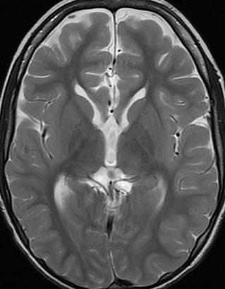
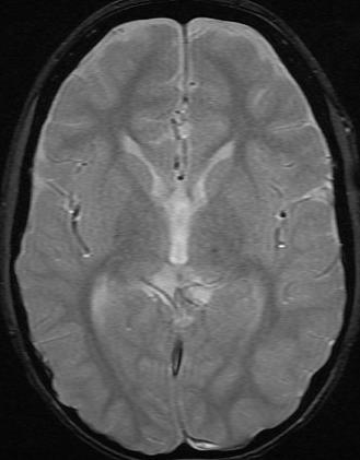
24 Cavernous Malformation AKA: Cavernous angioma, cavernoma, cavernous hemangioma 2 types: Inherited: Multiple & bilateral Sporadic

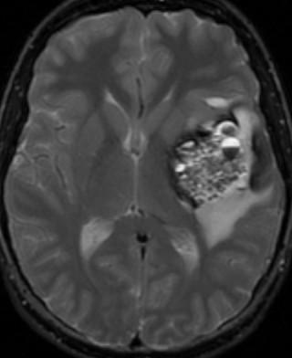
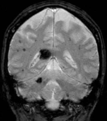
25 Cavernous Malformation: Imaging T2

26 Cavernous Malformation: Imaging T2
27 Cavernous Malformation T2
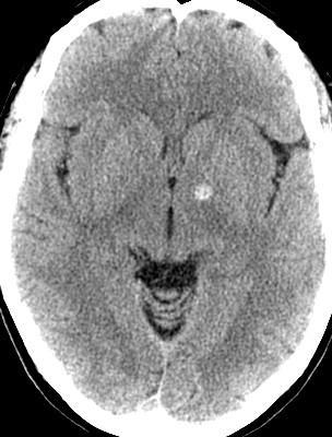
28 Cavernous Malformation NECT T1+Gd

29 Cavernous Malformation NECT T2 T1

30 Cavernous Malformation Risk of hemorrhage: %/year More common in posterior fossa lesions In patients with prior hemorrhage annual rate of rehemorrhage 4.5% Treatment: Observation: Asymptomatic or inaccessible lesions Surgical excision Radiosurgery: Progressively symptomatic but surgically inaccessible T2
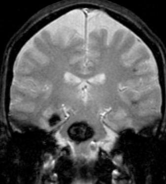
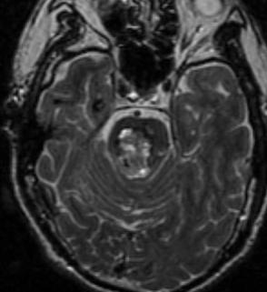
31 Cavernous Malformation T2 MPGR, GRE, SWI = T2* weighted sequences
32 Teaching Point Cavernous malformation is a no flow or angiographically occult lesion that may be associated with a developmental venous anomaly (prenatal venous obstruction).


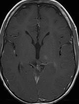
33 Developmental Venous Anomaly (DVA)


34 Developmental Venous Anomaly Isolated or associated with cavernous malformation Hemorrhage unusual T1+Gd


35 DVA Imaging: CT Calcification & ischemia may occur in the region drained most likely due to chronic venous obstructive disease Rare for DVA to cause venous complications NECT CECT CECT


36 Images courtesy Jason Schroeder, MD DVA Imaging: MRI T2 SWI Surrounding T2 hyperintensity May occur in asymptomatic patients Acute edema from venous thrombosis Gliosis from chronic outflow obstruction T1+Gd

37 T1+Gd DVA

38 DVA: Treatment NONE! Removal may cause venous infarction T1+Gd
39 Teaching Point DVAs are benign incidental findings and are not to be confused with AVMs.
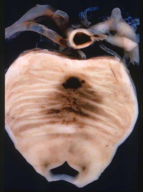
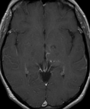
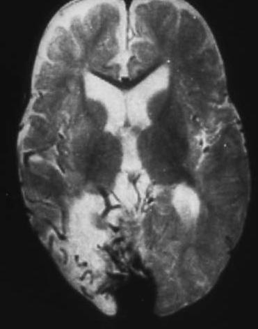
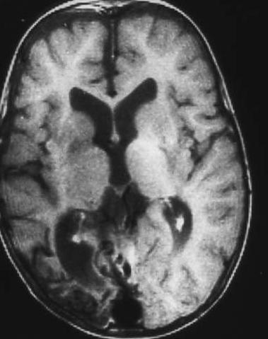
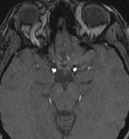
40 Capillary Telangiectasia
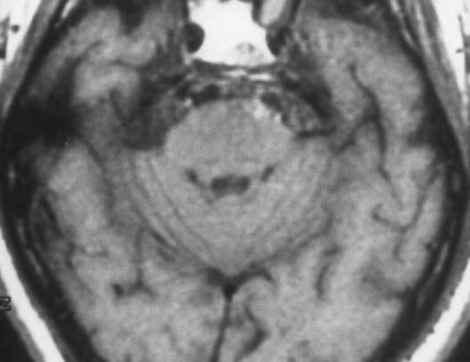
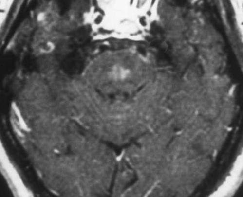
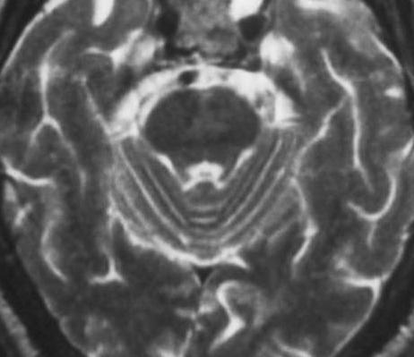
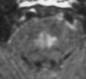
41 Capillary Telangiectasia T1 T2 T1+Gd

42 Brainstem Glioma
43 Capillary Telangiectasia T2 T1+C T2*

44 Capillary Telangiectasia T1+Gd E
45 Teaching Point Capillary telangiectasias are also benign incidental findings and are best seen on T2*-weighted or postgadolinium images.
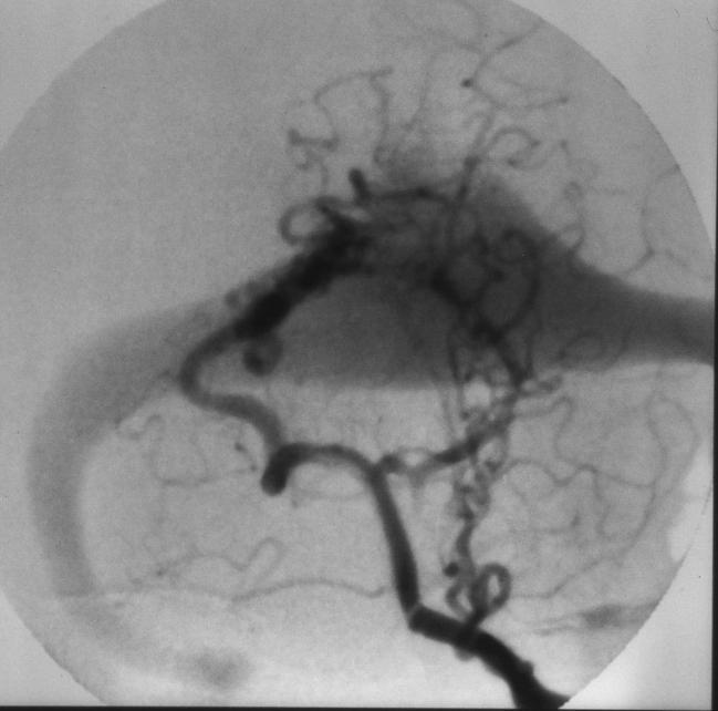
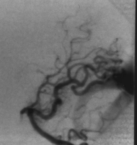
46 Vascular Malformations Arteriovenous malformation (AVM) Classic AVM (intraaxial nidus) Pial arteriovenous fistula (pavf) Dural arteriovenous fistula (davf) Developmental venous anomaly (DVA) Cavernous malformation Capillary telangiectasia Shunt Mixed Malformation = Combo No Shunt
47 Arteriovenous Fistulas Distinguished from AVMs by presence of a direct high flow fistula between artery & vein Pial AVF (pavf) Dural AVF (davf) Cavernous carotid fistula (CCF) Vein of Galen malformation These are less common than intraaxial AVMs in the head (reverse is true in the spine).

48 davf Arteriovenous shunts within dura 10-15% of intracranial vascular malformations 2 types: Adult: Tiny vessels in wall of thrombosed dural venous sinus typically middle aged & older patients Usually acquired - trauma Infantile: Multiple high-flow AVshunts involving several thrombosed dural sinuses SSFSE Fetal MRI


49 AVF SSFSE T1
50 davf Grading: Cognard Classification Type I: In sinus wall, normal antegrade venous drainage Type II: In main sinus A: Reflux into sinus B: Reflux into cortical veins: 10-20% hemorrhage Type III: Direct cortical drainage 40% hemorrhage Type IV: Direct cortical drainage + venous ectasia 2/3 hemorrhage Type V: Spinal perimedullary venous drainage Progressive myelopathy
51 davf Grading: Lalwani et al Type I Type II Type III Type IV

52 davf Most common near skull base Transverse sinus most common Hemorrhage incidence: 2-4% per year Spontaneous closure rare Most are type I

53 CECT davf Imaging: CT


54 davf Imaging: MRI T2 T1+Gd!!! MRA: May be negative MRV: Occluded sinus, collateral flow

55 davf T2 T1 T1

56 davf: Conventional Angiography Multiple arterial feeders Dural/transosseous branches from ECA: most common Tentorial/dural branches from ICA or VA Dural sinus may be thrombosed Flow reversal in dural sinus/cortical veins risk of hemorrhage Tortuous engorged pial veins pseudophlebitic pattern

57 davf CECT Pseudophlebitic pattern
58 Teaching Point Dural AVFs are extra-axial lesions usually located in the wall of a venous sinus. Involvement of cortical veins is the major risk factor for hemorrhage.
59 Carotid Cavernous Fistula (CCF) The cavernous sinus is the second most common location for a dural AVF. This is also known as an indirect CCF. Fistula between arteries to the dural wall and the venous sinus. A hole in the cavernous internal carotid artery would cause a high flow AVF. This is also known as a direct CCF. Trauma or ruptured aneurysm.


60 Venous Drainage SOV Superficial Middle Cerebral V. Uncal v. Cerebellar SPS Pterygoid & basilar plexus IPS

61 CCF: CT Imaging Non-Contrast


62 T1 CCF: MRI

63 CCF: Venous arterialization T1+Gd T2
64 CCF: Arteriovenous Shunting SOV IMAX Indirect Courtesy Steven Goldstein, MD
65 CCF: Treatment Endovascular Surgical resection Observation: Indirect CCF (dural AVF) Cortical venous reflux is risk factor for future hemorrhage
66 Teaching Point Carotid-cavernous fistulas may be direct (hole in cavernous ICA) or indirect (davf in the dural wall of the cavernous sinus).

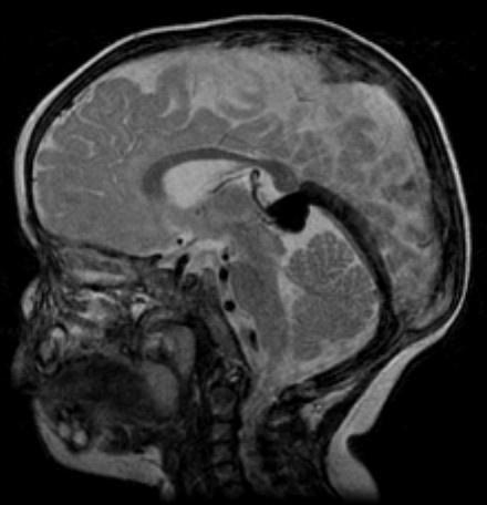
67 Vein of Galen Malformation (VOGM) Neonatal > infant presentation Rare adult presentation Classification: Choroidal: Multiple feeders from pericallosal, choroidal, & thalmoperforating arteries Mural: Few feeders from collicular or posterior choroidal arteries Drains MPV in 50% Falcine Sinus T1


68 VOGM is a type of AVF < 1% of cerebral vascular malformations CECT Venous Pouch
69 VOGM

70 VOG Malformation: Prenatal US

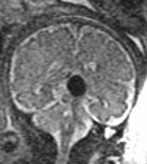
71 VOG SSFSE T2

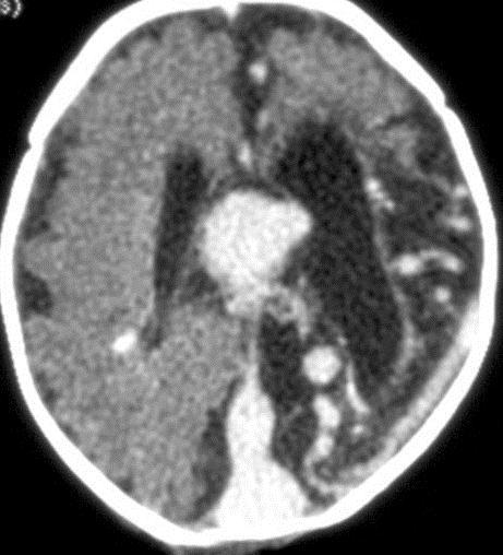
72 VOGM: CT Findings CECT NCCT

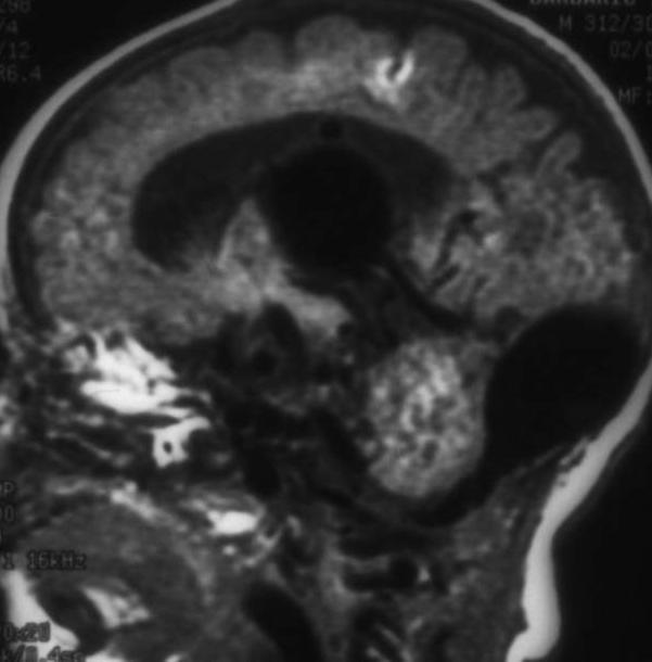
73 VOGM: MR Imaging T2 T1
74 VOGM: Angiography Choroidal or mural Dural venous sinus anomalies Falcine sinus in 50% +/- absence or stenosis of other sinuses
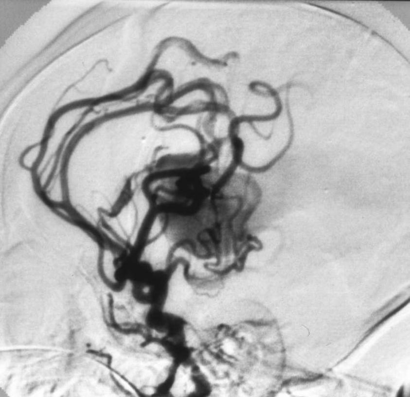
75 VOGM: Angiography Choroidal

76 VOGM: Treatment Choroidal Medical therapy for congestive heart failure until 5 or 6 mo 5-6 mo: Transcatheter embolization Mural Transcatheter embolization performed later

77 CECT VOGM
78 Teaching Point Vein of Galen aneurysm is really a pial AVF (choroidal arteries to median prosencephalic vein).

79 Sinus Pericranii Communication between extracranial venous system & dural venous sinus Rare May be congenital or acquired


80 Sinus Pericranii CECT CT: Single/multiple bone defects Vascular enhancement Conventional angiogram: Seen during venous phase

81 T1+Gd MRV Sinus Pericranii

82 Sinus Pericranii Spontaneous regression rare Risk of hemorrhage Treatment Surgery Endovascular T1+Gd
83 Teaching Point Sinus pericranii is a scalp varix with an anomalous communication to the underlying dural venous sinus.
84 Vascular Malformations Intraaxial Arteriovenous malformation (AVM) Parenchymal or ventricular nidus Cavernous malformation Developmental venous anomaly (DVA) Capillary telangiectasia Extraaxial Arteriovenous fistula Pial on surface of brain (e.g. HHT or VOG) Dural in wall of venous sinus (e.g. transverse) Carotid-cavernous fistula Direct (hole in ICA) Indirect (dural AVF) Sinus pericranii
85 What imaging pattern is associated with higher risk of hemorrhage in a patient with dural arteriovenous fistula (davf)? 1. Pseudosubarachnoid sign 2. Pseudophlebitic pattern 3. Palm tree" appearance 4. Brush stroke" appearance

86 Regarding cavernous malformations, why might intravenous contrast be administered? 1. Evaluate the full extent of the lesion 2. Assess for presence of other cavernous malformations 3. Look for associated developmental venous anomaly 4. Assess arterial and venous supply T1+Gd Cavernous Malformations
87 Which imaging finding would be most characteristic of a capillary telangiectasia? 1. Mild to moderate mass effect 2. Faint stippled appearance on T1 post contrast 3. High signal on T1 weighted imaging 4. Presence of a hemosiderin rim
88 Objectives Describe the typical imaging appearance of the four intraaxial vascular malformations. Understand their clinical significance and the possible treatment options. List a few extraaxial vascular malformations.
89 The End Thank you!
Vascular Malformations of the Brain: A Review of Imaging Features and Risks
 Vascular Malformations of the Brain: A Review of Imaging Features and Risks Comprehensive Neuroradiology: Best Practices October 27-30, 2016 Sudhakar R. Satti, MD Associate Director Neurointerventional
Dural Arteriovenous Malformations and Fistulae (DAVM S DAVF S)
") Jorge Guedes Campos NEUROIMAGING DEPARTMENT HOSPITAL SANTA MARIA UNIVERSITY OF LISBON PORTUGAL DEFINITION region of arteriovenous shunting confined to a leaflet of packymeninges often adjacent to a major
Jorge Guedes Campos NEUROIMAGING DEPARTMENT HOSPITAL SANTA MARIA UNIVERSITY OF LISBON PORTUGAL DEFINITION region of arteriovenous shunting confined to a leaflet of packymeninges often adjacent to a major
Overview of Cerebrovascular Malformations
 Overview of Cerebrovascular Malformations Pursuit of Neurovascular Excellence 8 th annual Barbara Albani, MD Chief, Neurointerventional Surgery Christiana Care Health Systems Newark, DE Financial Disclosures
MASSIVE EPISTAXIS IN A NEONATE: A SYMPTOM OF VEIN OF GALEN MALFORMATION!
 CASE REPORT MASSIVE EPISTAXIS IN A NEONATE: A SYMPTOM OF VEIN OF GALEN MALFORMATION! Shagufta Wahab 1, Rizwan Ahmad Khan 2, Manjari Thapa Manger 3 1. Radiodiagnosis, Aligarh Muslim University, Aligarh,
Brain Arteriovenous Malformations Endovascular Therapy and Associated Therapeutic Protocols Jorge Guedes Cabral de Campos
 Endovascular Therapy and Associated Therapeutic Protocols Jorge Guedes Cabral de Campos Neuroradiology Department Hospital de Santa Maria University of Lisbon CEREBRAL AVM CLINICAL / EPIDEMIOLOGY Brain
Brain AVM with Accompanying Venous Aneurysm with Intracerebral and Intraventricular Hemorrhage
 Cronicon OPEN ACCESS EC PAEDIATRICS Case Report Brain AVM with Accompanying Venous Aneurysm with Intracerebral and Intraventricular Hemorrhage Dimitrios Panagopoulos* Neurosurgical Department, University
A.J. Hauer Intracranial dural arteriovenous fistulae
 A.J. Hauer 27-06-2018 Intracranial dural arteriovenous fistulae Dural arteriovenous fistulae (davfs) epidemiology Pathological anastomoses (within the dural leaflets) between meningeal arteries and dural
CASE OF THE WEEK PROFESSOR YASSER METWALLY
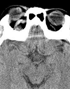
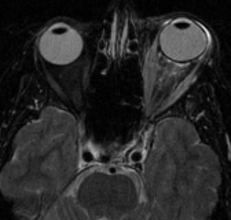
 CLINICAL PICTURE CLINICAL PICTURE: CASE OF THE WEEK PROFESSOR YASSER METWALLY A 29 years old male patients presented with proptosis, ecchymoses of the left eye with both subjective and objective bruit
Retrospective analytical six months study of vascular abnormalities of brain
 International Journal of Advances in Medicine http://www.ijmedicine.com pissn 2349-3925 eissn 2349-3933 Research Article DOI: http://dx.doi.org/10.18203/2349-3933.ijam20160185 Retrospective analytical
Contents. 1 Embryological and Anatomical Introduction... 1
 1 Embryological and Anatomical Introduction.... 1 1.1 Preliminary Remarks.................... 1 1.2 Leptomeninges....................... 21 1.3 Subpial Space........................ 22 1.3.1 Anatomy...........................
Vascular Malformations of the Brain. William A. Cox, M.D. Forensic Pathologist/Neuropathologist. September 8, 2014
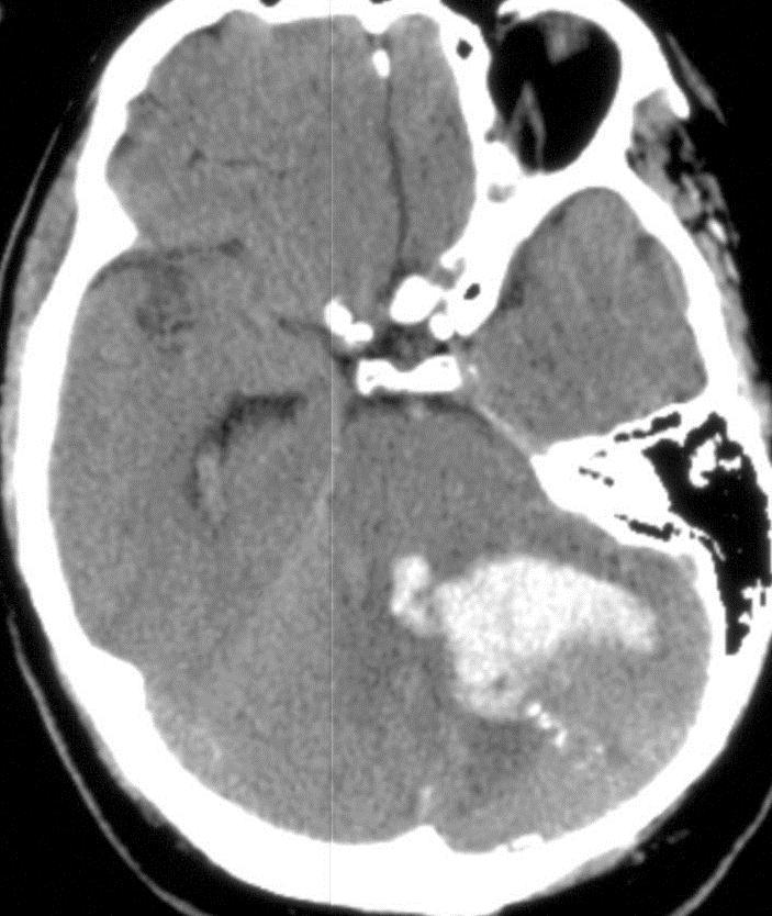
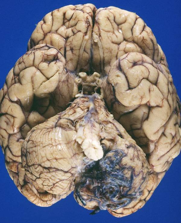
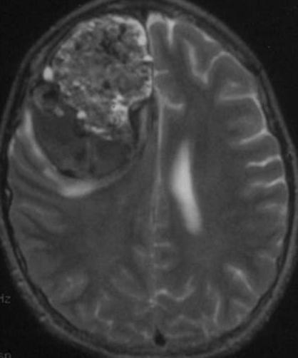
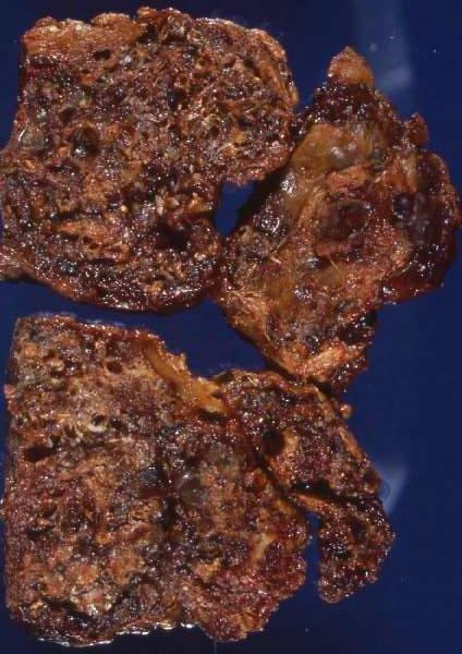
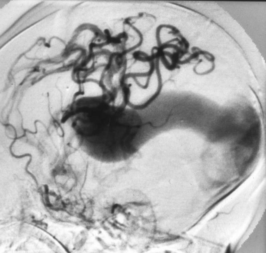
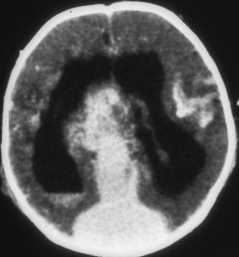
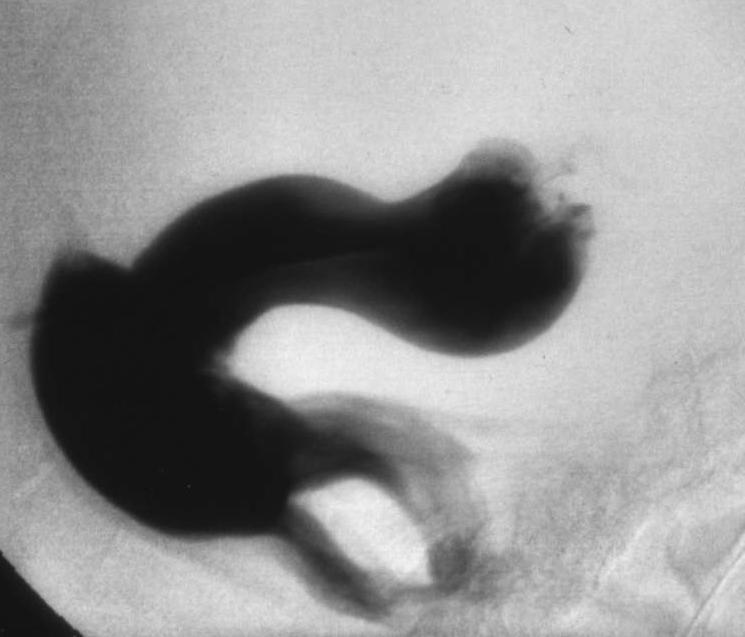
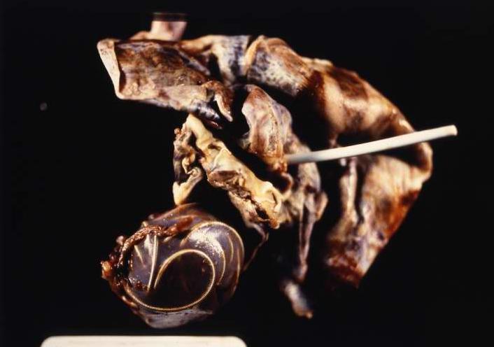
 Vascular Malformations of the Brain William A. Cox, M.D. Forensic Pathologist/Neuropathologist September 8, 2014 Vascular malformations of the brain are classified into four principal groups: arteriovenous
Cerebrovascular Malformations in the Elderly Indications for Treatment
 Cerebrovascular Malformations in the Elderly Indications for Treatment Johanna T. Fifi, MD, FAHA, FSVIN Director of Endovascular Ischemic Stroke Assistant Professor of Neurology, Neurosurgery, and Radiology
Diagnosis and Management of AVM in the Pregnant Patient
 Diagnosis and Management of AVM in the Pregnant Patient Wade Cooper, D.O. University of Michigan Assistant Professor Departments of Neurology & Anesthesiology Disclosures Wade Cooper - None Developmental
What Is an Arteriovenous malformation (AVM)?
?") American Society of Neuroradiology What Is an Arteriovenous malformation (AVM)? From the Cerebrovascular Imaging and Intervention Committee of the American Heart Association Cardiovascular Council Randall
American Society of Neuroradiology What Is an Arteriovenous malformation (AVM)? From the Cerebrovascular Imaging and Intervention Committee of the American Heart Association Cardiovascular Council Randall
Neurosurgical decision making in structural lesions causing stroke. Dr Rakesh Ranjan MS, MCh, Dip NB (Neurosurgery)
") Neurosurgical decision making in structural lesions causing stroke Dr Rakesh Ranjan MS, MCh, Dip NB (Neurosurgery) Subarachnoid Hemorrhage Every year, an estimated 30,000 people in the United States experience
Neurosurgical decision making in structural lesions causing stroke Dr Rakesh Ranjan MS, MCh, Dip NB (Neurosurgery) Subarachnoid Hemorrhage Every year, an estimated 30,000 people in the United States experience
Spinal Vascular Lesions
 Spinal Vascular Lesions Spinal Vascular Lesions Spinal cord infarction Hemangioblastoma Cavernous malformation Vascular malformations (Type 1-4) Spinal artery aneurysm Troy Hutchins, MD Assistant Professor
Pediatric Neurointervention: Vein of Galen Malformations
 Pediatric Neurointervention: Vein of Galen Malformations Johanna T. Fifi, M.D. Assistant Professor of Neurology, Neurosurgery, and Radiology Icahn School of Medicine at Mount Sinai November 9 th, 2014
Diffuse Proliferative Cerebral Angiopathy: A case report and review of the literature
 Diffuse Proliferative Cerebral Angiopathy: A case report and review of the literature Rohit 1*, Poh Sun Goh 1 1. Department of Radiology, National University hospital, Singapore * Correspondence: Dr. Rohit,
General Data. Gender: Female Birthday and age: 1932/11/03, 73 y/o Occupation: house keeper Date of Admission: 2005/03/30
 General Data Gender: Female Birthday and age: 1932/11/03, 73 y/o Occupation: house keeper Date of Admission: 2005/03/30 Chief Complain Dizziness and light headache for recent 1 year. Present illness Hypertension
Historical perspective
 SPINAL AVM Introduction Vascular malformations of spinal cord are a rare clinical entity, representing 5% of all primary spinal cord lesions, with arteriovenous malformations(avm) & cavernous malformations
VASCULAR MALFORMATIONS. Owen Samuels, MD Adam Webb, MD Emory University
 VASCULAR MALFORMATIONS Owen Samuels, MD Adam Webb, MD Emory University Introduction Brain and spinal cord vascular malformations can be separated into five main categories: 1) Arteriovenous malformation,
Marc Norman, Ph.D. - Do Not Use without Permission 1. Cerebrovascular Accidents. Marc Norman, Ph.D. Department of Psychiatry
 Cerebrovascular Accidents Marc Norman, Ph.D. Department of Psychiatry Neuropsychiatry and Behavioral Medicine Neuropsychology Clinical Training Seminar 1 5 http://www.nlm.nih.gov/medlineplus/ency/images/ency/fullsize/18009.jpg
Vascular malformations: Venous malformations anomalous veins drain normal brain tissue for 65% of all cases 2.5%. was 0, 3% per year
 Vascular malformations: 1. Venous malformations: congenital venous anomalies pathologically characterised by anomalous veins (thickened and hyalinised walls) separated by normal brain. These anatomically
Skull radiographs... 5 CT... 5 MRI... 15
 INTRACRANIAL VASCULAR MALFORMATIONS Vas30 (1) Intracranial Vascular Malformations Last updated: September 5, 2017 ARTERIOVENOUS MALFORMATIONS (AVM)... 2 PATHOLOGY, PATHOPHYSIOLOGY... 2 Hemodynamics...
Intracranial spontaneous hemorrhage mechanisms, imaging and management
 Intracranial spontaneous hemorrhage mechanisms, imaging and management Dora Zlatareva Department of Diagnostic Imaging Medical University, Sofia, Bulgaria Intracranial hemorrhage (ICH) ICH 15% of strokes
Spontaneous Obliteration of Pial Arteriovenous Malformations: A Review of 27 Cases
 AJNR Am J Neuroradiol :, March 00 Spontaneous Obliteration of Pial Arteriovenous Malformations: A Review of ases Maneesh. Patel, Timothy J. Hodgson, Andras A. Kemeny, and David M. Forster BAKGROUND AND
Vein of Galen Malformation Joseph Junewick, MD FACR
 Vein of Galen Malformation Joseph Junewick, MD FACR 04/14/2018 History Midline cystic intracranial mass on prenatal ultrasound. Diagnosis Vein of Galen Malformation Discussion In normal fetal development,
The outcome of treatment for arteriovenous malformations of the brain: A five-year retrospective series from the Philippines
 Neurology Asia 2006; 11 : 91 96 ORIGINAL ARTICLES The outcome of treatment for arteriovenous malformations of the brain: A five-year retrospective series from the Philippines Roland Mark M GIGATARAS MD,
North Oaks Trauma Symposium Friday, November 3, 2017
 Traumatic Intracranial Hemorrhage Aaron C. Sigler, DO, MS Neurosurgery Tulane Neurosciences None Disclosures Overview Anatomy Epidural hematoma Subdural hematoma Cerebral contusions Outline Traumatic ICH
Endovascular Treatment of Cerebral Arteriovenous Malformations. Bs. Nguyễn Ngọc Pi Doanh- Bs Đặng Ngọc Dũng Khoa Ngoại Thần Kinh
 Endovascular Treatment of Cerebral Arteriovenous Malformations Bs. Nguyễn Ngọc Pi Doanh- Bs Đặng Ngọc Dũng Khoa Ngoại Thần Kinh Stroke Vascular Malformations of the Brain Epidemiology: - Incidence: 0.1%,
Enhancement of Cranial US: Utility of Supplementary Acoustic Windows and Doppler Harriet J. Paltiel, MD
 Enhancement of Cranial US: Utility of Supplementary Acoustic Windows and Doppler Harriet J. Paltiel, MD Boston Children s Hospital Harvard Medical School None Disclosures Conventional US Anterior fontanelle
[(PHY-3a) Initials of MD reviewing films] [(PHY-3b) Initials of 2 nd opinion MD]
![[(PHY-3a) Initials of MD reviewing films] [(PHY-3b) Initials of 2 nd opinion MD]](/thumbs/89/98619893.jpg "[(PHY-3a) Initials of MD reviewing films] [(PHY-3b) Initials of 2 nd opinion MD]") 2015 PHYSICIAN SIGN-OFF (1) STUDY NO (PHY-1) CASE, PER PHYSICIAN REVIEW 1=yes 2=no [strictly meets case definition] (PHY-1a) CASE, IN PHYSICIAN S OPINION 1=yes 2=no (PHY-2) (PHY-3) [based on all available
2015 PHYSICIAN SIGN-OFF (1) STUDY NO (PHY-1) CASE, PER PHYSICIAN REVIEW 1=yes 2=no [strictly meets case definition] (PHY-1a) CASE, IN PHYSICIAN S OPINION 1=yes 2=no (PHY-2) (PHY-3) [based on all available
EMBOLIZATION OF ARTERIOVENOUS FISTULA AFTER RADIOSURGERY FOR MULTIPLE CEREBRAL ARTERIOVENOUS MALFORMATIONS
 Arteriovenous fistula after radiosurgery for multiple CAVM EMBOLIZATION OF ARTERIOVENOUS FISTULA AFTER RADIOSURGERY FOR MULTIPLE CEREBRAL ARTERIOVENOUS MALFORMATIONS Chao-Bao Luo, Wan-Yuo Guo, Michael
7/5/2016. Neonatal high-output cardiac failure. Case 1 POSTNATAL STRATEGIES FOR CEREBRAL ATERIOVENOUS MALFORMATIONS
 John Deveikis, M.D. POSTNATAL STRATEGIES FOR CEREBRAL ATERIOVENOUS MALFORMATIONS JULY, 2016 Neonatal high-output cardiac failure Tachypnea, tachycardia, hypotension, failure to thrive When congenital heart
Adult Choroidal Vein of Galen Malformation
 Adult Choroidal Vein of Galen Malformation Thomas A. Tomsick, Robert J. Ernst, John M. Tew, Thomas G. Brott, and John C. Breneman Summary: We report staged embolization and stereotactic radiation in a
Peripheral Spinal Cord Hypointensity on T2-weighted MR Images: A Reliable Imaging Sign of Venous Hypertensive Myelopathy
 AJNR Am J Neuroradiol 21:781 786, April 2000 Peripheral Spinal Cord Hypointensity on T2-weighted MR Images: A Reliable Imaging Sign of Venous Hypertensive Myelopathy Robert W. Hurst and Robert I. Grossman
Complex dural arteriovenous fistulas. Results of combined endovascular and neurosurgical treatment in 16 patients
 J Neurosurg 71:352-358,1989 Complex dural arteriovenous fistulas Results of combined endovascular and neurosurgical treatment in 16 patients STANLEY L. BARNWELL, M.D., PH.D., VAN V. HALBACH, M.D., RANDALL
Radiographic and statistical analysis of Brain Arteriovenous Malformations.
 Radiographic and statistical analysis of Brain Arteriovenous Malformations. Poster No.: C-0996 Congress: ECR 2017 Type: Educational Exhibit Authors: C. E. Rodriguez 1, A. Lopez Moreno 1, D. Sánchez Paré
Journal of Radiology Case Reports
 Pediatric Holohemispheric Developmental Venous Anomaly: Definitive characterization by 3D Susceptibility Weighted Magnetic Resonance Angiography Michael A. Casey 1, Sourabh Lahoti 2, Ajeet Gordhan 2* 1.
Transverse-Sigmoid Sinus Dural Arteriovenous Malformations
 Transverse-Sigmoid Sinus Dural Arteriovenous Malformations Kenan I. Amautovic, M.D., and Ali F. Krisht, M.D. '-...--- Learning Objectives: After reading this article, the participant should: 1. Have an
Official Journal of the American Osteopathic College of Radiology NEUROIMAGING. Editor-in Chief: William T. O Brien, Sr., D.O.
 ISSN: 2165-3259 JOCR Official Journal of the merican Osteopathic College of Radiology NEUROIMGING Editor-in Chief: William T. O rien, Sr., D.O. January 2012, Vol. 1, Issue 1 JOCR bout the Journal ims and
NEURORADIOLOGY Part I
 NEURORADIOLOGY Part I Vörös Erika University of Szeged Department of Radiology SZEGED BRAIN IMAGING METHODS Plain film radiography Ultrasonography (US) Computer tomography (CT) Magnetic resonance imaging
Occlusive hyperemia: a theory for the hemodynamic complications following resection of intracerebral arteriovenous malformations
 J Neurosurg 78: 167-175, 1993 Occlusive hyperemia: a theory for the hemodynamic complications following resection of intracerebral arteriovenous malformations NAYEF R. F. AL-RODHAN, M.D., PH.D., THORALF
Dilemma in Imaging Diagnosis, Endovascular Management and Complications
 Vascular anomaly at the craniocervical junction presenting with sub arachnoid hemorrhage: Dilemma in Imaging Diagnosis, Endovascular Management and Complications Ajeet 1* 1. Department of Radiology, St
Spontaneous occlusion of a cerebral arteriovenous malformation after subtotal endovascular embolisation
 206 Chiriac et al Spontaneous occlusion of a cerebral arteriovenous malformation Spontaneous occlusion of a cerebral arteriovenous malformation after subtotal endovascular embolisation A. Chiriac, N. Dobrin*,
Pearls and Pitfalls in Neuroradiology of Cerebrovascular Disease The Essentials with MR and CT
 Pearls and Pitfalls in Neuroradiology of Cerebrovascular Disease The Essentials with MR and CT Val M. Runge, MD Wendy R. K. Smoker, MD Anton Valavanis, MD Control # 823 Purpose The focus of this educational
PTA 106 Unit 1 Lecture 3
 PTA 106 Unit 1 Lecture 3 The Basics Arteries: Carry blood away from the heart toward tissues. They typically have thicker vessels walls to handle increased pressure. Contain internal and external elastic
Intracranial dural arteriovenous fistulas (DAVFs) with retrograde
 with retrograde") ORIGINAL RESEARCH W.J. van Rooij M. Sluzewski G.N. Beute Dural Arteriovenous Fistulas with Cortical Venous Drainage: Incidence, Clinical Presentation, and Treatment BACKGROUND AND PURPOSE: Our purpose
ORIGINAL RESEARCH W.J. van Rooij M. Sluzewski G.N. Beute Dural Arteriovenous Fistulas with Cortical Venous Drainage: Incidence, Clinical Presentation, and Treatment BACKGROUND AND PURPOSE: Our purpose
The superior ophthalmic vein approach for the treatment of carotid-cavernous fistulas: our first experience
 230 Chiriac et al - Superior ophthalmic vein approach The superior ophthalmic vein approach for the treatment of carotid-cavernous fistulas: our first experience A. Chiriac, N. Dobrin 1, Georgiana Ion
C. Douglas Phillips, MD FACR Director of Head and Neck Imaging Weill Cornell Medical College NewYork-Presbyterian Hospital
 C. Douglas Phillips, MD FACR Director of Head and Neck Imaging Weill Cornell Medical College NewYork-Presbyterian Hospital I have no financial disclosures Understand range of pathology that may present
SPINAL EPIDURAL ARTERIOVENOUS MALFORMATIONS: REPORT OF A CASE WITH DISCUSSION OF CLASSIFICATION AND TREATMENT
 SPINAL EPIDURAL ARTERIOVENOUS MALFORMATIONS: REPORT OF A CASE WITH DISCUSSION OF CLASSIFICATION AND TREATMENT Caitlin M. Clark, B.A. and W. Craig Clark, M.D., Ph.D., FAANS, FACS, FICS INTRODUCTION True
Classification and Endovascular Management of Pediatric Cerebral Vascular Malformations
 Classification and Endovascular Management of Pediatric Cerebral Vascular Malformations Timo Krings, MD, PhD, FRCP(C) a,b,c, *, Sasikhan Geibprasert, MD d,e, Karel terbrugge, MD, FRCP(C) a KEYWORDS Endovascular
Dural arteriovenous fistula discovered in patient presenting with recent head trauma
 ISSN 1507-6164 DOI: 10.12659/AJCR.889610 Received: 2013.07.25 Accepted: 2013.08.08 Published: 2013.10.28 Dural arteriovenous fistula discovered in patient presenting with recent head trauma Authors Contribution:
Supratentorial cerebral arteriovenous malformations : a clinical analysis
 Original article: Supratentorial cerebral arteriovenous malformations : a clinical analysis Dr. Rajneesh Gour 1, Dr. S. N. Ghosh 2, Dr. Sumit Deb 3 1Dept.Of Surgery,Chirayu Medical College & Research Centre,
Advanced Vascular Imaging: Pulsatile Tinnitus. Disclosures. Pulsatile Tinnitus: Differential Diagnosis. Pulsatile Tinnitus
 Advanced Vascular Imaging: Pulsatile Tinnitus Patrick Turski MD, Zach Clark MD, Tabby Kennedy MD The Objectives of this presentation are to: Review the differential diagnosis of pulsatile tinnitus Discuss
Untangling Cerebral Dural Arteriovenous Fistulas
 Untangling Cerebral Dural Arteriovenous Fistulas Bradley A. Gross, MD Assistant Professor, Dept of Neurosurgery, University of Pittsburgh September 2017 davfs Definition Clinical Presentation Natural History
Venous Stroke with Intracranial Dural Arteriovenous Fistula
 Case Reports 24 Venous Stroke with Intracranial Dural Arteriovenous Fistula Tzung-Wen Chiang 1,2, Shung-Lon Lai 1, Leang-Kai Chang 2, Yung-Yee Chang 1, Min-Yu Lan 1, Yeh-Lin Kuo 3, Chen-Chung Lu 3, and
Dementia Resulting from Dural Arteriovenous Fistulas: The Pathologic Findings of Venous Hypertensive Encephalopathy
 AJNR Am J Neuroradiol 19:1267 1273, August 1998 Dementia Resulting from Dural Arteriovenous Fistulas: The Pathologic Findings of Venous Hypertensive Encephalopathy Robert W. Hurst, Linda J. Bagley, Steven
A Case of Carotid-Cavernous Fistula
 A Case of Carotid-Cavernous Fistula By : Mohamed Elkhawaga 2 nd Year Resident of Ophthalmology Alexandria University A 19 year old male patient came to our outpatient clinic, complaining of : -Severe conjunctival
Blood Supply. Allen Chung, class of 2013
 Blood Supply Allen Chung, class of 2013 Objectives Understand the importance of the cerebral circulation. Understand stroke and the types of vascular problems that cause it. Understand ischemic penumbra
Identifying Cerebrovascular Disorders. Wengui Yu, MD, PhD Department of Neurology, University of California, Irvine
 Identifying Cerebrovascular Disorders Wengui Yu, MD, PhD Department of Neurology, University of California, Irvine Objectives Review different types of cerebrovascular disorders. Briefly discuss etiology,
Cryptogenic Enlargement Of Bilateral Superior Ophthalmic Veins
 ISPUB.COM The Internet Journal of Radiology Volume 18 Number 1 Cryptogenic Enlargement Of Bilateral Superior Ophthalmic Veins K Kragha Citation K Kragha. Cryptogenic Enlargement Of Bilateral Superior Ophthalmic
Transarterial Occlusion of Solitary Intracerebral Arteriovenous Fistulas
 747 Transarterial Occlusion of Solitary Intracerebral Arteriovenous Fistulas Van V. Halbach 1 2 Randall T. Higashida 1 2 Grant B. Hieshima 1 2 Carl W. Hardin 1 Christopher F. Dowd 1 Stanley L. Barnwell
Vein of Galen Aneurysms
 Interventional Neuroradiology 7 (Suppll): 99103, 2001 Vein of Galen Aneurysms Experience with Eleven Cases. KOIYAA, H. NAKAJIA,. NISHIKAWA, K. YAANAKA, Y. IWAI, T. YASUI, T. ORIKAWA*, S. KITANO*, H. SAKAOTO*,A.
Dural Arteriovenous Fistula of the Cavernous Sinus Presenting with Progressive Venous Congestion of the Pons and Cerebrum: Report of one case
 Dural Arteriovenous Fistula of the Cavernous Sinus Presenting with Progressive Venous Congestion of the Pons and Cerebrum: Report of one case Soo-Bin Yim, M.D., Jong-Sung Kim, M.D., Yang Kwon,M.D.*, Choong-Gon
Three Cases of Dural Arteriovenous Fistula of the Anterior Condylar Vein within the Hypoglossal Canal
 AJNR Am J Neuroradiol 20:2016 2020, November/December 1999 Case Report Three Cases of Dural Arteriovenous Fistula of the Anterior Condylar Vein within the Hypoglossal Canal Robert Ernst, Robert Bulas,
WLNC 2018 ISTANBUL CASES
 WLNC 2018 ISTANBUL CASES WLNC 2018 KOBE / ISTANBUL CASES PT 1 NK 62 Y F Presented with dizziness 2 years ago MR-DSA: Falcotentorial Dural AVF WLNC 2018 KOBE / ISTANBUL CASES MRI 2017 WLNC 2018 KOBE / ISTANBUL
Bilateral Carotid and Vertebral Rete Mirabile Presenting with a Prominent Anterior Spinal Artery Mimicking a Spinal Dural AV Fistula at MRI
 Case Report http://dx.doi.org/10.3348/kjr.2011.12.6.740 pissn 1229-6929 eissn 2005-8330 Korean J Radiol 2011;12(6):740-744 Bilateral Carotid and Vertebral Rete Mirabile Presenting with a Prominent Anterior
Neuroradiology: Imaging and Stroke
 Neuroradiology: Imaging and Stroke Stroke 2017 William Gallmann January 28, 2017 Stroke Arterial ischemia/infarct accounts for ~85% Cerebral venous occlusions - 0.5-1% Spontaneous intracranial hemorrhage
Transvenous Embolization of Cavernous Sinus Dural Arteriovenous Fistulas with Shunts Involving the Laterocavernous Sinus
 Journal of Neuroendovascular Therapy 2017; 11: 1 7 Online November 9, 2016 DOI: 10.5797/jnet.oa.2016-0062 Transvenous Embolization of Cavernous Sinus Dural Arteriovenous Fistulas with Shunts Involving
2. Subarachnoid Hemorrhage
 Causes: 2. Subarachnoid Hemorrhage A. Saccular (berry) aneurysm - Is the most frequent cause of clinically significant subarachnoid hemorrhage is rupture of a saccular (berry) aneurysm. B. Vascular malformation
UPSTATE Comprehensive Stroke Center. Neurosurgical Interventions Satish Krishnamurthy MD, MCh
 UPSTATE Comprehensive Stroke Center Neurosurgical Interventions Satish Krishnamurthy MD, MCh Regional cerebral blood flow is important Some essential facts Neurons are obligatory glucose users Under anerobic
Cerebellar Hemorrhage due to a Direct Carotid Cavernous Fistula after Surgery for Maxillary Cancer
 Case Report J Korean Neurosurg Soc 60 (1) : 89-93, 2017 https://doi.org/10.3340/jkns.2015.1206.001 pissn 2005-3711 eissn 1598-7876 Cerebellar Hemorrhage due to a Direct Carotid Cavernous Fistula after
41 year old female with headache. Elena G. Violari MD and Leo Wolansky MD
 41 year old female with headache Elena G. Violari MD and Leo Wolansky MD ? Dural Venous Sinus Thrombosis with Hemorrhagic Venous Infarct Acute intraparenchymal hematoma measuring ~3 cm in diameter centered
Diagnosis of Subarachnoid Hemorrhage (SAH) and Non- Aneurysmal Causes
 and Non- Aneurysmal Causes") Diagnosis of Subarachnoid Hemorrhage (SAH) and Non- Aneurysmal Causes By Sheila Smith, MD Swedish Medical Center 1 Disclosures I have no disclosures 2 Course Objectives Review significance and differential
Diagnosis of Subarachnoid Hemorrhage (SAH) and Non- Aneurysmal Causes By Sheila Smith, MD Swedish Medical Center 1 Disclosures I have no disclosures 2 Course Objectives Review significance and differential
MR Imaging of Spinal Cord Arteriovenous Malformations at 0.5 T: Study of 34 Cases
 833 MR Imaging of Spinal Cord Arteriovenous Malformations at 0.5 T: Study of 34 Cases D. Dormont 1 F. Gelbert 2 E. Assouline 2 D. Reizine 2 A. Helias 2 M. C. Riche 2 J. Chiras 1 J. Sories 1 J. J. Merland
Interventions in the Management of Acute Stroke. Dr Md Shafiqul Islam Associate Professor Neurosurgery Dhaka Medical College Hospital
 Interventions in the Management of Acute Stroke Dr Md Shafiqul Islam Associate Professor Neurosurgery Dhaka Medical College Hospital Acute stroke intervention Number of stroke patients increasing day by
Selective disconnection of cortical venous reflux as treatment for cranial dural arteriovenous fistulas
 J Neurosurg 101:31 35, 2004 Selective disconnection of cortical venous reflux as treatment for cranial dural arteriovenous fistulas J. MARC C. VAN DIJK, M.D., PH.D., KAREL G. TERBRUGGE, M.D., ROBERT A.
SPINAL AVM: CLASSIFICATION AND MANAGEMENT STRATEGIES. Presented by : Anuj K Tripathi
 SPINAL AVM: CLASSIFICATION AND MANAGEMENT STRATEGIES Presented by : Anuj K Tripathi SPINAL AVM Spinal vascular malformations represent a rare and insufficiently studied pathological entity Great difficulties
Division of Neurological Surgery, Barrow Neurological Institute, St. Joseph s Hospital and Medical Center, Phoenix, Arizona
 clinical article J Neurosurg 122:876 882, 2015 The role of microscope-integrated near-infrared indocyanine green videoangiography in the surgical treatment of intracranial dural arteriovenous fistulas
ISCHEMIC STROKE IMAGING
 ISCHEMIC STROKE IMAGING ผศ.พญ พญ.จ ร ร ตน ธรรมโรจน ภาคว ชาร งส ว ทยา คณะแพทยศาสตร มหาว ทยาล ยขอนแก น A case of acute hemiplegia Which side is the abnormality, right or left? Early Right MCA infarction
Case Conference: Neuroradiology. Case 1: Tumor Case 1: 22yo F w/ HA and prior Seizures
 Case Conference: Neuroradiology Case 1: 22yo F w/ HA and prior Seizures David E. Rex, MD, PhD Stanford University Hospital Department of Radiology Case 1: Tumor Most likely gangiloglioma, oligodendroglioma,
10 May Disclosure. + Outline. Case-based approach to nontraumatic intracranial hemorrhage. Kathleen R. Fink, MD University of Washington
 Kathleen R. Fink, MD University of Washington 5 th Nordic Emergency Radiology Course May 21, 2015 Disclosure My spouse receives research salary support from: Bracco BayerHealthcare Guerbet Outline Case-based
AVM and AV fistula. Effective diagnostic imaging for spinal arteriovenous fistula
 FPⅤ-1 Effective diagnostic imaging for spinal arteriovenous fistula Ryohei Miyazaki, Hidetoshi Murata, Mitsuru Sato, Takahiro Tanaka, Nobutaka Shimizu, Jun Suenaga, Tetsuya Yamamoto Yokohama City University
Disclosure. + Outline. What is a stroke? Role of imaging in stroke Ischemic stroke Venous infarct Current topics
 + Kathleen R. Fink, MD University of Washington 5 th Nordic Emergency Radiology Course May 21, 2015 + Disclosure My spouse receives research salary support from: Bracco BayerHealthcare Guerbet Thank you
Original Article Pial arteriovenous fistulas: two pediatric cases and a literature review
 Int J Clin Exp Med 2016;9(5):7855-7862 www.ijcem.com /ISSN:1940-5901/IJCEM0019732 Original Article Pial arteriovenous fistulas: two pediatric cases and a literature review Lei Feng *, Yunzhen Liu *, Jun
Spontaneous Closure of Dural Arteriovenous Fistulas: Report of Three Cases and Review of the Literature
 AJNR Am J Neuroradiol 22:992 996, May 2001 Case Report Spontaneous Closure of Dural Arteriovenous Fistulas: Report of Three Cases and Review of the Literature Alain Luciani, Emmanuel Houdart, Charbel Mounayer,
OBJECTIVES. At the end of the lecture, students should be able to: List the cerebral arteries.
 DR JAMILA EL MEDANY OBJECTIVES At the end of the lecture, students should be able to: List the cerebral arteries. Describe the cerebral arterial supply regarding the origin, distribution and branches.
Modern treatment of brain arteriovenous malformation
 ORIGINAL RESEARCH W.J. van Rooij M. Sluzewski G.N. Beute Brain AVM Embolization with Onyx BACKGROUND AND PURPOSE: To report the initial experience by using a new liquid embolic agent (Onyx) for embolization
Navigation-guided Burr Hole Aspiration Surgery for Acute Cerebellar Infarction
 FPⅣ-1 Navigation-guided Burr Hole Aspiration Surgery for Acute Cerebellar Infarction Eun-Sung Park, Dae-Won Kim, Sung-Don Kang Department of Neurosurgery, School of Medicine, Wonkwang University, Iksan,
Differences between CS-DAVF and TCCF to reveal and redefine CS-DAVF
 Pan et al. Chinese Neurosurgical Journal (2018) 4:26 https://doi.org/10.1186/s41016-018-0121-z CHINESE MEDICAL ASSOCIATION COMMENTARY Differences between CS-DAVF and TCCF to reveal and redefine CS-DAVF
Endovascular Treatment of Cavernous Sinus Dural Arteriovenous Fistulas by Direct Puncture of Facial Vein
 J Radiol Sci 2011; 36: 129-134 Endovascular Treatment of Cavernous Sinus Dural Arteriovenous Fistulas by Direct Puncture of Facial Vein Shih-Wei Hsu 1 Yeh-Lin Kuo 1 Min-Hsiung Cheng 2 Department of Diagnostic
Non-Traumatic Neuro Emergencies
 Department of Radiology University of California San Diego Non-Traumatic Neuro Emergencies John R. Hesselink, M.D. Nontraumatic Neuroemergencies 1. Acute focal neurological deficit 2. Worst headache of
Dural arteriovenous fistulas (DAVFs) are abnormal. Long-term angiographic results of endovascularly cured intracranial dural arteriovenous fistulas
 are abnormal. Long-term angiographic results of endovascularly cured intracranial dural arteriovenous fistulas") clinical article J Neurosurg 124:1123 1127, 2016 Long-term angiographic results of endovascularly cured intracranial dural arteriovenous fistulas Sudheer Ambekar, MD, Brandon G. Gaynor, MD, Eric C. Peterson,
clinical article J Neurosurg 124:1123 1127, 2016 Long-term angiographic results of endovascularly cured intracranial dural arteriovenous fistulas Sudheer Ambekar, MD, Brandon G. Gaynor, MD, Eric C. Peterson,
Cerebro-vascular stroke
 Cerebro-vascular stroke CT Terminology Hypodense lesion = lesion of lower density than the normal brain tissue Hyperdense lesion = lesion of higher density than normal brain tissue Isodense lesion = lesion
7 TI - Radiosurgery of angiographically occult vascular malformations. AU - Kida Y, et al.
 1 TI - Cerebral arteriovenous malformation in pregnancy: presentation and neurologic, obstetric, and ethical significance. AU - Finnerty JJ, et al. SO - Am J Obstet Gynecol. 1999 Aug;181(2):296-303. Review.
CENTRAL NERVOUS SYSTEM TRAUMA and Subarachnoid Hemorrhage. By: Shifaa AlQa qa
 CENTRAL NERVOUS SYSTEM TRAUMA and Subarachnoid Hemorrhage By: Shifaa AlQa qa Subarachnoid Hemorrhage Causes: Rupture of a saccular (berry) aneurysm Vascular malformation Trauma Hematologic disturbances
Uncommon Symptomatic Cerebral Vascular Malformations
 Uncommon Symptomatic Cerebral Vascular Malformations Mauro Bergui and Gianni Boris Bradac Summary: We describe three cases of unusual vascular malformations in which the most relevant angiographic findings
HEAD AND NECK IMAGING. James Chen (MS IV)
") HEAD AND NECK IMAGING James Chen (MS IV) Anatomy Course Johns Hopkins School of Medicine Sept. 27, 2011 OBJECTIVES Introduce cross sectional imaging of head and neck Computed tomography (CT) Review head
HEAD AND NECK IMAGING James Chen (MS IV) Anatomy Course Johns Hopkins School of Medicine Sept. 27, 2011 OBJECTIVES Introduce cross sectional imaging of head and neck Computed tomography (CT) Review head
Cerebral arteriovenous malformations in children: radiology assesment
 Cerebral arteriovenous malformations in children: radiology assesment Poster No.: C-1588 Congress: ECR 2015 Type: Scientific Exhibit Authors: J. S. Gaete, A. Sanchez-Montanez Garcia-Carpintero, E. Vasquez,