Scanner Spectral. Double Energie et Comptage Photonique. Principes et Premiers Résultats
|
|
|
- Ethel Wiggins
- 5 years ago
- Views:
Transcription


1 Scanner Spectral Double Energie et Comptage Photonique Principes et Premiers Résultats Philippe Douek, Loic Boussel, Monica Sigovan, Salim Si-Mohamed Daniel Bar ness
2 Plan 1. Rappel Techniques: Attenuation des RX 2. Double Energie: 1. Principes 2. Applications CV 3. Perspectives: SPCCT Scanner à Comptage Photonique:

 Cardio-vascular and coronary applications but still some limitations Reduced dose of contrast")
3 Computed Tomography CT: Imaging technology widely used in the world CT: Majors improvements the last 10 years Large detectors: Improved workflow with faster acquisitions Improved diagnosis (PE, Stroke, Emergency Polytrauma etc..) Cardio-vascular and coronary applications but still some limitations Reduced dose of contrast agent Iterative reconstructions Reduced dose with improved S/N 3


4 X-Ray: Photons of High Energy

5 X-Ray Tube Spectrum

6 X Ray and matter Interactions Probability of the photoelectric interaction is proportional to the atomic number Z (P Ph Z 3 ). Probability of a Compton interaction is almost independent of atomic number Z and is directly proportional to the number of electrons per gram (electron density) of the material.
 ( E) ( E) f ( E) f ( E) P c Single X-ray acquisition cannot always help in the tissue characterization (finding out the contribution of each effect:")
7 X-Ray Attenuation X-ray attenuation depends on the incident X-ray energy and on the effective atomic number of the traversed tissue Different tissues exhibit different combinations of photoelectric absorption and Compton scattering ( E) ( E) ( E) f ( E) f ( E) P c Single X-ray acquisition cannot always help in the tissue characterization (finding out the contribution of each effect: photoelectric and Compton) and may lead to similar HU for different tissues P P c c Calcium or Iodine? Calcium or Iodine?
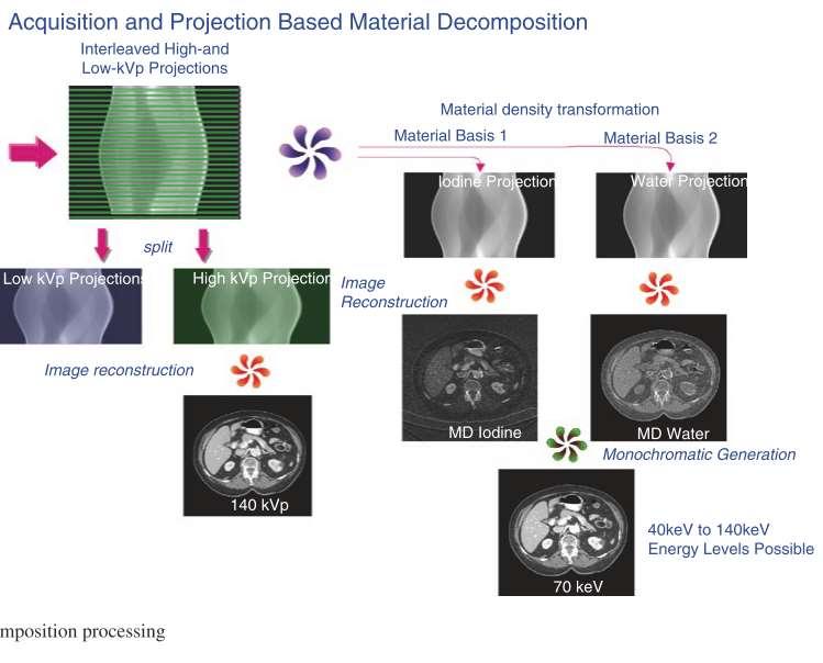
8 Double énergie Objectif : apporter une information spectrale supplémentaire Séparation hautes et basses énergies :
9 Double énergie Objectif : apporter une information spectrale supplémentaire Séparation hautes et basses énergies :

10 Double énergie Objectif : apporter une information spectrale supplémentaire Séparation hautes et basses énergies :
11 Double énergie Technology Paths to Dual-Energy Acquisition Dual Source kv Switch Dual Spin Detection Based No spectral mode: Spectral mode: Spectral mode: Spectral mode: SPECTRAL ALWAYS needs to be pre-selected needs to be pre-selected needs to be 120 kvp & 140 kvp 2 tubes (80 0r 100/140 kvp) Fast kv switching: 80/140kVp 1st 80kVp Tube ma modulation Image Space Projection Space 2nd 140kVp Dose Neutral (interpolations) Image Space Projection Space
12 Double énergie Objectif : apporter une information spectrale supplémentaire Séparation hautes et basses énergies : Emission du tube polychromatique Séparation des matériaux : Iode, calcium Algorithmes de prédiction probabiliste (likelihood) Cartographie : VNC, Energies (40->200 kv), iode, calcium Correction du beam hardening
13


14 Photoelectric - Compton Decomposition Material pairs Material Specific Images CT Image Calcium Image Iodine Image Calcium-Iodine pair

15 Photoelectric - Compton Decomposition Material pairs Material Specific Images CT Image Iodine image Water image Water-Iodine pair
 ( E) ( E) f ( E) f ( E) P c P")
 Raw")

16 Photoelectric - Compton Decomposition Virtual Mono Energetic Imaging ( E) ( E) ( E) f ( E) f ( E) P c P P c c Beam Hardening Correction De-noising Prep PhotoE Image FBP MCI Low kev PhotoE P Raw (LE) Raw (HE) Prep Compton Image Linear Combination With f p and f c FBP Compton c High kev Beam Hardening Correction De-noising

17 Virtual Monochromatic Spectral Imaging with Fast Kilovoltage Switching: Improved Image Quality as Compared with That Obtained with Conventional 120-kVp * 62-keV 67-keV *Radiology 2012 Matzumoto et al 72-keV VMS imaging conventional 120-kVp CT

18 Photoelectric - Compton Decomposition Virtual Mono Energetic Imaging 200 kev kev 40 kev


19 Photoelectric - Compton Decomposition Virtual Mono Energetic Imaging Conventional 120 kv CT Image Iodine low kev 55 kev Mono-Energy CT Image Conventional 120 kv CT Image 55 kev Mono-Energy CT Image <Eff. E> ROI: ± 25.9


20 Photoelectric - Compton Decomposition Virtual Mono Energetic Imaging Artefact high kev Conventional 120 kv CT Image 55 kev Mono-Energy CT Image 120kVp 200keV

21 Impact of monochromatic coronary computed tomography angiography from single-source dual-energy CT on coronary stenosis quantification J. Stehli et al. / Journal of Cardiovascular Computed Tomography 10 (2016)

22 Impact of monochromatic coronary computed tomography angiography from single-source dual-energy CT on coronary stenosis quantification J Stehli et al. / Journal of Cardiovascular Computed Tomography 10 (2016)

23 Impact of monochromatic coronary computed tomography angiography from single-source dual-energy CT on coronary stenosis quantification J. Stehli et al. / Journal of Cardiovascular Computed Tomography 10 (2016)

24 Diagnostic Accuracy of Rapid Kilovolt Peak Switching Dual-Energy CT Coronary Angiography in Patients With a High Calcium Score JA C C : C A R D I OV A S C U L A R IMA G I N G,
 Reconstruction itérative : dose et amélioration du contraste/détection iode * Radiology.")
25 Perfusion Réalisation d'une double acquisition* : Repos (coronaires) Stress pharmacologique ou effort(basse dose) Reconstruction itérative : dose et amélioration du contraste/détection iode * Radiology Jan;270(1):25-46
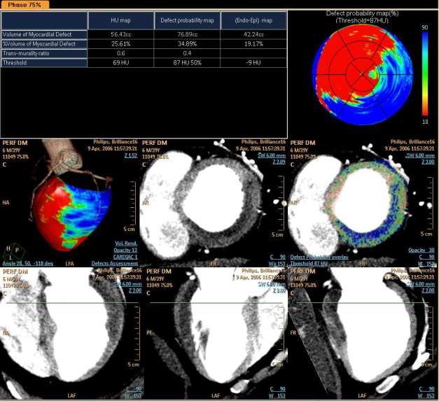
26 Coro-scanner Perfusion

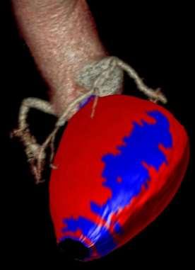
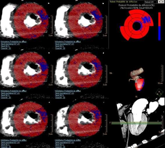
27 Analyse cardiaque Evaluation d un défect de perfusion.

. Vliegenthart R et al.")
28 50-year-old man with recurrent chest pain after prior myocardial infarction in left anterior descending artery territory and surgical revascularization.a, Short-axis images of SPECT (A) and MRI (B) examinations at rest show subendocardial perfusion defect (arrows). Vliegenthart R et al. AJR 2012;199:S54-S by American Roentgen Ray Society


29 50-year-old man with recurrent chest pain after prior myocardial infarction in left anterior descending artery territory and surgical revascularization., Corresponding short-axis cross-section of contrastenhanced dual-energy CT study at rest, reconstructed as merged gray-scale image Vliegenthart R et al. AJR 2012;199:S54-S63 with superimposed iodine distribution color map.
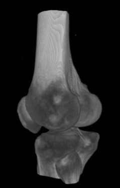
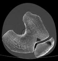
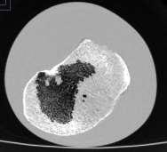
 * AJR 2010;")
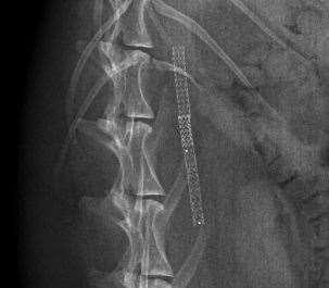
30 Amélioration de la quantification Perfusion Double énergie Correction du beam hardening (paroi postérieure +++) * AJR 2010; 195:

31 Dual-energy CT might be a better way for optimising myocardial and coronary artery imaging Pan et al, International Journal of Cardiology March 2016


32 Spectral Photon Counting CT SPCCT Imaging all the Photons... Common Scintillating Detector Photon counting detector

 Photon")


33 Detector Photo Diode Photo Diode X-Ray Tube Spectrum Dual Energy CT Dual Layer Detector (PHILIPS) Photon counting CT Direct Conversion Detector Low density/high Light Output Scintillator Direct Conversion Detector High Output Scintillator h e h e h e Integrating ASIC Counting ASIC

34 Schematic diagram of energy discrimination
35 Potential Benefits = High Spatial Resolution = K-edge Imaging = Multiple material characterization = Precise energy separation = Low-contrast resolution = Low Dose


36 Biological material: Methods and Materials 8 calcified atherosclerotic plaques 10 lipid-rich atherosclerotic plaques Filled and immerged into an Iodine solution Photon Counting Multi-Energy CT: 70 kev, 20 mas/slice FOV: 60 mm Resolution: 0.1x0.1x0.2 mm 3 Scan time: 200 sec/slice Photon counting CT, Philips, Germany 36

37 Results 37 Significant differences between all elements for Photoelectric absorption and Iodine concentration (p<0.008) No difference for Compton scattering between vessel wall, perivascular fat and lipid-rich plaques

38 Results Iodine map and calcifications CT-like image Iodine map 38 L Boussel Ph Douek, BJR 2014

39 Molecular imaging 39
* For characterization of macrophage")

40 Molecular imaging Gold high-density lipoprotein nanoparticle contrast agent (Au-HDL)* For characterization of macrophage burden, calcification and stenosis of atherosclerotic plaques In Apolipoprotein E knockout mice Comode et al Radiology
: 9635 9639.")
41 Molecular imaging Fibrin using bismuth loaded nanoparticles D Pan et al Computed Tomography in Color: NanoK-Enhanced Spectral CT Angew Chem Int Ed Engl December 10; 49(50):
![Installation of 1 st ict-based preclinical SPCCT prototype @ CERMEP Lyon Parameter Specification Platform Philips ict Tube voltages [kvp] 80, 100, 120 Tube currents](/docs-images/92/108996770/images/42-0.jpg "[ma] 10-100 Focal spot [mmx mm] 0.6 x 0.7 Gantry rotation [s] 1.")
42 Installation of 1 st ict-based preclinical SPCCT CERMEP Lyon Parameter Specification Platform Philips ict Tube voltages [kvp] 80, 100, 120 Tube currents [ma] Focal spot [mmx mm] 0.6 x 0.7 Gantry rotation [s] 1.0 Spatial Resolution [lp/cm] > 20 FOV [mm] 168 # energy bins > 2 Sensor Material Sensor Thickness CZT 2 mm
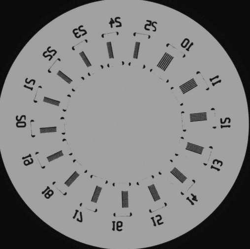

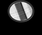

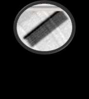
43 Spatial Resolution Line-Pairs plastic phantom Scan of anatomical leg phantom

![Voxel Size: 0.2 x 0.2 x 0.25 [mm] W2000L400 0.2x0.25 R=1.5mm R=3.4mm W2000L400 0.](/docs-images/92/108996770/images/44-1.jpg "2x0.25")
44 Spatial Resolution Stenosis Phantom = Scan Parameter : - Scan Type: Axial (stack) - 120kVp 50mA - Rotation 1sec = Reconstruction: - HU Image - Filter: Standard - Voxel Size: 0.2 x 0.2 x 0.25 [mm] W2000L x0.25 R=1.5mm R=3.4mm W2000L x0.25 Z =5.25 Z =11 Mean:1850 Z=5.25 W2000L x0.2x0.25 Z=11 W2000L x0.2x0.25


45 Spatial Resolution Stent Standard CT SPCCT ww1500 wl470 Voxel Size: 0.1 x 0.1 x 0.1 ww2000 wl800 Voxel Size: 0.1 x 0.1 x 0.1 ww1500 wl470 Voxel Size: 0.1 x 0.1 x 0.1 ww2000 wl800 Voxel Size: 0.1 x 0.1 x 0.1

46 X-Ray Rabbit X-RAY

47 Acquisition In-Vivo SPCCT : Contrast Acquisition : Axial, Z coverage = 2 mm, 120 kvp, 100 ma Reconstruction : FOV 80, Matrix size 1024, pixel size= 0,08, slice thickness 0,1 mm, Detail filter

48 Acquisition In-Vivo SPCCT : Contrast Figure 3: Volume rendering of 2mm coverage with media contrast agent
![Material Decomposition Contrast Specificity Images Axial : 120kVp 100mA HU WL0 WW1000 : SW 2mm : Standard Water [mg/cc] WL790 WW2764 : SW 2mm : Standard Gadolinium [mg/cc]](/docs-images/92/108996770/images/49-0.jpg "WL3.64 WW4.38 : SW 2mm : Smooth Iodine [mg/cc] WL18.28 WW35.26 : SW 2mm : Standard Gold [mg/cc] WL4.76 WW6.")
49 Material Decomposition Contrast Specificity Images Axial : 120kVp 100mA HU WL0 WW1000 : SW 2mm : Standard Water [mg/cc] WL790 WW2764 : SW 2mm : Standard Gadolinium [mg/cc] WL3.64 WW4.38 : SW 2mm : Smooth Iodine [mg/cc] WL18.28 WW35.26 : SW 2mm : Standard Gold [mg/cc] WL4.76 WW6.02 : SW 2mm : Smooth Partnership with: University of Pennsylvania

 injected")
50 Rabbit Gold Contrast HU WL60 WW600 : SW 2mm : Standard Gold WL1.5 WW3.2 : SW 2mm 11.2cc Gold (peg 65mg/cc ) injected 120 kvp 100 ma


51 Rabbit Gadolinium Contrast HU WL60 WW360w : SW2mm Gadolinium [mg/cc] WL580 WW180 : SW2mm 50 seconds after Gadolinium Injection. = Cavity : 2.92 [mg/cc] = Parenchyma: 1.63 [mg/cc]
 (gif) : 30 secondes of acquisition starting")

")
 and past injection of gold (gif) : 30")
52 Rabbit 2 CARDIAC ANGIOGRAPHY Parameters : - Thickness 2 mm - No Filter - L1172; W311 Fig 1 : Cardiac angiography after dynamic injection of Gold (12 ml at 1 cc/s) (gif) : 30 secondes of acquisition starting 5 s after injection, 15 cycles of 2 secondes Parameters : - Thickness 2 mm - Gaussian filter 2 mm Fig 3 : Reconstruction of the Kedge of Gold. Cardiac angiography after dynamic injection of Gold (12 ml at 1 cc/s) (gif) : 30 secondes of acquisition, 15 cycles of 2 secondes Fig 2 : Cardiac angiography after dynamic injection of Iodine (3 ml at 1 cc/s) and past injection of gold (gif) : 30 secondes of acquisition, 15 cycles of 2 secondes Fig 4 : Reconstruction of the Kedge of Gold. Cardiac angiography after dynamic injection of Iodine (3 ml at 1 cc/s) and past injection of gold (gif) : 30 secondes of acquisition, 15 cycles of 2 secondes
 and past injection")
 and past injection of gold")
53 Rabbit 2 DISCRIMINATION GOLD/IODINE Fig 1 : Dynamic angiography during 30 s with reconstruction of Kedge of Gold after injection of iodine (3 ml at 1 cc/s) and past injection of gold (12 ml) (gif) Fig 2 : Dynamic angiography during 30 s with reconstruction of material decomposition of Iodine after injection of iodine (3 ml at 1 cc/s) and past injection of gold (12 ml) (gif)


54 HU Material Decomposition Contrast Specificity Images Applications of Dual Contrast Agents I Gd I Gadolinium Iodine iodine phantom unenhanced polyp Axial : 120kVp 50mA HU: WL-350 WW1400 Gd : WL545 WW45 I : WL690 WW255 gadolinium enhanced polyp Partnership with: Technical University Munich
 injected 120 kvp")
55 Rabbit Gold & Iodine Contrast 3cc Iodine (400 mg/cc) injected 120 kvp 100 ma

56 Rabbit Gold & Iodine Contrast
57 HORIZON 2020 HORIZON 2020 SPCCT


 Quantitative imaging (follow-up)")
 Ex-vivo, pre-clinical (hours, 100 µa) In-vivo")
58 H2020 SPCCT :Lyon university coordinator To develop and validate a widely accessible, new quantitative and analytical imaging technology combining: Spectral Photon Counting Computed Tomography (SPCCT) AND Dedicated Contrast Agents To accurately detect, characterize and monitor neurovascular and cardiovascular disease Ultra-low dose imaging CA dose reduction (reduction of entire scans) Quantitative imaging (follow-up) Functional imaging (K-edge) Higher spatial resolution Clinical (sub-seconds, 1000mA) Ex-vivo, pre-clinical (hours, 100 µa) In-vivo pre-clinical (seconds, 100 ma)


59 Acknowledgements & Collaborators Loic Boussel Daniel Bar-Ness Monica Sigovan Salim Si Mohamed Franck Lavenne Marlene Wiart Yves berthezene Caroline Bouillot Jean-Baptiste Langlois Peter B. Noël David Cormode Frédéric LEROUGE Simon Rit Jean-Michel Létang Gloria Vilches Freixas Philippe Coulon Bracco research Philips Hamburg Philips Haifa

60 60 Thank you
Dual-Energy CT: The Technological Approaches
 Dual-Energy CT: The Technological Approaches Dushyant Sahani, M.D Director of CT Associate Professor of Radiology Massachusetts General Hospital Harvard Medical School Email-dsahani@partners.org Disclosure
An Introduction to Dual Energy Computed Tomography
 An Introduction to Dual Energy Computed Tomography Michael Riedel University of Texas Health Science Center at San Antonio Introduction The idea of computed tomography (CT) was first introduced in the
Les Outils Cliniques de Demain en Scanner Cardiaque. Cardiaque Status en ECR 2018 From Diagnosis to Prognosis
 ECR 2018 From Diagnosis to Prognosis ECR 2018 From Diagnosis to Prognosis Thursday, March 1, 2018/08:30-10:00/Room N Les Outils Cliniques de Demain en Scanner Cardiaque Cardiaque Status en 2018 Rodrigo
Usefulness of New CT Technologies for Interventional Cardiovascular Procedures
 Usefulness of New CT Technologies for Interventional Cardiovascular Procedures Ronen Rubinshtein, MD FACC FESC Department of Cardiovascular Medicine Lady Davis Carmel Medical Center & Technion Israel Institute
Ultrasound. Computed tomography. Case studies. Utility of IQon Spectral CT in. cardiac imaging
 Ultrasound Computed tomography Case studies Utility of IQon Spectral CT in cardiac imaging Cardiac imaging is a challenging procedure where it is necessary to image a motion-free heart. This requires a
Simon Nepveu 1, Irina Boldeanu 1, Yves Provost 1, Jean Chalaoui 1, Louis-Mathieu Stevens 2,3, Nicolas Noiseux 2,3, Carl Chartrand-Lefebvre 1,3
 Coronary Artery Bypass Graft Imaging with CT Angiography and Iterative Reconstruction: Quantitave Evaluation of Radiation Dose Reduction and Image Quality Simon Nepveu 1, Irina Boldeanu 1, Yves Provost
Typical PET Image. Elevated uptake of FDG (related to metabolism) Lung cancer example: But where exactly is it located?
 Lung cancer example: But where exactly is it located?") Typical PET Image Elevated uptake of FDG (related to metabolism) Lung cancer example: But where exactly is it located? PET/CT Oncology Imaging Anatometabolic fusion images are useful in the management
Typical PET Image Elevated uptake of FDG (related to metabolism) Lung cancer example: But where exactly is it located? PET/CT Oncology Imaging Anatometabolic fusion images are useful in the management
Dual-Energy 101: Principles, Methods and Dose
 Dual-Energy 101: Principles, Methods and Dose Juan Carlos Ramirez-Giraldo, Ph.D Staff Scien2st, Collabora2ons Manager SE Region ISCT San Francisco, 2017 Siemens Medical Solu2ons USA, Inc., 2017 Page 1
True Dual Energy. Dr. Stefan Ulzheimer, Siemens Healthcare GmbH. DEfinitely Siemens
 DEfinitely Siemens True Dual Energy Dr. Stefan Ulzheimer, Siemens Healthcare GmbH International version. Not for distribution in the US. Unrestricted Siemens AG 2015 All rights reserved. The products/features
Cardiac Computed Tomography
 Cardiac Computed Tomography Authored and approved by Koen Nieman Stephan Achenbach Francesca Pugliese Bernard Cosyns Patrizio Lancellotti Anastasia Kitsiou Contents CARDIAC COMPUTED TOMOGRAPHY Page 1.
Dual Energy CT of the Heart: Perfusion and Beyond
 Dual Energy CT of the Heart: Perfusion and Beyond U. Joseph Schoepf, MD, FAHA, FSCBT MR, FSCCT Professor of Radiology, Medicine, and Pediatrics Director of Cardiovascular Imaging Disclosures Consultant
X-Ray & CT Physics / Clinical CT
 Computed Tomography-Basic Principles and Good Practice X-Ray & CT Physics / Clinical CT INSTRUCTORS: Dane Franklin, MBA, RT (R) (CT) Office hours will be Tuesdays from 5pm to 6pm CLASSROOM: TIME: REQUIRED
Computed tomography. Department of Radiology, University Medical School, Szeged
 Computed tomography Department of Radiology, University Medical School, Szeged voxel +1-4 +2 +5 +3 +1 0-2 pixel -2 0 +1-4 -6 +5 +2 +1 Department of Radiology, University Medical School, Szeged
A Snapshot on Nuclear Cardiac Imaging
 Editorial A Snapshot on Nuclear Cardiac Imaging Khalil, M. Department of Physics, Faculty of Science, Helwan University. There is no doubt that nuclear medicine scanning devices are essential tool in the
Alessandro Albonico Philips
 Alessandro Albonico Philips Alessandro.albonico@philips.com Noise (Standard Deviation in HU) Virtually noise-free Characteristic of a true knowledge-based IR 80 70 Standard Recon idose4 Level6 1 mm Slice
Dual Energy CT of the Liver
 34th Annual Course October 2011 Washington, DC Dual Energy CT of the Liver Vassilios Raptopoulos, MD Beth Israel Deaconess Medical Center Harvard Medical School Dual Energy CT (DECT) Different materials
CT Perfusion. U. Joseph Schoepf, MD, FAHA, FSCBT MR, FSCCT Professor of Radiology, Medicine, and Pediatrics Director of Cardiovascular Imaging
 CT Perfusion U. Joseph Schoepf, MD, FAHA, FSCBT MR, FSCCT Professor of Radiology, Medicine, and Pediatrics Director of Cardiovascular Imaging Disclosures Consultant for / research support from Bayer Bracco
Combined Anatomical and Functional Imaging with Revolution * CT
 GE Healthcare Case studies Combined Anatomical and Functional Imaging with Revolution * CT Jean-Louis Sablayrolles, M.D. Centre Cardiologique du Nord, Saint-Denis, France Case 1 Whole Brain Perfusion and
New Technologies for Cardiac CT. Geoffrey D. Rubin, MD, MBA, FACR, FNASCI Duke University
 1996 New Technologies for Cardiac CT Geoffrey D. Rubin, MD, MBA, FACR, FNASCI Duke University New Technology The Long View Levels of Efficacy Endpoint Examples 1: Technical Imaging resolution 2: Diagnostic
Cardiac Imaging Tests
 Cardiac Imaging Tests http://www.medpagetoday.com/upload/2010/11/15/23347.jpg Standard imaging tests include echocardiography, chest x-ray, CT, MRI, and various radionuclide techniques. Standard CT and
BioMedical quantitative X-Ray Imaging. Emmanuel Brun Researcher Inserm Université Grenoble Alpes
 BioMedical quantitative X-Ray Imaging Emmanuel Brun Researcher Inserm Université Grenoble Alpes 1 Outline Introduction K-Edge Imaging Patient imaging at the European synchrotron Medical Phase Contrast
Coronary Artery Calcification
 Coronary Artery Calcification Julianna M. Czum, MD OBJECTIVES CORONARY ARTERY CALCIFICATION Julianna M. Czum, MD Dartmouth-Hitchcock Medical Center 1. To review the clinical significance of coronary heart
Quantitative Imaging of Transmural Vasa Vasorum Distribution in Aortas of ApoE -/- /LDL -/- Double Knockout Mice using Nano-CT
 Quantitative Imaging of Transmural Vasa Vasorum Distribution in Aortas of ApoE -/- /LDL -/- Double Knockout Mice using Nano-CT M. Kampschulte 1, M.D.; A. Brinkmann 1, M.D.; P. Stieger 4, M.D.; D.G. Sedding
CT Myocardial Perfusion: Is there Added Value to Coronary CT?
 CT Myocardial Perfusion: Is there Added Value to Coronary CT? U. Joseph Schoepf, MD, FAHA, FSCBT MR, FSCCT Professor of Radiology, Medicine, and Pediatrics Director of Cardiovascular Imaging Disclosures
University of Groningen. Quantitative CT myocardial perfusion Pelgrim, Gert
 University of Groningen Quantitative CT myocardial perfusion Pelgrim, Gert IMPORTANT NOTE: You are advised to consult the publisher's version (publisher's PDF) if you wish to cite from it. Please check
Cardiac CT - Coronary Calcium Basics Workshop II (Basic)
") Cardiac CT - Coronary Calcium Basics Workshop II (Basic) J. Jeffrey Carr, MD, MSCE Dept. of Radiology & Public Health Sciences Wake Forest University School of Medicine Winston-Salem, NC USA No significant
Cardiac CT - Coronary Calcium Basics Workshop II (Basic) J. Jeffrey Carr, MD, MSCE Dept. of Radiology & Public Health Sciences Wake Forest University School of Medicine Winston-Salem, NC USA No significant
Dual-Energy Imaging of Bone Marrow Edema on a Dedicated Multi-Source Cone-Beam CT System for the Extremities
 Dual-Energy Imaging of Bone Edema on a Dedicated Multi-Source Cone-Beam CT System for the Extremities W Zbijewski, 1 A Sisniega, 1 JW Stayman, 1 N Packard, 2 J Yorkston, 2 G Thawait, 3 S Demehri, 3 J Fritz,
General Cardiovascular Magnetic Resonance Imaging
 2 General Cardiovascular Magnetic Resonance Imaging 19 Peter G. Danias, Cardiovascular MRI: 150 Multiple-Choice Questions and Answers Humana Press 2008 20 Cardiovascular MRI: 150 Multiple-Choice Questions
Dual-Energy CT Applications in Radiation Therapy
 THE UNIVERSITY OF WISCONSIN MADISON Dual-Energy CT Applications in Radiation Therapy - Jessica Miller 1 Disclosures Funding provided by Siemens Medical 2 Learning objectives General principles of dual
Adapted Transfer Function Design for Coronary Artery Evaluation
 Adapted Transfer Function Design for Coronary Artery Evaluation Sylvia Glaßer 1, Steffen Oeltze 1, Anja Hennemuth 2, Skadi Wilhelmsen 3, Bernhard Preim 1 1 Department of Simulation and Graphics, University
Integrated PET/CT systems State of the art and Clinical Applications
 Integrated PET/CT systems State of the art and Clinical Applications V. Bettinardi Nuclear Medicine Dep. Scientific Institute San Raffaele Hospital Milan Italy The announcement of a new Diagnostic Imaging
Low-dose and High-resolution Cardiac Imaging with Revolution CT
 GE Healthcare Case study Low-dose and High-resolution Cardiac Imaging with Revolution CT Prof. Philipp A. Kaufmann, M.D. Ronny R. Buechel, M.D. Fran Mikulicic, M.D. Dominik C. Benz, M.D. University of
Coronary Artery Imaging. Suvipaporn Siripornpitak, MD Inter-hospital Conference : Rajavithi Hospital
 Coronary Artery Imaging Suvipaporn Siripornpitak, MD Inter-hospital Conference : Rajavithi Hospital Larger array : cover scan area Detector size : spatial resolution Rotation speed : scan time Retrospective
Cone-Beam CT for MSK Extremities
 8/6/0 Diagnostic Image Quality Evaluation of an Extremity Cone-Beam CT Scanner: Pre-Clinical and First Clinical Results Abdullah Muhit Wojciech Zbijewski, J Webster Stayman John Yorkston, Nathan Packard,
Cardiac CTA Prospective Gating Broad Beam
 Cardiac CTA Prospective Gating Broad Beam ACQUISITION- Broad Beam Gating: Prospective Non Contrast Scan-Calcium Score Patient Position Supine Feet First into Gantry Heart Isocenter Scanogram AP and Lateral
State-of-the-Art SPECT/CT: Cardiac Imaging
 State-of-the-Art SPECT/CT: Cardiac Imaging Ernest V Garcia*, PhD Endowed Professor in Cardiac Imaging Director, Nuclear Cardiology R&D Laboratory Disclosure: Dr. Garcia receives royalties from the sale
Toshiba Aquillion 64 CT Scanner. Phantom Center Periphery Center Periphery Center Periphery
 Comparison of radiation dose and imaging performance for the standard Varian x-ray tube and the Richardson Healthcare ALTA750 replacement tube for the Toshiba Aquillion CT scanners. by Robert L. Dixon,
Cardiac CT Lowering the Dose Dramatically
 Cardiac CT Lowering the Dose Dramatically U. Joseph Schoepf, MD, FAHA, FSCBT MR, FSCCT Professor of Radiology, Medicine, and Pediatrics Director of Cardiovascular Imaging Disclosures Consultant for / research
CT Myocardial Perfusion
 1 CT Myocardial Perfusion Ting-Yim Lee, PhD, FCCPM, FCOMP Aaron So, PhD, FSCCT Gerald Wisenberg, MD, FRCPC Ali Islam, MD, FRCPC Lawson Health Research Institute Robarts research Institute The University
Dual Energy CT Aortography: Can We Reduce Iodine Dose??
 Dual Energy CT Aortography: Can We Reduce Iodine Dose?? William P. Shuman MD, FACR FSCBTMR Department of Radiology University of Washington SCBTMR Annual Course Boston, October 10, 2012 Conflict of Interest
Is Coronary Stent Assessment Improved with Spectral Analysis of Dual Energy CT? 1
 Is Coronary Stent Assessment Improved with Spectral Analysis of Dual Energy CT? 1 Ethan J. Halpern, MD, David J. Halpern, Jeffrey H. Yanof, PhD, Sigal Amin-Spector, PhD, David Fischman, MD, Galit Aviram,
Triple Rule-out using 320-row-detector volume MDCT: A comparison of the wide volume and helical modes
 Triple Rule-out using 320-row-detector volume MDCT: A comparison of the wide volume and helical modes Poster No.: C-0488 Congress: ECR 2012 Type: Authors: Keywords: DOI: Scientific Exhibit E.-J. Kang,
Translating Protocols Across Patient Size: Babies to Bariatric
 Translating Protocols Across Patient Size: Babies to Bariatric Cynthia H. McCollough, PhD, FACR, FAAPM Professor of Radiologic Physics Director, CT Clinical Innovation Center Department of Radiology Mayo
Optical Coherence Tomography
 Optical Coherence Tomography Disclosure Information Demetrius Lopes MD The following relationships exist related to this presentation: University Grant/Research Support: Rush University Industry Grant
Disclosure Information
 Coronary CTA Pearls and Pitfalls Ricardo C. Cury, MD, FSCCT, FAHA, FACC Chairman of Radiology Radiology Associates of South Florida Director of Cardiac Imaging Miami Cardiac and Vascular Institute Past-President
RADIATION PROTECTION IN DIAGNOSTIC AND INTERVENTIONAL RADIOLOGY. L19: Optimization of Protection in Mammography
 IAEA Training Material on Radiation Protection in Diagnostic and Interventional Radiology RADIATION PROTECTION IN DIAGNOSTIC AND INTERVENTIONAL RADIOLOGY L19: Optimization of Protection in Mammography
Neuro CT What s a Good Head Exam?
 Neuro CT What s a Good Head Exam? Rajiv Gupta, PhD, MD Neuroradiology Massachusetts General Hospital Harvard Medical School Outline What we need to see? Routine Head CT protocols Dose optimization strategies
Validation of CT Perfusion Imaging Against Invasive Angiography and FFR on a 320-MDCT Scanner
 Validation of CT Perfusion Imaging Against Invasive Angiography and FFR on a 320-MDCT Scanner Zhen Qian, Gustavo Vasquez, Sarah Rinehart, Parag Joshi, Eric Krivitsky, Anna Kalynych, Dimitri Karmpaliotis,
Biases affecting tumor uptake measurements in FDG-PET
 Biases affecting tumor uptake measurements in FDG-PET M. Soret, C. Riddell, S. Hapdey, and I. Buvat Abstract-- The influence of tumor diameter, tumor-tobackground activity ratio, attenuation, spatial resolution,
THE TUFFEST STUFF CT REGISTRY REVIEW Live Lecture Seminar SATURDAY CURRICULUM
 1. The CT Imaging Chain-10 major components & their functions a. The x-ray tube b. Generator c. Filter d. Pre-patient collimator e. Pre-detector collimator f. Detector system g. Analog to digital converter
Simulations of Preclinical andclinical Scans in Emission Tomography, Transmission Tomography and Radiation Therapy. Using GATE
 GATE Simulations of Preclinical andclinical Scans in Emission Tomography, Transmission Tomography and Radiation Therapy Using GATE Quick tour & Highlights! GATE Training, INSTN-Saclay, October 2015 Albertine
Radiation Dose Reduction: Should You Use a Bismuth Breast Shield?
 Radiation Dose Reduction: Should You Use a Bismuth Breast Shield? Lincoln L. Berland, M.D., F.A.C.R. Michael V. Yester, Ph.D. University of Alabama at Birmingham Breast Radiation on CT Use of chest CT
8/3/2016. Consultant for / research support from: Astellas Bayer Bracco GE Healthcare Guerbet Medrad Siemens Healthcare. Single Energy.
 U. Joseph Schoepf, MD Prof. (h.c.), FAHA, FSCBT-MR, FNASCI, FSCCT Professor of Radiology, Medicine, and Pediatrics Director, Division of Cardiovascular Imaging Consultant for / research support from: Astellas
Computed tomography Acceptance testing and dose measurements
 Computed tomography Acceptance testing and dose measurements Jonas Andersson Medical Physicist, Ph.D. Department of Radiation Sciences University Hospital of Norrland, Umeå Sweden Contents The Computed
Perspectives of new imaging techniques for patients with known or suspected coronary artery disease
 Perspectives of new imaging techniques for patients with known or suspected coronary artery disease Department of Cardiology, Leiden University Medical Center, Leiden, The Netherlands Correspondence: Jeroen
Computed Tomography of the Coronary Arteries
 Cardiology Update DAVOS 2011 Computed Tomography of the Coronary Arteries Anders Persson M.D., Ph.D Director, Assoc. Professor Center for Medical Image Science and Visualization Linköping University SWEDEN
Correlation of Cardiac CTA to Conventional Cardiac Angiography in Diagnosing Coronary Artery Stenosis in a Community Based Center
 Correlation of Cardiac CTA to Conventional Cardiac Angiography in Diagnosing Coronary Artery Stenosis in a Community Based Center Mathieu Sabbagh, R3 Michigan State University Radiology Garden City Hospital
SOMATOM Drive System Owner Manual Dosimetry and imaging performance report
 www.siemens.com/healthcare SOMATOM Drive System Owner Manual Dosimetry and imaging performance report Table of contents 1 Dosimetry and imaging performance report 5 1.1 Dose information 5 1.1.1 General
Radiation Dose Reduction Strategies in Coronary CT Angiography
 Radiation Dose Reduction Strategies in Coronary CT Angiography Noor Diyana Osman, PhD noordiyana@usm.my Contents: Introduction Radiation dosimetry in CT Radiation risk associated with coronary CT angiography
Radiation Detection and Measurement
 Radiation Detection and Measurement Range of charged particles (e.g.,!: µm; ": mm) Range of high energy photons (cm) Two main types of interactions of high energy photons Compton scatter Photoelectric
Photon Attenuation Correction in Misregistered Cardiac PET/CT
 Photon Attenuation Correction in Misregistered Cardiac PET/CT A. Martinez-Möller 1,2, N. Navab 2, M. Schwaiger 1, S. G. Nekolla 1 1 Nuklearmedizinische Klinik der TU München 2 Computer Assisted Medical
Low Dose Era in Cardiac CT
 Low Dose Era in Cardiac CT DIANA E. LITMANOVICH, MD Department of Radiology Beth Israel Deaconess Medical Center Harvard Medical School Disclosures Neither I nor my immediate family members have a financial
CURRENT CT DOSE METRICS: MAKING CTDI SIZE-SPECIFIC
 CURRENT CT DOSE METRICS: MAKING CTDI SIZE-SPECIFIC Keith Strauss, MSc, FAAPM, FACR Cincinnati Children s Hospital University of Cincinnati College of Medicine Acknowledgments John Boone, PhD Michael McNitt-Grey,
CVIA. Dual-Layer Computed Tomography in Cardiovascular Imaging INTRODUCTION
 CVIA REVIEW ARTICLE pissn 2508-707X / eissn 2508-7088 https://doi.org/10.22468/cvia.2018.00066 CVIA 2018;2(2):49-57 Received: March 15, 2018 Revised: April 2, 2018 Accepted: April 9, 2018 Corresponding
Fundamentals, Techniques, Pitfalls, and Limitations of MDCT Interpretation and Measurement
 Fundamentals, Techniques, Pitfalls, and Limitations of MDCT Interpretation and Measurement 3 rd Annual Imaging & Physiology Summit November 20-21, 21, 2009 Seoul, Korea Wm. Guy Weigold, MD, FACC Cardiovascular
How do the Parameters affect Image Quality and Dose for Abdominal CT? Image Review
 How do the Parameters affect Image Quality and Dose for Abdominal CT? Image Review Mannudeep K. Kalra, MD, DNB Massachusetts General Hospital Harvard Medical School Financial Disclosure This presentation
CT Imaging of Atherosclerotic Plaque. William Stanford MD Professor-Emeritus Radiology University of Iowa College of Medicine Iowa City, IA
 CT Imaging of Atherosclerotic Plaque William Stanford MD Professor-Emeritus Radiology University of Iowa College of Medicine Iowa City, IA PREVALENCE OF CARDIOVASCULAR DISEASE In 2006 there were 80 million
A new method for radiation dose reduction at cardiac CT with multi-phase data-averaging and non-rigid image registration: preliminary clinical trial
 A new method for radiation dose reduction at cardiac CT with multi-phase data-averaging and non-rigid image registration: preliminary clinical trial Poster No.: C-0595 Congress: ECR 2013 Type: Authors:
COMPUTED TOMOGRAPHY COURSE
 CT Radiography RAD 421 4 th year semester 2 Course Lecture Tutorial Practical Credit hours CT Radiography 2-1 2 Course Description The course explores the basic physical and technical principles of CT
Ultralow Dose Chest CT with MBIR
 Ultralow Dose Chest CT with MBIR Ella A. Kazerooni, M.D. Professor & Director Cardiothoracic Radiology Associate Chair for Clinical Affairs University of Michigan Disclosures Consultant: GE Healthcare
Biomarkers and the Future of. John R. Votaw CBIS 5 th Year Anniversary Celebration/Look to the future February 8, 2013
 Biomarkers and the Future of Radiology John R. Votaw CBIS 5 th Year Anniversary Celebration/Look to the future February 8, 2013 Statistics/Radiology Collaboration The utility of Radiologic procedures
Pushing the limits of cardiac CT. Steven Dymarkowski Radiology / Medical Imaging Research Centre
 Pushing the limits of cardiac CT Steven Dymarkowski Radiology / Medical Imaging Research Centre 5 X 2013 Introduction Rapid technological advances and new clinical applications in cardiovascular imaging
Fused monochromatic imaging acquired by single source dual energy CT in hepatocellular carcinoma during arterial phase: an initial experience
 Original Article Fused monochromatic imaging acquired by single source dual energy CT in hepatocellular carcinoma during arterial phase: an initial experience Shun-Yu Gao, Xiao-Peng Zhang, Yong Cui, Ying-Shi
Medical Imaging. Alex Elliott Western Infirmary Glasgow
 Medical Imaging Alex Elliott Western Infirmary Glasgow History of medical imaging X-rays - Roentgen, 1895 Nuclear medicine - Cassen, 1951 Ultrasound Donald, 1962 SPECT - Kuhl, Edwards, 1963 PET Ter-Pogossian,
Implementation of the 2012 ACR CT QC Manual in a Community Hospital Setting BRUCE E. HASSELQUIST, PH.D., DABR, DABSNM ASPIRUS WAUSAU HOSPITAL
 Implementation of the 2012 ACR CT QC Manual in a Community Hospital Setting BRUCE E. HASSELQUIST, PH.D., DABR, DABSNM ASPIRUS WAUSAU HOSPITAL Conflict of Interest Disclaimer Employee of Aspirus Wausau
Managing Radiation Risk in Pediatric CT Imaging
 Managing Radiation Risk in Pediatric CT Imaging Mahadevappa Mahesh, MS, PhD, FAAPM, FACR, FACMP, FSCCT. Professor of Radiology and Cardiology Johns Hopkins University School of Medicine Chief Physicist
Cardiac CT Techniques in Neonates (and infants)
") Cardiac CT Techniques in Neonates (and infants) Siddharth P. Jadhav, MD Director, Body CT and MRI Edward B. Singleton Department of Pediatric Radiology Texas Children s Hospital Disclosures None Objectives
Cardiac CT Techniques in Neonates (and infants) Siddharth P. Jadhav, MD Director, Body CT and MRI Edward B. Singleton Department of Pediatric Radiology Texas Children s Hospital Disclosures None Objectives
Bone Densitometry Radiation dose: what you need to know
 Bone Densitometry Radiation dose: what you need to know John Damilakis, PhD Associate Professor and Chairman University of Crete, Iraklion, Crete, GREECE Estimation of bone status using X-rays Assessment
Breast CT and Dosimetry
 2013 ICTP/IAEA Training Course on Radiation Protection of Patients Trieste Breast CT and Dosimetry John M. Boone, Ph.D., FAAPM, FSBI, FACR Professor and Vice Chair (Research) of Radiology Professor of
CARDIAC IMAGING FOR SUBCLINICAL CAD
 CARDIAC IMAGING FOR SUBCLINICAL CAD WHY DON'T YOU ADOPT MORE SMART TECHNIQUE? Whal Lee, M.D. Seoul National University Hospital Department of Radiology We are talking about Coronary artery Calcium scoring,
Introduction. Cardiac Imaging Modalities MRI. Overview. MRI (Continued) MRI (Continued) Arnaud Bistoquet 12/19/03
 MRI (Continued) Arnaud Bistoquet 12/19/03") Introduction Cardiac Imaging Modalities Arnaud Bistoquet 12/19/03 Coronary heart disease: the vessels that supply oxygen-carrying blood to the heart, become narrowed and unable to carry a normal amount
Introduction Cardiac Imaging Modalities Arnaud Bistoquet 12/19/03 Coronary heart disease: the vessels that supply oxygen-carrying blood to the heart, become narrowed and unable to carry a normal amount
GE Healthcare. Rad Rx. White Paper
 GE Healthcare Rad Rx White Paper Introduction This publication is part of a series of white papers aimed at communicating the importance of each component in the image chain of a PET/CT study. From data
Fellows on this rotation are expected to attend nuclear conferences and multimodality imaging conference.
 Rotation: Imaging 1 Imaging 1 provides COCATS Level 1 experience for nuclear cardiology (including SPECT and PET) and cardiac CT. Fellows will administer, process, and read cardiac nuclear studies with
Modifi ed CT perfusion contrast injection protocols for improved CBF quantifi cation with lower temporal sampling
 Investigations and research Modifi ed CT perfusion contrast injection protocols for improved CBF quantifi cation with lower temporal sampling J. Wang Z. Ying V. Yao L. Ciancibello S. Premraj S. Pohlman
Evidence for myocardial CT perfusion imaging in the diagnosis of hemodynamically significant coronary artery disease
 Editorial Evidence for myocardial CT perfusion imaging in the diagnosis of hemodynamically significant coronary artery disease Zhonghua Sun Discipline of Medical Imaging, Department of Imaging and Applied
Role of DE-CT in Oncology
 Role of DE-CT in Oncology Dushyant Sahani, M.D Director of CT Associate Professor of Radiology Massachusetts General Hospital Harvard Medical School Email-dsahani@partners.org Disclosure Research Grant
Cardiovascular Imaging
 Cardiovascular Imaging Cardiovascular Imaging Cardio and Vascular Imaging Vascularization / Angiogenesis Cardiovascular Imaging metabolic imaging of the heart myocardial perfusion imaging Cardiac CT Vascularization
Coronary angiography is the standard way of visualizing
 Clinical Investigation and Reports Coronary Artery Fly-Through Using Electron Beam Computed Tomography Peter M.A. van Ooijen, MSc; Matthijs Oudkerk, MD, PhD; Robert J.M. van Geuns, MD; Benno J. Rensing,
Diagnostic and Prognostic Value of Coronary Ca Score
 Diagnostic and Prognostic Value of Coronary Ca Score Dr. Ghormallah Alzahrani Cardiac imaging division, Adult Cardiology department Prince Sultan Cardiac Center ( PSCC) Madina, June 2 Coronary Calcium
The Final 10-Year Follow-up Results from the Bari Randomized Trial J Am Coll Cardiol (2007) 49;1600-6
 49;1600-6") The Final 10-Year Follow-up Results from the Bari Randomized Trial J Am Coll Cardiol (2007) 49;1600-6 n&list_uids=17433949 64-Multislice Detector Computed Tomography Coronary Angiography as Potential Alternative
The Final 10-Year Follow-up Results from the Bari Randomized Trial J Am Coll Cardiol (2007) 49;1600-6 n&list_uids=17433949 64-Multislice Detector Computed Tomography Coronary Angiography as Potential Alternative
Acute Management of Pulmonary Embolism
 Acute Management of Pulmonary Embolism Dr Alex West Respiratory Consultant Guy s and St Thomas Hospital London Declarations - none Order of Play Up date in Diagnostic Imaging - CTPA and V:Q SPECT Sub-massive
Giorgio Ascenti Angelo Vanzulli Carlo Catalano Rendon C. Nelson. CT of the. Retroperitoneum. From Conventional to Multi-energy Imaging
 CT of the Giorgio Ascenti Angelo Vanzulli Carlo Catalano Rendon C. Nelson Retroperitoneum From Conventional to Multi-energy Imaging 123 CT of the Retroperitoneum Giorgio Ascenti Angelo Vanzulli Carlo Catalano
SPECT or PET for Cardiovascular Screening in High-Risk Patients
 SPECT or PET for Cardiovascular Screening in High-Risk Patients Paeng, Jin Chul MD PhD Department of Nuclear Medicine Seoul National University Hospital Contents Recent Development in SPECT and PET Technology
Anthem Blue Cross and Blue Shield Virginia Advanced Imaging Procedures Requiring Precertification Revised 02/13/2013
 Anthem Blue Cross and Blue Shield Virginia Advanced Imaging Procedures Requiring Precertification Revised 02/13/2013 Modality and CT Head CTA Head: Cerebrovascular MRI Head MRA Head: Cerebrovascular Functional
Using Coronary Artery Calcium Score in the Quest for Cardiac Health. Robert J. Hage, D.O.
 Using Coronary Artery Calcium Score in the Quest for Cardiac Health Robert J. Hage, D.O. Heart disease is the leading cause of death in the United States in both men and women. About 610,000 people die
Why is CT Dose of Interest?
 Why is CT Dose of Interest? CT usage has increased rapidly in the past decade Compared to other medical imaging CT produces a larger radiation dose. There is direct epidemiological evidence for a an increase
ESTABLISHING DRLs in PEDIATRIC CT. Keith Strauss, MSc, FAAPM, FACR Cincinnati Children s Hospital University of Cincinnati College of Medicine
 ESTABLISHING DRLs in PEDIATRIC CT Keith Strauss, MSc, FAAPM, FACR Cincinnati Children s Hospital University of Cincinnati College of Medicine CT Dose Indices CTDI INTRODUCTION CTDI 100, CTDI w, CTDI vol
Radiology Review Course Hotel del Coronado Coronado, California
 37 th Annual Radiology Review Course Hotel del Coronado Coronado, California Saturday, April 22, 2017 - AM TABLE OF CONTENTS Saturday, April 22, 2017 - AM 7:00 AM 7:30 AM Coffee and Pastries for Registrants
An Epidemic of Heart Disease The Silent Killer. Professor Mike Kirby FRCP University of Hertfordshire Institute of Diabetes for Older People
 An Epidemic of Heart Disease The Silent Killer Professor Mike Kirby FRCP University of Hertfordshire Institute of Diabetes for Older People Initial clinical presentation of CAD Angina MI Atypical CP Death
SPECIFIC PRINCIPLES FOR DOSE REDUCTION IN HEAD CT IMAGING. Rajiv Gupta, MD, PhD Neuroradiology, Massachusetts General Hospital Harvard Medical School
 SPECIFIC PRINCIPLES FOR DOSE REDUCTION IN HEAD CT IMAGING Rajiv Gupta, MD, PhD Neuroradiology, Massachusetts General Hospital Harvard Medical School OUTLINE 1 st Presentation: Dose optimization strategies