Neuro CT What s a Good Head Exam?
|
|
|
- Albert Stewart
- 5 years ago
- Views:
Transcription


1 Neuro CT What s a Good Head Exam? Rajiv Gupta, PhD, MD Neuroradiology Massachusetts General Hospital Harvard Medical School
2 Outline What we need to see? Routine Head CT protocols Dose optimization strategies Some newer tricks using Dual Energy CT


3 58 year old, new onset of neurologic symptoms Where is the Abnormality? CT: Equivocal for an infarct


4 Acute Infarct in the ACA territory CT Angiography CTA SI: Reduced DWI perfusion
5 Requirements Good gray-white differentiation (8-10 HU)

6 Different Case: Young patient after trauma
7 Requirements Good gray-white differentiation Proper cupping correction


8 11 month old female that presented with gagging after trauma to oropharynx with drum stick.
9 Diagnosis Retropharyngeal abscess Etiology Suppurative bacterial lymphadenitis S. aureus, Strep B, oral flora Foreign body perforation Trauma
10 Requirements Good gray-white differentiation Proper cupping correction Good soft tissue discrimination


11 44 year old fell from a tree

12 2 months later

13 Surgical Specimen Take-home Points: - Wood may appear as air - Accurate HU calibration is important


14 Another Case: Pencil vs Globe
15 Requirements Good gray-white differentiation Proper cupping correction Good soft tissue discrimination Accurate, reliable HU calibration

16 History of Trauma Subtle right occipital fracture, only visible on thin slices, and sharp kernel
17 Requirements Good gray-white differentiation Proper cupping correction Good soft tissue discrimination Accurate, reliable HU calibration High spatial resolution and MTF


18 4 year old with 2 week history of headaches, abdominal pain, anorexia and vomiting. Recent antibiotics for sinusitis. Artifact or Pathology?


19 Imaging 1 Week Later CT w/ Contrast T1 Post


20 Pathology: Burkitt Lymphoma B-cell lymphoma


21 Beam Hardening Effective beam energy increases. Beam hardens as it penetrates. Higher E => lower. Cupping artifact thickness
22 Requirements Good gray-white differentiation Proper cupping correction Good soft tissue discrimination Accurate, reliable HU calibration High spatial resolution and MTF Artifact-free posterior fossa and skull base
23 The Chest X-ray of the Brain Must support quick, confident read. Main Culprits Poor SNR Poor CNR Poor spatial resolution Improper recon kernel Improper protocol Artifacts Motion Scatter Beam hardening Windmill

24 Sample MGH 64-slice Head CT Protocol (Minor Variations between Scanners) Helical, 120kV, 120kV, 250mA, Auto-mA, Pitch 0.5, Pitch ST 1.25, 0.5, interval ST 1.25, interval 0.625
25 CT Dose Reduction Strategies Use another modality! Optimize acquisition Tailor to clinical requisition Lower technique Minimize artifacts Dual Energy acquisition Optimize reconstruction / post-processing FBP vs newer iterative reconstruction kernals Filters, e.g. metal streak reduction Optimize readout Coronal Reformats Optimal PACS display Appropriate window/level settings Michael Lev, MGH
26 Optimizing Dose: Basic Principles Lowest possible mas is proportional to: Degree of intrinsic tissue contrast Acceptable level of image noise Noise ~ 1 / SQRT (mas) Tailor the protocol to the clinical question E.g.: 30 mas for sinus CT, FESS planning Mulkens et al, AJR May 2005 Loubele et al, Radiat Prot Dosimetry 2005 E.g.: 30 mas for pituitary CT, transphenoidal sx Michael Lev, MGH

 Mullins, Lev, et al.")
27 50% mas Reduction? Slightly Noisier, but OK for F/U 170 ma 90 ma Dept wide study ma by 50% for all CT s Unchanged HU, GW conspicuity 22% decreased CNR (attributable to noise) Mullins, Lev, et al. Comparison of image quality between conventional and low dose NCCT. AJNR, Apr 2004.
28 Optimizing Dose: Adaptive ma modulation Varies ma both in radial and axial direction Substantial dose reductions have been reported % decrease depends on baseline protocol Smith, Dillon, Wintermark et al. Radiology 2008 More useful for neck than head, in our experience Wide range of thickness in shoulders Noise index values of 11.4 and 20.2, result in 20% and 34% dose reduction, respectively Russell, Anzai et al, Seattle. AJNR 2008
29 Optimizing Dose: Other Considerations Lower kv Increased photoelectric effect Higher HU iodine Avoid rescanning same region E.g., head and temporal bone, face and sinuses (? billing) Maximize quality parameters Decrease motion artifact: speed, sedation Remove extraneous hardware Optimize contrast bolus; right sided Angle gantry though clips, fillings Brown, Lustrin, Lev, Taveras et al. AJR 1999
 LOW DOSE + FBP LOW DOSE + 100% Improved Image Quality, Lower Dose Courtesy of Shervin Kamalian and GE")
30 Technology Assessment Institute: Summit on CT Dose Iterative Reconstruction Algorithms ASIR (GE), IRIS, SAFIRE (Siemens): (MBIR --- Model Based Iterative Recon) LOW DOSE + FBP LOW DOSE + 100% Improved Image Quality, Lower Dose Courtesy of Shervin Kamalian and GE Healthcare


31 Sample CT Dose Reduction at 30% ASIR H E A D S P I N E
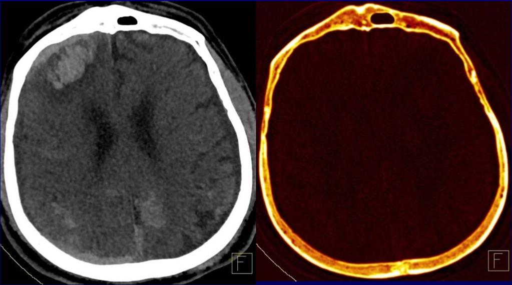
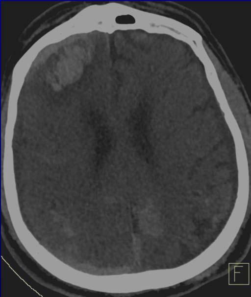
32 Single vs Dual Energy CT A single CT Number (HU) Prior knowledge for material separation Unable to distinguish materials with same HU Blood vs. dilute contrast Blood vs. diffuse mineralization Uric acid vs. Ca oxalate Calcification vs. gouty tophus

33 Dual Energy Principles Total attenuation decreases with increasing energy Decrease is characteristic for each material Depends on photon energy and material density X-ray absorption depends on the inner electron shells DECT is sensitive to atomic number and density DECT is not sensitive to chemical binding C. Leidecker, Siemens
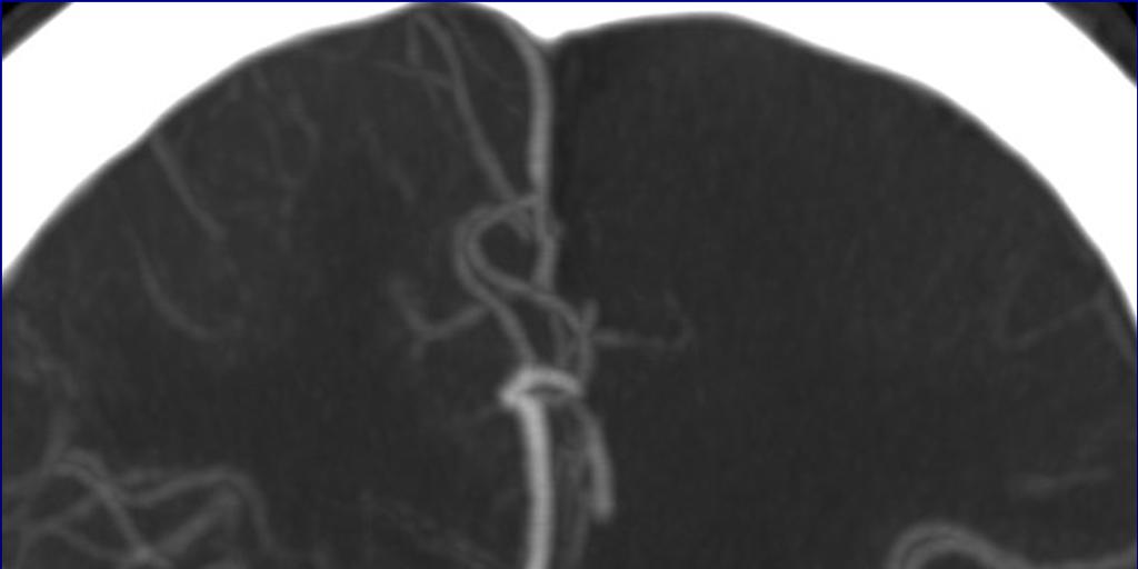
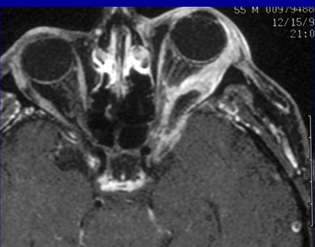
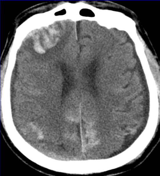
34 Dual Energy Systems Siemens: Dual Source CT (Definition) Two X-ray sources, two detectors, simultaneous acquisition Operate one source at 80kV and the other at 140kV General Electric: Discovery Gemstone (HD-750) Single Source, single detector, on a fast gantry Rapidly alternate the single tube between 80kV and 140kV 140kV 80kV
 Projections close to each other in time => separation in projection domain Tube current constant (i.e., not optimized for each kv).")
35 Siemens: Dual Source CT (Definition) Simultaneous acquisition Optimized tube current Projections: 90 deg apart One detector smaller than other 140kV DECT: Pro and Cons 80kV General Electric: Discovery Gemstone (HD-750) Projections close to each other in time => separation in projection domain Tube current constant (i.e., not optimized for each kv).
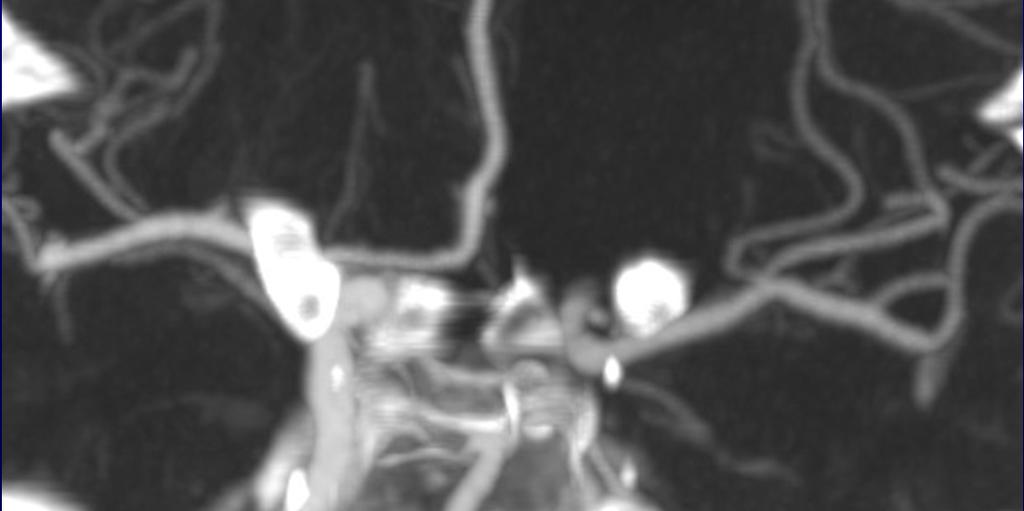
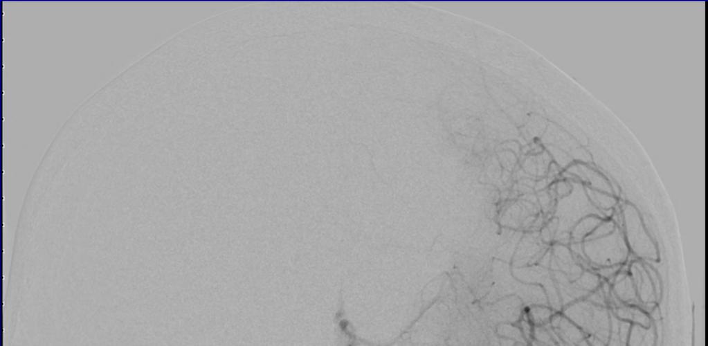
36 Clinical Case 79 yo man with acute onset of right hemiparesis and aphasia Received IV t-pa at OSH Transferred to MGH for further management At MGH, CTA showed: - emboli in the distal left ICA - occlusion of left M1
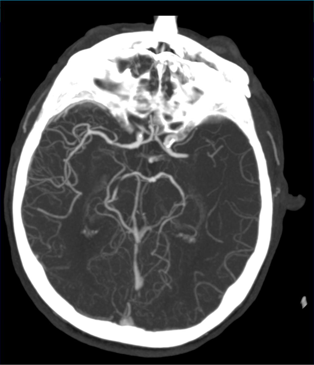
37
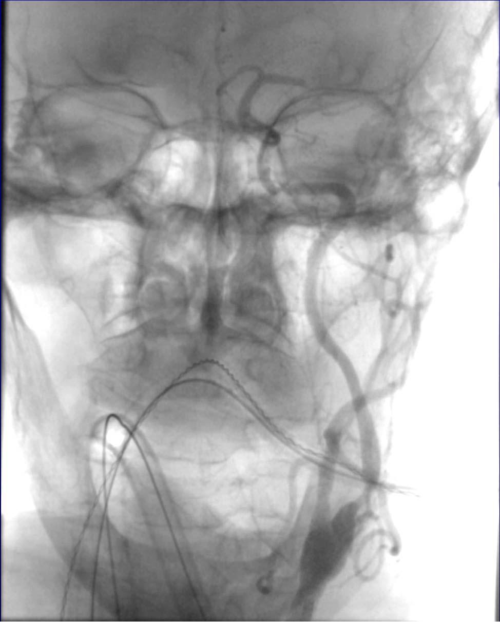
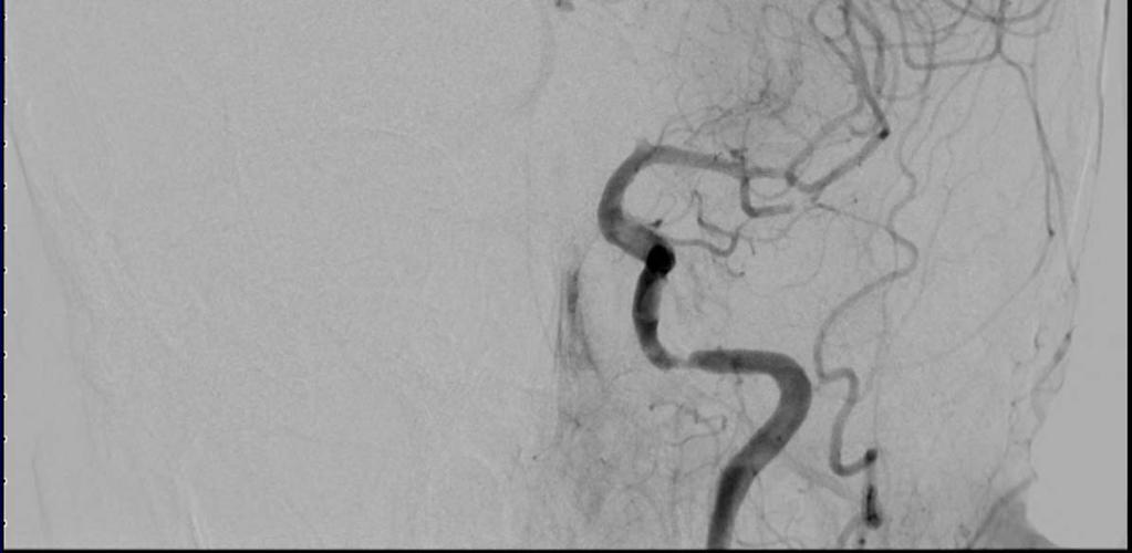
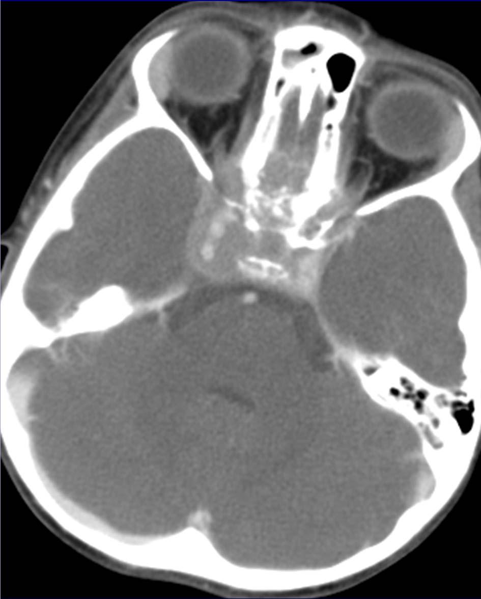
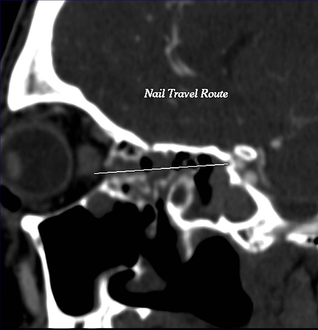
38 Cath Lab: Recanalization


39 80 kv Post-op: 80kV and 140 kv Images


40 VNC and Iodine Overlay Images VNC Iodine
41 Intra-parenchymal Hemorrhage A B C D Single Energy Iodine Overlay Virtual Non-contrast Follow-up Image
42 Virtual Monochromatic Images Are they clinically useful?

43 27 year-old male presents to ER complaining of sudden onset of severe right sided eye pain while using a weed whacker


44 27 year-old after a weed whacker accident


45 Is the left optic nerve involved?


46 Metal artifacts, identical W/L setting 80 kv 140 kv


47 Monochromatic 190kV Images The left optic nerve is spared!

48 Pre-Op CTA Nail entered the bony covering Carotid artery spared!

49 Post-Op CTA
50 Outcome Nail removed with globe intact Post-Op Evaluation: Minor conjunctival laceration No optic nerve injury bilaterally 20/20 vision bilaterally! Take Home points: Dual-energy CT helps Wear Eye protection
51 MGH Head CT Take Home Points Dose well below ACR guidelines Configure protocol to the clinical need Avoid orbits if possible Especially important in serial scans: ICU, pediatrics But don t sacrifice diagnostic accuracy! Pediatrics 125 ma used, < HALF the adult dose Strategy: screen with low dose CT, confirm with MRI Age < 18 Axial vs helical mode? Axial, arguably, has > SNR for otherwise fixed settings No real speed advantage to helical Helical = more reformat/recon options (e.g., coronals) Dual Energy CT Effective tool for material discrimination Quantitative tool Both sensitive and specific for Hemorrhage vs. Iodine Limited by saturation and other artifacts
SPECIFIC PRINCIPLES FOR DOSE REDUCTION IN HEAD CT IMAGING. Rajiv Gupta, MD, PhD Neuroradiology, Massachusetts General Hospital Harvard Medical School
 SPECIFIC PRINCIPLES FOR DOSE REDUCTION IN HEAD CT IMAGING Rajiv Gupta, MD, PhD Neuroradiology, Massachusetts General Hospital Harvard Medical School OUTLINE 1 st Presentation: Dose optimization strategies
Dual-Energy CT: The Technological Approaches
 Dual-Energy CT: The Technological Approaches Dushyant Sahani, M.D Director of CT Associate Professor of Radiology Massachusetts General Hospital Harvard Medical School Email-dsahani@partners.org Disclosure
Combined Anatomical and Functional Imaging with Revolution * CT
 GE Healthcare Case studies Combined Anatomical and Functional Imaging with Revolution * CT Jean-Louis Sablayrolles, M.D. Centre Cardiologique du Nord, Saint-Denis, France Case 1 Whole Brain Perfusion and
X-Ray & CT Physics / Clinical CT
 Computed Tomography-Basic Principles and Good Practice X-Ray & CT Physics / Clinical CT INSTRUCTORS: Dane Franklin, MBA, RT (R) (CT) Office hours will be Tuesdays from 5pm to 6pm CLASSROOM: TIME: REQUIRED
Fundamentals, Techniques, Pitfalls, and Limitations of MDCT Interpretation and Measurement
 Fundamentals, Techniques, Pitfalls, and Limitations of MDCT Interpretation and Measurement 3 rd Annual Imaging & Physiology Summit November 20-21, 21, 2009 Seoul, Korea Wm. Guy Weigold, MD, FACC Cardiovascular
B. CT protocols for the spine
 B. CT protocols for the spine Poster No.: A-003 Congress: ECR 2010 Type: Invited Speaker Topic: Neuro Authors: B. Tins; Oswestry/UK Keywords: CT, spine, diagnostic imaging protocol DOI: 10.1594/ecr2010/A-003
Ultralow Dose Chest CT with MBIR
 Ultralow Dose Chest CT with MBIR Ella A. Kazerooni, M.D. Professor & Director Cardiothoracic Radiology Associate Chair for Clinical Affairs University of Michigan Disclosures Consultant: GE Healthcare
Translating Protocols Across Patient Size: Babies to Bariatric
 Translating Protocols Across Patient Size: Babies to Bariatric Cynthia H. McCollough, PhD, FACR, FAAPM Professor of Radiologic Physics Director, CT Clinical Innovation Center Department of Radiology Mayo
Ultrasound. Computed tomography. Case studies. Utility of IQon Spectral CT in. cardiac imaging
 Ultrasound Computed tomography Case studies Utility of IQon Spectral CT in cardiac imaging Cardiac imaging is a challenging procedure where it is necessary to image a motion-free heart. This requires a
Emerging Applications in Musculoskeletal CT Imaging
 Emerging pplications in Musculoskeletal CT Imaging y K Murali MD(RD), PDCC, Director of Interventional Radiology, G. Francis DMRD, DN (RD), Consultant Radiologist, and R. Madan, MS, MD, Consultant Radiologist,
STRUCTURED EDUCATION REQUIREMENTS EFFECTIVE: JANUARY 1, 2016
 Computed Tomography The purpose of structured education is to provide the opportunity for individuals to develop mastery of discipline-specific knowledge that, when coupled with selected clinical experiences,
CT - Brain Examination
 CT - Brain Examination Submitted by: Felemban 1 CT - Brain Examination The clinical indication of CT brain are: a) Chronic cases (e.g. headache - tumor - abscess) b) ER cases (e.g. trauma - RTA - child
Toshiba Aquillion 64 CT Scanner. Phantom Center Periphery Center Periphery Center Periphery
 Comparison of radiation dose and imaging performance for the standard Varian x-ray tube and the Richardson Healthcare ALTA750 replacement tube for the Toshiba Aquillion CT scanners. by Robert L. Dixon,
Managing Radiation Risk in Pediatric CT Imaging
 Managing Radiation Risk in Pediatric CT Imaging Mahadevappa Mahesh, MS, PhD, FAAPM, FACR, FACMP, FSCCT. Professor of Radiology and Cardiology Johns Hopkins University School of Medicine Chief Physicist
Low-Dose CT: Clinical Studies & the Radiologist Perspective
 Low-Dose CT: Clinical Studies & the Radiologist Perspective RD-ASiR RD-MBIR SD-FBP RD=0.35 msv (80% dose reduction) Perry J. Pickhardt, MD UW School of Medicine & Public Health Low-Dose CT: Clinical Overview
Clinical Image Gallery Next Generation Volume 1
 Clinical Image Gallery Next Generation Volume 1 Dr. Russell Bull Royal Bournemouth Hospital, Bournemouth, United Kingdom After long experience with the first generation, a next generation Aquilion ONE
The Computed Tomography Examination
 CONTENT SPECIFICATIONS The Computed Tomography Examination The purpose of The American Registry of Radiologic Technologists (ARRT ) Computed Tomography Examination is to assess the knowledge and cognitive
Dual Energy CT Aortography: Can We Reduce Iodine Dose??
 Dual Energy CT Aortography: Can We Reduce Iodine Dose?? William P. Shuman MD, FACR FSCBTMR Department of Radiology University of Washington SCBTMR Annual Course Boston, October 10, 2012 Conflict of Interest
CT of the Head, Spine, and Cerebral Vessels
 CT of the Head, Spine, and Cerebral Vessels Objectives Determine specific imaging plane used to acquire or reformat CT scan, i.e. sagittal, coronal, transverse, and offaxis or oblique. Assess and evaluate
CT SCAN PROTOCOL. Shoulder
 CT SCAN PROTOCOL Shoulder Purpose and Summary CT images made with this protocol are used to provide the orthopedic surgeon with a detailed 3D anatomical reconstruction of the patient s scapula and proximal
Doses from Cervical Spine Computed Tomography (CT) examinations in the UK. John Holroyd and Sue Edyvean
 examinations in the UK. John Holroyd and Sue Edyvean") Doses from Cervical Spine Computed Tomography (CT) examinations in the UK John Holroyd and Sue Edyvean Why a new dose survey? Number of enquires received concerning the current NDRL Concern that could
Doses from Cervical Spine Computed Tomography (CT) examinations in the UK John Holroyd and Sue Edyvean Why a new dose survey? Number of enquires received concerning the current NDRL Concern that could
Metal Artefact Reduction in CT
 Metal Artefact Reduction in CT DANIEL MARRINER Metal Artefact Reduction in CT Metal Artefact Clinical Indications for MAR SEMAR and How It Works Technical Considerations Case Studies utilising SEMAR Metal
Imaging Acute Stroke and Cerebral Ischemia
 Department of Radiology University of California San Diego Imaging Acute Stroke and Cerebral Ischemia John R. Hesselink, M.D. Causes of Stroke Arterial stenosis Thrombosis Embolism Dissection Hypotension
Cardiac Computed Tomography
 Cardiac Computed Tomography Authored and approved by Koen Nieman Stephan Achenbach Francesca Pugliese Bernard Cosyns Patrizio Lancellotti Anastasia Kitsiou Contents CARDIAC COMPUTED TOMOGRAPHY Page 1.
Typical PET Image. Elevated uptake of FDG (related to metabolism) Lung cancer example: But where exactly is it located?
 Lung cancer example: But where exactly is it located?") Typical PET Image Elevated uptake of FDG (related to metabolism) Lung cancer example: But where exactly is it located? PET/CT Oncology Imaging Anatometabolic fusion images are useful in the management
Typical PET Image Elevated uptake of FDG (related to metabolism) Lung cancer example: But where exactly is it located? PET/CT Oncology Imaging Anatometabolic fusion images are useful in the management
An Introduction to Dual Energy Computed Tomography
 An Introduction to Dual Energy Computed Tomography Michael Riedel University of Texas Health Science Center at San Antonio Introduction The idea of computed tomography (CT) was first introduced in the
Les Outils Cliniques de Demain en Scanner Cardiaque. Cardiaque Status en ECR 2018 From Diagnosis to Prognosis
 ECR 2018 From Diagnosis to Prognosis ECR 2018 From Diagnosis to Prognosis Thursday, March 1, 2018/08:30-10:00/Room N Les Outils Cliniques de Demain en Scanner Cardiaque Cardiaque Status en 2018 Rodrigo
Cardiac CT - Coronary Calcium Basics Workshop II (Basic)
") Cardiac CT - Coronary Calcium Basics Workshop II (Basic) J. Jeffrey Carr, MD, MSCE Dept. of Radiology & Public Health Sciences Wake Forest University School of Medicine Winston-Salem, NC USA No significant
Cardiac CT - Coronary Calcium Basics Workshop II (Basic) J. Jeffrey Carr, MD, MSCE Dept. of Radiology & Public Health Sciences Wake Forest University School of Medicine Winston-Salem, NC USA No significant
Cardiac CT Lowering the Dose Dramatically
 Cardiac CT Lowering the Dose Dramatically U. Joseph Schoepf, MD, FAHA, FSCBT MR, FSCCT Professor of Radiology, Medicine, and Pediatrics Director of Cardiovascular Imaging Disclosures Consultant for / research
Pediatric CT Protocols (18 years old or less)
") Pediatric CT Protocols (18 years old or less) Ped1: Head CT Ped2: Cervical spine CT Ped3: Sinus CT Ped4: Neck CT Ped5: Chest CT Ped6: Abdomen and pelvis CT Ped7: Thoracic or lumbar spine CT Ped8: Extremity
Pediatric CT Protocols (18 years old or less) Ped1: Head CT Ped2: Cervical spine CT Ped3: Sinus CT Ped4: Neck CT Ped5: Chest CT Ped6: Abdomen and pelvis CT Ped7: Thoracic or lumbar spine CT Ped8: Extremity
Dual-Energy 101: Principles, Methods and Dose
 Dual-Energy 101: Principles, Methods and Dose Juan Carlos Ramirez-Giraldo, Ph.D Staff Scien2st, Collabora2ons Manager SE Region ISCT San Francisco, 2017 Siemens Medical Solu2ons USA, Inc., 2017 Page 1
MEDICAL POLICY EFFECTIVE DATE: 12/18/08 REVISED DATE: 12/17/09, 03/17/11, 05/19/11, 05/24/12, 05/23/13, 05/22/14
 MEDICAL POLICY SUBJECT: CT (COMPUTED TOMOGRAPHY) PAGE: 1 OF: 5 If the member's subscriber contract excludes coverage for a specific service it is not covered under that contract. In such cases, medical
Low Dose Era in Cardiac CT
 Low Dose Era in Cardiac CT DIANA E. LITMANOVICH, MD Department of Radiology Beth Israel Deaconess Medical Center Harvard Medical School Disclosures Neither I nor my immediate family members have a financial
BrightSpeed Elite CT with ASiR: Comparing Dose & Image Quality Rule Out Pulmonary Embolism on Initial & Follow-Up Exam
 GE Healthcare BrightSpeed Elite CT with ASiR: Comparing Dose & Image Quality Rule Out Pulmonary Embolism on Initial & Follow-Up Exam Michael Swack, MD Diagnostic Radiologist Irvington Radiologists, PC
THE TUFFEST STUFF CT REGISTRY REVIEW Live Lecture Seminar SATURDAY CURRICULUM
 1. The CT Imaging Chain-10 major components & their functions a. The x-ray tube b. Generator c. Filter d. Pre-patient collimator e. Pre-detector collimator f. Detector system g. Analog to digital converter
CT Perfusion. U. Joseph Schoepf, MD, FAHA, FSCBT MR, FSCCT Professor of Radiology, Medicine, and Pediatrics Director of Cardiovascular Imaging
 CT Perfusion U. Joseph Schoepf, MD, FAHA, FSCBT MR, FSCCT Professor of Radiology, Medicine, and Pediatrics Director of Cardiovascular Imaging Disclosures Consultant for / research support from Bayer Bracco
Customizing Contrast Injection for Body MDCT: Algorithmic Approach
 Customizing Contrast Injection for Body MDCT: Algorithmic Approach Lincoln L. Berland, M.D., F.A.C.R. University of Alabama at Birmingham Before Contrast Prep and Hydration Hydration single most important
Dual-Energy CT Applications in Radiation Therapy
 THE UNIVERSITY OF WISCONSIN MADISON Dual-Energy CT Applications in Radiation Therapy - Jessica Miller 1 Disclosures Funding provided by Siemens Medical 2 Learning objectives General principles of dual
Acknowledgments. A Specific Diagnostic Task: Lung Nodule Detection. A Specific Diagnostic Task: Chest CT Protocols. Chest CT Protocols
 Personalization of Pediatric Imaging in Terms of Needed Indication-Based Quality Per Dose Acknowledgments Duke University Medical Center Ehsan Samei, PhD Donald Frush, MD Xiang Li PhD DABR Cleveland Clinic
NEURORADIOLOGY Part I
 NEURORADIOLOGY Part I Vörös Erika University of Szeged Department of Radiology SZEGED BRAIN IMAGING METHODS Plain film radiography Ultrasonography (US) Computer tomography (CT) Magnetic resonance imaging
True Dual Energy. Dr. Stefan Ulzheimer, Siemens Healthcare GmbH. DEfinitely Siemens
 DEfinitely Siemens True Dual Energy Dr. Stefan Ulzheimer, Siemens Healthcare GmbH International version. Not for distribution in the US. Unrestricted Siemens AG 2015 All rights reserved. The products/features
How do the Parameters affect Image Quality and Dose for Abdominal CT? Image Review
 How do the Parameters affect Image Quality and Dose for Abdominal CT? Image Review Mannudeep K. Kalra, MD, DNB Massachusetts General Hospital Harvard Medical School Financial Disclosure This presentation
Implementation of the 2012 ACR CT QC Manual in a Community Hospital Setting BRUCE E. HASSELQUIST, PH.D., DABR, DABSNM ASPIRUS WAUSAU HOSPITAL
 Implementation of the 2012 ACR CT QC Manual in a Community Hospital Setting BRUCE E. HASSELQUIST, PH.D., DABR, DABSNM ASPIRUS WAUSAU HOSPITAL Conflict of Interest Disclaimer Employee of Aspirus Wausau
COMPUTED TOMOGRAPHY COURSE
 CT Radiography RAD 421 4 th year semester 2 Course Lecture Tutorial Practical Credit hours CT Radiography 2-1 2 Course Description The course explores the basic physical and technical principles of CT
Neuroradiology MR Protocols
 Neuroradiology MR Protocols Brain protocols N 1: Brain MRI without contrast N 2: Pre- and post-contrast brain MRI N 3 is deleted N 4: Brain MRI without or pre-/post-contrast (seizure protocol) N 5: Pre-
Cardiac CT Techniques in Neonates (and infants)
") Cardiac CT Techniques in Neonates (and infants) Siddharth P. Jadhav, MD Director, Body CT and MRI Edward B. Singleton Department of Pediatric Radiology Texas Children s Hospital Disclosures None Objectives
Cardiac CT Techniques in Neonates (and infants) Siddharth P. Jadhav, MD Director, Body CT and MRI Edward B. Singleton Department of Pediatric Radiology Texas Children s Hospital Disclosures None Objectives
EXAMINATION CONTENT SPECIFICATIONS ARRT BOARD APPROVED: JANUARY 2017 IMPLEMENTATION DATE: JULY 1, 2017
 EXAMINATION CONTENT SPECIFICATIONS Computed Tomography The purpose of the computed tomography examination is to assess the knowledge and cognitive skills underlying the intelligent performance of the tasks
FOR CMS (MEDICARE) MEMBERS ONLY NATIONAL COVERAGE DETERMINATION (NCD) FOR MAGNETIC RESONANCE IMAGING:
 MEMBERS ONLY NATIONAL COVERAGE DETERMINATION (NCD) FOR MAGNETIC RESONANCE IMAGING:") National Imaging Associates, Inc. Clinical guidelines SINUS MRI Original Date: November 2007 Page 1 of 5 CPT Codes: 70540, 70542, 70543 Last Review Date: July 2014 NCD 220.2 MRI Last Effective Date: July
National Imaging Associates, Inc. Clinical guidelines SINUS MRI Original Date: November 2007 Page 1 of 5 CPT Codes: 70540, 70542, 70543 Last Review Date: July 2014 NCD 220.2 MRI Last Effective Date: July
(Non-EKG Gated) CTA Thoracic Aorta = CTA Chest
 CTA Thoracic Aorta = CTA Chest") (Non-EKG Gated) CTA Thoracic Aorta = CTA Chest Reviewed By: Dan Verdini, MD, Rachael Edwards, MD Last Reviewed: January 2019 Contact: (866) 761-4200, Option 1 In accordance with the ALARA principle, TRA
(Non-EKG Gated) CTA Thoracic Aorta = CTA Chest Reviewed By: Dan Verdini, MD, Rachael Edwards, MD Last Reviewed: January 2019 Contact: (866) 761-4200, Option 1 In accordance with the ALARA principle, TRA
Stroke Imaging Basics. Jeremy Hopkin M.D.
 Stroke Imaging Basics Jeremy Hopkin M.D. Goals Introduce the basic physical properties of imaging used in stroke. Understand why each modality is used in the setting of stroke. Understand some strengths
General Imaging. Imaging modalities. Incremental CT. Multislice CT Multislice CT [ MDCT ]
![General Imaging. Imaging modalities. Incremental CT. Multislice CT Multislice CT [ MDCT ]](/thumbs/76/74079340.jpg "General Imaging. Imaging modalities. Incremental CT. Multislice CT Multislice CT [ MDCT ]") General Imaging Imaging modalities Conventional X-rays Ultrasonography [ US ] Computed tomography [ CT ] Radionuclide imaging Magnetic resonance imaging [ MRI ] Angiography conventional, CT,MRI Interventional
General Imaging Imaging modalities Conventional X-rays Ultrasonography [ US ] Computed tomography [ CT ] Radionuclide imaging Magnetic resonance imaging [ MRI ] Angiography conventional, CT,MRI Interventional
Blunt Carotid Injury- CT Angiography is Adequate For Screening. Kelly Knudson, M.D. UCHSC April 3, 2006
 Blunt Carotid Injury- CT Angiography is Adequate For Screening Kelly Knudson, M.D. UCHSC April 3, 2006 CT Angiography vs Digital Subtraction Angiography Blunt carotid injury screening is one of the very
Ask EuroSafe Imaging. Tips & Tricks. Paediatric Imaging Working Group. Shielding in pediatric CT
 Ask EuroSafe Imaging Tips & Tricks Paediatric Imaging Working Group Shielding in pediatric CT Claudio Granata (IRCCS Istituto Giannina Gaslini, IT) Joana Santos (ESTeSC-Coimbra Health School, PT) Elina
HI-Res Extremity Sensation 16
 Page 1 Routine Extremity - (2/14/2013) CTDI: ~20 mgy per acquisition Used for evaluation of: Humerus Forearm Femur Knee Tib/Fib Billing: 1. CT Upper/Lower Extremity of concern without contrast, with contrast,
Computed tomography Acceptance testing and dose measurements
 Computed tomography Acceptance testing and dose measurements Jonas Andersson Medical Physicist, Ph.D. Department of Radiation Sciences University Hospital of Norrland, Umeå Sweden Contents The Computed
ESTABLISHING DRLs in PEDIATRIC CT. Keith Strauss, MSc, FAAPM, FACR Cincinnati Children s Hospital University of Cincinnati College of Medicine
 ESTABLISHING DRLs in PEDIATRIC CT Keith Strauss, MSc, FAAPM, FACR Cincinnati Children s Hospital University of Cincinnati College of Medicine CT Dose Indices CTDI INTRODUCTION CTDI 100, CTDI w, CTDI vol
CT Myocardial Perfusion: Is there Added Value to Coronary CT?
 CT Myocardial Perfusion: Is there Added Value to Coronary CT? U. Joseph Schoepf, MD, FAHA, FSCBT MR, FSCCT Professor of Radiology, Medicine, and Pediatrics Director of Cardiovascular Imaging Disclosures
RADIOLOGY TEACHING CONFERENCE
 RADIOLOGY TEACHING CONFERENCE John Athas, MD Monica Tadros, MD Columbia University, College of Physicians & Surgeons Department of Otolaryngology- Head & Neck Surgery September 27, 2007 CT SCAN IMAGING
Dual Energy CT of the Heart: Perfusion and Beyond
 Dual Energy CT of the Heart: Perfusion and Beyond U. Joseph Schoepf, MD, FAHA, FSCBT MR, FSCCT Professor of Radiology, Medicine, and Pediatrics Director of Cardiovascular Imaging Disclosures Consultant
After the Chest X-Ray:
 After the Chest X-Ray: What To Do Next Alan S. Brody Professor of Radiology and Pediatrics Chief of Thoracic Imaging Cincinnati Children s Hospital Cincinnati, Ohio USA What Should We Do Next? CT scan?
Hi RES Extremity - (04/18/2011) CTDI: ~13 mgy per acquisition Used for evaluation of: Ankle Elbow Hand Wrist Foot /Calcaneous Toes Fingers
 CTDI: ~13 mgy per acquisition Used for evaluation of: Ankle Elbow Hand Wrist Foot /Calcaneous Toes Fingers") P a g e 1 Hi RES Extremity - (04/18/2011) CTDI: ~13 mgy per acquisition Used for evaluation of: Ankle Elbow Hand Wrist Foot /Calcaneous Toes Fingers Billing: 1. CT Upper/Lower Extremity of concern without
P a g e 1 Hi RES Extremity - (04/18/2011) CTDI: ~13 mgy per acquisition Used for evaluation of: Ankle Elbow Hand Wrist Foot /Calcaneous Toes Fingers Billing: 1. CT Upper/Lower Extremity of concern without
Gemstone Spectral Imaging quantifies lesion characteristics for a confident diagnosis
 GE Healthcare Gemstone Spectral Imaging quantifies lesion characteristics for a confident diagnosis CT clinical case study lesion characterization Desiree Morgan, MD Vice Chair of Clinical Research Professor
Diagnostic improvement from average image in acute ischemic stroke
 Diagnostic improvement from average image in acute ischemic stroke N. Magne (1), E.Tollard (1), O. Ozkul- Wermester (2), V. Macaigne (1), J.-N. Dacher (1), E. Gerardin (1) (1) Department of Radiology,
CT Guided Procedures And Interesting Cases. Stephen Kim, MD Diagnostic and Interventional Radiology
 CT Guided Procedures And Interesting Cases Stephen Kim, MD Diagnostic and Interventional Radiology CT guided procedure benefits Precise lesion targeting Clear image guidance for needle placement Immediate
A Snapshot on Nuclear Cardiac Imaging
 Editorial A Snapshot on Nuclear Cardiac Imaging Khalil, M. Department of Physics, Faculty of Science, Helwan University. There is no doubt that nuclear medicine scanning devices are essential tool in the
STRUCTURED EDUCATION REQUIREMENTS IMPLEMENTATION DATE: JULY 1, 2017
 STRUCTURED EDUCATION REQUIREMENTS IMPLEMENTATION DATE: JULY 1, 2017 Computed Tomography The purpose of structured education is to provide the opportunity for individuals to develop mastery of discipline-specific
SOMATOM Drive System Owner Manual Dosimetry and imaging performance report
 www.siemens.com/healthcare SOMATOM Drive System Owner Manual Dosimetry and imaging performance report Table of contents 1 Dosimetry and imaging performance report 5 1.1 Dose information 5 1.1.1 General
Disclosure Information
 Coronary CTA Pearls and Pitfalls Ricardo C. Cury, MD, FSCCT, FAHA, FACC Chairman of Radiology Radiology Associates of South Florida Director of Cardiac Imaging Miami Cardiac and Vascular Institute Past-President
Pediatric chest HRCT using the idose 4 Hybrid Iterative Reconstruction Algorithm: Which idose level to choose?
 Journal of Physics: Conference Series PAPER OPEN ACCESS Pediatric chest HRCT using the idose 4 Hybrid Iterative Reconstruction Algorithm: Which idose level to choose? To cite this article: M Smarda et
8/3/2016. Consultant for / research support from: Astellas Bayer Bracco GE Healthcare Guerbet Medrad Siemens Healthcare. Single Energy.
 U. Joseph Schoepf, MD Prof. (h.c.), FAHA, FSCBT-MR, FNASCI, FSCCT Professor of Radiology, Medicine, and Pediatrics Director, Division of Cardiovascular Imaging Consultant for / research support from: Astellas
Effective Utilization of Imaging. John V. Roberts, M.D. Premier Radiology Abdominal Imaging
 Effective Utilization of Imaging John V. Roberts, M.D. Premier Radiology Abdominal Imaging Safety Contrast and Radiation What to order Abdomen/Pelvis Brain/Spine Chest Musculoskeletal Ob/Gyn Head and Neck
Aquilion ONE: Pediatric Imaging. Richard Mather, PhD. Senior Manager, CT Clinical Science Toshiba America Medical Systems, Inc.
 Aquilion ONE: Pediatric Imaging Richard Mather, PhD Senior Manager, CT Clinical Science Toshiba America Medical Systems, Inc. The use of CT in pediatric diagnostic procedures has increased significantly
Integrated PET/CT systems State of the art and Clinical Applications
 Integrated PET/CT systems State of the art and Clinical Applications V. Bettinardi Nuclear Medicine Dep. Scientific Institute San Raffaele Hospital Milan Italy The announcement of a new Diagnostic Imaging
Dual Energy CT of the Liver
 34th Annual Course October 2011 Washington, DC Dual Energy CT of the Liver Vassilios Raptopoulos, MD Beth Israel Deaconess Medical Center Harvard Medical School Dual Energy CT (DECT) Different materials
Imaging Stroke: Is There a Stroke Equivalent of the ECG? Albert J. Yoo, MD Director of Acute Stroke Intervention Massachusetts General Hospital
 Imaging Stroke: Is There a Stroke Equivalent of the ECG? Albert J. Yoo, MD Director of Acute Stroke Intervention Massachusetts General Hospital Disclosures Penumbra, Inc. research grant (significant) for
Biomarkers and the Future of. John R. Votaw CBIS 5 th Year Anniversary Celebration/Look to the future February 8, 2013
 Biomarkers and the Future of Radiology John R. Votaw CBIS 5 th Year Anniversary Celebration/Look to the future February 8, 2013 Statistics/Radiology Collaboration The utility of Radiologic procedures
Cardiac Imaging Tests
 Cardiac Imaging Tests http://www.medpagetoday.com/upload/2010/11/15/23347.jpg Standard imaging tests include echocardiography, chest x-ray, CT, MRI, and various radionuclide techniques. Standard CT and
Low-dose and High-resolution Cardiac Imaging with Revolution CT
 GE Healthcare Case study Low-dose and High-resolution Cardiac Imaging with Revolution CT Prof. Philipp A. Kaufmann, M.D. Ronny R. Buechel, M.D. Fran Mikulicic, M.D. Dominik C. Benz, M.D. University of
TORNIER BLUEPRINT. 3D Planning + PSI SCAN PROTOCOL
 TORNIER BLUEPRINT 3D Planning + PSI SCAN PROTOCOL Contents 3 Introduction 3 Patient preparation 3 Scanning instructions 4 Image instructions 5 Scanning parameters 6 Technical instructions 2 BLUEPRINT 3D
CT Myocardial Perfusion
 1 CT Myocardial Perfusion Ting-Yim Lee, PhD, FCCPM, FCOMP Aaron So, PhD, FSCCT Gerald Wisenberg, MD, FRCPC Ali Islam, MD, FRCPC Lawson Health Research Institute Robarts research Institute The University
Case Conference: Neuroradiology. Case 1: Tumor Case 1: 22yo F w/ HA and prior Seizures
 Case Conference: Neuroradiology Case 1: 22yo F w/ HA and prior Seizures David E. Rex, MD, PhD Stanford University Hospital Department of Radiology Case 1: Tumor Most likely gangiloglioma, oligodendroglioma,
B-Flow, Power Doppler and Color Doppler Ultrasound in the Assessment of Carotid Stenosis: Comparison with 64-MD-CT Angiography
 Med. J. Cairo Univ., Vol. 85, No. 2, March: 805-809, 2017 www.medicaljournalofcairouniversity.net B-Flow, Power Doppler and Color Doppler Ultrasound in the Assessment of Carotid Stenosis: Comparison with
Radiation Dose Reduction Strategies in Coronary CT Angiography
 Radiation Dose Reduction Strategies in Coronary CT Angiography Noor Diyana Osman, PhD noordiyana@usm.my Contents: Introduction Radiation dosimetry in CT Radiation risk associated with coronary CT angiography
CTA Pulmonary Embolism CTA Chest W (arterial)
") CTA Pulmonary Embolism CTA Chest W (arterial) Reviewed By: Rachael Edwards, MD; Anna Ellermeier, MD; Brett Mollard, MD Last Reviewed: January 2019 Contact: (866) 761-4200, Option 1 In accordance with the
CTA Pulmonary Embolism CTA Chest W (arterial) Reviewed By: Rachael Edwards, MD; Anna Ellermeier, MD; Brett Mollard, MD Last Reviewed: January 2019 Contact: (866) 761-4200, Option 1 In accordance with the
AAPM Multimodality Medical Imaging -I
 Monday, July 28, 2008 Imaging Continuing Education Course CE-Imaging: Multimodality Medical Imaging - I Case History 46 year old female with melanoma. Multimodality Imaging Clinical Perspective PET-CT
Modifi ed CT perfusion contrast injection protocols for improved CBF quantifi cation with lower temporal sampling
 Investigations and research Modifi ed CT perfusion contrast injection protocols for improved CBF quantifi cation with lower temporal sampling J. Wang Z. Ying V. Yao L. Ciancibello S. Premraj S. Pohlman
The diagnostic value of Computed Tomography in evaluation of maxillofacial Trauma
 The diagnostic value of Computed Tomography in evaluation of maxillofacial Trauma Qais H. Muassa FICMS College of Dentistry, Babylon University Ibrahim S. Gataa, BDS, FICMS College of Dentistry, Sulaimania
PINPOINTING RADIATION THERAPY WITH THE PRECISION OF MR.
 GE Healthcare PINPOINTING RADIATION THERAPY WITH THE PRECISION OF MR. MR Radiation Oncology Suite MAXIMIZE YOUR PRECISION. HELP MINIMIZE PATIENT COMPLICATIONS. Our goal in MR radiation oncology is to
Computed tomography. Department of Radiology, University Medical School, Szeged
 Computed tomography Department of Radiology, University Medical School, Szeged voxel +1-4 +2 +5 +3 +1 0-2 pixel -2 0 +1-4 -6 +5 +2 +1 Department of Radiology, University Medical School, Szeged
Role of the Radiologist
 Diagnosis and Treatment of Blunt Cerebrovascular Injuries NORDTER Consensus Conference October 22-24, 2007 Clint W. Sliker, M.D. University of Maryland Medical Center R Adams Cowley Shock Trauma Center
8/1/2017. Financial Disclosures. Dose Tracking at MGH. How Dose Tracking Affected Protocol Optimization in a Tertiary Quaternary Healthcare Center
 How Dose Tracking Affected Protocol Optimization in a Tertiary Quaternary Healthcare Center Mannudeep K. Kalra, MD Webster Center for Quality and Safety Massachusetts General Hospital Harvard Medical School
TORNIER. scan protocol V2.1. Tornier Upper Extremities
 TORNIER TM scan protocol V2.1 Tornier Upper Extremities Contents 4 Introduction 4 Patient Preparation 4 Scanning Instructions 5 Image Instructions 6 Scanning Parameters 7 Technical Instructions Scan Protocol
CT angiography techniques. Boot camp
 CT angiography techniques Boot camp Overview Basic concepts Contrast administration arterial opacification Time scan acquisition during the arterial phase Protocol examples Helical non-gated CTA Pulmonary
Radiation dose reduction in computed tomography: techniques and future perspective
 REVIEW Radiation dose reduction in computed tomography: techniques and future perspective Despite universal consensus that computed tomography (CT) overwhelmingly benefits patients when used for appropriate
2
 1 2 3 4 5 6 7 8 9 10 11 12 13 Cine loop of tomosynthesis slice images through the chest. 14 Standard PA chest radiograph (left) and single slice from the tomosynthesis image dataset (right) of a patient
Bringing the power of imaging to your patient. Portable full body 32-slice CT scanner
 Bringing the power of imaging to your patient Portable full body 32-slice CT scanner Point-of-care CT imaging Your multi-departmental imaging solution Orthopedic surgery Arthroplasty Musculoskeletal disorders
MRI and CT of the CNS
 MRI and CT of the CNS Dr.Maha ELBeltagy Assistant Professor of Anatomy Faculty of Medicine The University of Jordan 2018 Computed Tomography CT is used for the detection of intracranial lesions. CT relies
To Shield or Not to Shield? Lincoln L. Berland, M.D.
 To Shield or Not to Shield? Lincoln L. Berland, M.D. Disclosures Consultant to: Nuance, Inc. Page 2 Breast Radiation on CT Use of chest CT has increased in women vulnerable to cancer induction by radiation.