AORTIC VALVE CASES. Richard L. Hallett, MD
|
|
|
- Kathlyn Dennis
- 6 years ago
- Views:
Transcription


1 AORTIC VALVE CASES Richard L. Hallett, MD Section Chief, Cardiovascular Imaging Northwest Radiology Network Indianapolis, IN Adjunct Assistant Professor of Radiology Stanford University Hospital and Clinics Stanford, CA NASCI 2015 San Diego, CA September 26, 2015
2 DISCLOSURES None HANDOUT: Choose folder: NASCI 2015
3 CASE ONE
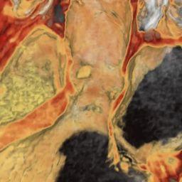
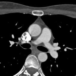
4 CASE ONE 32 yr old male Atypical CP, equivocal stress echo Cath: No vessel coming off R sinus, concern for anomalous coronary artery


5 CASE ONE
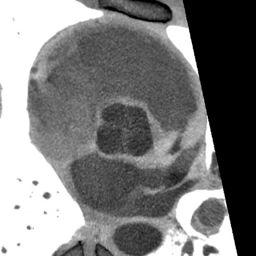
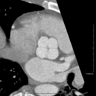


6 QUADRACUSPID AORTIC VALVE
7 QUESTION The most common complication of Quadricuspid Aortic Valve (QAV) is: A. Valvular aortic stenosis B. Aortic regurgitation C. Atrial fibrillation D. Left ventricular hypertrophy Douglas, H., Moore, M., & Purvis, J. (2012). Comprehensive assessment of a quadricuspid aortic valve and coronary arteries by multidetector cardiac CT. Heart, 98(24),
8 QUESTION: ANSWER The most common complication of Quadricuspid Aortic Valve (QAV) is: A. Valvular aortic stenosis B. Aortic regurgitation C. Atrial fibrillation D. Left ventricular hypertrophy Douglas, H., Moore, M., & Purvis, J. Heart, 2012: 98(24),
9 QUADRICUSPID AORTIC VALVE (QAV) Rare, 1/6000 aortic valve surgery patients 1 M = F, avg. age at Dx ~ 50 Classification by size of cusps 2 Most common: 3 same size + 1 smaller cusp (type B) Echo: X -shaped SAX view CT/MR: confirmatory; perform planimetry and/or flow measurement 3 1. Douglas, H., Moore, M., & Purvis, J. (2012) Heart 2012; 98(24), Hurwitz L, et al. Am J Cardiol 1973; 31(5) Khan SK, Tamin SS, Araoz PA. J Comput Assist Tomogr. 35 (5):
 623-626.")
10 CLASSIFICATION OF QAV 1 A B C D E F G 4 equal sized cusps 3 equal + 1 smaller (most common) 2 equal + 2 equal smaller 1 large + 2 intermediate + 1 smaller 3 equal + 1 larger 2 equal large + 2 smaller unequal sizes 4 unequal sized cusps 1. Hurwitz L, et al. Am J Cardiol 1973; 31(5)
11 QUADRICUSPID AORTIC VALVE (QAV) Usually isolated, but can be associated with: Single or Anomalous Coronary Arteries Displacement of coronary ostia (from addl cusp) HCM / Subaortic Stenosis PDA, VSD Endocarditis 1. Jagganath AD, et al. Echocardiography 2011; 28(9), Zhu J, et al. J Cardiothor Surg 2013; 8(1) Tutarel O. J. Heart Valve Dis. 2004;13 (4):
12 QUADRICUSPID AORTIC VALVE (QAV) Complications: Aortic Insufficiency (#1) Up to 75% at time of Dx LVH Conduction problems (BBB) TX: Reconstruction and/or Surgical valve replacement 1. Jagganath AD, et al. Echocardiography 2011; 28(9), Zhu J, et al. J Cardiothor Surg 2013; 8(1) Tutarel O. J. Heart Valve Dis. 2004;13 (4):
13 CASE TWO


14 CASE TWO 68 yo Male, embolic lesions in kidneys on CT Technically difficult echo exam,? AoV lesion

15
16
17 CASE TWO: DDX Vegetation Thrombus Tumor Degenerated valve tissue
18 CASE TWO: PAPILLARY FIBROELASTOMA
19 QUESTION: Papillary Fibroelastoma is: A. the most common cardiac tumor B. potentially malignant C. more common in females D. responsible for 75% of valvular tumors
20 QUESTION: ANSWER Papillary Fibroelastoma is: A. the most common cardiac tumor B. potentially malignant C. more common in females D. responsible for 75% of valvular tumors Grebenc ML, Rosado de Christenson ML, Burke AP, Green CE, Galvin JR. Radiographics. 2000;20(4):
21 PAPILLARY FIBROELASTOMA Avg age 60 M=F #1 neoplasm of cardiac valves (prevalence only ~ 0.02%) #2 or 3 cardiac neoplasm overall (myxoma, lipoma) Often Asx but can present w/ embolic disease (tumor or bland), TIA/stroke, dyspnea, sudden cardiac death Imaging Diagnosis! Grebenc ML, Rosado de Christenson Ml et al. Radiographics. 2000;20(4): Kumbala, D, Sharp, T, Kamalesh M. Angiology, 2008; 59(5),
:1073 103. Kumbala, D, Sharp, T, Kamalesh M.")
22 PAPILLARY FIBROELASTOMA Path: gelatinous, avascular papilloma covered by single layer epithelium sea anemone surface: but can be obscured by surface thrombus Mobile, pedunculated lesions w/ connection to endothelium by stalk Grebenc ML, Rosado de Christenson Ml et al. Radiographics. 2000;20(4): Kumbala, D, Sharp, T, Kamalesh M. Angiology, 2008; 59(5),
 Grebenc ML, Rosado de Christenson Ml et al. Radiographics. 2000;20(4):1073 103. Kumbala, D, Sharp, T, Kamalesh M.")
23 PAPILLARY FIBROELASTOMA Aortic >> mitral > tricuspid > pulmonic Can also occur on endocardial surfaces of atria / ventricles Mobility is independent predictor of non-fatal and fatal events (surgical treatment) Grebenc ML, Rosado de Christenson Ml et al. Radiographics. 2000;20(4): Kumbala, D, Sharp, T, Kamalesh M. Angiology, 2008; 59(5),


24 PULMONIC VALVE FIBROELASTOMA
25 CASE THREE
 Previous aortic valve")
26 CASE THREE 45 yr old female 330 lb, 5 2 Previous pacer (vent rate 75) Previous aortic valve replacement
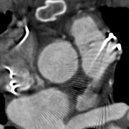
27 CASE THREE New onset CHF Abnormal echo possible lesion on prosthetic aortic valve CTA requested to assess valve. And.CCTA requested for pre-op coronary clearance
28 TECHNICAL ISSUES How to scan with low enough noise to fully assess valve and coronaries Impact of paced HR Pacer wire artifacts?

29


 5mm")
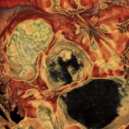
30 VEGETATION ON ST. JUDE PROSTHETIC AORTIC VALVE (330 LB PATIENT) 5mm MPR 3mm MINIP
31 QUESTION In order to improve image quality in larger patients, one should: A. Use filtered back projection reconstruction B. Utilize ECG pulsing C. Utilize weight-based contrast medium dosing D. Scan at 100 kv to save dose
32 QUESTION: ANSWER In order to improve image quality in larger patients, one should: A. Use filtered back projection reconstruction B. Utilize ECG pulsing C. Utilize weight-based contrast medium dosing D. Scan at 100 kv to save dose 1. Fleischmann D. How to design injection protocols for multiple detector-row CT angiography (MDCTA). Eur Radiol Dec 1;15 Suppl 5:E60 5.
33 PROSTHETIC VALVE DYSFUNCTION SX: Heart Murmur Heart Failure Fever Stroke DOE Angina Habets, J., Mali, WPTM, & Budde, RPJ (2012). Radiographics, 32(7):
34 PROSTHETIC VALVE DYSFUNCTION Echo primary imaging tool Fluoroscopy can visualize stuck leaflets CT useful if echo limited (obese, COPD) Leaflet excursion Perivalvular abscess Mycotic aneurysms
35 PROSTHETIC VALVE DYSFUNCTION: CT IMAGING Retrospective ECG gating useful for motion No ECG pulsing Ni / Ti alloys : GOOD (St. Jude: Nickel alloy) Cobalt Chrome: BAD (Bjork Shiley) Most Bioprosthetic valves are well assessed Habets, J., Mali, WPTM, & Budde, RPJ (2012). Radiographics, 32(7):
36 TECHNICAL TIPS FOR IMAGING LARGE PATIENTS - 1 Consider 140 kv voltage (trade-offs) Less blooming artifact from metal Less image noise More dose Scan at thicker initial collimation Slow gantry rotation time (~ 0.5 sec)
37 TECHNICAL TIPS FOR IMAGING LARGE PATIENTS -2 Use iterative reconstruction techniques WEIGHT-BASED Contrast medium flow-rates and volume Radiation dose: HIGH but re-do valve surgery for dysfunction has mortality up to 15%!!
38 CONCLUSIONS Quadricuspid aortic valve is visualized as an X on echo and CT/MR, and is associated with AI Aortic valve fibroelastoma is the most common valvular tumor Vegetations and lesions of prosthetic valves can be well assessed on ECG-synchronized CTA Adaptations / tradeoffs necessary for imaging valves in larger patients


39 THANKS FOR YOUR ATTENTION! Special Thanks to: Albert Hsiao, MD, PhD Dominik Fleischmann, MD HANDOUT: Choose folder: NASCI 2015
Richard L. Hallett, MD
 Richard L. Hallett, MD Chief, Cardiovascular Imaging Northwest Radiology Network Indianapolis, IN Adjunct Assistant Professor of Radiology Stanford University Hospital and Clinics Stanford, CA NASCI 2016
TSDA Boot Camp September 13-16, Introduction to Aortic Valve Surgery. George L. Hicks, Jr., MD
 TSDA Boot Camp September 13-16, 2018 Introduction to Aortic Valve Surgery George L. Hicks, Jr., MD Aortic Valve Pathology and Treatment Valvular Aortic Stenosis in Adults Average Course (Post mortem data)
Index. radiologic.theclinics.com. Note: Page numbers of article titles are in boldface type.
 Index Note: Page numbers of article titles are in boldface type. A ALCAPA. See Anomalous left coronary artery from the pulmonary artery. Angiosarcoma computed tomographic assessment of, 809 811 Anomalous
Valvular Imaging Optimizing Data Acquisition and Interpretation
 Valvular Imaging Optimizing Data Acquisition and Interpretation Suhny Abbara, MD Director Cardiovascular Imaging Section, Massachusetts General Hospital Assistant Professor, Harvard Medical School Sabbara@Partners.org
Case 47 Clinical Presentation
 93 Case 47 C Clinical Presentation 45-year-old man presents with chest pain and new onset of a murmur. Echocardiography shows severe aortic insufficiency. 94 RadCases Cardiac Imaging Imaging Findings C
Post-Op Aorta: Differentiating Normal Post-Op vs. Complications. Linda C. Chu, MD Assistant Professor of Radiology Johns Hopkins University
 Post-Op Aorta: Differentiating Normal Post-Op vs. Complications Linda C. Chu, MD Assistant Professor of Radiology Johns Hopkins University No disclosures Disclosures Goals and Objectives To review CT technique
Noncoronary Cardiac MDCT
 Noncoronary Cardiac MDCT David A. Bluemke, M.D., Ph.D. Professor, of Radiology and Medicine Johns Hopkins University School of Medicine Baltimore, Maryland Toshiba Disclosures Grant support Noncoronary
Adult Echocardiography Examination Content Outline
 Adult Echocardiography Examination Content Outline (Outline Summary) # Domain Subdomain Percentage 1 2 3 4 5 Anatomy and Physiology Pathology Clinical Care and Safety Measurement Techniques, Maneuvers,
Disclosure Information
 Coronary CTA Pearls and Pitfalls Ricardo C. Cury, MD, FSCCT, FAHA, FACC Chairman of Radiology Radiology Associates of South Florida Director of Cardiac Imaging Miami Cardiac and Vascular Institute Past-President
New Technologies for Cardiac CT. Geoffrey D. Rubin, MD, MBA, FACR, FNASCI Duke University
 1996 New Technologies for Cardiac CT Geoffrey D. Rubin, MD, MBA, FACR, FNASCI Duke University New Technology The Long View Levels of Efficacy Endpoint Examples 1: Technical Imaging resolution 2: Diagnostic
PROSTHETIC VALVE BOARD REVIEW
 PROSTHETIC VALVE BOARD REVIEW The correct answer D This two chamber view shows a porcine mitral prosthesis with the typical appearance of the struts although the leaflets are not well seen. The valve
Cardiac Computed Tomography
 Cardiac Computed Tomography Authored and approved by Koen Nieman Stephan Achenbach Francesca Pugliese Bernard Cosyns Patrizio Lancellotti Anastasia Kitsiou Contents CARDIAC COMPUTED TOMOGRAPHY Page 1.
Image Library Case Listing:
 Image Library Case Listing: 1. Giant left atrial myxoma with mitral valve damage 2. Type A aortic dissection 3. Primum ASD 4. Aortic Transection from motor vehicle accident 5. Snake thrombus in right atrium
Fundamentals, Techniques, Pitfalls, and Limitations of MDCT Interpretation and Measurement
 Fundamentals, Techniques, Pitfalls, and Limitations of MDCT Interpretation and Measurement 3 rd Annual Imaging & Physiology Summit November 20-21, 21, 2009 Seoul, Korea Wm. Guy Weigold, MD, FACC Cardiovascular
Review of Cardiac Imaging Modalities in the Renal Patient. George Youssef
 Review of Cardiac Imaging Modalities in the Renal Patient George Youssef ECHO Left ventricular hypertrophy (LVH) assessment Diastolic dysfunction Stress ECHO Cardiac CT angiography Echocardiography - positives
Echocardiographic Evaluation of the Cardiomyopathies. Stephanie Coulter, MD, FACC, FASE April, 2016
 Echocardiographic Evaluation of the Cardiomyopathies Stephanie Coulter, MD, FACC, FASE April, 2016 Cardiomyopathies (CMP) primary disease intrinsic to cardiac muscle Dilated CMP Hypertrophic CMP Infiltrative
Mitral Valve Disease. Prof. Sirchak Yelizaveta Stepanovna
 Mitral Valve Disease Prof. Sirchak Yelizaveta Stepanovna Fall 2008 Mitral Valve Stenosis Lecture Outline Mitral Stenosis Mitral Regurgitation Etiology Pathophysiology Clinical features Diagnostic testing
Cardiac Masses. Cardiac Masses: Considerations. Dennis A. Tighe, MD, FASE. University of Massachusetts Medical School Worcester, MA 4/16/2018
 Cardiac Masses Dennis A. Tighe, MD, FASE University of Massachusetts Medical School Worcester, MA Cardiac Masses: Considerations Definition of the mass Nature Location Benign or malignant Presentation
Advanced Imaging MRI and CTA
 Advanced Imaging MRI and CTA Who and why may benefit. Matthew W. Martinez, M.D. FACC Lehigh Valley Health Network Director, Cardiovascular Imaging Learning Objectives Review basics of CMR and CTA Review
M-Mode Echocardiography Is it still Alive? Itzhak Kronzon, MD,FASE. Sampling Rate M-Mode: 1800 / sec 2D: 30 / sec
 M-Mode Echocardiography Is it still Alive? Itzhak Kronzon, MD,FASE Honoraria: Philips Classical M-mode Echocardiography M-Mode offers better time and image resolution. Sampling Rate M-Mode: 1800 / sec
Artifact reduction strategies for prosthetic heart valve CT imaging
 Int J Cardiovasc Imaging (2012) 28:2099 2108 DOI 10.1007/s10554-012-0041-5 ORIGINAL PAPER Artifact reduction strategies for prosthetic heart valve CT imaging Jesse Habets Petr Symersky Tim Leiner Bas A.
The Special Echo Case
 Praxis-orientierter Luzerner Fortbildungskurs, 14. Januar 2006 The Special Echo Case Michel Romanens, Cardiology Consultant, Cantonal Hospital and RDC Olten 83 year old female subject with an incident
Cardiac Masses. Dennis A. Tighe, MD, FASE. University of Massachusetts Medical School Worcester, MA
 Cardiac Masses Dennis A. Tighe, MD, FASE University of Massachusetts Medical School Worcester, MA Cardiac Masses: Considerations Definition of the mass Nature Location Benign or malignant Presentation
General Cardiovascular Magnetic Resonance Imaging
 2 General Cardiovascular Magnetic Resonance Imaging 19 Peter G. Danias, Cardiovascular MRI: 150 Multiple-Choice Questions and Answers Humana Press 2008 20 Cardiovascular MRI: 150 Multiple-Choice Questions
April 16, 09:00-09:15 중앙대학교 윤신원
 April 16, 09:00-09:15 중앙대학교 윤신원 When to perform Echocardiography in IE? Vegetations?(pathologic Whatever the level hallmark) of suspicion Intracardiac abscess? Confirm or R/O at the Earliest opportunity.
Supplementary Appendix
 Supplementary Appendix This appendix has been provided by the authors to give readers additional information about their work. Supplement to: Kang D-H, Kim Y-J, Kim S-H, et al. Early surgery versus conventional
Endocarditis and Its Complications: The Role of Echocardiography
 Endocarditis and Its Complications: The Role of Echocardiography Pravin Patil, MD FACC FASE Associate Professor of Medicine Director, Cardiovascular Disease Training Program Lewis Katz School of Medicine
For more information about how to cite these materials visit
 Author: Michael Shea, M.D., 2008 License: Unless otherwise noted, this material is made available under the terms of the Creative Commons Attribution Share Alike 3.0 License: http://creativecommons.org/licenses/by-sa/3.0/
Adult Cardiac Surgery
 Adult Cardiac Surgery Mahmoud ABU-ABEELEH Associate Professor Department of Surgery Division of Cardiothoracic Surgery School of Medicine University Of Jordan Adult Cardiac Surgery: Ischemic Heart Disease
New Cardiovascular Devices and Interventions: Non-Contrast MRI for TAVR Abhishek Chaturvedi Assistant Professor. Cardiothoracic Radiology
 New Cardiovascular Devices and Interventions: Non-Contrast MRI for TAVR Abhishek Chaturvedi Assistant Professor Cardiothoracic Radiology Disclosure I have no disclosure pertinent to this presentation.
Echocardiography after stroke - where to look
 Echocardiography after stroke - where to look Vuyisile T. Nkomo, MD,MPH, FACC, FASE Joint Cardiac Imaging Society of South Africa/Mayo Clinic Echocardiography Workshop 2017 2016 MFMER slide-1 Disclosures
Ultrasound. Computed tomography. Case studies. Utility of IQon Spectral CT in. cardiac imaging
 Ultrasound Computed tomography Case studies Utility of IQon Spectral CT in cardiac imaging Cardiac imaging is a challenging procedure where it is necessary to image a motion-free heart. This requires a
RC 612B 3 December Richard L. Hallett, MD
 RC 612B 3 December 2015 0830 1000 Richard L. Hallett, MD Chief, Cardiovascular Imaging Northwest Radiology Network Indianapolis, IN Adjunct Assistant Professor Radiology Stanford University Stanford, CA
Coronary Artery Anomalies from Birth to Adulthood; the Role of CT Coronary Angiography in Sudden Cardiac Death Screening
 Coronary Artery Anomalies from Birth to Adulthood; the Role of CT Coronary Angiography in Sudden Cardiac Death Screening E O Dwyer 1, C O Brien 1, B Loo 1, A Snow Hogan 1, O Buckley1 2, B 1. Department
Valvular Heart Disease. Dr. HANAN ALBACKR
 Valvular Heart Disease Dr. HANAN ALBACKR Valvular Heart Disease Format for this lecture IMPORTANT CLINICAL INFO know for boards, tests and clinical practice Spectrum of VHD Aortic Valve Mitral Valve Tricuspid
Cardiology. the Sounds: #7 HCM. LV Outflow Obstruction: Aortic Stenosis. (Coming Soon - HCM)
") A Cardiology HCM LV Outflow Obstruction: Aortic Stenosis (Coming Soon - HCM) the Sounds: #7 Howard J. Sachs, MD www.12daysinmarch.com E-mail: Howard@12daysinmarch.com Aortic Valve Disorders Stenosis Regurgitation
A Cardiology HCM LV Outflow Obstruction: Aortic Stenosis (Coming Soon - HCM) the Sounds: #7 Howard J. Sachs, MD www.12daysinmarch.com E-mail: Howard@12daysinmarch.com Aortic Valve Disorders Stenosis Regurgitation
Heart on Fire: Infective Endocarditis. Objectives. Disclosure 8/27/2018. Mary McGreal DNP, RN, ANP-c, CCRN
 Heart on Fire: Infective Endocarditis Mary McGreal DNP, RN, ANP-c, CCRN Objectives Discuss the incidence of infective endocarditis? Discuss the pathogenesis of infective endocarditis? Discuss clinical
9/8/2009 < 1 1,2 3,4 5,6 7,8 9,10 11,12 13,14 15,16 17,18 > 18. Tetralogy of Fallot. Complex Congenital Heart Disease.
 Current Indications for Pediatric CTA S Bruce Greenberg Professor of Radiology Arkansas Children s Hospital University of Arkansas for Medical Sciences greenbergsbruce@uams.edu 45 40 35 30 25 20 15 10
pulmonary valve on, 107 pulmonary valve vegetations on, 113
 INDEX Adriamycin-induced cardiomyopathy, 176 Amyloidosis, 160-161 echocardiographic abnormalities in, 160 intra-mural tumors similar to, 294 myocardial involvement in, 160-161 two-dimensional echocardiography
By the end of this session, the student should be able to:
 Valvular Heart disease HVD By Dr. Ashraf Abdelfatah Deyab VHD- Objectives By the end of this session, the student should be able to: Define and classify valvular heart disease. Enlist the causes of acquired
When Should I Order a Stress Test or an Echocardiogram
 When Should I Order a Stress Test or an Echocardiogram Updates in Cardiology 2015 March 7, 2015 Donald L. Lappé, MD, FAHA, FACC Chairman, Cardiovascular Department Medical Director, Intermountain Cardiovascular
New Imaging for Aortic Valve Disease. Anthony DeMaria Judy and Jack White Chair Director, Sulpizio CV Center University of California, San Diego
 New Imaging for Aortic Valve Disease Anthony DeMaria Judy and Jack White Chair Director, Sulpizio CV Center University of California, San Diego Imaging in Aortic Stenosis Valve morphology calcification
Multimodality Imaging of Right-Sided (Tricuspid Valve) Papillary Fibroelastoma: Recognition of a Surgically Remediable Disease
 Papillary Fibroelastoma: Recognition of a Surgically Remediable Disease") Published online: September 21, 2013 1662 6575/13/0063 0485$38.00/0 This is an Open Access article licensed under the terms of the Creative Commons Attribution-NonCommercial 3.0 Unported license (CC BY-NC)
Published online: September 21, 2013 1662 6575/13/0063 0485$38.00/0 This is an Open Access article licensed under the terms of the Creative Commons Attribution-NonCommercial 3.0 Unported license (CC BY-NC)
Pushing the limits of cardiac CT. Steven Dymarkowski Radiology / Medical Imaging Research Centre
 Pushing the limits of cardiac CT Steven Dymarkowski Radiology / Medical Imaging Research Centre 5 X 2013 Introduction Rapid technological advances and new clinical applications in cardiovascular imaging
Coronary Artery Imaging. Suvipaporn Siripornpitak, MD Inter-hospital Conference : Rajavithi Hospital
 Coronary Artery Imaging Suvipaporn Siripornpitak, MD Inter-hospital Conference : Rajavithi Hospital Larger array : cover scan area Detector size : spatial resolution Rotation speed : scan time Retrospective
The use of Cardiac CT and MRI in Clinical Practice
 The use of Cardiac CT and MRI in Clinical Practice Matthew W. Martinez, MD Assistant Professor of Medicine LVPG - Lehigh Valley Heart Specialists Lehigh Valley Health Network Oct. 3, 2009 DISCLOSURE Relevant
HISTORY. Question: How do you interpret the patient s history? CHIEF COMPLAINT: Dyspnea of two days duration. PRESENT ILLNESS: 45-year-old man.
 HISTORY 45-year-old man. CHIEF COMPLAINT: Dyspnea of two days duration. PRESENT ILLNESS: His dyspnea began suddenly and has been associated with orthopnea, but no chest pain. For two months he has felt
Radiology of the respiratory/cardiac diseases (part 2)
") Cardiology Cycle - Lecture 6 436 Teams Radiology of the respiratory/cardiac diseases (part 2) Objectives Done By Team Leaders: Khalid Alshehri Hanin Bashaikh Team Members: Leena Alwakeel Aroob Alhuthail
Cardiology Cycle - Lecture 6 436 Teams Radiology of the respiratory/cardiac diseases (part 2) Objectives Done By Team Leaders: Khalid Alshehri Hanin Bashaikh Team Members: Leena Alwakeel Aroob Alhuthail
ICE: Echo Core Lab-CRF
 APPENDIX 1 ICE: Echo Core Lab-CRF Study #: - Pt Initials: 1. Date of study: / / D D M M M Y Y Y Y 2. Type of Study: TTE TEE 3. Quality of Study: Poor Moderate Excellent Ejection Fraction 4. Ejection Fraction
Low Dose Era in Cardiac CT
 Low Dose Era in Cardiac CT DIANA E. LITMANOVICH, MD Department of Radiology Beth Israel Deaconess Medical Center Harvard Medical School Disclosures Neither I nor my immediate family members have a financial
Congenital heart disease. By Dr Saima Ali Professor of pediatrics
 Congenital heart disease By Dr Saima Ali Professor of pediatrics What is the most striking clinical finding in this child? Learning objectives By the end of this lecture, final year student should be able
PRINCIPLES OF ENDOCARDITIS
 015 // Endocarditis CONTENTS 140 Principles of Endocarditis 141 Native Valve Endocarditis 143 Complications of Native Valve Endocarditis 145 Right Heart Endocarditis 145 Prosthetic Valve Endocarditis 146
Etiology, Classification & Management. Sheba Medical Center Cardiology Department Matthew Wright St. George s University of London
 Etiology, Classification & Management Sheba Medical Center Cardiology Department Matthew Wright St. George s University of London Introduction World Health Organization (1995): Diseases of myocardium (heart
Detailed Order Request Checklists for Cardiology
 Next Generation Solutions Detailed Order Request Checklists for Cardiology 8600 West Bryn Mawr Avenue South Tower Suite 800 Chicago, IL 60631 www.aimspecialtyhealth.com Appropriate.Safe.Affordable 2018
Cardiac Mass and Mass-like Structures
 KSE 2017 Basic Echo Review Course (4) Nov 26, 2017 Cardiac Mass and Mass-like Structures Sun Hwa Lee, MD, PhD Chonbuk National University Hospital & Medical School Introduction Although cardiac tumors
Imaging of the Heart Todd Tessendorf MD FACC
 Imaging of the Heart Todd Tessendorf MD FACC Outline Imaging Modalities for Structural Heart Disease ECHO, MRI Imaging Modalities for Ischemic Heart Disease SPECT, PET, CCTA Show lots of pretty pictures
DISCLOSURE. Echocardiography in Systemic Diseases: Questions. Relevant Financial Relationship(s) None. Off Label Usage None 5/7/2018
 None. Off Label Usage None 5/7/2018") Echocardiography in Systemic Diseases: Questions Sunil Mankad, MD, FACC, FCCP, FASE Associate Professor of Medicine Mayo Clinic College of Medicine Director, Transesophageal Echocardiography Associate
Echocardiography in Systemic Diseases: Questions Sunil Mankad, MD, FACC, FCCP, FASE Associate Professor of Medicine Mayo Clinic College of Medicine Director, Transesophageal Echocardiography Associate
results in stenosis or insufficiency (regurgitation or incompetence), or both.
, or both.") results in stenosis or insufficiency (regurgitation or incompetence), or both. The outcome of valvular disease depends on : 1-the valve involved 2-the degree of impairment 3-the cause of its development
results in stenosis or insufficiency (regurgitation or incompetence), or both. The outcome of valvular disease depends on : 1-the valve involved 2-the degree of impairment 3-the cause of its development
Long-term results (22 years) of the Ross Operation a single institutional experience
 of the Ross Operation a single institutional experience") Long-term results (22 years) of the Ross Operation a single institutional experience Authors: Costa FDA, Schnorr GM, Veloso M,Calixto A, Colatusso D, Balbi EM, Torres R, Ferreira ADA, Colatusso C Department
Long-term results (22 years) of the Ross Operation a single institutional experience Authors: Costa FDA, Schnorr GM, Veloso M,Calixto A, Colatusso D, Balbi EM, Torres R, Ferreira ADA, Colatusso C Department
Cardiac CT Techniques in Neonates (and infants)
") Cardiac CT Techniques in Neonates (and infants) Siddharth P. Jadhav, MD Director, Body CT and MRI Edward B. Singleton Department of Pediatric Radiology Texas Children s Hospital Disclosures None Objectives
Cardiac CT Techniques in Neonates (and infants) Siddharth P. Jadhav, MD Director, Body CT and MRI Edward B. Singleton Department of Pediatric Radiology Texas Children s Hospital Disclosures None Objectives
True cryptogenic stroke
 True cryptogenic stroke Arne Lindgren, MD, PhD Dept of Clinical Sciences Lund, Neurology, Lund University Dept of Neurology and Rehabilitation Medicine Skåne University Hospital Lund, Sweden Disclosures
PERIPHERAL CTA. Richard L. Hallett, MD
 RC 812B Lakeside E351 1 December 2017 0830 1000 Richard L. Hallett, MD Chief, Cardiovascular Imaging Northwest Radiology Network Indianapolis, IN Adjunct Assistant Professor Radiology Cardiovascular Imaging
Current Indications for Cardiac MRI: What You See is What You Get?
 Current Indications for Cardiac MRI: What You See is What You Get? Javier Ganame, MD, PhD, FASE No disclosures Cardiology Update, Niagara, Sept 24th, 2016 The Ideal Diagnostic Technique Easy to apply Accurate
Case # 1. Page: 8. DUKE: Adams
 Case # 1 Page: 8 1. The cardiac output in this patient is reduced because of: O a) tamponade physiology O b) restrictive physiology O c) coronary artery disease O d) left bundle branch block Page: 8 1.
CARDIOLOGY GRAND ROUNDS
 CARDIOLOGY GRAND ROUNDS Presentation: Speakers: Percutaneous Repair of Paravalvular Prosthetic Regurgitation Paul Sorajja, MD Director of the Center for Valve and Structural Heart Disease Minneapolis Heart
Choosing the Appropriate Stress Test: Brett C. Stoll, MD, FACC February 24, 2018
 Choosing the Appropriate Stress Test: Brett C. Stoll, MD, FACC February 24, 2018 Choosing the Appropriate Stress Test: Does it Really Matter? Brett C. Stoll, MD, FACC February 24, 2018 Conflicts of Interest
The 2014 Mayo Approach to the Management of HCM and Non-Compaction
 The 2014 Mayo Approach to the Management of HCM and Non-Compaction R A Nishimura MD MACC MACP Judd and Mary Morris Leighton Professor Mayo Clinic No disclosures or conflict of interest CP1288794-1 Let
left atrial myxoma causes paradoxical motion of the catheter; posterior
 Am JRoentgenolla6:II55-II58, 1976 ABNORMAL LEFT VENTRICULAR CATHETER MOTION: AN ANCILLARY ANGIOGRAPHIC SIGN OF LEFT ATRIAL MYXOMA ABsTRACT: J. M. RAU5CH, R. T. REINKE, K. L. PETERSON,2 AND C. B. HIGGINs
CT angiography techniques. Boot camp
 CT angiography techniques Boot camp Overview Basic concepts Contrast administration arterial opacification Time scan acquisition during the arterial phase Protocol examples Helical non-gated CTA Pulmonary
Severe aortic stenosis should be operated before symptom onset CONTRA. Helmut Baumgartner
 Severe aortic stenosis should be operated before symptom onset CONTRA Helmut Baumgartner Westfälische Wilhelms-Universität Münster Adult Congenital and Valvular Heart Disease Center Dept. of Cardiology
Chapter 5 Section 1.1. Diagnostic Radiology (Diagnostic Imaging)
") Radiology Chapter 5 Section 1.1 Issue Date: March 7, 1986 Authority: 32 CFR 199.4(a), (b)(2)(x), (c)(2)(viii), (e)(14) and 32 CFR 199.6(d)(2) 1.0 CPT 1 PROCEDURE CODES 70010-72292, 73000-76499, 77071-77084,
Radiology Chapter 5 Section 1.1 Issue Date: March 7, 1986 Authority: 32 CFR 199.4(a), (b)(2)(x), (c)(2)(viii), (e)(14) and 32 CFR 199.6(d)(2) 1.0 CPT 1 PROCEDURE CODES 70010-72292, 73000-76499, 77071-77084,
Advanced CT acquisition protocol with a third-generation. dual-source CT scanner (DSCT) and iterative reconstruction
 and iterative reconstruction") Eur Radiol (2018) 28:2159 2168 https://doi.org/10.1007/s00330-017-5163-7 CARDIAC Advanced CT acquisition protocol with a third-generation dual-source CT scanner and iterative reconstruction technique for
Eur Radiol (2018) 28:2159 2168 https://doi.org/10.1007/s00330-017-5163-7 CARDIAC Advanced CT acquisition protocol with a third-generation dual-source CT scanner and iterative reconstruction technique for
Index of subjects. effect on ventricular tachycardia 30 treatment with 101, 116 boosterpump 80 Brockenbrough phenomenon 55, 125
 145 Index of subjects A accessory pathways 3 amiodarone 4, 5, 6, 23, 30, 97, 102 angina pectoris 4, 24, 1l0, 137, 139, 140 angulation, of cavity 73, 74 aorta aortic flow velocity 2 aortic insufficiency
MI Acute occlusion of the proximal left anterior descending (LAD) artery is the cause of 40% to 50% of all MIs. *
 artery is the cause of 40% to 50% of all MIs. *") MI *33% -50% die before hospital lethal arrhythmia Sudden Cardiac Death. * Arrhythmias are caused by electrical abnormalities of the ischemic myocardium and conduction system. *Acute occlusion of the proximal
MI *33% -50% die before hospital lethal arrhythmia Sudden Cardiac Death. * Arrhythmias are caused by electrical abnormalities of the ischemic myocardium and conduction system. *Acute occlusion of the proximal
EKG Competency for Agency
 EKG Competency for Agency Name: Date: Agency: 1. The upper chambers of the heart are known as the: a. Atria b. Ventricles c. Mitral Valve d. Aortic Valve 2. The lower chambers of the heart are known as
Cardiac CT Lowering the Dose Dramatically
 Cardiac CT Lowering the Dose Dramatically U. Joseph Schoepf, MD, FAHA, FSCBT MR, FSCCT Professor of Radiology, Medicine, and Pediatrics Director of Cardiovascular Imaging Disclosures Consultant for / research
Echo Assessment Pre-TAVI
 Disclosure Statement of Financial Interest Within the past 12 months, I or my spouse/partner have had a financial Interest /arrangement or affiliation with the organization(s) listed below Echocardiographic
You Won t Believe What I Saw on. Disclosures. Goals. Dimensions 2013 October 18 th Michael Pfeiffer, MD. No Financial Disclosures
 You Won t Believe What I Saw on that ECHO! Dimensions 2013 October 18 th Michael Pfeiffer, MD Disclosures No Financial Disclosures Goals Review unusual and unique echocardiographic images. Briefly present
Heart Valve disease: MR. AS tough patient When to echo, When to refer, What s new
 Heart Valve disease: MR. AS tough patient When to echo, When to refer, What s new B. Sonnenberg UAH Cardiology CME Day 5 May 2015 Disclosures Speaker s or Advisory Boards: none Research grants: none (co-investigator
Combined Anatomical and Functional Imaging with Revolution * CT
 GE Healthcare Case studies Combined Anatomical and Functional Imaging with Revolution * CT Jean-Louis Sablayrolles, M.D. Centre Cardiologique du Nord, Saint-Denis, France Case 1 Whole Brain Perfusion and
Valvular Heart Disease Mitral Stenosis
 Valvular Heart Disease Mitral Stenosis A 75 year old woman with loud first heart sound and mid-diastolic murmur Chronic dyspnea Class 2/4 Fatigue Recent orthopnea/pnd Nocturnal palpitation Pedal edema
ρ = 4(νp)2 Scale -200 to 200 V = m/s Grad = 34 mmhg V = 1.9 m/s Grad = 14 mmhg Types
2 Scale -200 to 200 V = m/s Grad = 34 mmhg V = 1.9 m/s Grad = 14 mmhg Types") Pre and Post Operative Evaluation of the Aorta and Aortic Valve Andrew J. Bierhals, MD The Pre and Post-Operative Evaluation of the Aorta and Aortic Valve Andrew Bierhals, MD, MPH Mallinckrodt Institute
Pre and Post Operative Evaluation of the Aorta and Aortic Valve Andrew J. Bierhals, MD The Pre and Post-Operative Evaluation of the Aorta and Aortic Valve Andrew Bierhals, MD, MPH Mallinckrodt Institute
Patient preparation and coronary CTA techniques. Gregory Kicska, M.D. Ph.D. University of Washington, Thoracic Imaging
 Patient preparation and coronary CTA techniques Gregory Kicska, M.D. Ph.D. University of Washington, Thoracic Imaging Overview 1. Patient preparation 2. Scanning techniques Patient preparation Preparation
Cardiac Imaging Tests
 Cardiac Imaging Tests http://www.medpagetoday.com/upload/2010/11/15/23347.jpg Standard imaging tests include echocardiography, chest x-ray, CT, MRI, and various radionuclide techniques. Standard CT and
Case Studies in Complex Endocarditis
 Case Studies in Complex Endocarditis Vera H. Rigolin, MD Professor of Medicine Northwestern University Feinberg School of Medicine Medical Director, Echocardiography Laboratory Northwestern Memorial Hospital
Improvement of Image Quality with ß-Blocker Premedication on ECG-Gated 16-MDCT Coronary Angiography
 16-MDCT Coronary Angiography Shim et al. 16-MDCT Coronary Angiography Sung Shine Shim 1 Yookyung Kim Soo Mee Lim Received December 1, 2003; accepted after revision June 1, 2004. 1 All authors: Department
Coronary Anomalies & Hemodynamic Identification
 Coronary Anomalies & Hemodynamic Identification David Stultz, MD Cardiology Fellow, PGY 6 May 2, 2006 Anomaly #1 Anomaly #2 Anomaly #3 Figure 18-27 Anomalous origin of the left circumflex artery.
Clinical significance of cardiac murmurs: Get the sound and rhythm!
 Clinical significance of cardiac murmurs: Get the sound and rhythm! Prof. dr. Gunther van Loon, DVM, PhD, Ass Member ECVDI, Dip ECEIM Dept. of Large Animal Internal Medicine Ghent University, Belgium Murmurs
NOT ANOTHER TALK ABOUT A - FIB
 NOT ANOTHER TALK ABOUT A - FIB CASES KUDOS AND A CHALLENGE Case 1 67 y/o female s/p R mastectomy 3 months earlier Second course of adjuvant chemotherapy Muga scan E.F. 35% What do we do next? Case 1 Cardiology
Clinical Indications for Echocardiography
 Clinical Indications for Echocardiography Echocardiography is widely utilised and potential applications are increasing with advances in technology. The aim of this document is two-fold: 1) To define clinical
, David Stultz, MD. Cardiac CT. David Stultz, MD Cardiology Fellow, PGY 6 March 28, 2006
 Cardiac CT David Stultz, MD Cardiology Fellow, PGY 6 March 28, 2006 Courtesy Tom Kracus Courtesy Kettering Tom Medical Kracus Cente Kettering Medical Center 2003-2006, David Stultz, MD Courtesy Tom Kracus
Optimal testing for coronary artery disease in symptomatic and asymptomatic patients
 Optimal testing for coronary artery disease in symptomatic and asymptomatic patients Alexandre C Ferreira, MD Clinical Chief of Cardiology Jackson Health System Director, Interventional Cardiology Training
Overview of cardiac and paracardiac aneurysms/pseudoaneurysms: Radiologist`s perspective. Presenting Authors. Ameya J Baxi, MD Carlos Restrepo, MD
 Overview of cardiac and paracardiac aneurysms/pseudoaneurysms: Radiologist`s perspective Presenting Authors Ameya J Baxi, MD Carlos Restrepo, MD Co-authors S. Martinez Jiminez, MD Disclaimer: We do not
Blank DISCLOSURES 1/17/2017 COMPLEX VALVE CASES CHALLENGES IN EVALUATING AND MANAGING MULTIVALVULAR HEART DISEASE ECHO HAWAII 1/23/17 NONE
 Blank COMPLEX VALVE CASES ECHO HAWAII 1/23/17 1 David A. Orsinelli, MD, FACC, FASE Professor, Internal Medicine Director, Structural Heart Imaging The Ohio State University Division of Cardiovascular Medicine
Overview of Surgical Approach to Mitral Valve Disease : Why Repair? Steven F. Bolling, MD Cardiac Surgery University of Michigan
 Overview of Surgical Approach to Mitral Valve Disease : Why Repair? Steven F. Bolling, MD Cardiac Surgery University of Michigan Degenerative MR is not Functional MR 2o - Functional MR : Ventricular Problem!!
ECG Gated CT Aorta in Transcatheter Aortic Valve Implantation
 ECG Gated CT Aorta in Transcatheter Aortic Valve Implantation Poster No.: C-2014 Congress: ECR 2014 Type: Educational Exhibit Authors: M. A. Ottesen; Oslo/NO Keywords: Cardiac, Arteries / Aorta, CT, CT-Angiography,
Pre-procedural CT angiography for Transcatheter Aortic Valve Implantation: What a Radiologist Needs to Know?
 Pre-procedural CT angiography for Transcatheter Aortic Valve Implantation: What a Radiologist Needs to Know? E O Dwyer, C O Brien, I Murphy, C Shortt, O Buckley Department of Radiology, AMNCH, Dublin,
What s New in Cardiac MRI
 What s New in Cardiac MRI Katie M. Hawthorne, MD Director, Cardiac MRI Main Line Health Philadelphia Cardiovascular Summit November 18, 2017 Cardiac MRI: Disclosure 2 Disclosures No financial disclosures
HISTORY. Question: What category of heart disease is suggested by this history? CHIEF COMPLAINT: Heart murmur present since early infancy.
 HISTORY 18-year-old man. CHIEF COMPLAINT: Heart murmur present since early infancy. PRESENT ILLNESS: Although normal at birth, a heart murmur was heard at the six week check-up and has persisted since
Richard L. Hallett, MD
 SCCT 2015 LAS VEGAS, NV 18 JULY 2015 Richard L. Hallett, MD Chief, Cardiovascular Imaging Northwest Radiology Network Indianapolis St. Vincent Heart Center of Indiana Adjunct Assistant Professor of Radiology