EAE Teaching Course. Magnetic Resonance Imaging. Competitive or Complementary? Sofia, Bulgaria, 5-7 April F.E. Rademakers
|
|
|
- Ernest Hicks
- 5 years ago
- Views:
Transcription

1 EAE Teaching Course Magnetic Resonance Imaging Competitive or Complementary? Sofia, Bulgaria, 5-7 April 2012 F.E. Rademakers
2 Complementary? Of Course
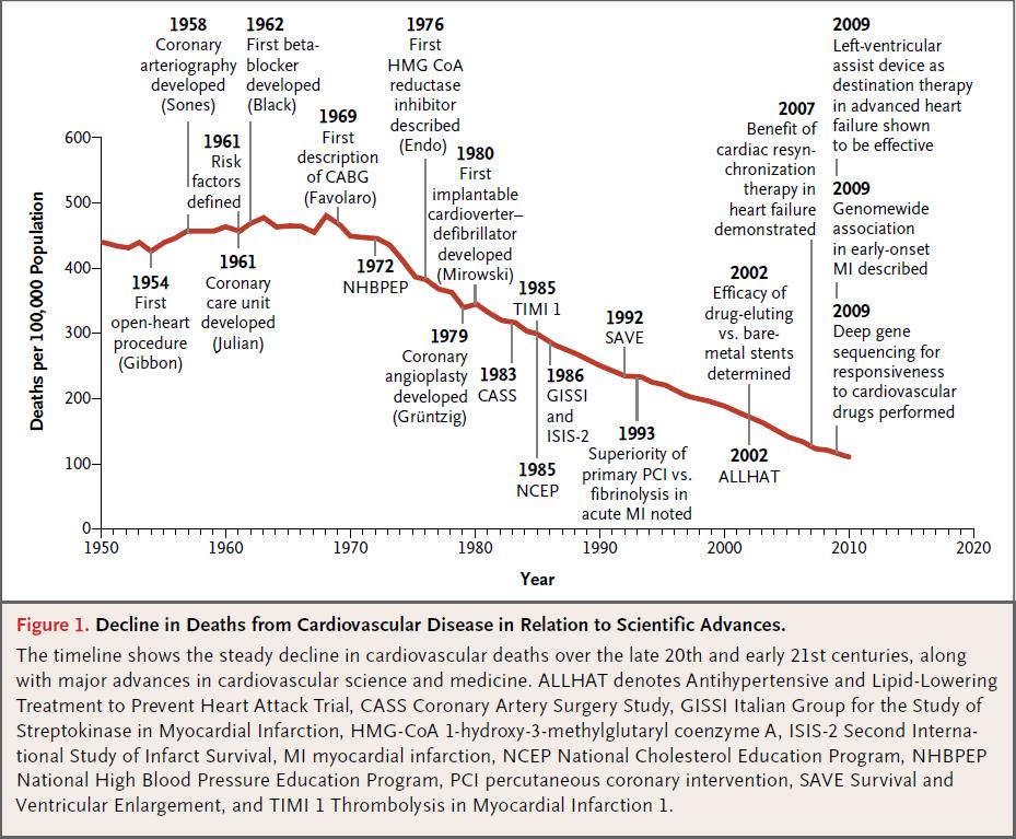
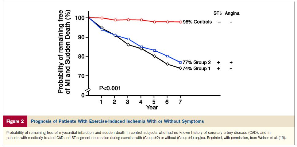
3 N Engl J Med 2012;366:54-63
4 Clinical relevance Treatment is warranted To alleviate symptoms To improve prognosis The balance between these is governed by Circumstances Age Expectations. Balance between risk and benefit Cost benefit / effectiveness / social acceptance
5
6 N Engl J Med 2012;366:54-63
7 Number of MI patients Acute Myocardial Infarctions Evolve Most Frequently From Plaques With Mild to Moderate Obstruction Ambrose et al Little et al Nobuyoshi et al Giroud et al ALL <50% 50-70% >70% E Falk, PK Shah, V Fuster. Circulation 1995;92:657
 74% Berenson Children 8% Middle-age 69% Arbustini 42% Thrombosis 8% Angelini 20%")
8 Coronary Lesions on Pathology Korean war 77% Vietnam 45% Velican 33% Prevalence Baroldi (40yrs) 74% Berenson Children 8% Middle-age 69% Arbustini 42% Thrombosis 8% Angelini 20% McGill 20%
9
10

 NC Rupture Site")

")

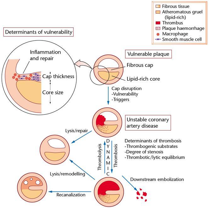
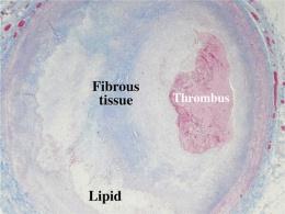
11 Main Causes of Coronary Thrombosis Rupture (65%) Erosion (30%) NC Rupture Site Th Th NC Th Pathologic Intimal Thickening (50%) Fibroatheroma (50%) Th Th Th Virmani R, et al. Arterioscler Thromb Vasc Biol 2000;20:1262
12 Frequency of Coronary Thrombi in Culprit Lesions by Decade in Men Dying Sudden Coronary Death Percent 7o * p=0.05 # * # * # * # p=0.05 Acute Thrombus Plaque Rupture Plaque Erosion Stable Plaque Age Range in Years R. Virmani

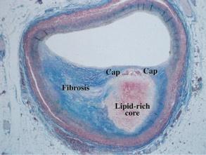
13 Rupture, Stenosis, Cap Thickness and Necrotic Core Arterioscler Thromb Vasc Biol. 2010;30:

 32,")
14 European Heart Journal (2011) 32,
15 European Heart Journal (2011) 32,
16 Circ Cardiovasc Interv. 2011;4:

17
18 Prognosis Ischemia Presence Extent + knowledge of viability Prevention of ACS Unstable plaque Evolution towards Heart Failure Arrhythmia risk Borderzone of necrosis with mixture of normal cells, fibrosis, persistent ischemia
19 What can be of help? Risk factors Symptoms Calcium score Stenosis severity Ischemia Can guide therapy
20 Stenosis classification 2 J Am Coll Cardiol 2010;55:

21 FFR in Left Main Disease Circulation. 2009;120:
22 Choice of treatment J Nucl Med 2006; 47:


23 J Am Coll Cardiol 2012;59:
24
25
26
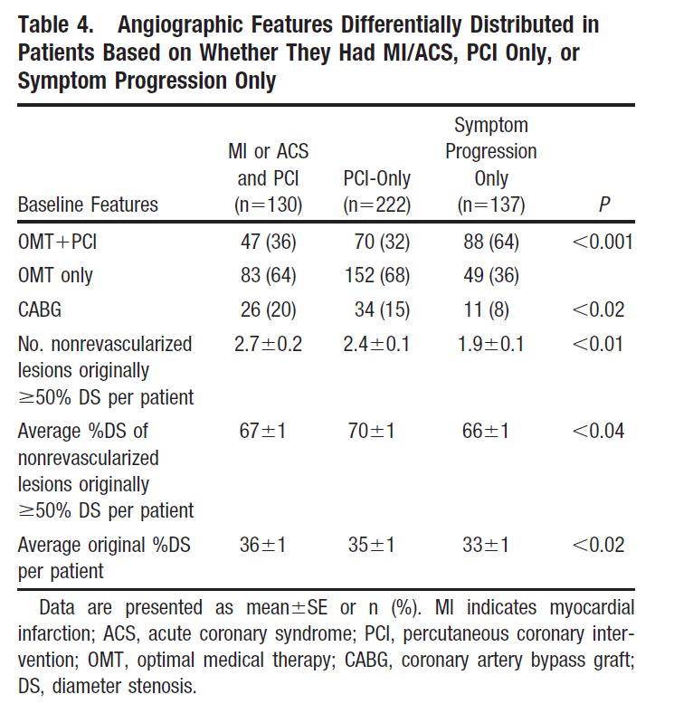
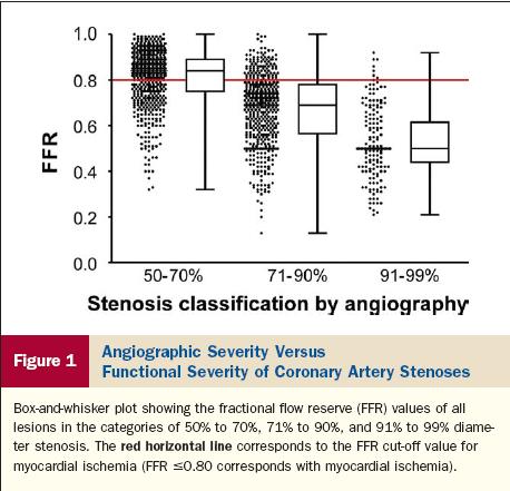
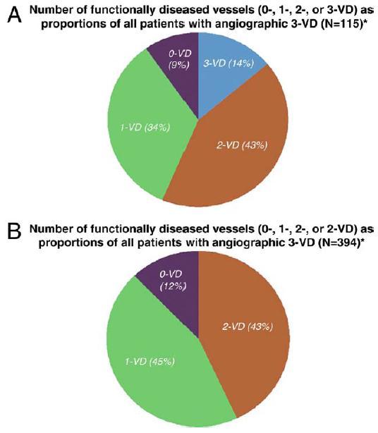
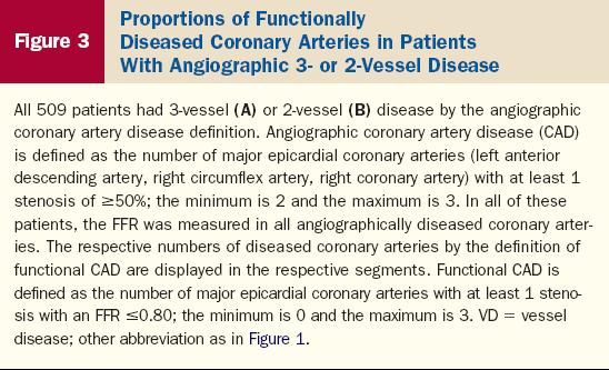
27 J Am Coll Cardiol 2012;59:825 35

28 J Am Coll Cardiol 2012;59:825 35
29 Teaching Cases JAN BOGAERT CLINICAL CARDIAC MRI S E C O N D E D I T I O N


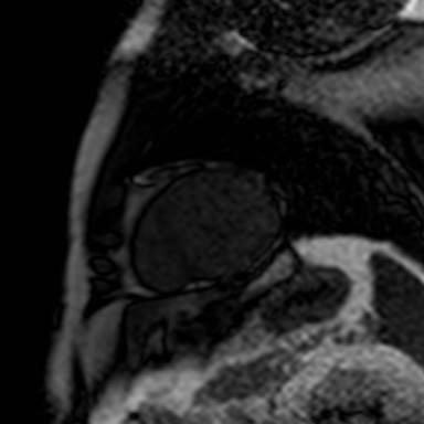
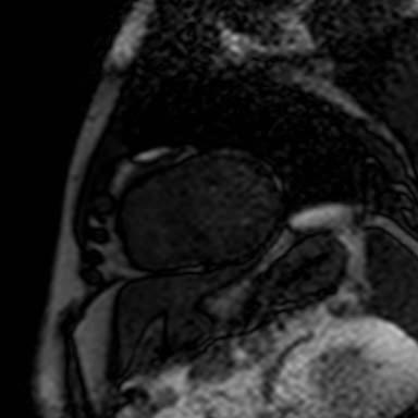
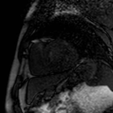
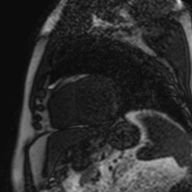
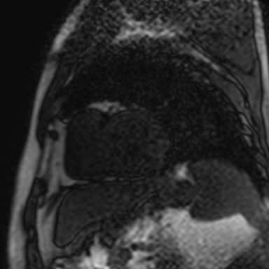
30 36-year-old man admitted with retrosternal pain, nicotine abuse. ECG: non-stemi / troponin I : 16 mg/l. Occlusion 1st lateral branch. Cardiac MRI: LV EDV 219 ml - EF 57%, mild hypokinesia in lateral wall, T2wimaging: edema in anterolateral wall (arrows, still frames upper row), with strong transmural enhancement on late Gd MRI (arrows, lower panels). Acute MI (Non-STEMI)


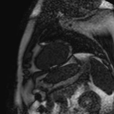
31 Idiopathic DCM 11-year-old girl presenting with DCM. Cardiac MRI shows severely dilated and dysfunctional ventricles (LV EDV 324 ml - EF 9% / RV EDV 520 ml EF 7%) with left-sided cardiac rotation / dilated atria and dilated IVC / mild pericardial effusion. Late Gd imaging shows diffuse strong enhancement of epicardial LV borders and diffuse RV enhancement. Myocardial biopsy shows myocardial replacement fibrosis. See Fig. 27 Heart Muscle Diseases
 and diffuse enhancement of RV wall (arrowheads, d,e,f) except the anterior part of RV free wall.")
32 RV Myocardial Infarction (2) T2w-imaging shows focal edema of LV inferoseptal wall (arrow, c) and diffuse edema in RV wall (arrowheads, c). Late Gd imaging shows focal transmural enhancement in LV inferoseptal wall (arrow, d) and diffuse enhancement of RV wall (arrowheads, d,e,f) except the anterior part of RV free wall. Findings of an acute MI involving both ventricles (limited LV, extensive RV) with a severe impact on RV function. See Fig. 31 Ischemic Heart Disease


 shows decreased LV function")
.")
33 Stress Cardiomyopathy (Apical Form) 72-year-old woman admitted with chest pain irradiating to left arm during walking. 1-2 mm Elevation in V3-V5. Mild increase of cardiac enzymes (Troponin I: 3.8 mg/l). No significant CAD on coronary angiography but akinesia of apical half of LV, resembling a Tako-Tsubo. Cardiac MRI (performed two days after the acute event) shows decreased LV function (EF 41%), with residual akinesia of the entire LV apex and diffuse myocardial edema on T2w-imaging in LV apex (right panel). No abnormal myocardial enhancement on late Gd imaging. Small amount of pericardial fluid and bilateral pleural fluid. Fig. 47 Heart Muscle Diseases


34 Stress Perfusion Defect in LAD Territory STRESS MPI REST MPI See Fig. 8 Myocardial Perfusion

 and function (EF 56%) at rest (left panel) but extensive")
 immediately after perfusion MRI shows severe dysfunction in non-enhanced")
 transmural enhancement in anterolateral wall (segments 1,7,12).")
35 Recurrent Ischemia Post-Revascularization (1) 43-year-old woman with recent history of CAD, with LAD stent for ACS complicated with LM dissection, urgent CABG (GSV end-to-side LAD, GSV side-to-side ramus angularis, GSD end-to-side LCx). Recurrent exercise-related interscapular chest pain. Cardiac MRI shows normal LV volumes (EDV 166 ml) and function (EF 56%) at rest (left panel) but extensive and long-lasting perfusion defect during persantine stress (segments 1,2,6,7,8,11,12,13,14). Real-time cine MRI (right panel) immediately after perfusion MRI shows severe dysfunction in non-enhanced mycardial regions. Late Gd imaging shows smal(ler) transmural enhancement in anterolateral wall (segments 1,7,12). Cardiac catheterization shows occlusion of LAD and venous graft to LAD with filling of distal LAD by RCA collaterals, slow flow in venous graft to LCx.
 in the hypoperfused myocardium")
36 Recurrent Ischemia Post-Revascularization (2) Stress perfusion imaging at 2 short-axis levels (a,b) shows extensive anterior (including anteroseptum and lateral wall, arrows, a,b). Cine imaging performed immediately following stress perfusion imaging shows severely impaired myocardial contractility (due to myocardial ischemia) in the hypoperfused myocardium (arrows, c,d).


37 Comprehensive MRI in IHD (1) 46-year-old patient with multi-vessel CAD and recent history of NSTEMI. Cardiac catherization shows occlusion of mid RCA and 2nd lateral branch LCx. Non-stenotic CAD in LAD. RCA filling by LAD collaterals. PCI 2nd lateral branch LCx. LV EDV 215 ml SV 111 ml EF 52%. Mild thinning of the LV mid anterolateral wall showing mild to moderate hypokinesia.
38 Comprehensive MRI in IHD (2) STRESS MPI REST MPI
.")

39 Comprehensive MRI in IHD (3) Rest MPI(previous slide) shows a perfusion defect in the LV anterolateral wall, corresponding to the area of enhancement on late Gd imaging. Stress MPI shows extensive and long-lasting perfusion defect in LV inferior wall (segments 3,4,9,10,15). Late Gd imaging (suboptimal image quality) shows almost complete transmural enhancement of the LV anterolateral wall (segments 6,12,16). Findings of anterolateral transmural MI, and extensive stress-induced perfusion defect in RCA territory. Rest cardiac volumes/function within normal limits.


40 Circ Cardiovasc Imaging published online February 16, 2012

41 J Am Coll Cardiol 2012;59:462 74

42 J Am Coll Cardiol 2012;59:462 74

43 J Am Coll Cardiol 2012;59:462 74
44 Conclusion Look for ischemia and viability Presence Extent Use the technique you know best ACT UPON YOUR FINDINGS
45
State of the Art. Advances in Cardiovascular Imaging. ESC Congres Stockholm September 1, 2010 Frank E. Rademakers, MD, PhD, FESC
 State of the Art Advances in Cardiovascular Imaging ESC Congres Stockholm September 1, 2010 Frank E. Rademakers, MD, PhD, FESC Coronary Artery Disease Content Patho Physiology Imaging requirements Economical
Coronary interventions
 Controversial issues in the management of ischemic heart failure Coronary interventions Maciej Lesiak Department of Cardiology, University Hospital in Poznan none DECLARATION OF CONFLICT OF INTEREST CHF
Current Guidelines for Diagnosis of AMI Chest pain ST change on EKG Cardiac Enzymes
 Noninvasive Cardiac Imaging in Myocardial Infarction Sangchol Lee Sungkyunkwan University Samsung Medical Center Current Guidelines for Diagnosis of AMI Chest pain ST change on EKG Cardiac Enzymes Do We
MRI ACS-ben. Tamás Simor MD, PhD, Med Hab. University of Pécs, Heart Institute
 MRI ACS-ben Tamás Simor MD, PhD, Med Hab Time Course of Changes in Infarct Size, Viable Myocardium, and LV Mass After Reperfused and Nonreperfused MI Blue lines denote reperfused myocardial infarction
Ischemic heart disease
 Ischemic heart disease Introduction In > 90% of cases: the cause is: reduced coronary blood flow secondary to: obstructive atherosclerotic vascular disease so most of the time it is called: coronary artery
Rational use of imaging for viability evaluation
 EUROECHO and other imaging modalities 2011 Rational use of imaging for viability evaluation Luc A. Pierard, MD, PhD, FESC, FACC Professor of Medicine Head, Department of Cardiology, CHU Liège, Belgium
The Value of Stress MRI in Evaluation of Myocardial Ischemia
 The Value of Stress MRI in Evaluation of Myocardial Ischemia Dr. Saeed Al Sayari, MBBS, EBCR, MBA Department of Radiology and Nuclear Medicine Mafraq Hospital, Abu Dhabi United Arab Emirates Introduction
Cardiac MRI: Cardiomyopathy
 Cardiac MRI: Cardiomyopathy Laura E. Heyneman, MD I do not have any relevant financial relationships with any commercial interests Cardiac MRI: Cardiomyopathy Laura E. Heyneman, MD Duke University Medical
J. Schwitter, MD, FESC Section of Cardiology
 J. Schwitter, MD, FESC Section of Cardiology CMR Center of the CHUV University Hospital Lausanne - CHUV Switzerland Centre de RM Cardiaque J. Schwitter, MD, FESC Section of Cardiology CMR Center of the
Malaysian Healthy Ageing Society
 Organised by: Co-Sponsored: Malaysian Healthy Ageing Society CAD INVESTIGATIONS PHYSIOLOGICAL / FUNCTIONAL VS ANATOMICAL / STRUCTURAL Disclosure iheal medical centre is a one stop cardiac centre with
The use of Cardiac CT and MRI in Clinical Practice
 The use of Cardiac CT and MRI in Clinical Practice Matthew W. Martinez, MD Assistant Professor of Medicine LVPG - Lehigh Valley Heart Specialists Lehigh Valley Health Network Oct. 3, 2009 DISCLOSURE Relevant
Pearls & Pitfalls in nuclear cardiology
 Pearls & Pitfalls in nuclear cardiology Maythinee Chantadisai, MD., NM physician Division of Nuclear Medicine, Department of radiology, KCMH Principle of myocardial perfusion imaging (MPI) Radiotracer
Imaging ischemic heart disease: role of SPECT and PET. Focus on Patients with Known CAD
 Imaging ischemic heart disease: role of SPECT and PET. Focus on Patients with Known CAD Hein J. Verberne Academic Medical Center, University of Amsterdam, Amsterdam, Netherlands International Conference
Acute Myocardial Infarction
 Acute Myocardial Infarction Hafeza Shaikh, DO, FACC, RPVI Lourdes Cardiology Services Asst.Program Director, Cardiology Fellowship Associate Professor, ROWAN-SOM Acute Myocardial Infarction Definition:
MR Assessment of Myocardial Viability
 MR Assessment of Myocardial Viability Definition of Viability Clinical Metabolism: Presence of glucose uptake Perfusion / Perfusion reserve Morphology: Wall thickness, wall thickening Contractility: Recovery
Left main coronary artery (LMCA): The proximal segment
: The proximal segment") Anatomy and Pathology of Left main coronary artery G Nakazawa Tokai Univ. Kanagawa, Japan 1 Anatomy Difinition Left main coronary artery (LMCA): The proximal segment RCA AV LAD LM LCX of the left coronary
Anatomy and Pathology of Left main coronary artery G Nakazawa Tokai Univ. Kanagawa, Japan 1 Anatomy Difinition Left main coronary artery (LMCA): The proximal segment RCA AV LAD LM LCX of the left coronary
Chronic Total Occlusions. Stephen Cook, MD Medical Director, Cardiac Catheterization Laboratory Oregon Heart & Vascular Institute
 Chronic Total Occlusions Stephen Cook, MD Medical Director, Cardiac Catheterization Laboratory Oregon Heart & Vascular Institute Financial Disclosures /see -tee-oh / abbr. Med. Chronic Total Occlusion,
The role of Magnetic Resonance Imaging in the diagnosis of viability & Coronary Artery Disease
 The role of Magnetic Resonance Imaging in the diagnosis of viability & Coronary Artery Disease G.P. Spanos, MSc, Phd Head of CardioVascular Imaging Tomographia Diagnostic Center Cardiovascular magnetic
Echo in CAD: Wall Motion Assessment
 Echo in CAD: Wall Motion Assessment Joe M. Moody, Jr, MD UTHSCSA and STVHCS October 2007 Relevant References ACC/AHA/ASE 2003 Guideline Update for the Clinical Application of Echocardiography Bayes de
Use of Nuclear Cardiology in Myocardial Viability Assessment and Introduction to PET and PET/CT for Advanced Users
 Use of Nuclear Cardiology in Myocardial Viability Assessment and Introduction to PET and PET/CT for Advanced Users February 1 5, 2011 University of Santo Tomas Hospital Angelo King A-V Auditorium Manila,
Advanced Imaging MRI and CTA
 Advanced Imaging MRI and CTA Who and why may benefit. Matthew W. Martinez, M.D. FACC Lehigh Valley Health Network Director, Cardiovascular Imaging Learning Objectives Review basics of CMR and CTA Review
Clinical Summary. Live Cases I - IX
 Clinical Summary Live Cases I - IX Case #1 2017/09/16 Age: 66 Target Vessel: RCA proximal Relevant Diagnosis: Single vessel CAD with normal LVEF Coronary Risk Factors: Hypertension, smoke (90py) ECG abnormality:
Clinical Summary. Live Cases I - IX
 Clinical Summary Live Cases I - IX Patient: Male, 66 years Diagnosis: Single vessel CAD with normal LVEF Target lesion: proximal RCA Coronary risk factor: Hypertension, smoke (90py) Clinical course: ECG
Heart disease remains the leading cause of morbidity and mortality in industrialized nations. It accounts for nearly 40% of all deaths in the United
 Heart disease remains the leading cause of morbidity and mortality in industrialized nations. It accounts for nearly 40% of all deaths in the United States, totaling about 750,000 individuals annually
ECG in coronary artery disease. By Sura Boonrat Central Chest Institute
 ECG in coronary artery disease By Sura Boonrat Central Chest Institute EKG P wave = Atrium activation PR interval QRS = Ventricle activation T wave= repolarization J-point EKG QT interval Abnormal repolarization
ST - segment Elevation Myocardial Infarction complicating an atypical Kawasaki disease
 ST - segment Elevation Myocardial Infarction complicating an atypical Kawasaki disease Raluca PRISECARU, Marc VINCENT, Steven VERCAUTEREN Brussels Heart Center, Brussels, Belgium Disclosure None Clinical
Imaging in Ischemic Heart Disease: Role of Cardiac MRI
 Imaging in Ischemic Heart Disease: Role of Cardiac MRI Chiara Bucciarelli Ducci MD, PhD, FESC, FRCP Consultant Senior Lecturer Cardiologist Bristol Heart Institute, University of Bristol, UK Chair elect,
Acute Myocardial Infarction. Willis E. Godin D.O., FACC
 Acute Myocardial Infarction Willis E. Godin D.O., FACC Acute Myocardial Infarction Definition: Decreased delivery of oxygen and nutrients to the myocardium Myocardial tissue necrosis causing irreparable
Current Indications for Cardiac MRI: What You See is What You Get?
 Current Indications for Cardiac MRI: What You See is What You Get? Javier Ganame, MD, PhD, FASE No disclosures Cardiology Update, Niagara, Sept 24th, 2016 The Ideal Diagnostic Technique Easy to apply Accurate
XVth Balkan Congress of Radiology Danubius Hotel Helia, October 2017, Budapest, Hungary
 XVth Balkan Congress of Radiology Danubius Hotel Helia, 12-14 October 2017, Budapest, Hungary Ružica Maksimović MRI in Myocarditis Faculty of Medicine, University of Belgrade, Centre for Radiology and
Hospital, 6 Lukon Road, Lukong Town, Changhua Shien, Taiwan 505, Taiwan.
 Volume 1, Issue 1 Image Article Resolution of Inferior Wall Ischemia after Successful Revascularization of LAD Lesion: The Value of Myocardial Perfusion Imaging in Guiding Management of Multi-vessel CAD
Culprit Lesion Remodeling and Long-term (> 5years) Prognosis in Patients with Acute Coronary Syndrome
 Prognosis in Patients with Acute Coronary Syndrome") Culprit Lesion Remodeling and Long-term (> 5years) Prognosis in Patients with Acute Coronary Syndrome Hiroyuki Okura*, MD; Nobuya Matsushita**,MD Kenji Shimeno**, MD; Hiroyuki Yamaghishi**, MD Iku Toda**,
Culprit Lesion Remodeling and Long-term (> 5years) Prognosis in Patients with Acute Coronary Syndrome Hiroyuki Okura*, MD; Nobuya Matsushita**,MD Kenji Shimeno**, MD; Hiroyuki Yamaghishi**, MD Iku Toda**,
3/27/2014. Introduction.
 Introduction. Myocardial perfusion & contractility becomes abnormal immediately after the onset of ischaemia, even before the development of the symptoms & ST segment changes. 1 Myocardial Wall Motion
Radiologic Assessment of Myocardial Viability
 November 2001 Radiologic Assessment of Myocardial Viability Joshua Moss, Harvard Medical School Year III Patient EF 66yo female with a 3-year history of intermittent chest pain previously relieved by sublingual
Sung A Chang Department of Internal Medicine, Division of Cardiology, Sungkyunkwan University School of Medicine, Samsung Medical Center
 CMR Perfusion and Viability A STICH Out of Time? Sung A Chang Department of Internal Medicine, Division of Cardiology, Sungkyunkwan University School of Medicine, Samsung Medical Center Can Imaging Improve
UPDATE ON THE MANAGEMENTACUTE CORONARY SYNDROME. DR JULES KABAHIZI, Psc (Rwa) Lt Col CHIEF CONSULTANT RMH/KFH 28 JUNE18
 Lt Col CHIEF CONSULTANT RMH/KFH 28 JUNE18") UPDATE ON THE MANAGEMENTACUTE CORONARY SYNDROME DR JULES KABAHIZI, Psc (Rwa) Lt Col CHIEF CONSULTANT RMH/KFH 28 JUNE18 INTRODUCTION The clinical entities that comprise acute coronary syndromes (ACS)-ST-segment
UPDATE ON THE MANAGEMENTACUTE CORONARY SYNDROME DR JULES KABAHIZI, Psc (Rwa) Lt Col CHIEF CONSULTANT RMH/KFH 28 JUNE18 INTRODUCTION The clinical entities that comprise acute coronary syndromes (ACS)-ST-segment
Fractional Flow Reserve: Basics, FAME 1, FAME 2. William F. Fearon, MD Associate Professor Stanford University Medical Center
 Fractional Flow Reserve: Basics, FAME 1, FAME 2 William F. Fearon, MD Associate Professor Stanford University Medical Center Conflict of Interest Advisory Board for HeartFlow Research grant from St. Jude
1. LV function and remodeling. 2. Contribution of myocardial ischemia due to CAD, and
 1 The clinical syndrome of heart failure in adults is commonly associated with the etiologies of ischemic and non-ischemic dilated cardiomyopathy, hypertrophic cardiomyopathy, hypertensive heart disease,
All About STEMIs. Presented By: Brittney Urvand, RN, BSN, CCCC. Essentia Health Fargo Cardiovascular Program Manager.
 All About STEMIs Presented By: Brittney Urvand, RN, BSN, CCCC Essentia Health Fargo Cardiovascular Program Manager Updated 10/2/2018 None Disclosures Objectives Identify signs and symptoms of a heart attack
Multimodality Imaging in Spontaneous Coronary Artery Dissection in the Peripartum Period
 Multimodality Imaging in Spontaneous Coronary Artery Dissection in the Peripartum Period Marysia Tweet, MD NASCI Annual Meeting October 18 th, 2016 2016 MFMER slide-1 DISCLOSURE No relevant financial relationship(s)
Coronary Artery Disease in the 21 st Century: An Integrated Approach Based on Science and Art
 Coronary Artery Disease in the 21 st Century: An Integrated Approach Based on Science and Art Harisios Boudoulas, MD, Dr, Dr. Hon. Professor, Honorary Professor, Academician Development of Coronary Artery
Imaging of Coronary Artery Disease: II
 Acta Radiológica Portuguesa, Vol.XIX, nº 74, pág. 45-51, Abr.-Jun., 2007 Imaging of Coronary Artery Disease: II Jean Jeudy University of Maryland School of Medicine Department of Diagnostic Radiology Armed
Assessment of plaque morphology by OCT in patients with ACS
 Assessment of plaque morphology by OCT in patients with ACS Takashi Akasaka, M.D. Department of Cardiovascular Medicine Wakayama, Japan Unstable plaque Intima Lipid core Plaque rupture and coronary events
Myocardial Infarction
 Myocardial Infarction MI = heart attack Defined as necrosis of heart muscle resulting from ischemia. A very significant cause of death worldwide. of these deaths, 33% -50% die before they can reach the
CASE from South Korea
 CASE from South Korea Bon-Kwon Koo, MD, PhD, Seoul, Korea Outpatient clinic of a non-interventional cardiologist F/56 Chief complaint: Angina with recent aggravation, CCS II~III Brief history: # Stroke
Abnormal, Autoquant Adenosine Myocardial Perfusion Heart Imaging. ID: GOLD Date: Age: 46 Sex: M John Doe Phone (310)
") Background: Reason: preoperative assessment of CAD, Shortness of Breath Symptom: atypical chest pain Risk factors: hypertension Under influence: a beta blocker Medications: digoxin Height: 66 in. Weight:
Background: Reason: preoperative assessment of CAD, Shortness of Breath Symptom: atypical chest pain Risk factors: hypertension Under influence: a beta blocker Medications: digoxin Height: 66 in. Weight:
Atypical pain and normal exercise test
 Atypical pain and normal exercise test F. Mut, M. Beretta Nuclear Medicine Service, Asociacion Española Montevideo, Uruguay Clinical history 67-year old male with several coronary risk factors. Atypical
Chronic Total Occlusion: A case for coronary artery bypass grafting
 Chronic Total Occlusion: A case for coronary artery bypass grafting Prof. Alfredo R Galassi MD, FESC, FACC, FSCAI Director of Cardiac Catheterization and Interventional Cardiology Unit Department of Medical
Acute heart failure in a patient with lower urinary tract infection Case report of an infection-induced Reverse Takotsubo syndrome
 Acute heart failure in a patient with lower urinary tract infection Case report of an infection-induced Reverse Takotsubo syndrome N.Μoschos, A.Dimitra, E.Tsakiri, D.Stavrianakis, A.Nouli CARDIOLOGY DEPARTMENT
Non-Invasive Evaluation of Coronary Vasospasm Using a Combined Hyperventilation and Cold-Pressure-Test Perfusion CMR Protocol
 Journal of Cardiovascular Magnetic Resonance (2007) 9, 759 764 Copyright c 2007 Informa Healthcare USA, Inc. ISSN: 1097-6647 print / 1532-429X online DOI: 10.1080/10976640701544662 Non-Invasive Evaluation
Fractional Flow Reserve. A physiological approach to guide complex interventions
 Fractional Flow Reserve A physiological approach to guide complex interventions What is FFR? Fractional Flow Reserve (FFR) is a lesion specific, physiological index determining the hemodynamic severity
CABG Surgery following STEMI
 CABG Surgery following STEMI Susana Harrington, MS,APRN-NP Cardio-Thoracic Surgery Nebraska Methodist Hospital February 15, 2018 2013 ACCF/AHA Guideline for the Management of ST-Elevation Myocardial Infarction:
Ventricular tachycardia and ischemia. Martin Jan Schalij Department of Cardiology Leiden University Medical Center
 Ventricular tachycardia and ischemia Martin Jan Schalij Department of Cardiology Leiden University Medical Center Disclosure: Research grants from: Boston Scientific Medtronic Biotronik Sudden Cardiac
Fourth Universal Definition of Myocardial Infarction (2018)
") EUSEM, Glasgow 2018 Fourth Universal Definition of Myocardial Infarction (2018) Univ.-Prof. Dr. Martin Möckel, Division of Emergency and Acute Medicine, Charité Universitätsmedizin Berlin 2 1 History of
EUSEM, Glasgow 2018 Fourth Universal Definition of Myocardial Infarction (2018) Univ.-Prof. Dr. Martin Möckel, Division of Emergency and Acute Medicine, Charité Universitätsmedizin Berlin 2 1 History of
Assessment of Vulnerable Plaque by IVUS and VH-IVUS
 Assessment of Vulnerable Plaque by IVUS and VH-IVUS Akiko Maehara, MD Director of Intravascular Imaging & Physiology Core Laboratories Associate Director of MRI/MDCT Core Laboratory Cardiovascular Research
Can Angiographic Complete Revascularization Improve Outcomes for Patients with Decreased LV Function? NO!
 Can Angiographic Complete Revascularization Improve Outcomes for Patients with Decreased LV Function? NO! Young-Hak Kim, MD, PhD Heart Institute, University of Ulsan College of Medicine Asan Medical Center,
Hybrid cardiac imaging Advantages, limitations, clinical scenarios and perspectives for the future
 Hybrid cardiac imaging Advantages, limitations, clinical scenarios and perspectives for the future Prof. Juhani Knuuti, MD, FESC Turku, Finland Disclosure: Juhani Knuuti, M.D. Juhani Knuuti, M.D. has financial
Cardiogenic Shock. Carlos Cafri,, MD
 Cardiogenic Shock Carlos Cafri,, MD SHOCK= Inadequate Tissue Mechanisms: Perfusion Inadequate oxygen delivery Release of inflammatory mediators Further microvascular changes, compromised blood flow and
4/11/2017. Cardiomyopathy. John Steuter, MD Bryan Heart. Disclosures. No Conflicts. Cardiomyopathy. WHO Classification
 Cardiomyopathy John Steuter, MD Bryan Heart Disclosures No Conflicts Cardiomyopathy WHO Classification Anatomy & physiology of the LV 1. Dilated Enlarged Systolic dysfunction 2. Hypertrophic Thickened
MultiTransmit 4D brings robustness to 3.0T cardiac imaging
 Publication for the Philips MRI Community Issue 44 SUMMER 2011 MultiTransmit 4D brings robustness to 3.0T cardiac imaging Bonn clinicians see a reduction in artifacts and inhomogeneities as a significant
Benefit of Performing PCI Based on FFR
 Benefit of Performing PCI Based on FFR William F. Fearon, MD Associate Professor Director, Interventional Cardiology Stanford University Medical Center Benefit of FFR-Guided PCI FFR-Guided PCI vs. Angiography-Guided
CCS/CAIC/CSCS Position Statement on Revascularization Multi-vessel CAD. Teo et al, Canadian Journal of Cardiology 2014;30:
 CCS/CAIC/CSCS Position Statement on Revascularization Multi-vessel CAD Teo et al, Canadian Journal of Cardiology 2014;30: 1482-1491 Parallel Paper: Canadian Cardiovascular Society Guidelines for the Diagnosis
Can IVUS Define Plaque Features that Impact Patient Care?
 Can IVUS Define Plaque Features that Impact Patient Care? A Pichard L Satler, K Kent, R Waksman, W Suddath, N Bernardo, N Weissman, M Angelo, D Harrington, J Lindsay, J Panza. Washington Hospital Center
Intervention: How and to which extent is technology helping us?
 Cardiological Society of India Congress 12th February 2016 Chennai, India Intervention: How and to which extent is technology helping us? SIMONE BISCAGLIA MD CARDIOVASCULAR INSTITUTE, FERRARA, ITALY Introduction
Cardiac MRI: Clinical Application to Disease
 Cardiac MRI: Clinical Application to Disease Jessi Smith, MD Cardiothoracic imaging, Indiana University Slides courtesy of Stacy Rissing, MD Outline Imaging planes Disease findings Pulse sequences used
Integrating IVUS, FFR, and Noninvasive Imaging to Optimize Outcomes. Gary S. Mintz, MD Cardiovascular Research Foundation
 Integrating IVUS, FFR, and Noninvasive Imaging to Optimize Outcomes Gary S. Mintz, MD Cardiovascular Research Foundation COURAGE Nuclear Substudy (n=314) Death/MI according the residual ischemia (SPECT)
Multislice coronary computed tomographic angiography in emergency department presentations of unsuspected acute myocardial infarction
 Journal of Cardiovascular Computed Tomography (2009) 3, 272 278 Case Report Multislice coronary computed tomographic angiography in emergency department presentations of unsuspected acute myocardial infarction
Supplementary material 1. Definitions of study endpoints (extracted from the Endpoint Validation Committee Charter) 1.
 1.") Rationale, design, and baseline characteristics of the SIGNIFY trial: a randomized, double-blind, placebo-controlled trial of ivabradine in patients with stable coronary artery disease without clinical
Rationale, design, and baseline characteristics of the SIGNIFY trial: a randomized, double-blind, placebo-controlled trial of ivabradine in patients with stable coronary artery disease without clinical
ADVANCED CARDIOVASCULAR IMAGING. Medical Knowledge. Goals and Objectives PF EF MF LF Aspirational
 Medical Knowledge Goals and Objectives PF EF MF LF Aspirational Know the basic principles of magnetic resonance imaging (MRI) including the role of the magnetic fields and gradient coil systems, generation
Surgical vs. Percutaneous Revascularization in Patients with Diabetes and Acute Coronary Syndrome
 Surgical vs. Percutaneous Revascularization in Patients with Diabetes and Acute Coronary Syndrome Chris C. Cook, MD Associate Professor of Surgery Director, CT Residency Program, WVU ACOI 10/17/18 No Disclosures
Optical Coherence Tomography for Intracoronary Imaging
 Optical Coherence Tomography for Intracoronary Imaging Lorenz Räber Stephan Windecker Department of Cardiology Swiss Cardiovascular Center and Clinical Trials Unit Bern Bern University Hospital, Switzerland
Perfusion, Viability, Edema and Hemorrhage: How it Can (and Should) Change Clinical Practice. Rohan Dharmakumar, Ph.D.
 Change Clinical Practice. Rohan Dharmakumar, Ph.D.") Perfusion, Viability, Edema and Hemorrhage: How it Can (and Should) Change Clinical Practice Rohan Dharmakumar, Ph.D. Director, Translational Cardiac Imaging Research Associate Director, Biomedical Imaging
Perfusion, Viability, Edema and Hemorrhage: How it Can (and Should) Change Clinical Practice Rohan Dharmakumar, Ph.D. Director, Translational Cardiac Imaging Research Associate Director, Biomedical Imaging
Typical chest pain with normal ECG
 Typical chest pain with normal ECG F. Mut, C. Bentancourt, M. Beretta Nuclear Medicine Service, Asociacion Española Montevideo, Uruguay Clinical history Male 41 y.o. Overweight, hypertension, high cholesterol,
Post PCI functional testing and imaging: case based lessons from FFR React
 Post PCI functional testing and imaging: case based lessons from FFR React Joost Daemen, MD, PhD, FESC Optics in Cardiology 2018 April 21st, 2018 10.15 10.30h Disclosure Statement of Financial Interest
Pathology of Vulnerable Plaque Angioplasty Summit 2005 TCT Asia Pacific, Seoul, April 28-30, 2005
 Pathology of Vulnerable Plaque Angioplasty Summit 25 TCT Asia Pacific, Seoul, April 28-3, 25 Renu Virmani, MD CVPath, A Research Service of the International Registry of Pathology Gaithersburg, MD Plaque
Takotsubo syndrome. Ευτυχία Σμπαρούνη, FACC, FESC
 Takotsubo syndrome Ευτυχία Σμπαρούνη, FACC, FESC Definition Takotsubo Apical ballooning Broken heart syndrome Stress cardiomyopathy Cathecholaminergic cardiomyopathy Epidemiology 1990 first report by Japanese
2017 Cardiology Survival Guide
 2017 Cardiology Survival Guide Chapter 4: Cardiac Catheterization/Percutaneous Coronary Intervention A cardiac catheterization involves a physician inserting a thin plastic tube (catheter) into an artery
Bifurcation stenting with BVS
 Bifurcation stenting with BVS Breaking the limits or just breaking the struts? Maciej Lesiak Department of Cardiology University Hospital in Poznan, Poland Disclosure Speaker s name: Maciej Lesiak I have
Safety of Single- Versus Multi-vessel Angioplasty for Patients with AMI and Multi-vessel CAD
 Safety of Single- Versus Multi-vessel Angioplasty for Patients with AMI and Multi-vessel CAD Mun K. Hong, MD Associate Professor of Medicine Director, Cardiovascular Intervention and Research Weill Cornell
Evaluation of Intermediate Coronary lesions: Can You Handle the Pressure? Jeffrey A Southard, MD May 4, 2013
 Evaluation of Intermediate Coronary lesions: Can You Handle the Pressure? Jeffrey A Southard, MD May 4, 2013 Disclosures Consultant- St Jude Medical Boston Scientific Speaker- Volcano Corporation Heart
Role of echocardiography in the assessment of ischemic heart disease 분당서울대학교병원윤연이
 Role of echocardiography in the assessment of ischemic heart disease 분당서울대학교병원윤연이 Outline Evaluation of Chest pain Evaluation of MI complications Prediction of Outcomes Evaluation of Chest pain Evaluation
Perioperative Management After Coronary Stenting: Risk Assessment Before Surgery. Christian Seiler No conflict of interest to declare.
 Perioperative Management After Coronary Stenting: Risk Assessment Before Surgery Christian Seiler No conflict of interest to declare PCI Long-Term Outcome Perioperative Management After Coronary Stenting:
Cardiac MRI: Clinical Application to Disease
 Cardiac MRI: Clinical Application to Disease Stacy Rissing, MD! Cardiothoracic imaging, Indiana University! Outline Imaging planes Disease findings Pulse sequences used for each indication Pathophysiology
I have no financial disclosures
 Manpreet Singh MD I have no financial disclosures Exercise Treadmill Bicycle Functional capacity assessment Well validated prognostic value Ischemic assessment ECG changes ST segments Arrhythmias Hemodynamic
Pathophysiology of Coronary Microvascular Dysfunction
 Pathophysiology of Coronary Microvascular Dysfunction Cheol Woong Yu, MD, PhD Cardiology Department Division of Internal Medicine Korea University Anam Hospital. Etiologies of Chest Pain without obstructive
Imaging congestive heart failure: role of coronary computed tomography angiography (CCTA)
") Imaging congestive heart failure: role of coronary computed tomography angiography (CCTA) Gianluca Pontone, MD, PhD, FESC, FSCCT Director of MR Unit Deputy Director of Cardiovascul CT Unit Clinical Cardiology
Imaging congestive heart failure: role of coronary computed tomography angiography (CCTA) Gianluca Pontone, MD, PhD, FESC, FSCCT Director of MR Unit Deputy Director of Cardiovascul CT Unit Clinical Cardiology
When Aspiration Thrombectomy Does Not Work? A A R O N W O N G N A T I O N A L H E A R T C E N T R E S I N G A P O R E
 When Aspiration Thrombectomy Does Not Work? A A R O N W O N G N A T I O N A L H E A R T C E N T R E S I N G A P O R E Thrombus in STEMI Over 70% of STEMI patients has angiographic evidence of thrombus
Case based learning: CMR in Heart Failure
 Case based learning: CMR in Heart Failure Milind Y Desai, MD FACC FAHA FESC Associate Professor of Medicine Heart and Vascular Institute, Cleveland Clinic Cleveland, OH Disclosures: none Use of Gadolinium
Testing the Asymptomatic CAD Patient: When and Why?
 Testing the Asymptomatic CAD Patient: When and Why? Timothy M. Bateman M.D. Co-Director, Cardiovascular Radiologic Imaging Mid America Heart Institute Professor of Medicine University of Missouri-Kansas
Σεμινάριο Ομάδων Εργασίας Fractional Flow Reserve (FFR) Σε ποιούς ασθενείς; ΔΗΜΗΤΡΗΣ ΑΥΖΩΤΗΣ Επιστ. υπεύθυνος Αιμοδυναμικού Τμήματος, Βιοκλινική
 Σε ποιούς ασθενείς; ΔΗΜΗΤΡΗΣ ΑΥΖΩΤΗΣ Επιστ. υπεύθυνος Αιμοδυναμικού Τμήματος, Βιοκλινική") ΕΛΛΗΝΙΚΗΚΑΡΔΙΟΛΟΓΙΚΗΕΤΑΙΡΕΙΑ Σεμινάριο Ομάδων Εργασίας 2011 Fractional Flow Reserve (FFR) Σε ποιούς ασθενείς; ΔΗΜΗΤΡΗΣ ΑΥΖΩΤΗΣ Επιστ. υπεύθυνος Αιμοδυναμικού Τμήματος, Βιοκλινική GUIDELINES ON MYOCARDIAL
ΕΛΛΗΝΙΚΗΚΑΡΔΙΟΛΟΓΙΚΗΕΤΑΙΡΕΙΑ Σεμινάριο Ομάδων Εργασίας 2011 Fractional Flow Reserve (FFR) Σε ποιούς ασθενείς; ΔΗΜΗΤΡΗΣ ΑΥΖΩΤΗΣ Επιστ. υπεύθυνος Αιμοδυναμικού Τμήματος, Βιοκλινική GUIDELINES ON MYOCARDIAL
FFR in unstable angina and after MI F
 FFR in unstable angina and after MI F June-Hong Kim, MD. PhD Cardiovascular center Pusan National University Yangsan Hospital FFR tells you physiologic stenosis severity rather than anatomical stenosis
FFR in Left Main Disease
 FFR in Left Main Disease William F. Fearon, MD Associate Professor of Medicine Director, Interventional Cardiology Stanford University Medical Center Why FFR instead of IVUS? Physiologic versus anatomic
Role of CMR in heart failure and cardiomyopathy
 Role of CMR in heart failure and cardiomyopathy Hajime Sakuma Department of Radiology, Mie University Late gadolinium enhancement (LGE) LGE MRI can demonstrate site of necrosis, fibrosis or deposition
Myocardial Infarction. Reading Assignment (p66-78 in Outline )
") Myocardial Infarction Reading Assignment (p66-78 in Outline ) Objectives 1. Why do ST segments go up or down in ischemia? 2. STEMI locations and culprit vessels 3. Why 15-lead ECGs? 4. What s up with avr?
Myocardial Infarction Reading Assignment (p66-78 in Outline ) Objectives 1. Why do ST segments go up or down in ischemia? 2. STEMI locations and culprit vessels 3. Why 15-lead ECGs? 4. What s up with avr?
Stable Angina: Indication for revascularization and best medical therapy
 Stable Angina: Indication for revascularization and best medical therapy Cardiology Basics and Updated Guideline 2018 Chang-Hwan Yoon, MD/PhD Cardiovascular Center, Department of Internal Medicine Bundang
Myocardial contusion injury (MCI) may occur as a rare
 may occur as a rare") Cardiovascular Images Myocardial Contusion in an 8-Year-Old Boy A Kick to the Heart Danielle M. Moyé, MD; Adrian K. Dyer, MD; Poonam P. Thankavel, MD Myocardial contusion injury (MCI) may occur as a rare
Cardiovascular Images Myocardial Contusion in an 8-Year-Old Boy A Kick to the Heart Danielle M. Moyé, MD; Adrian K. Dyer, MD; Poonam P. Thankavel, MD Myocardial contusion injury (MCI) may occur as a rare
Pregnancy in Patients with a History of Spontaneous Coronary Artery Dissection (SCAD)
") Pregnancy in Patients with a History of Spontaneous Coronary Artery Dissection (SCAD) Marysia Tweet, MD The 4 th International Congress on Cardiac Problems in Pregnancy February 29 th, 2016 2015 MFMER
Pregnancy in Patients with a History of Spontaneous Coronary Artery Dissection (SCAD) Marysia Tweet, MD The 4 th International Congress on Cardiac Problems in Pregnancy February 29 th, 2016 2015 MFMER
Dr Felix Keng. Imaging of the heart is technically difficult because: Role of Cardiac MSCT. Current: Cardiac Motion Respiratory Motion
 Siemens Philips Dr Felix Keng GE Toshiba Role of Cardiac MSCT Current: Structural / congenital heart imaging Extra-cardiac / Great vessel imaging Volumes and ejection fractions (cine + gating) Calcium
Cardiovascular Nursing Practice: A Comprehensive Resource Manual and Study Guide for Clinical Nurses 2 nd Edition
 Cardiovascular Nursing Practice: A Comprehensive Resource Manual and Study Guide for Clinical Nurses 2 nd Edition Table of Contents Volume 1 Chapter 1: Cardiovascular Anatomy and Physiology Basic Cardiac