Pathology of Vulnerable Plaque Angioplasty Summit 2005 TCT Asia Pacific, Seoul, April 28-30, 2005
|
|
|
- Adela Higgins
- 6 years ago
- Views:
Transcription


1 Pathology of Vulnerable Plaque Angioplasty Summit 25 TCT Asia Pacific, Seoul, April 28-3, 25 Renu Virmani, MD CVPath, A Research Service of the International Registry of Pathology Gaithersburg, MD


2 Plaque Rupture 55-6% Plaque Morphology of Thrombi in SCD th th Plaque Erosion 3-35% th th Calcified Nodule 2-77 % th
3 Clinical and Morphologic Difference in Plaques Associated with Luminal Thrombi Plaque rupture Plaque erosion Calcified nodule Lumen Th Necrotic core Lumen Th Th 45-55% thrombi in SCD M>F, Older, Ca ++ Eccentric = concentric Greater % stenosis Macs, T cells,hladr 35-4% thrombi in SCD M=F, younger Usually eccentric Lesser % stenosis SMC rich, proteoglycans 4-7% thrombi in SCD, calcified plates M>F, older, mid RCA Usually eccentric Stenosis variable Nodules of bone


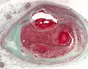
4 A B Th NC NC Gross and Light Microscopic Features of Plaque Rupture 6% of Thrombi in Sudden Coronary Death Are form Plaque Rupture D F Th SMCs T cells C NC E G Th MΦ HLA-DR Fig 3-1


5 Plaque erosion in a 33 year-old female complaining of chest pain for two-weeks and discharged from the emergency room with a diagnoses of anxiety. A B C D NC E SMCs F MΦ T-cells PLT Fibrin


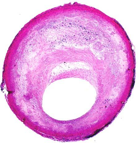
6 Development of Human Coronary Atherosclerosis Intimal thickening Intimal xanthoma Pathologic intimal thickening Fibrous cap atheroma Thin-cap Fibroatheroma LP NC NC FC Smooth muscle cells Macrophage foam cells Extracellular lipid Cholesterol clefts Necrotic core Calcified plaque Hemorrhage Thrombus Healed thrombus Collagen
7 What is a Vulnerable Plaque? Plaque morphology underlying luminal thrombi represents a vulnerable plaque Thin-cap Fibroatheroma Plaque rupture Pathologic intimal thickening Fibroatheroma Calcified plates with bone formation Calcified nodule - surface thrombus Plaque Erosion

8 Thin cap Fibroatheroma is a Precursor lesions of Plaque Rupture TCFA Plaque Rupture nc Thin fibrous cap? *Ruptured cap Th th * nc
9 Thin-Cap Atheroma (Vulnerable Plaque) Components Necrotic core Thin fibrous cap (< 65 µm) Cap infiltrated by macrophages and lymphocytes Cap composition type 1 collagen and few smooth muscle cells


10 A Non-Hemodynamically Limiting Thin-cap Fibroatheroma A NC B NC C Movat H&E D E Fig 2-2 MΦ SMCs
11 Morphologic Characteristics of Plaque Rupture and Thin-cap Fibroatheromas Plaque type Necrotic Core (%) Fibrous cap Thickness (µm) MΦs (%) SMCs (%) T- lymph Calcification Score Rupture 34±17 23±19 26±2.2±.4 4.9± ±1.3 Thin-cap Fibroatheroma 23±17 <65µm 14±1 6.6± ±1.4.97±1.1 P value.1.5 ns ns.14 Mean values are represented ± standard deviation. Abbreviations: MΦs= macrophages, SMCs= smooth muscle cells, T-lymph= T-lymphocytes Kolodgie F, et al. Current Opinion in Cardiology 21;16:285


12 Morphological Variants of the Thin-Cap Fibroatheroma Insignificant Plaque burden Large eccentric necrotic core Large concentric necrotic core Healed Rupture(s) NC NC NC NC NC NC NC Kolodgie F, et al. Current Opinion in Cardiology 21;16:285
13 Vulnerable Plaque Characteristics A % cross sectional area luminal narrowing fibrous atheroma Type of plaque thin cap hemorrhage rupture healed rupture B % thin cap atheroma <5% 5-74% Cross sectional luminal narrowing Mean % x-sectional area For TCFA is 6% 8% of thin cap atheromas occur in arteries with < 75% x-sectional area luminal narrowing (<5% diameter reduction)
14 Plaque Ruptures (%) (n= 9) Thin-Cap Fibroatheromas (%) n= 16) C A Distribution of Ruptures and Thin-Cap Atheromas by Plaque Area or Lipid Core Size Fig ± 9%* 71 ± 13%* 77 ± 15%* % <1 %1-25 %25-5 >5% Lipid core as % plaque area 65 ± 17%* 71 ± 17%* 79 ± 13%* 77 ± 9%* 76 ± 14%* % <1 %1-25 %25-5 >5% Plaque Ruptures (%) (n= 9) Thin-Cap Fibroatheromas (%) n= 16) D B ± 9%* 73 ± 13%* 77 ± 16%* 78 ± 12%* area<1 area 1-3 area 3-5 area > 5 Cross sectional lipid core area, mm 2 65 ± 15%* 74 ± 15%* 78 ± 12%* 77 ± 13%* area<1 area 1-3 area 3-5 area > 5 * = mean cross sectional area luminal narrowing

15 Do TCFAs lead to plaque progression? Movat pentachrome Sirius red Sirius red with polarized light
16 Mean % Stenosis Increases with Number of Prior Rupture Sites but the Increase with Each New Rupture is Small (<2%) Mean % stenosis A Healed Rupture Sites Number of prior ruptures, healed rupture sites Mean % stenosis B Acute Rupture Sites Number of prior ruptures, acute rupture sites Burke, A P et al. Circulation 21;13:
17 A Total Sections (%) Fig < 5 Percentage of Cross-Sectional Sectional-Area Narrowing by Plaque Morphology Thin cap Acute rupture < Healed rupture < Cross-Sectional-Area Narrowing (% ) B C X-SCA Narrowing (%) New lumen - Old lumen (%) Occlusive P=.5 N=12 N=19 Thrombus Non-Occlusive No. Healed Ruptures
18 Necrotic core size, sum mm 2, independent of plaque area, morphometrically determined, at maximal luminal narrowing of 3 major epicardial arteries P=.1 Mean necrotic core area mm P=.3 Normal TC/HDL-C TC/HDL-C > 7 TC/HDL-C >5, <7
19 Relationship of Fibrous Cap Thickness to Macrophage Infiltration P =.3 P =.6 Cell Mean for % Kp-1 1 Less than 65 µm 66-2 µm 21-3 µm More than 3 µm
20 Correlation of Fibrous Cap Thickness and Macrophage Infiltration % kp Fibrous cap thickness mm R 2 =.117 P = <.1
21 Relationship of Fibrous Cap Thickness to Underlying Percent Necrotic Core % Necrotic Core 5 Less than 65 µm 66-2 µm 21-3 µm More than 3 µm


22 A Correlation of X-Ray X Calcification with Plaque type LM B Speckled RCA LAD C Fragmented D Diffuse
23 Calcified Matrix Determined Histologically Severe Coronary Artery Disease, n=36, yrs Coronary Arteries Serially Sectioned Mean calcification score fibroatheroma Thin cap rupture erosion healed rupture Pathologic intimal thickening
24 Proportion and types of unstable plaques, by approximate distance from ostium % distribution proximal mid distal 1 Fibroatheroma hemorrhage into plaque thin cap acute rupture healed rupture
25 Frequency and Location of Unstable Lesions: Thin-cap Atheromas, Acute and Healed Ruptures in the Coronary Circulation Thin-Cap Fibroatheroma Frequency (%) Thin-cap Fibroatheroma Total Lesions= Healed Rupture Frequency (%) Plaque Rupture Frequency (%) Healed Plaque Rupture Total Lesions= Acute Plaque Rupture Total Lesions= plad prc plcx LM LOM drc dlad dlcx Kolodgie F, et al. Current Opinion in Cardiology 21;16:285
26 A. Mean Number of TCFAs Fig 2-7 P=.9 P=.1 P=.4 P=.5 3 s 4 s 5 s 6 s 7 s B. Mean Number of TCFAs Frequency of Thin-Cap Fibroatheromas by Decades 3 s 4 s 5 s 6 s 7 s No acute MI With acute MI
27 Number of Thin-Cap Atheromas in Various Coronary Syndromes in Males and Females Fig 2-11 Mean Number of Thin-Cap Atheromas MI SCD Incidental Erosion Male Female
28 Frequency of thin cap atheroma, by mechanism of death noncoronary erosion stable plaque healed rupture acute rupture Number of thin cap atheromas


29 LCx Plaque rupture A RCA LCx C nc G nc nc Thin cap Fibroatheroma F E Thin cap fibroatheroma B th nc LAD E nc F 43-year old WM collapsed at work and could not be resuscitated. nc D Fig. 11
30 Comparison of the Length, Necrotic Core Area and % Necrotic core/plaque Area Dimensions Fibroatheroma Thin-cap Atheroma Plaque Rupture Length, mm, mean/range 6 mm (range 1-18 mm) 8 mm (range 2-16 mm) 9 mm (range mm) Necrotic core area mm2 1.2 ± ± ± 5.5 % necrotic core/plaque area 15 ± 2 % 23 ± 17 % 34 ± 17 %


31 Serial Sections of a Thin-Cap Fibroatheroma Cut 25 mm apart A B C D E F G H Fig 2-16

")


32 Serial Coronary Sections (mm) Demonstrating Multiple Vulnerable Plaques and Rupture Sites Proximal LCx A B C D E F G H Distal I J K L
33 A. 3 IEL-Expected IEL (mm 2 ) Erosion Stable Thin cap atheroma Plaque hemorrhage Acute rupture Healed rupture Total occlusion IEL-Expected IEL (/plaque area) Erosion Stable Thin cap atheroma Plaque hemorrhage Acute rupture Healed rupture Total occlusion Remodeling in Varying Coronary Lesion Morphologies B
34 Mean Number of Thin Cap Fibroatheromas and Serum Cholesterol in Men P=.1 Fig TC<21 mg/dl & ratio<5 P=.1 TC>21 or ratio>5 P=.2 TC >21 mg/dl& ratio >5 P=.2 All cases Total Population Whites Blacks
35 Mean Number of Thin-Cap Fibroatheromas in 51 Women with SCD and Severe Coronary Disease A Comparison of Risk Factors Age Cholesterol P<.1 Diabetes <5 yrs >5 yrs P=.3 Normal Smoking TC >21 mg/dl ratio>5.8.6 P=.2 >1 GlyHgb.8.6 P=.9.4 Normal Fig 2-18 Glycohemoglobin. Non- Smoker Smoker
36 Serum hs-crp correlated with Immunohistochemical staining intensity of Plaques and with TCFA CRP Low CRP group (<1.µg/mL) CRP staining intensity of plaques* Mean number of thin cap atheroma High hs-crp group (>3.2µg/mL) *Grading of staining intensity was assessed on macrophages and Lipid core. A quantitative score of to 4 was applied to each. A sum of the 2 scores resulted in overall grading system of to 8
37 Thin Cap Fibroatheroma- A plaque vulnerable to rupture? Definition Frequency is higher in AMI than SCD, >males than females Higher prevalence in the presence of high TC, low HDL-C, high TC/HDL-C ration, high hs CRP (>3.2 mg/dl) Location in SCD, proximal and mid LAD, RCA, and LCX Length 2-22 mm ( mean 8 mm) % luminal narrowing (8% of TCFAs occur in lesions <5% diameter stenosis) % necrotic core is <25% of plaque area in 7% of TCFAs Calcification is not a marker of TCFA
Imaging Overview for Vulnerable Plaque: Data from IVUS Trial and An Introduction to VH-IVUS Imgaging
 Imaging Overview for Vulnerable Plaque: Data from IVUS Trial and An Introduction to VH-IVUS Imgaging Gary S. Mintz,, MD Cardiovascular Research Foundation New York, NY Today, in reality, almost everything
TVA_C02.qxd 8/8/06 10:27 AM Page 19 PART 2. Pathology
 TVA_C2.qxd 8/8/6 :27 AM Page 19 2 PART 2 Pathology TVA_C2.qxd 8/8/6 :27 AM Page TVA_C2.qxd 8/8/6 :27 AM Page 21 2 CHAPTER 2 The pathology of vulnerable plaque Renu Virmani, Allen P Burke, James T Willerson,
Pathology of the Vulnerable Plaque
 Journal of the American College of Cardiology Vol. 47, No. 8 Suppl C 2006 by the American College of Cardiology Foundation ISSN 0735-1097/06/$32.00 Published by Elsevier Inc. doi:10.1016/j.jacc.2005.10.065
Left main coronary artery (LMCA): The proximal segment
: The proximal segment") Anatomy and Pathology of Left main coronary artery G Nakazawa Tokai Univ. Kanagawa, Japan 1 Anatomy Difinition Left main coronary artery (LMCA): The proximal segment RCA AV LAD LM LCX of the left coronary
Anatomy and Pathology of Left main coronary artery G Nakazawa Tokai Univ. Kanagawa, Japan 1 Anatomy Difinition Left main coronary artery (LMCA): The proximal segment RCA AV LAD LM LCX of the left coronary
Assessment of Vulnerable Plaque by IVUS and VH-IVUS
 Assessment of Vulnerable Plaque by IVUS and VH-IVUS Akiko Maehara, MD Director of Intravascular Imaging & Physiology Core Laboratories Associate Director of MRI/MDCT Core Laboratory Cardiovascular Research
Pathology of Coronary Artery Disease
 Pathology of Coronary Artery Disease Seth J. Kligerman, MD Pathology of Coronary Artery Disease Seth Kligerman, MD Assistant Professor Medical Director of MRI University of Maryland Department of Radiology
Imaging Atheroma The quest for the Vulnerable Plaque
 Imaging Atheroma The quest for the Vulnerable Plaque P.J. de Feijter 1. Department of Cardiology 2. Department of Radiology Coronary Heart Disease Remains the Leading Cause of Death in the U.S, Causing
Can IVUS Define Plaque Features that Impact Patient Care?
 Can IVUS Define Plaque Features that Impact Patient Care? A Pichard L Satler, K Kent, R Waksman, W Suddath, N Bernardo, N Weissman, M Angelo, D Harrington, J Lindsay, J Panza. Washington Hospital Center
Vulnerable Plaque Pathophysiology, Detection, and Intervention. VP: A Local Problem or Systemic Disease. Erling Falk, Denmark
 Vulnerable Plaque Pathophysiology, Detection, and Intervention VP: A Local Problem or Systemic Disease Erling Falk, Denmark Vulnerable Plaque Pathophysiology, Detection, and Intervention VP: A Local Problem
Invasive Coronary Imaging Modalities for Vulnerable Plaque Detection
 Invasive Coronary Imaging Modalities for Vulnerable Plaque Detection Gary S. Mintz, MD Cardiovascular Research Foundation New York, NY Greyscale IVUS studies have shown Plaque ruptures do not occur randomly
State of the Art. Advances in Cardiovascular Imaging. ESC Congres Stockholm September 1, 2010 Frank E. Rademakers, MD, PhD, FESC
 State of the Art Advances in Cardiovascular Imaging ESC Congres Stockholm September 1, 2010 Frank E. Rademakers, MD, PhD, FESC Coronary Artery Disease Content Patho Physiology Imaging requirements Economical
Assessment of plaque morphology by OCT in patients with ACS
 Assessment of plaque morphology by OCT in patients with ACS Takashi Akasaka, M.D. Department of Cardiovascular Medicine Wakayama, Japan Unstable plaque Intima Lipid core Plaque rupture and coronary events
Added Value of Invasive Coronary Imaging for Plaque Rupture and Erosion
 Assessment of Coronary Plaque Rupture and Erosion Added Value of Invasive Coronary Imaging for Plaque Rupture and Erosion Yukio Ozaki, MD, PhD, FACC, FESC Cardiology Dept., Fujita Health Univ. Toyoake,
Plaque Characteristics in Coronary Artery Disease. Chourmouzios Arampatzis MD, PhD, FESC
 Plaque Characteristics in Coronary Artery Disease Chourmouzios Arampatzis MD, PhD, FESC Disclosure Statement of Financial Interest Regarding this Presentation NONE Atherosclerosis Model proposed by Stary
2yrs 2-6yrs >6yrs BMS 0% 22% 42% DES 29% 41% Nakazawa et al. J Am Coll Cardiol 2011;57:
 Pathology of In-stent Neoatherosclerosis in BMS and DES 197 BMS, 103 SES, and 106 PES with implant duration >30 days The incidence of neoatherosclerosis was significantly greater in DES (31%) than BMS
Assessment of vulnerable plaque by OCT
 Assessment of vulnerable plaque by OCT Comparison with histology and possible clinical applications Takashi Akasaka, M.D. Department of Cardiovascular Medicine Wakayama, Japan Identification of vulnerable
OCT Findings: Lesson from Stable vs Unstable Plaques
 ANGIOPLASTY SUMMIT TCTAP 2010 Imaging Workshop OCT Findings: Lesson from Stable vs Unstable Plaques Giulio Guagliumi MD Ospedali Riuniti di Bergamo, Italy DISCLOSURE OF FINANCIAL INTERESTS Consultant Boston
Usefulness of OCT during coronary intervention
 Usefulness of OCT during coronary intervention Takashi Akasaka, M.D. Department of Cardiovascular Medicine Wakayama, Japan Predictors at 12 Months of Stent Thrombosis and Target Lesion Revascularization
Sites of Atherosclerosis In order of Frequency
 Pathological Features of Peripheral Atherosclerosis: Implication for Device Development G Nakazawa Tokai Univ. Kanagawa, Japan 1 Sites of Atherosclerosis In order of Frequency carotid (3) (3) Coronary
1st Department of Cardiology, University of Athens, Hippokration Hospital, Athens, Greece
 Konstantinos Toutouzas, Maria Riga, Antonios Karanasos, Eleftherios Tsiamis, Andreas Synetos, Maria Drakopoulou, Chrysoula Patsa, Georgia Triantafyllou, Aris Androulakis, Christodoulos Stefanadis 1st Department
as a Mechanism of Stent Failure
 In-Stent t Neoatherosclerosis e osc e os s as a Mechanism of Stent Failure Soo-Jin Kang MD., PhD. University of Ulsan College of Medicine, Heart Institute Asan Medical Center, Seoul, Korea Disclosure I
Optical Coherence Tomography for Intracoronary Imaging
 Optical Coherence Tomography for Intracoronary Imaging Lorenz Räber Stephan Windecker Department of Cardiology Swiss Cardiovascular Center and Clinical Trials Unit Bern Bern University Hospital, Switzerland
Dynamics of intracoronary thrombosis in STEMI and sudden death patients Kramer, M.C.A.
 UvA-DARE (Digital Academic Repository) Dynamics of intracoronary thrombosis in STEMI and sudden death patients Kramer, M.C.A. Link to publication Citation for published version (APA): Kramer, M. C. A.
OCT; Comparative Imaging Results with IVUS, VH and Angioscopy
 OCT; Comparative Imaging Results with IVUS, VH and Angioscopy Takashi Akasaka, M.D. Department of Cardiovascular Medicine Wakayama, Japan Comparison among coronary imaging techniques OCT IVUS MRI CAG Angioscopy
Catch-up Phenomenon: Insights from Pathology
 Catch-up Phenomenon: Insights from Pathology Michael Joner, MD CVPath Institute Inc. Gaithersburg, MD USA Path Lessons learned from the BMS and DES (1 st Gen) era Neointimal Thickness [mm] In Stent Re
Histopathology: Vascular pathology
 Histopathology: Vascular pathology These presentations are to help you identify basic histopathological features. They do not contain the additional factual information that you need to learn about these
Review Article Pathologic Etiologies of Late and Very Late Stent Thrombosis following First-Generation Drug-Eluting Stent Placement
 ombosis Volume 2012, Article ID 608593, 16 pages doi:10.1155/2012/608593 Review Article Pathologic Etiologies of Late and Very Late Stent ombosis following First-Generation Drug-Eluting Stent Placement
MR Imaging of Atherosclerotic Plaques
 MR Imaging of Atherosclerotic Plaques Yeon Hyeon Choe, MD Department of Radiology, Samsung Medical Center, Sungkyunkwan University, Seoul MRI for Carotid Atheroma Excellent tissue contrast (fat, fibrous
This review will reconsider the current paradigm for
 Lessons From Sudden Coronary Death A Comprehensive Morphological Classification Scheme for Atherosclerotic Lesions Renu Virmani, Frank D. Kolodgie, Allen P. Burke, Andrew Farb, Stephen M. Schwartz This
Drug Eluting Stent DES Pathology Update What we know, what we do not know
 Drug Eluting Stent DES Pathology Update What we know, what we do not know 13 th Summit TCT Asia 25 th April 2008 Renu Virmani, MD. CVPath Institute Inc. Gaithersburg, MD Disclosure Statement of Financial
Blood Vessels. Dr. Nabila Hamdi MD, PhD
 Blood Vessels Dr. Nabila Hamdi MD, PhD ILOs Understand the structure and function of blood vessels. Discuss the different mechanisms of blood pressure regulation. Compare and contrast the following types
Ischemic heart disease
 Ischemic heart disease Introduction In > 90% of cases: the cause is: reduced coronary blood flow secondary to: obstructive atherosclerotic vascular disease so most of the time it is called: coronary artery
Chapter 43 Noninvasive Coronary Plaque Imaging
 hapter 43 Noninvasive oronary Plaque Imaging NIRUDH KOHLI The goal of coronary imaging is to define the extent of luminal narrowing as well as composition of an atherosclerotic plaque to facilitate appropriate
Can We Identify Vulnerable Patients & Vulnerable Plaque?
 Can We Identify Vulnerable Patients & Vulnerable Plaque? We Know Enough to Treat High-Risk Lesions? Takashi Akasaka, MD, PhD Department of Cardiovascular Medicine, Japan Disclosure Statement of Financial
The PROSPECT Trial. A Natural History Study of Atherosclerosis Using Multimodality Intracoronary Imaging to Prospectively Identify Vulnerable Plaque
 The PROSPECT Trial Providing Regional Observations to Study Predictors of Events in the Coronary Tree A Natural History Study of Atherosclerosis Using Multimodality Intracoronary Imaging to Prospectively
Cardiovascular Research Foundation and Columbia University Medical Center, New York.
 Virtual Histology Intravascular Ultrasound Analysis of Non-culprit Attenuated Plaques Detected by Grayscale Intravascular Ultrasound in Patients with Acute Coronary Syndromes Xiaofan Wu, Akiko Maehara,
Between Coronary Angiography and Fractional Flow Reserve
 Visual-Functional Mismatch Between Coronary Angiography and Fractional Flow Reserve Seung-Jung Park, MD., PhD. University of Ulsan, College of Medicine Asan Medical Center, Seoul, Korea Visual - Functional
Three main types of vascular calcification have been
 ATVB in Focus Vascular Calcification: Basic Mechanisms to Clinical Perspectives Series Editors: Ann Marie Schmidt and Renu Virmani Has Our Understanding of Calcification in Human Coronary Atherosclerosis
Coronary Artery Thermography
 Coronary Artery Thermography The 10th Anniversary, Interventional Vascular Therapeutics Angioplasty Summit 2005 TCT Asia Pacific Christodoulos Stefanadis Professor of Cardiology Athens Medical School In
Culprit Lesion Remodeling and Long-term (> 5years) Prognosis in Patients with Acute Coronary Syndrome
 Prognosis in Patients with Acute Coronary Syndrome") Culprit Lesion Remodeling and Long-term (> 5years) Prognosis in Patients with Acute Coronary Syndrome Hiroyuki Okura*, MD; Nobuya Matsushita**,MD Kenji Shimeno**, MD; Hiroyuki Yamaghishi**, MD Iku Toda**,
Culprit Lesion Remodeling and Long-term (> 5years) Prognosis in Patients with Acute Coronary Syndrome Hiroyuki Okura*, MD; Nobuya Matsushita**,MD Kenji Shimeno**, MD; Hiroyuki Yamaghishi**, MD Iku Toda**,
Pathology of Second-Generation Everolimus-Eluting Stents Versus First-Generation Sirolimus- and Paclitaxel-Eluting Stents in Humans
 Pathology of Second-Generation Everolimus-Eluting Stents Versus First-Generation Sirolimus- and Paclitaxel-Eluting Stents in Humans Fumiyuki Otsuka, MD, PhD; Marc Vorpahl, MD; Masataka Nakano, MD; Jason
Acute Coronary Syndromes Compendium. Acute Coronary Syndromes: Pathology, Diagnosis, Genetics, Prevention, and Treatment
 Acute Coronary Syndromes Compendium Circulation Research Compendium on Acute Coronary Syndromes Acute Coronary Syndromes: Pathology, Diagnosis, Genetics, Prevention, and Treatment Mechanisms of Plaque
The PROSPECT Trial. A Natural History Study of Atherosclerosis Using Multimodality Intracoronary Imaging to Prospectively Identify Vulnerable Plaque
 The PROSPECT Trial Providing Regional Observations to Study Predictors of Events in the Coronary Tree A Natural History Study of Atherosclerosis Using Multimodality Intracoronary Imaging to Prospectively
FESC, FACC, MAHA, MSCAI, MEAPSI, ESH
 «Απεικόνιση και φυσιολογία στο αιμοδυναμικό εργαστήριο». Eυάλωτη αθηρωματική πλάκα. Πού βρισκόμαστε? Ηλίας Α. Σανίδας MD, PhD, FESC, FACC, MAHA, MSCAI, MEAPSI, ESH Specialist Επεμβατικός Kαρδιολόγος Επιμελητής,
Multimodality Imaging Atlas of Coronary Atherosclerosis
 JCC: CRDIOVSCUR IMGING VO. 3, NO. 8, 2010 2010 BY THE MERICN COEGE OF CRDIOOGY FOUNDTION ISSN 0735-1097/$36.00 PUBISHED BY ESEVIER INC. DOI:10.1016/j.jcmg.2010.06.006 IMGING VIGNETTE Multimodality Imaging
CHAPTER (2) THE VULNERABLE PLAQUE
 THE VULNERABLE PLAQUE") CHAPTER (2) THE VULNERABLE PLAQUE UNSTABLE OR HIGH RISK ATHEROSCLEROTIC PLAQUE - Definition and Composition - Plaque Destabilization and Disruption - Fate of Disrupted Plaque - Clinical Presentation -
CHAPTER (2) THE VULNERABLE PLAQUE UNSTABLE OR HIGH RISK ATHEROSCLEROTIC PLAQUE - Definition and Composition - Plaque Destabilization and Disruption - Fate of Disrupted Plaque - Clinical Presentation -
Ambiguity in Detection of Necrosis in IVUS Plaque Characterization Algorithms and SDH as Alternative Solution
 Ambiguity in Detection of Necrosis in IVUS Plaque Characterization Algorithms and SDH as Alternative Solution Amin Katouzian, Ph.D., Debdoot Sheet, M.S., Abouzar Eslami, Ph.D., Athanasios Karamalis, M.Sc.,
EAE Teaching Course. Magnetic Resonance Imaging. Competitive or Complementary? Sofia, Bulgaria, 5-7 April F.E. Rademakers
 EAE Teaching Course Magnetic Resonance Imaging Competitive or Complementary? Sofia, Bulgaria, 5-7 April 2012 F.E. Rademakers Complementary? Of Course N Engl J Med 2012;366:54-63 Clinical relevance Treatment
Failure of positive. Recanalization and CTO formation. TCFA rupture with (fatal) thrombotic occlusion. TCFA Lipid pool
 thrombotic occlusion. TCFA Lipid pool") Vulnerable Plaque features on coronary CT Jin Ho Choi, MD, PhD Department of Internal Medicine, Emergency Medicine Samsung Medical Center, Sungkyunkwan University School of Medicine, Seoul, Korea IPS /
Vulnerable Plaque features on coronary CT Jin Ho Choi, MD, PhD Department of Internal Medicine, Emergency Medicine Samsung Medical Center, Sungkyunkwan University School of Medicine, Seoul, Korea IPS /
Cardiac CT Angiography
 Cardiac CT Angiography Dr James Chafey, Radiologist Why do we need a better test for C.A.D? 1. CAD is the leading cause of death in the US CAD 31% Cancer 23% Stroke 7% 2. The prevalence of atherosclerosis
04RC2. The biology of vulnerable plaques. Jozef L. Van Herck 1, Christiaan J. Vrints 1, Arnold G. Herman 2
 04RC2 The biology of vulnerable plaques Jozef L. Van Herck 1, Christiaan J. Vrints 1, Arnold G. Herman 2 1 Department of Cardiology, Antwerp University Hospital, Edegem, Belgium 2 Department of Pharmacology,
Coronary Artery Imaging. Suvipaporn Siripornpitak, MD Inter-hospital Conference : Rajavithi Hospital
 Coronary Artery Imaging Suvipaporn Siripornpitak, MD Inter-hospital Conference : Rajavithi Hospital Larger array : cover scan area Detector size : spatial resolution Rotation speed : scan time Retrospective
High-risk vulnerable plaques. Kostis Raisakis G.Gennimatas General Hospital of Athens
 High-risk vulnerable plaques. Kostis Raisakis G.Gennimatas General Hospital of Athens Overview: 1 Definition-Pathology 2 3 Diagnostic Strategies Invasive Non Invasive Prognostic Value of Detection 4 Treatment
Dr Rodney Itaki Lecturer Anatomical Pathology Discipline. University of Papua New Guinea School of Medicine & Health Sciences Division of Pathology
 Arterial Diseases Dr Rodney Itaki Lecturer Anatomical Pathology Discipline University of Papua New Guinea School of Medicine & Health Sciences Division of Pathology Disease Spectrum Arteriosclerosis Atherosclerosis
IVUS Analysis. Myeong-Ki. Hong, MD, PhD. Cardiac Center, Asan Medical Center University of Ulsan College of Medicine, Seoul, Korea
 IVUS Analysis Myeong-Ki Hong, MD, PhD Cardiac Center, Asan Medical Center University of Ulsan College of Medicine, Seoul, Korea Intimal disease (plaque) is dense and will appear white Media is made of
A Novel Low Pressure Self Expanding Nitinol Coronary Stent (vprotect): Device Design and FIH Experience
: Device Design and FIH Experience") A Novel Low Pressure Self Expanding Nitinol Coronary Stent (vprotect): Device Design and FIH Experience Juan F. Granada, MD Medical Director, Skirball Center for Cardiovascular Research The Cardiovascular
A Novel Low Pressure Self Expanding Nitinol Coronary Stent (vprotect): Device Design and FIH Experience Juan F. Granada, MD Medical Director, Skirball Center for Cardiovascular Research The Cardiovascular
Gary S. Mintz,, MD. IVUS Observations in Acute (vs Chronic) Coronary Artery Disease: Structure vs Function
 Coronary Artery Disease: Structure vs Function") Gary S. Mintz,, MD IVUS Observations in Acute (vs Chronic) Coronary Artery Disease: Structure vs Function Important IVUS Observations: Remodeling Originally used (first by Glagov) ) to explain atherosclerosis
Gary S. Mintz,, MD IVUS Observations in Acute (vs Chronic) Coronary Artery Disease: Structure vs Function Important IVUS Observations: Remodeling Originally used (first by Glagov) ) to explain atherosclerosis
actually rupture! Challenges to the vulnerable plaque concept
 An Update on the Pathogenesis of the Acute Coronary Syndromes Peter Libby Brigham & Women s Hospital Harvard Medical School ADVANCES IN HEART DISEASE University of California San Francisco December 20,
Percutaneous Intervention of Unprotected Left Main Disease
 Percutaneous Intervention of Unprotected Left Main Disease Technical feasibility and Clinical outcomes Seung-Jung Park, MD, PhD, FACC Professor of Internal Medicine Asan Medical Center, Seoul, Korea Unprotected
Characterization of coronary plaques with combined use of intravascular ultrasound, virtual histology and optical coherence tomography
 Heart International 2010; volume 5:e12 Characterization of coronary plaques with combined use of intravascular ultrasound, virtual histology and optical coherence tomography Guillermo Sánchez-Elvira, 1
Plaque Imaging: What It Can Tell Us. Kenneth Snyder, MD, PhD L Nelson Hopkins MD FACS Elad Levy MD MBA FAHA FACS Adnan Siddiqui MD PhD
 Plaque Imaging: What It Can Tell Us Kenneth Snyder, MD, PhD L Nelson Hopkins MD FACS Elad Levy MD MBA FAHA FACS Adnan Siddiqui MD PhD Buffalo Disclosure Information FINANCIAL DISCLOSURE: Research and consultant
Elevated C-Reactive Protein Values and Atherosclerosis in Sudden Coronary Death. Association With Different Pathologies
 Elevated C-Reactive Protein Values and Atherosclerosis in Sudden Coronary Death Association With Different Pathologies Allen P. Burke, MD; Russell P. Tracy, PhD; Frank Kolodgie, PhD; Gray T. Malcom, PhD;
ATHEROSCLEROSIS. Secondary changes are found in other coats of the vessel wall.
 ATHEROSCLEROSIS Atherosclerosis Atherosclerosis is a disease process affecting the intima of the aorta and large and medium arteries, taking the form of focal thickening or plaques of fibrous tissue and
Smooth muscle pharmacology & interventional cardiology
 Smooth muscle pharmacology & interventional cardiology By: Pascal Bernatchez!!!!! LAST LECTURE Classic Vascular pharmacology -chronic -systemic Local Vascular pharmacology -acute -targeted High blood pressure
The 10 th International & 15 th National Congress on Quality Improvement in Clinical Laboratories
 The 10 th International & 15 th National Congress on Quality Improvement in Clinical Laboratories Cardiac biomarkers in atherosclerosis Najma Asadi MD-APCP Ross and Colleagues in 1973: Response to Injury
The Site of Plaque Rupture in Native Coronary Arteries
 Journal of the American College of Cardiology Vol. 46, No. 2, 2005 2005 by the American College of Cardiology Foundation ISSN 0735-1097/05/$30.00 Published by Elsevier Inc. doi:10.1016/j.jacc.2005.03.067
Prevalence of Coronary Artery Disease: A Tertiary Care Hospital Based Autopsy Study
 Article History Received: 03 Feb 2016 Revised: 05 Feb 2016 Accepted: 08 Feb 2016 *Correspondence to: Dr. Alpana Jain Senior demonstrator SMS Medical College, Jaipur, Rajasthan, INDIA. dr.alpana.jain@gmail.com
Fielder XT: Initial and. Department of Cardiology, Asan Medical Center, Ulsan University of college of medicine
 Fielder XT: Initial and Professional Use for CTO Seung-Whan Lee, MD, PhD D t t f C di l A M di l C t Department of Cardiology, Asan Medical Center, Ulsan University of college of medicine Plastic-Jacket
Atherosclerosis is a chronic disease that affects medium
 Classification of Human Carotid Atherosclerotic Lesions With In Vivo Multicontrast Magnetic Resonance Imaging Jian-Ming Cai, MD, PhD; Thomas S. Hatsukami, MD; Marina S. Ferguson, BS; Randy Small, BS; Nayak
Arteriosclerosis & Atherosclerosis
 Arteriosclerosis & Atherosclerosis Arteriosclerosis = hardening of arteries = arterial wall thickening + loss of elasticity 3 types: -Arteriolosclerosis -Monckeberg medial sclerosis -Atherosclerosis Arteriosclerosis,
Atherosclerosis 229 (2013) 124e129. Contents lists available at SciVerse ScienceDirect. Atherosclerosis
 124e129. Contents lists available at SciVerse ScienceDirect. Atherosclerosis") Atherosclerosis 229 (2013) 124e129 Contents lists available at SciVerse ScienceDirect Atherosclerosis journal homepage: www.elsevier.com/locate/atherosclerosis Coronary calcification identifies the vulnerable
Atherosclerosis 229 (2013) 124e129 Contents lists available at SciVerse ScienceDirect Atherosclerosis journal homepage: www.elsevier.com/locate/atherosclerosis Coronary calcification identifies the vulnerable
STABILITY Stabilization of Atherosclerotic plaque By Initiation of darapladib TherapY. Harvey D White on behalf of The STABILITY Investigators
 STABILITY Stabilization of Atherosclerotic plaque By Initiation of darapladib TherapY Harvey D White on behalf of The STABILITY Investigators Lipoprotein- associated Phospholipase A 2 (Lp-PLA 2 ) activity:
Assessment of vulnerable and unstable carotid atherosclerotic plaques on endarterectomy specimens
 2028 Assessment of vulnerable and unstable carotid atherosclerotic plaques on endarterectomy specimens DOINA BUTCOVAN 1,2, VERONICA MOCANU 2, DANA BARAN 2, DIANA CIURESCU 1,3 and GRIGORE TINICA 1,3 1 Department
CLINICAL APPLICATIONS OF OPTICAL COHERENCE TOMOGRAPHY. Konstantina P. Bouki, FESC 2 nd Department of Cardiology General Hospital Of Nikea, Pireaus
 CLINICAL APPLICATIONS OF OPTICAL COHERENCE TOMOGRAPHY Konstantina P. Bouki, FESC 2 nd Department of Cardiology General Hospital Of Nikea, Pireaus OPTICAL COHERENCE TOMOGRAPHY (OCT) IVUS and OCT IVUS OCT
Coronary Atherosclerosis In Jammu Region - A Random Postmortem Study
 ORIGINAL ARTICLE Coronary Atherosclerosis In Jammu Region - A Random Postmortem Study Sindhu Sharma, Jagriti Singh, P. Angmo, Chavi, K.K. Kaul Abstract Atherosclerosis is a complex and common disease contributing
THE EFFECT OF CALCIFIED PLAQUE ON STRESS WITHIN A FIBROUS THIN CAP ATHEROMA IN AN ATHEROSCLEROTIC CORONARY ARTERY USING FINITE ELEMENT ANALYSIS (FEA)
") THE EFFECT OF CALCIFIED PLAQUE ON STRESS WITHIN A FIBROUS THIN CAP ATHEROMA IN AN ATHEROSCLEROTIC CORONARY ARTERY USING FINITE ELEMENT ANALYSIS (FEA) A Thesis Presented to the Faculty of California Polytechnic
THE EFFECT OF CALCIFIED PLAQUE ON STRESS WITHIN A FIBROUS THIN CAP ATHEROMA IN AN ATHEROSCLEROTIC CORONARY ARTERY USING FINITE ELEMENT ANALYSIS (FEA) A Thesis Presented to the Faculty of California Polytechnic
Cottrell Memorial Lecture. Has Reversing Atherosclerosis Become the New Gold Standard in the Treatment of Cardiovascular Disease?
 Cottrell Memorial Lecture Has Reversing Atherosclerosis Become the New Gold Standard in the Treatment of Cardiovascular Disease? Stephen Nicholls MBBS PhD @SAHMRI_Heart Disclosures Research support: AstraZeneca,
CPIS So-Yeon Choi, MD., PhD. Department of Cardiology Ajou University School of MedicineSuwon, Korea
 So-Yeon Choi, MD., PhD. Department of Cardiology Ajou University School of MedicineSuwon, Korea Coronary Artery Imaging The ideal coronary imaging technology would be capable of identifying not only vessel
CT Imaging of Atherosclerotic Plaque. William Stanford MD Professor-Emeritus Radiology University of Iowa College of Medicine Iowa City, IA
 CT Imaging of Atherosclerotic Plaque William Stanford MD Professor-Emeritus Radiology University of Iowa College of Medicine Iowa City, IA PREVALENCE OF CARDIOVASCULAR DISEASE In 2006 there were 80 million
Pattern of calcification: intimal vs. medial, and difference below and above the knee. Aloke Finn, MD CVPath Institute Inc. Gaithersburg, MD.
 Pattern of calcification: intimal vs. medial, and difference below and above the knee Aloke Finn, MD CVPath Institute Inc. Gaithersburg, MD. USA Conflict of Interest Declaration Institution grant/research
The Dynamic Nature of Coronary Artery Lesion Morphology Assessed by Serial Virtual Histology Intravascular Ultrasound Tissue Characterization
 Journal of the American College of Cardiology Vol. 55, No. 15, 2010 2010 by the American College of Cardiology Foundation ISSN 0735-1097/10/$36.00 Published by Elsevier Inc. doi:10.1016/j.jacc.2009.07.078
Journal of the American College of Cardiology Vol. 45, No. 12, by the American College of Cardiology Foundation ISSN /05/$30.
 Journal of the American College of Cardiology Vol. 45, No. 12, 2005 2005 by the American College of Cardiology Foundation ISSN 0735-1097/05/$30.00 Published by Elsevier Inc. doi:10.1016/j.jacc.2004.09.080
Tissue Characterization of Coronary Plaques Using Intravascular Ultrasound/Virtual Histology
 REVIEW Korean Circulation J 2006;36:553-558 ISSN 1738-5520 c 2006, The Korean Society of Circulation Tissue Characterization of Coronary Plaques Using Intravascular Ultrasound/Virtual Histology Jang-Ho
IVUS Virtual Histology. Listening through Walls D. Geoffrey Vince, PhD The Cleveland Clinic Foundation
 IVUS Virtual Histology Listening through Walls D. Geoffrey Vince, PhD Disclosure VH is licenced to Volcano Therapeutics Grant funding from Pfizer, Inc. Grant funding from Boston-Scientific Most Myocardial
Pathology of Cardiovascular Interventions. Body and Disease 2011
 Pathology of Cardiovascular Interventions Body and Disease 2011 Coronary Artery Atherosclerosis Intervention Goals: Acute Coronary Syndromes: Treat plaque rupture and thrombosis Significant Disease: Prevent
Title for Paragraph Format Slide
 Title for Paragraph Format Slide Presentation Title: Month Date, Year Atherosclerosis A Spectrum of Disease: February 12, 2015 Richard Cameron Padgett, MD Executive Medical Director, OHVI Pt RB Age 38
PCI for Left Anterior Descending Artery Ostial Stenosis
 PCI for Left Anterior Descending Artery Ostial Stenosis Why do you hesitate PCI for LAD ostial stenosis? LAD Ostial Lesion Limitations of PCI High elastic recoil Involvement of the distal left main coronary
FFR CT : Beyond FFR. Bon-Kwon Koo, MD, PhD. Seoul National University Hospital, Seoul, Korea. Seoul National University Hospital Cardiovascular Center
 FFR CT : Beyond FFR Bon-Kwon Koo, MD, PhD, Seoul, Korea Patient-specific non-invasive coronary hemodynamic assessment Non-invasive, Pt-specific Hemodynamics Pressure Pressure difference Pressure gradient
Innate Immunity in Atherosclerosis
 Innate Immunity in Atherosclerosis Peter Libby Brigham & Women s Hospital Harvard Medical School IAS Amsterdam May 26, 2015 ACS Stable demand angina Characteristics of Atherosclerotic Plaques Associated
DEB experience in Gachon Universtiy Gil Hospital (in ISR) Soon Yong Suh MD., PhD. Heart Center Gachon University Gil Hospital Seoul, Korea.
 Soon Yong Suh MD., PhD. Heart Center Gachon University Gil Hospital Seoul, Korea.") DEB experience in Gachon Universtiy Gil Hospital (in ISR) Soon Yong Suh MD., PhD. Heart Center Gachon University Gil Hospital Seoul, Korea. In-stent restenosis (ISR) Remains important issue even in the
DEB experience in Gachon Universtiy Gil Hospital (in ISR) Soon Yong Suh MD., PhD. Heart Center Gachon University Gil Hospital Seoul, Korea. In-stent restenosis (ISR) Remains important issue even in the
Calcified Aortic Sinotubular Ridge: A Source of Coronary Ostial Stenosis or Embolism
 1510 JACC Vol. 12, No, 6 December 1988:1510--4 Calcified Aortic Sinotubular Ridge: A Source of Coronary Ostial Stenosis or Embolism KEVIN J. TVETER, MD, JESSE E. EDWARDS, MD, FACC St, Paul, Minnesota This
Quantification of Coronary Arterial Narrowing at Necropsy in Acute Transmural Myocardial Infarction
 Quantification of Coronary Arterial Narrowing at Necropsy in Acute Transmural Myocardial Infarction Analysis and Comparison of Findings in 27 Patients and 22 Controls WILLIAM C. ROBERTS, M.D., AND ANCIL
Pathophysiology of Cardiovascular System. Dr. Hemn Hassan Othman, PhD
 Pathophysiology of Cardiovascular System Dr. Hemn Hassan Othman, PhD hemn.othman@univsul.edu.iq What is the circulatory system? The circulatory system carries blood and dissolved substances to and from
UNDERSTANDING ATHEROSCLEROSIS
 UNDERSTANDING ATHEROSCLEROSIS UNDERSTANDING ATHEROSCLEROSIS ARTERIES Arteries are blood vessels that carry oxygenated blood to all the organs of the body. Arteries are made up of three important layers:
Biomechanics and Inflammation in Atherosclerotic Plaque Erosion and Plaque Rupture: Implications for Cardiovascular Events in Women
 Biomechanics and Inflammation in Atherosclerotic Plaque Erosion and Plaque Rupture: Implications for Cardiovascular Events in Women Ian Campbell, Emory University Jonathan D. Suever, Emory University Lucas
Le espressioni della placca che preoccupano il clinico: progressione o vulnerabilità? CardioLUCCA Marzo 2017
 Le espressioni della placca che preoccupano il clinico: progressione o vulnerabilità? CardioLUCCA Marzo 2017 F Prati San Giovanni H. and CLI Foundation, Rome Euro Image Research Che cosa preoccupa il cardiologo
OCT GUIDED TREATMENT OF CALCIFIED LESIONS RICHARD SHLOFMITZ, MD CHAIRMAN OF DEPT. OF CARDIOLOGY ST. FRANCIS HOSPITAL ROSLYN, NEW YORK
 OCT GUIDED TREATMENT OF CALCIFIED LESIONS RICHARD SHLOFMITZ, MD CHAIRMAN OF DEPT. OF CARDIOLOGY ST. FRANCIS HOSPITAL ROSLYN, NEW YORK Disclosure Statement of Financial Interest Within the past 12 months,
Imaging the Vulnerable Plaque. David A. Dowe, MD Atlantic Medical Imaging
 Imaging the Vulnerable Plaque David A. Dowe, MD Atlantic Medical Imaging Why is this so important? The Acute Situation Coronary disease-important Diagnosis of cardiovascular disease cost $148 billion in
OCT-Based Diagnosis and Management of STEMI Associated With Intact Fibrous Cap
 JACC: CARDIOVASCULAR IMAGING VOL. 6, NO. 3, 2013 2013 BY THE AMERICAN COLLEGE OF CARDIOLOGY FOUNDATION ISSN 1936-878X/$36.00 PUBLISHED BY ELSEVIER INC. http://dx.doi.org/10.1016/j.jcmg.2012.12.007 CONCEPTS