Anterior communicating artery aneurysm surgery: Determining the most appropriate. head position.
|
|
|
- Curtis Turner
- 5 years ago
- Views:
Transcription
1 Anterior communicating artery aneurysm surgery: Determining the most appropriate head position. Mevci Ozdemir, MD a, Ayhan Comert, MD b, Hasan Caglar Ugur, MD, PhD, c, Gokmen Kahilogullari, MD, PhD c, R. Shane Tubbs, MS, PA-C, PhD, d, Nihat Egemen, MD, c a Ergani State Hospital, Neurosurgery Clinic, Diyarbakir, Turkey b Ankara University, Faculty of Medicine, Department of Anatomy, Ankara, Turkey c Ankara University, Faculty of Medicine, Department of Neurosurgery, Ankara, Turkey d Pediatric Neurosurgery, Children s Hospital, Birmingham, AL, USA Running head: Head position in anterior communicating artery aneurysm surgery Corresponding Author Ayhan Comert, MD Department of Anatomy, Ankara University, Faculty of Medicine Sihhiye, Ankara, Turkey comertayhan@yahoo.com Tel: Fax:
2 Abstract Head positioning and the degree of rotation for anterior communicating artery aneurysm surgery is controversial. With this study, we aimed to give a broad description of head positioning for various aneurysm dome projections. Methods: Using a corrosion-cast technique, a three-dimensional arterial tree was demonstrated, an anterior communicating artery region aneurysm model was prepared, and pictures were taken at various angles. Brain arteries of 3 cadaveric heads filled with colored latex were studied bilaterally. Following this procedure, a pterional surgical approach was performed. The aneurysm model was developed on cadavers for their intraoperative position and for each projection; pictures demonstrating the microscopic surgical view were taken at 15 intervals. All dissections were performed using a surgical microscope (Carl-Zeiss, NC 4- Germany), and for each dome projection, determination of the most suitable head positioning was ascertained. Results: According to our observations, 30 of rotation was found as the most suitable position for the anterior projection. This angle was the best position for controlling the contralateral A1 and A2 parts of the anterior cerebral artery with the aneurysm neck. A1 perforators and ipsilateral A1 and A2 segments were the most important structures to be protected. In addition, all arterial branches of this region were identified and their relationships were documented. Between A1 and A2, the best view of the aneurysm neck was obtained with 15 rotation with a posterior projection, while 30 was the best angle for a superior projection. Aneurysms projecting inferiorly necessitated the greatest rotation at 45. Conclusions: Aiming at minimally invasive procedures, we tried to improve the surgeon s orientation during anterior communicating artery surgeries. Detailed arterial anatomical 2
3 knowledge of each dome projection may reduce intraoperative complications and concomitant morbidity. Key Words: Anterior communicating artery aneurysms, surgical head position, subarachnoid hemorrhage, parenchymal hematoma, ventricular hematoma Abbreviations: A1, Proximal segment of anterior cerebral artery; A2, postcommunicating segment of anterior cerebral artery; ACoA, Anterior communicating artery; ACoAA, Anterior communicating artery aneurysm. 3
4 Introduction The success of an operation is dependent not only on advanced microsurgical instruments, timing and correct indications but also surgical positioning. 7,17,18 Position is especially important when minimal tissue ecartation is desired. In aneurysm surgery, minimal tissue manipulation should be done in order to prevent the development of postoperative vasospasm. However, a review of the literature demonstrated a range of suggestions for anterior communicating artery aneurysm (ACoAA) surgical positioning. 3,4,10,14 In this study, we aimed to determine the most appropriate positions in aneurysm surgery on the ACoA by creating a cadaver model. Materials and Methods This study was performed at Ankara University Faculty of Medicine, Department of Anatomy, Surgical Neuroanatomy Laboratory and the Department of Neurosurgery operating room. First, a corrosion injection-molding technique, which was previously applied to other arteries, was used to show three-dimensional anatomy of the brain arteries. 15,16 A fresh human brain was irrigated by cannulating the internal carotid and basilar arteries. These vessels were filled with colored polyester blend. For this purpose, polyester (50 ml), pigments and catalyst (2.5 ml) were mixed in a bottle at room temperature and an accelerator (2.5 ml) was added. Then, this mixture was injected through the cannulae within a period not exceeding 7 minutes. The brains were stored in containers filled with diluted sulfuric acid for the corrosion process and debris removed with water. This method provided a threedimensional view of the brain arteries (Fig. 1). After placing a synthetic substance for creating an aneurysm model, camera images (Fuji Finepix S7000) were obtained at different angles. The relationship between vascular structures adjacent to the ACoAA was evaluated (Fig 2). After corrosion images were taken, a cadaver model was developed. Three human cadavers 4
5 were used in the study. All cadavers were male with a mean age of 60.3 (52, 61 and 68 years, respectively). Cadavers were filled with colored latex and fixed using 10% formaldehyde. All dissections were performed using surgical instruments and a three-pin head rest to place the head for skull fixation. The hair was generously shaved. The skin incision curves began at the level of the zygoma about 5 mm anterior to the tragus. The incision was continued in a straight line superiorly for 8-10 cm and then curved gently anteriorly to reach the level of the normal hairline at the midline. The scalp and temporalis muscle were lifted as a single flap and the craniotomy was performed with three burr holes. The sphenoid ridge and the bone overlying the anterior temporal lobe were then removed. The dura was opened in a curvilinear fashion and retracted over the base. The dissection proceeded under the operating microscope (Carl-Zeiss, NC-4, Germany). The sylvian fissure was opened. Both the A1 segment, the proximal portion of the A2 segment and its branches, the ACoA, the recurrent artery of Heubner, and the orbitofrontal, frontopolar and perforating arteries were dissected. Arachnoid bands were carefully cleaned of branches and perforating arteries. A small resection was performed of the ipsilateral gyrus rectus. The falx cerebri was defined as the midline. Images were obtained for each projection of the aneurysm fundus (superior, inferior, anterior, posterior) at 15 intervals from 0 up to 45 by turning the head under the microscope. The most suitable head position for each projection of the aneurysm was then determined. Results Anterior Projection The aneurysm was located superior to the ACoA and projected forward to the distal part of the anterior cerebral artery but this did not include the distal A2 segments. The aneurysm 5
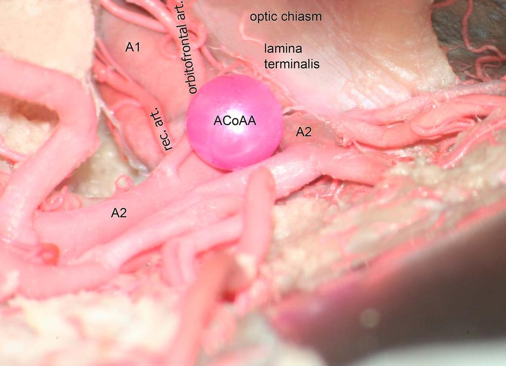
6 fundus was located inferior to the optic nerve and medial to the recurrent artery of Heubner. The most important anatomic structure in this projection was the contralateral A2 segment. Zero Degree (0 ): The ipsilateral recurrent artery of Heubner and the orbitofrontal and frontopolar arteries were larger than in the surgical field, and the aneurysm neck remained covered by the fundus in this view. Fifteen Degrees (15 ): In this view, the fundus of the aneurysm prevented visualization of the neck of the aneurysm. Because the fundus was located slightly more laterally, the aneurysm neck began to partially emerge but the view was still insufficient for clipping the aneurysm neck. Thirty Degrees (30 ): In this view, both A1 and A2 segments and the recurrent artery of Heubner were clearly seen. The neck of the aneurysm was in the middle of the surgical exposure. A 30 angle was found to be the most suitable head position for the anterior projection of the ACoAA (Fig 3). Forty-five Degrees (45 ): In this view, visualization of the aneurysm neck was adequate, but the contralateral A2 segment was covered by the aneurysm fundus. In addition, the ipsilateral recurrent artery of Heubner and the orbitofrontal and frontopolar arteries were too prominent in the surgical field. Posterior Projection The aneurysm fundus protected by the occipital lobe, and was located posterior to the A2 segment. It was covered by the contralateral orbitofrontal and frontopolar arteries. The perforating branches of the ACoA were located at the base of the aneurysm neck. The most important anatomical structures in this projection were the hypothalamic arteries. 6
7 Zero Degree (0 ): In this view, the aneurysm was not clearly seen because of the perforating arteries of the ACoA and A2; only a part of the aneurysm base could be seen. There was no corridor for approaching the aneurysm. Fifteen Degrees (15 ): In this view, perforating branches of the ACoAA and fundus of the aneurysm could be more easily seen. There was a surgical corridor toward the aneurysm between the A1 and A2 segments. This was found to be the most suitable head position for anteriorly projecting ACoAA (Fig 4). Thirty Degrees (30 ): After rotating the ipsilateral A2 segment, the aneurysm could be visualized slightly better but the neck of aneurysm could not be seen sufficiently for clipping. The perforating branches of the ACoA were too prominent in the surgical area in this view. Forty-five Degrees (45 ): The aneurysm was completely covered by perforating arteries and the orbitofrontal artery. The neck of the aneurysm could not be clearly visualized. Superior Projection The aneurysm was located between both A2 segments and the aneurysm fundus projected in the same direction as the A2 segments. The aneurysm was covered by the contralateral A2 segment and the proximal segment of the recurrent artery of Heubner. The most important anatomic structures in this projection were the recurrent artery of Heubner and the A2 segment. Zero Degree (0 ): In this view, the recurrent artery of Heubner and the A2 segment were clearly seen. The aneurysm fundus was seen between both A2 segments but the aneurysm neck could not be clearly seen. 7
8 Fifteen Degrees (15 ): Visualization of the aneurysm was slightly better but the aneurysm neck was still not clearly seen. Thirty Degrees (30 ): After 30 of rotation, the contralateral A2 segment, recurrent artery of Heubner and neck of the aneurysm were clearly seen. The most suitable head position a superiorly projecting ACoAA was found to be the 30 angle (Fig 5, 6). Forty-five Degrees (45 ): In 45 of rotation, the ipsilateral recurrent artery of Heubner was seen to emerge entirely into the surgical field. The contralateral A2 segment and recurrent artery of Heubner prevented visualization of the aneurysm. Inferior Projection The aneurysm fundus projected toward the skull base in this view and the most important anatomic structures seen were the hypothalamic arteries. In this projection, the recurrent artery of Heubner and the A1 and A2 segments were seen much more clearly viewed compared to the other projections. Zero Degree (0 ): In this view, the aneurysm fundus was seen but the aneurysm neck could not be clearly seen. Fifteen Degrees (15 ): In this view, the ACoA and A1 segment were located in the middle of this view. The neck of the aneurysm could not be visualized in this projection. Thirty Degrees (30 ): In this view, the aneurysm neck was seen but the perforating branches of the ACoA posed a serious obstacle. 8
9 Forty-five Degrees (45 ): In this view, the aneurysm neck and perforating branches of the ACoA were visualized better. The most suitable head position for the inferior projection of the ACoAA was found to be the 45 angle (Fig 7). Discussion Establishment of head position is one of the most important steps in ACoAA surgery. 1,2,5,7,13 Aneurysm control should be done by clipping the aneurysm with minimal tissue manipulation and dissection in the best possible head orientation. A number of different head positions for ACoAA surgery have been described in the literature. For example, Meyer et al. 6 suggested that the head must be rotated a maximum of 30 towards the opposite side, and that this angle should not be exceeded. Yaşargil 17,18 and Roux et al. 8 emphasized the need to position the head rotated 30 and Tamargo et al. 12 stated that ACoAA surgery required an angle between Sekhar et al. 11 and Samson et al. 9 suggested head positions of 45 and 60, respectively. We classified ACoAA separately according to 4 different projections, and attempted to determine the most suitable degree of head rotation for each projection. Optimal head positions for anterior and superior projections of the ACoAA were obtained at 30. With 30 rotation, the A1 segment, A2 segment, recurrent artery of Heubner, and aneurysm neck were clearly seen for the anterior projection. For the superior projection, the contralateral A2, recurrent artery of Heubner and aneurysm neck were also clearly seen with 30 of rotation. In the literature, Yasargil et al. 17,18 proposed 30 of rotation. Roux et al. 8 also reported that the head should be rotated 30 in order to keep the contralateral A2 out of the field. For a posterior projection, the most suitable head position was found to be 15. In this view, the origin of the perforating arteries and aneurysm neck could be clearly visualized. A 9
10 review of the literature revealed that the nearest similar position was suggested by Meyer et al 6. For an inferiorly projected ACoAA, the most suitable head position was found to be 45. In this view, the aneurysm neck and perforating branches were clearly seen. Tamargo et al. 12 and Sekhar et al. 11 also suggested 45 of head rotation with such aneurysms. Conclusion In this study, we emphasize that each aneurysm dome projection of the ACoAA should be individually considered and the head position adjusted accordingly. The use of appropriate head positions during surgery will prevent the development of postoperative ischemic complications and will increase the success of surgery by preventing unnecessary tissue manipulation. 10
11 References: 1. Botterell EH, Lougheed WM, Scott JW, Vandewater SL. Hypothermia, and Interruption of Carotid, or Carotid and Vertebral Circulation, in the Surgical Management of Intracranial Aneurysms 1. J Neurosurg Special Supplements, 2010;112(2): Hassan T, Hassan AI, Ahmed YM. Influence of parent vessel dominancy on fluid Dynamics of anterior communicating artery aneurysms. Acta Neurochir, 2011; 153(2): Hernesniemi J, Dashti R, Lehecka M, Niemelä M, Rinne J, Lehto H, Ronkainen A, Koivisto T, Jääskeläinen JE. Microneurosurgical management of anterior communicating artery aneurysms. Surgical Neurology 2008; 70: Juvela S, Porras M, Heiskanen O. Natural history of unruptured intracranial aneurysms: a long-term follow-up study. J Neurosurg 1993; 79: Kashimura H, Kubo Y, Ogasawara K, Kakino S, Yoshida K, Ogawa A.Easy Dissection Of The Interhemispheric Fissure For Treatment Of The Anterior Communicating Artery Aneurysm By The Pterional Approach.World Neurosurgery, 2010; 64: Meyer FB. Atlas of Neurosurgery. In Meyer FB, eds. Philadelphia: Mayo Foundation, 1999; Perlmutter D, Rhoton AL Jr. Microsurgical anatomy of the anterior cerebral anterior communicating-recurrent artery complex. J Neurosurg 1976; 45: Roux PD, Winn HR. Anterior Communicating Artery Aneurysms: Surgical Techniques. In Batjer HH, eds. Cerebrovascular Disease. New York: Lippincott, 1996;
12 9. Samson DS, Batjer HH. Aneurysms of the anterior communicating artery. In: Sanson D, Batjer HH, eds. Intracranial Aneurysm Surgery: Techniques. New York: Futura, 1990; Schievink WI, Piepgras DG, Wirth FP. Rupture of previously documented small asymptomatic saccular intracranial aneurysms. Report of three cases. J Neurosurg 1992; 76: Sekhar LN, Natarajan SK, Britz GW, Ghodke B. Microsurgical management of anterior communicating artery aneurysms. Neurosurgery 2007; 61 (Suppl 2): S Tamargo R, Haroun R, Rigamonti D. Anterior communicating and anterior cerebral artery aneurysms. In: Winn R, editor. Youmans neurological surgery. Philadelphia (Pa): Saunders Elsevier; p Taylor CL, Yuan Z, Selman WR, Ratcheson RA, Rimm AA. Cerebral arterial aneurysm formation and rupture in 20,767 elderly patients: hypertension and other risk factors. J Neurosurg 1995; 83: Wiebers DO, Whisnant JP, Sundt TM Jr, O'Fallon WM. The significance of unruptured intracranial saccular aneurysms. J Neurosurg 1987; 66: Yalcin B, Kirici Y, Ozan H. The sinus node artery: anatomic investigations based on injection-corrosion of 60 sheep hearts. Interact Cardiovasc Thorac Surg Jun;3(2): Yalcin B, Komesli GH, Ozgok Y, Ozan H. Vascular anatomy of normal and undescended testes: surgical assessment of anastomotic channels between testicular and deferential arteries. Urology Oct;66(4): Yasargil M. Microneurosurgery, Vol. II. Stuttgart: Georg Thieme Verlag; 1984; Yasargil M. Microneurosurgery, Vol. I. Stuttgart: Georg Thieme Verlag; 1984;
13 Figure Legends Figure 1: Appearance of the brain arteries after corrosion technique. ICA: internal carotid artery; MCA: middle cerebral artery; ACA: anterior cerebral artery Figure 2: Appearance of the aneurysm model. AcoAA, anterior communicating artery aneurysm model; MCA: middle cerebral artery; DACA:distal anterior cerebral artery. Figure 3: Anteriorly projected ACoAA and view of related vascular structures in 30 of head rotation. AcoAA: Anterior communicating artery aneurysm model; rec. art: recurrent artery of Heubner; orbitofrontal art.:orbitofrontal artery. Figure 4: Posteriorly projected ACoAA and view of related vascular structures in 15 of head rotation. AcoAA: Anterior communicating artery aneurysm model; rec. art: recurrent artery of Heubner; orbitofrontal art.:orbitofrontal artery; perf. art. perforating arteries. Figure 5: Superiorly projected ACoAA and view of related vascular structures in 30 of head rotation. AcoAA: Anterior communicating artery aneurysm model; rec. art: recurrent artery of Heubner; orbitofrontal art.:orbitofrontal artery. Figure 6: Clipping view of superiorly projected ACoAA in 30 of head rotation. AcoAA: anterior communicating artery aneurysm model AcoAA: Anterior communicating artery aneurysm model; rec. art: recurrent artery of Heubner; orbitofrontal art.:orbitofrontal artery. Figure 7: Inferiorly projected ACoAA and view of related vascular structures in 45 of head rotation. ACoAA: Anterior communicating artery aneurysm model; rec. art: recurrent artery of Heubner; orbitofrontal art.:orbitofrontal artery; A1. perf.: perforator arteries originating from the proximal segment (A1) of the anterior cerebral artery. 13


14

15


16

17
Contralateral clipping of bilateral middle cerebral artery aneurysms. Case report
 Romanian Neurosurgery Volume XXXI Number 1 2017 January - March Article Contralateral clipping of bilateral middle cerebral artery aneurysms. Case report Georgiana Ion, Alexandru Chiriac, Ziyad Faiyad,
5. COMMON APPROACHES. Each of the described approaches is also demonstrated on supplementary videos, please see Appendix 2.
 5. COMMON APPROACHES Each of the described approaches is also demonstrated on supplementary videos, please see Appendix 2. 5.1. LATERAL SUPRAORBITAL APPROACH The most common craniotomy approach used in
Ruptured Cerebral Aneurysm of the Anterior Circulation
 Original Articles * Division of Neurosurgery Department of Surgery Ruptured Cerebral Aneurysm of the Anterior Circulation Management and Microsurgical Treatment Ossama Al-Mefty, MD* ABSTRACT Based on the
Surgical Neurology International
 Surgical Neurology International OPEN ACCESS For entire Editorial Board visit : http://www.surgicalneurologyint.com Editor: James I. Ausman, MD, PhD University of California, Los Angeles, CA, USA Case
Surgical anatomy of the juxta dural ring area
 J Neurosurg 89:250 254, 1998 Surgical anatomy of the juxta dural ring area SUSUMU OIKAWA, M.D., KAZUHIKO KYOSHIMA, M.D., AND SHIGEAKI KOBAYASHI, M.D. Department of Neurosurgery, Shinshu University School
Extracranial-to-Intracranial Bypass Using Radial Artery Grafting for Complex Skull Base Tumors: Technical Note
 Extracranial-to-Intracranial Bypass Using Radial Artery Grafting for Complex Skull Base Tumors: Technical Note Saleem I. Abdulrauf, M.D., F.A.C.S. 1 ABSTRACT The management of complex skull base tumors
Anterior Communicating Artery Aneurysms. Surgical approaches Review
 Anterior Communicating Artery Aneurysms. Surgical approaches Review Hirotoshi Sano Dept. of Neurosurgery Fujita Health University Introduction There are two main types of approaches for anterior communicating
Surgical anatomy of the juxtadural ring area
 Surgical anatomy of the juxtadural ring area Susumu Oikawa, M.D., Kazuhiko Kyoshima, M.D., and Shigeaki Kobayashi, M.D. Department of Neurosurgery, Shinshu University School of Medicine, Matsumoto, Japan
Management of Giant Aneurysm
 Management of Giant Aneurysm Paul P. Huang, M.D. and Jafar J. Jafar, M.D. Department of Neurosurgery New York University Medical Center New York, New York, USA Introduction Giant intracranial aneurysms
Title Review of the Literature. Honda, Masaru; Ando, Takeo. Issue Date Right
 NAOSITE: Nagasaki University's Ac Title Author(s) Proximal Anterior Cerebral Artery A Review of the Literature Honda, Masaru; Ando, Takeo Citation Acta medica Nagasakiensia, 57(3), p Issue Date 2013-02
Neurosurgical Techniques
 Neurosurgical Techniques EBEN ALEXANDER, JR., M.D., EDITOR Supratentorial Skull Flaps GuY L. ODOM, M.D., AND BARNES WOODHALL,!V[.D. Department of Surgery, Division of Neurosurgery, Duke University Medical
Pichayen Duangthongpon MD*, Chaiwit Thanapaisal MD*, Amnat Kitkhuandee MD*, Kowit Chaiciwamongkol MD**, Vilaiwan Morthong MD**
 The Relationships between Asterion, the Transverse-Sigmoid Junction, the Superior Nuchal Line and the Transverse Sinus in Thai Cadavers: Surgical Relevance Pichayen Duangthongpon MD*, Chaiwit Thanapaisal
Single center experience and technical nuances in the treatment of distal anterior cerebral artery aneurysms
 Romanian Neurosurgery Volume XXXI Number 1 2017 January - March Article Single center experience and technical nuances in the treatment of distal anterior cerebral artery aneurysms Dorin Nicolae Gherasim,
Pterional-subolfactory Approach for Treatment of High Positioned Anterior Communicating Artery Aneurysms
 Journal of Cerebrovascular and Endovascular Neurosurgery ISSN 2234-8565, EISSN 2287-3139, http://dx.doi.org/10.7461/jcen.2013.15.3.177 Clinical Article Pterional-subolfactory Approach for Treatment of
Presented by- Dr. Vivek Tandon
 Presented by- Dr. Vivek Tandon Basic principles of aneurysm surgery Hemostasisof the extradural portion should be meticulous. Hand rests, instrumentholders and self retaining retractors must be checked
Principles Arteries & Veins of the CNS LO14
 Principles Arteries & Veins of the CNS LO14 14. Identify (on cadaver specimens, models and diagrams) and name the principal arteries and veins of the CNS: Why is it important to understand blood supply
Effect of early operation for ruptured aneurysms on prevention of delayed ischemic symptoms
 J Neurosurg 57:622-628, 1982 Effect of early operation for ruptured aneurysms on prevention of delayed ischemic symptoms MAMORU TANEDA, M.D. Department of Neurosurgery, Hanwa Memorial Hospital, Osaka,
Moyamoya Syndrome with contra lateral DACA aneurysm: First Case report with review of literature
 Romanian Neurosurgery Volume XXXI Number 3 2017 July-September Article Moyamoya Syndrome with contra lateral DACA aneurysm: First Case report with review of literature Ashish Kumar Dwivedi, Pradeep Kumar,
An Unruptured Anterior Communicating Artery Aneurysm with Bilateral Infraoptic Anterior Cerebral Arteries. Case Report and Review of the Literature
 An Unruptured Anterior Communicating Artery Aneurysm with Bilateral Infraoptic Anterior Cerebral Arteries. Case Report and Review of the Literature The Harvard community has made this article openly available.
Department of Neurosurgery, Research Center for Neurosurgical Robotic Systems, Kyungpook National University, Daegu, Korea
 Technical Note J Korean Neurosurg Soc 60 (2) : 250-256, 2017 https://doi.org/10.3340/jkns.2016.0910.009 pissn 2005-3711 eissn 1598-7876 Pterional or Subfrontal ccess for Proximal Vascular ontrol in nterior
A Modified Mini-Pterional (Subfrontal-Suprapterional) Approach to MCA Bifurcation Aneurysms with Minimal Dissection of the Temporal Muscle
 Approach to MCA Bifurcation Aneurysms with Minimal Dissection of the Temporal Muscle") DOI: 10.5137/1019-5149.JTN.16472-15.1 Received: 24.11.2015 / Accepted: 16.2.2016 Published Online: 03.08.2016 Technical Note A Modified Mini-Pterional (Subfrontal-Suprapterional) Approach to MCA Bifurcation
DOI: 10.5137/1019-5149.JTN.16472-15.1 Received: 24.11.2015 / Accepted: 16.2.2016 Published Online: 03.08.2016 Technical Note A Modified Mini-Pterional (Subfrontal-Suprapterional) Approach to MCA Bifurcation
Intracranial-to-intracranial vascular anastomosis created using a microanastomotic device for the treatment of distal middle cerebral artery aneurysms
 J Neurosurg 97:486 491, 2002 Intracranial-to-intracranial vascular anastomosis created using a microanastomotic device for the treatment of distal middle cerebral artery aneurysms Technical note DAVID
Azygos anterior cerebral artery aneurysm with subarachnoid hemorrhage
 Chowdhury et al. Neuroimmunol Neuroinflammation 2018;5:39 DOI: 10.20517/2347-8659.2018.37 Neuroimmunology and Neuroinflammation Letter to Editor Open ccess zygos anterior cerebral artery aneurysm with
Arterial vascularisation of the anterior
 Original Article Singapore Med J 2011; 52(6) 410 Arterial vascularisation of the anterior perforated substance Sen T, Esmer A F, Acar H I, Karahan S T, Tuccar E ABSTRACT Introduction: The arteries of the
Open clipping surgery to treat cerebral aneurysms. Head-up display may facilitate safe keyhole surgery for cerebral aneurysm clipping
 CLINICAL ARTICLE J Neurosurg 129:883 889, 2018 Head-up display may facilitate safe keyhole surgery for cerebral aneurysm clipping Terushige Toyooka, MD, PhD, Naoki Otani, MD, PhD, Kojiro Wada, MD, PhD,
History of revascularization
 History of revascularization Author (year) Kredel, 1942 Woringer& Kunlin, 1963 Donaghy& Yasargil, 1968 Loughheed 1971 Kikuchini & Karasawa1973 Karasawa, 1977 Story, 1978 Sundt, 1982 EC/IC bypass study
Effect of clot removal on cerebral vasospasm TETSUJI INAGAWA, M.D., MITSUO YAMAMOTO, M.D., AND KAZUKO KAMIYA, M.D.
 J Neurosurg 72:224-230, 1990 Effect of clot removal on cerebral vasospasm TETSUJI INAGAWA, M.D., MITSUO YAMAMOTO, M.D., AND KAZUKO KAMIYA, M.D. Department of Neurosurgery, Shimane Prefectural Central Hospital,
Alessandro Della Puppa
 Intraoperative measurement of arterial blood flow in complex cerebral aneurysms surgery Studio flussimetrico intra-operatorio nel clipping degli aneurismi complessi Alessandro Della Puppa NEUROSURGERY
OBJECTIVES. At the end of the lecture, students should be able to: List the cerebral arteries.
 DR JAMILA EL MEDANY OBJECTIVES At the end of the lecture, students should be able to: List the cerebral arteries. Describe the cerebral arterial supply regarding the origin, distribution and branches.
I T IS well known that aneurysms occur at
 The Lateral Perforating Branches of the Anterior and Middle Cerebral Arteries* HARRY A. KAPLAN, M.D. Division of Neurosurgery, Seton Hall College of Medicine, and Jersey City Medical Center, Jersey City,
Penetration of the Optic Nerve or Chiasm by Anterior Communicating Artery Aneurysms. - Three Case Reports-
 Penetration of the Optic Nerve or Chiasm by Anterior Communicating Artery Aneurysms. - Three Case Reports- Tetsuyoshi Horiuchi 1, Toshiya Uchiyama 1, Yoshikazu Kusano 1, Maki Okada 1, Kazuhiro Hongo 1,
Comparison between modified lateral supraorbital approach and pterional approach in the surgical treatment of middle cerebral artery aneurysms
 Chen et al. Chinese Neurosurgical Journal (2018) 4:4 https://doi.org/10.1186/s41016-018-0110-2 CHINESE MEDICAL ASSOCIATION CHINESE NEUROSURGICAL SOCIETY RESEARCH Open Access Comparison between modified
Clip-Grafts for Aneurysm and Small Vessel Surgery*
 J. Neurosurg. / Volume 31 / July, 1969 Clip-Grafts for Aneurysm and Small Vessel Surgery* Part 3: Clinical Experience in Intracranial Internal Carotid Artery Aneurysms THORALF M. SUNDT, JR., M.D., AND
Vasculature and Aneurysm Design
 Vasculature and Aneurysm Design Amy Swartz November 18, 2016 Team 23: In-vitro Tube Model System ME450 Section 3 Dr. David Trevas and Dr. Sarah Oman Introduction The primary goal of this capstone project
Interpositional carotid artery bypass strategies in the surgical management of aneurysms and tumors of the skull base
 Neurosurg Focus 14 (3):Article 2, 2003, Click here to return to Table of Contents Interpositional carotid artery bypass strategies in the surgical management of aneurysms and tumors of the skull base JAMES
Preoperative Grading Systems of Spontaneous Subarachnoid Hemorrhage
 KISEP KOR J CEREBROVASCULAR DISEASE March 2000 Vo. 2, No 1, page 24-9 자발성지주막하출혈환자의수술전등급 황성남 Preoperative Grading Systems of Spontaneous Subarachnoid Hemorrhage Sung-Nam Hwang, MD Department of Neurosurgery,
Carotid cave aneurysms of the internal carotid artery
 J Neurosurg 70:216-221, 1989 Carotid cave aneurysms of the internal carotid artery SHIGEAKI KOBAYASHI, M.D., KAZUHIKO KYOSHIMA, M.D., HIROHIKO GIBO, M.D., SATHYARANJANDAS A. HEGDE, M.D., TOSHIKI TAKEMAE,
As a group, basilar artery aneurysms are
 OPERATIVE NUANCES ORBITOZYGOMATIC APPROACH TO BASILAR APEX ANEURYSMS Frank P.K. Hsu, M.D., Ph.D. Division of Neurological Surgery, St. Joseph s Hospital and Medical Center, Phoenix, Arizona Richard E.
Surgical techniques and procedures for cerebrovascular surgery. Surgery for the AVF at the cranio-cervical junction and high cervical spine
 VS-1 Surgery for the AVF at the cranio-cervical junction and high cervical spine Hiroyuki Kinouchi University of Yamanashi, Department of Neurosurgery Dural AVFs have been recognized as common type of
Endoscopic anatomical study on anterior communicating artery aneurysm surgery by endonasal transphenoidal approach
 Ma et al. Chinese Neurosurgical Journal (2016) 2:27 DOI 10.1186/s41016-016-0042-7 CHINESE NEUROSURGICAL SOCIETY RESEARCH CHINESE MEDICAL ASSOCIATION Endoscopic anatomical study on anterior communicating
Unit 18: Cranial Cavity and Contents
 Unit 18: Cranial Cavity and Contents Dissection Instructions: The calvaria is to be removed without damage to the dura mater which is attached to the inner surface of the calvaria. Cut through the outer
Depicting Cerebral Veins by Three-Dimensional CT Angiography before Surgical Clipping of Aneurysms
 AJNR Am J Neuroradiol 23:85 91, January 2001 Depicting Cerebral Veins by Three-Dimensional CT Angiography before Surgical Clipping of Aneurysms Makio Kaminogo, Hideyuki Hayashi, Hideki Ishimaru, Minoru
PTA 106 Unit 1 Lecture 3
 PTA 106 Unit 1 Lecture 3 The Basics Arteries: Carry blood away from the heart toward tissues. They typically have thicker vessels walls to handle increased pressure. Contain internal and external elastic
TRANSVERSE SECTION PLANE Scalp 2. Cranium. 13. Superior sagittal sinus
 TRANSVERSE SECTION PLANE 1 1. Scalp 2. Cranium 3. Superior sagittal sinus 4. Dura mater 5. Falx cerebri 6. Frontal lobes of the cerebrum 7. Middle meningeal artery 8. Cortex, grey matter 9. Cerebral vessels
EndoBlade Soft Tissue Release System
 Surgical Technique Endoscopic Gastroc Recession Endoscopic Plantar Fascia Release EndoBlade Soft Tissue Release System Endoscopic Gastroc Recession Arthrex has developed a comprehensive, completely disposable
KEY WORDS: Microsurgical anatomy, Olfactory nerve, Pterional approach. Neurosurgery 57[ONS Suppl 1]:ONS-17 ONS-21, 2005
![KEY WORDS: Microsurgical anatomy, Olfactory nerve, Pterional approach. Neurosurgery 57[ONS Suppl 1]:ONS-17 ONS-21, 2005](/thumbs/83/88796898.jpg "KEY WORDS: Microsurgical anatomy, Olfactory nerve, Pterional approach. Neurosurgery 57[ONS Suppl 1]:ONS-17 ONS-21, 2005") Salvatore Cardali, M.D. Alberto Romano, M.D. Filippo Flavio Angileri, M.D., Ph.D. Alfredo Conti, M.D. Domenico La Torre, M.D. Oreste de Divitiis, M.D. Domenico d Avella, M.D. Manfred Tschabitscher, M.D.
Salvatore Cardali, M.D. Alberto Romano, M.D. Filippo Flavio Angileri, M.D., Ph.D. Alfredo Conti, M.D. Domenico La Torre, M.D. Oreste de Divitiis, M.D. Domenico d Avella, M.D. Manfred Tschabitscher, M.D.
THE ANGULAR TRACT: AN ANATOMICAL
 British Journal of Oral Surgery (1981) 19, 116-120 0 The British Association of Oral Surgeons 0007-117X/81/00170116$02.00 THE ANGULAR TRACT: AN ANATOMICAL OF SURGICAL SIGNIFICANCE STRUCTURE HAITHEM A.
NEURO IMAGING 2. Dr. Said Huwaijah Chairman of radiology Dep, Damascus Univercity
 NEURO IMAGING 2 Dr. Said Huwaijah Chairman of radiology Dep, Damascus Univercity I. EPIDURAL HEMATOMA (EDH) LOCATION Seventy to seventy-five percent occur in temporoparietal region. CAUSE Most likely caused
Superficial Temporal Artery to Middle Cerebral Artery Bypass
 Superficial Temporal Artery to Middle Cerebral Artery Bypass David W. Newell, M.D. 1 ABSTRACT The superficial temporal artery to middle artery bypass is a technique that allows the blood supply from the
Rupture of Very Small Intracranial Aneurysms: Incidence and Clinical Characteristics
 Journal of Cerebrovascular and Endovascular Neurosurgery pissn 2234-8565, eissn 2287-3139, http://dx.doi.org/10.7461/jcen.2015.17.3.217 Original Article Rupture of Very Small Intracranial Aneurysms: Incidence
Anastomosis of the superficial temporal artery to the distal anterior cerebral artery with interposed cephalic vein graft
 J Neurosurg 58~25-429, 1983 Anastomosis of the superficial temporal artery to the distal anterior cerebral artery with interposed cephalic vein graft Case report RYOJI ISHII, M.D., TETSUO KOIKE, M.D.,
For Emergency Doctors. Dr Suzanne Smallbane November 2011
 For Emergency Doctors Dr Suzanne Smallbane November 2011 A: Orbit B: Sphenoid Sinus C: Temporal Lobe D: EAC E: Mastoid air cells F: Cerebellar hemisphere A: Frontal lobe B: Frontal bone C: Dorsum sellae
A STUDY OF TEMPORAL BRANCHES OF MIDDLE CEREBRAL ARTERY
 J. Anat. Sciences, 23(2) : Dec. 2015, 15-21 Original Article A STUDY OF TEMPORAL BRANCHES OF MIDDLE CEREBRAL ARTERY Medha Das*, Shirin Jahan*, Pranjal Pankaj** * Department of Anatomy, Rama Medical College,
We describe some of the conceptual nuances and
 Suprasellar Meningiomas Ivan Ciric, M.D., Sami Rosenblatt, M.D. Division of Neurosurgery, Evanston Northwestern Healthcare, Evanston Hospital, Northwestern University Medical School, Evanston, Illinois
Distal anterior cerebral artery (DACA) aneurysms are. Case Report
 aneurysms are. Case Report") 248 Formos J Surg 2010;43:248-252 Distal Anterior Cerebral Artery Aneurysm: an Infrequent Cause of Transient Ischemic Attack Followed by Diffuse Subarachnoid Hemorrhage: Report of a Case Che-Chuan Wang
248 Formos J Surg 2010;43:248-252 Distal Anterior Cerebral Artery Aneurysm: an Infrequent Cause of Transient Ischemic Attack Followed by Diffuse Subarachnoid Hemorrhage: Report of a Case Che-Chuan Wang
ANATOMICAL CONSIDERATION OF THE INTRACRANIAL PORTION OF MIDDLE MENIGEAL ARTERY IN THAIS
 ANATOMICAL CONSIDERATION OF THE INTRACRANIAL PORTION OF MIDDLE MENIGEAL ARTERY IN THAIS Kritsana Namonta, 1 Lanaprai Kwathai, 1 Thanaporn Rungruang, 1 Vipavadee Chaisuksunt, 2 Wandee Apinhasmit, 3 Supin
Surgery of Middle Cerebral Artery (MCA) Aneurysm
 Aneurysm") Surgery of Middle Cerebral Artery (MCA) Aneurysm 15 Fusao Ikawa 15.1 Sign and Symptoms Middle cerebral artery (MCA) aneurysm is one of the most popular cerebral aneurysm. The incidence of ruptured MCA
Surgery of Middle Cerebral Artery (MCA) Aneurysm 15 Fusao Ikawa 15.1 Sign and Symptoms Middle cerebral artery (MCA) aneurysm is one of the most popular cerebral aneurysm. The incidence of ruptured MCA
T HE direct surgical approach to an aneurysm on
 J Neurosurg 66:500-505, 1987 Aneurysms of the basilar artery trunk KENICHIRO SUGITA, M.D., SHIGEAKI KOBAYASHI, M.D., TOSHIKI TAKEMAE, M.D., TSUYOSHI TADA, M.D., AND YUICHIRO TANAKA, M.D. Department of
Studying Aneurysm Devices in the Intracranial Neurovasculature
 Studying Aneurysm Devices in the Intracranial Neurovasculature The benefits and risks of treating unruptured aneurysms depend on the anatomical location. One approach to studying devices to treat unruptured
Microsurgery for ruptured cerebellar arteriovenous malformations
 European Review for Medical and Pharmacological Sciences Microsurgery for ruptured cerebellar arteriovenous malformations S.-F. GONG 1,2, X.-B. WANG 1,3, Y.-Q. LIAO 1,2, T.-P. JIANG 1,2, J.-B. HE 1,2,
Small and medium size intracranial aneurysms - a 5 years retrospective analysis trial and multimodal treatment
 Romanian Neurosurgery (2015) XXIX 4: 417-426 417 DOI: 10.1515/romneu-2015-0057 Small and medium size intracranial aneurysms - a 5 years retrospective analysis trial and multimodal treatment Valentin Munteanu
Intrapetrous Internal Carotid Artery
 James C. Andrews, M.D., Neil A. Martin, M.D., Keith Black, M.D., Vincent F Honrubia, M.D., and Donald P Becker, M.D. Midd le Cranial Fossa Transtemporal Approach to the Intrapetrous Internal Carotid Artery
Residence of Discipline of Neurosurgery of Hospital da Santa Casa de Misericórdia of Sao Paulo Sao Paulo, Brazil
 Cronicon OPEN ACCESS NEUROLOGY Research Article Efficacy of the Lamina Terminalis Fenestration Associated With the Liliequist Membrane Fenestration in Reducing Shunt-Dependent Hydrocephalus Following Aneurysm
Alexander C Vlantis. Selective Neck Dissection 33
 05 Modified Radical Neck Dissection Type II Alexander C Vlantis Selective Neck Dissection 33 Modified Radical Neck Dissection Type II INCISION Various incisions can be used for a neck dissection. The incision
NIH Public Access Author Manuscript J Am Coll Radiol. Author manuscript; available in PMC 2013 June 24.
 NIH Public Access Author Manuscript Published in final edited form as: J Am Coll Radiol. 2010 January ; 7(1): 73 76. doi:10.1016/j.jacr.2009.06.015. Cerebral Aneurysms Janet C. Miller, DPhil, Joshua A.
Multiple intracranial aneurysms: incidence and outcome in a series of 357 patients
 450 Sergiu Gaivas et al Multiple intracranial aneurysms Multiple intracranial aneurysms: incidence and outcome in a series of 357 patients Sergiu Gaivas 1, Daniel Rotariu 1, Bogdan Iliescu 2, Faiyad Ziyad
Ruptured fusiform aneurysm of the proximal anterior cerebral artery in young patient - case report
 286 Ion et al Ruptured fusiform aneurysm of the proximal anterior cerebral artery Ruptured fusiform aneurysm of the proximal anterior cerebral artery in young patient - case report Georgiana Ion*, A. Chiriac,
Classical CNS Disease Patterns
 Classical CNS Disease Patterns Inflammatory Traumatic In response to the trauma of having his head bashed in GM would have experienced some of these features. NOT TWO LITTLE PEENY WEENY I CM LACERATIONS.
Optimum Sylvian Fissure Dissection Needed for Anterior Circulation Aneurysms
 Med. J. Cairo Univ., Vol. 84, No. 2, June: 49-54, 2016 www.medicaljournalofcairouniversity.net Optimum Sylvian Fissure Dissection Needed for Anterior Circulation Aneurysms MOHAMED A. GABR, M.Sc.; NASSER
ANASTAMOSIS FOR BRAIN STEM ISCHEMIA/Khodadad et al.
 ANASTAMOSIS FOR BRAIN STEM ISCHEMIA/Khodadad et al. visualization of the posterior inferior cerebellar artery. The patient, now 11 months post-operative, has shown further neurological improvement since
Introduction. BY W. R. HUDGINS, M.D.,* AND JULIO H. GARCIA, M.D.t. Abstract: ADDITIONAL KEY WORDS focal cerebral ischemia
 Transorbital Approach to the Middle Cerebral Artery of the Squirrel Monkey: A Technique for Experimental Cerebral Infarction Applicable to Ultrastructural Studies BY W. R. HUDGINS, M.D.,* AND JULIO H.
Chapter XII: Temporal Expanding Processes, Including Those in the Sylvian Fissure and the Insula
 Acta Radiologica ISSN: 0001-6926 (Print) (Online) Journal homepage: https://www.tandfonline.com/loi/iaro20 Chapter XII: Temporal Expanding Processes, Including Those in the Sylvian Fissure and the Insula
OHSU Neurological Surgery Education. Neurosurgical Basics. Copyright Oregon Health & Science University Department of Neurological Surgery
 OHSU Neurological Surgery Education Neurosurgical Basics Chapter 1 Basic Principles: Cranial Three-Point Head Fixation Proper and safe application of a three-point rigid cranial fixation device provides
Chapter Five. Anosmia after aneurysmal subarachnoid hemorrhage. M.J.H. Wermer, M. Donswijk, P. Greebe, B. Verweij and G.J.E.
 Chapter Anosmia after aneurysmal subarachnoid hemorrhage M.J.H. Wermer, M. Donswijk, P. Greebe, B. Verweij and G.J.E. Rinkel Abstract Background and purpose Anosmia has an important impact on well-being,
Transcranial Doppler (Basic Step) Dae-il Chang, M.D., Sung Sang Yoon, M.D. Department of Neurology, College of Medicine, Kyunghee university
 Dae-il Chang, M.D., Sung Sang Yoon, M.D. Department of Neurology, College of Medicine, Kyunghee university") Transcranial Doppler (Basic Step) Dae-il Chang, M.D., Sung Sang Yoon, M.D. Department of Neurology, College of Medicine, Kyunghee university Principles of Doppler Ultrasonography Major target Speed & direction
Transcranial Doppler (Basic Step) Dae-il Chang, M.D., Sung Sang Yoon, M.D. Department of Neurology, College of Medicine, Kyunghee university Principles of Doppler Ultrasonography Major target Speed & direction
FIG The inferior and posterior peritoneal reflection is easily
 PSOAS HITCH, BOARI FLAP, AND COMBINATION OF PSOAS 7 HITCH AND BOARI FLAP The psoas hitch procedure, Boari flap, and transureteroureterostomy are useful operative procedures for reestablishing continuity
Evaluation of the Lenticulostriate Arteries with Rotational Angiography and 3D Reconstruction
 AJNR Am J Neuroradiol 26:306 312, February 2005 Evaluation of the Lenticulostriate Arteries with Rotational Angiography and 3D Reconstruction Hyun-Seung Kang, Moon Hee Han, Bae Ju Kwon, O-Ki Kwon, Sung
ARTICLE COVERSHEET LWW_CONDENSED-FLA(8.125X10.875) SERVER-BASED
 SERVER-BASED") ARTICLE COVERSHEET LWW_CONDENSED-FLA(8.125X10.875) SERVER-BASED Template version : 7.0 Revised: 03/03/2013 Article : SCS204391 Typesetter : jesona Date : Monday May 27th 2013 Time : 17:05:50 Number of
ARTICLE COVERSHEET LWW_CONDENSED-FLA(8.125X10.875) SERVER-BASED Template version : 7.0 Revised: 03/03/2013 Article : SCS204391 Typesetter : jesona Date : Monday May 27th 2013 Time : 17:05:50 Number of
HEAD AND NECK IMAGING. James Chen (MS IV)
") HEAD AND NECK IMAGING James Chen (MS IV) Anatomy Course Johns Hopkins School of Medicine Sept. 27, 2011 OBJECTIVES Introduce cross sectional imaging of head and neck Computed tomography (CT) Review head
HEAD AND NECK IMAGING James Chen (MS IV) Anatomy Course Johns Hopkins School of Medicine Sept. 27, 2011 OBJECTIVES Introduce cross sectional imaging of head and neck Computed tomography (CT) Review head
ANATOMY OF THE VEIN OF LABBE: A CADAVERIC STUDY
 Original Research Article ANATOMY OF THE VEIN OF LABBE: A CADAVERIC STUDY Ashwini C Appaji * 1, Murali Mohan 2, Roopa Kulkarni 3, R N Kulkarni 4. ABSTRACT Background: Vein of Labbe is the major inferior
Surgical treatment and perioperative management of intracranial aneurysms in Chinese patients with ischemic cerebrovascular diseases: a case series
 Zheng and Wu BMC Neurology (2018) 18:142 https://doi.org/10.1186/s12883-018-1147-8 RESEARCH ARTICLE Open Access Surgical treatment and perioperative management of intracranial aneurysms in Chinese patients
TRANS-SULCAL MICROSURGICAL APPROACH TO INTRACEREBRAL LESIONS
 Trakia Journal of Sciences, No 3, pp 277-282, 2014 Copyright 2014 Trakia University Available online at: http://www.uni-sz.bg ISSN 1313-7050 (print) doi:10.15547/tjs.2014.03.009 ISSN 1313-3551 (online)
Surgical clipping of a dissecting aneurysm of the precommunicating segment of the anterior cerebral artery: a case report and review of the literature
 de Divitiis et al. Journal of Medical Case Reports (2015) 9:117 DOI 10.1186/s13256-015-0604-x CASE REPORT JOURNAL OF MEDICAL CASE REPORTS Open Access Surgical clipping of a dissecting aneurysm of the precommunicating
Case Report 1. CTA head. (c) Tele3D Advantage, LLC
 Tele3D Advantage, LLC") Case Report 1 CTA head 1 History 82 YEAR OLD woman with signs and symptoms of increased intra cranial pressure in setting of SAH. CT Brain was performed followed by CT Angiography of head. 2 CT brain Extensive
Case Report 1 CTA head 1 History 82 YEAR OLD woman with signs and symptoms of increased intra cranial pressure in setting of SAH. CT Brain was performed followed by CT Angiography of head. 2 CT brain Extensive
Anatomy of the Middle Cerebral Artery: The Temporal Branches
 Anatomy of the Middle Cerebral Artery: The Temporal Branches BY W. BRADFORD DeLONG, M.D., F.A.C.S. Abstract: Anatomy of the Middle Cerebral Artery: The Temporal Branches Nineteen out of 23 middle cerebral
Cerebral revascularization: past, present and future
 Cerebral revascularization: past, present and future Tomás Funes 1 MD, Asis Lorente Muñoz 2 MD, Federico Fernández Molina 3 MD, Paula Ypa 3 MD, Francisco Mannara 4 MD, Luis González Martínez 2 MD, PhD
Incidence of Superficial Sylvian Vein Compromise and Postoperative Effects on CT Imaging after Surgical Clipping of Middle Cerebral Artery Aneurysms
 AJNR Am J Neuroradiol 26:2019 2026, September 2005 Incidence of Superficial Sylvian Vein Compromise and Postoperative Effects on CT Imaging after Surgical Clipping of Middle Cerebral Artery Aneurysms Bruce
What Is the Significance of a Large Number of Ruptured Aneurysms Smaller than 7 mm in Diameter?
 online ML Comm www.jkns.or.kr 10.3340/jkns.2009.45.2.85 J Korean Neurosurg Soc 45 : 85-89, 2009 Print ISSN 2005-3711 On-line ISSN 1598-7876 Copyright 2009 The Korean Neurosurgical Society Clinical Article
Neuroanatomy Dr. Maha ELBeltagy Assistant Professor of Anatomy Faculty of Medicine The University of Jordan 2018
 Neuroanatomy Dr. Maha ELBeltagy Assistant Professor of Anatomy Faculty of Medicine The University of Jordan 2018 Blood Supply of Brain and Spinal Cord Arterial Supply of Brain The brain receives blood
Literature Review: Neurosurgery
 NANOS 2018 Kona, Hawaii Literature Review: Neurosurgery Neil R. Miller, MD FACS Frank B. Walsh Professor of Neuro-Ophthalmology Professor of Ophthalmology, Neurology & Neurosurgery Johns Hopkins University
Superior Orbital Rim Approach for Anterior Communicating Artery Aneurysms: A Surgical Series of 27 Patients
 J Korean Med Sci 2003; 18: 566-72 ISSN 1011-8934 Copyright The Korean Academy of Medical Sciences Superior Orbital Rim Approach for Anterior Communicating Artery Aneurysms: A Surgical Series of 27 Patients
Eyebrow craniotomy for anterior skull base lesions: how I do it
 Acta Neurochir (2013) 155:99 106 DOI 10.1007/s00701-012-1552-5 HOW I DO IT - NEUROSURGICAL TECHNIQUES Eyebrow craniotomy for anterior skull base lesions: how I do it Zsolt Zador & Kanna Gnanalingham Received:
Anatomical observations of the subarachnoid cisterns of the brain during surgery
 Anatomical observations of the subarachnoid cisterns of the brain during surgery M. GAZI YASARGIL~ M.D., KONSTANTIN KASDAGLIS, M.D., KEWAL K. JAIN, M.D., AND HANS'PETER WEBER University Neurosurgical Clinic,
Supratentorial cerebral arteriovenous malformations : a clinical analysis
 Original article: Supratentorial cerebral arteriovenous malformations : a clinical analysis Dr. Rajneesh Gour 1, Dr. S. N. Ghosh 2, Dr. Sumit Deb 3 1Dept.Of Surgery,Chirayu Medical College & Research Centre,
The Far Lateral Approach to Skull Base: in the Context of Head and Neck Cancer
 Review article The Far Lateral Approach to Skull Base: in the Context of Head and Neck Cancer 1Dr. Jaspreet Singh Badwal, 2 Dr. Upkardeep Singh, 3 Dr. Neha Bharti, 4 Dr.Shivani Garg, 5 Dr.Simarpreet Singh
T HE principal objective of the bifrontal
 Bifrontal craniotomy for anterior communicating artery aneurysms J. LAWRENCE POOL, M.D. Department of Neurological Surgery, College of Physicians and Surgeons, Columbia University, New York, New York The
Intracranial vascular anastomosis using the microanastomotic system
 J Neurosurg 89:676 681, 1998 Intracranial vascular anastomosis using the microanastomotic system Technical note DAVID W. NEWELL, M.D., ANDREW T. DAILEY, M.D., AND STEPHEN L. SKIRBOLL, M.D. Department of
Trigger factors for rupture of intracranial aneurysms in relation to patient and aneurysm characteristics
 J Neurol (2012) 259:1298 1302 DOI 10.1007/s00415-011-6341-1 ORIGINAL COMMUNICATION Trigger factors for rupture of intracranial aneurysms in relation to patient and aneurysm characteristics Monique H. M.
T HE presence and significance of collateral
 The 0ccipital-Vertebral Anastomosis MANNIE M. SCHECIITER, M.D. Section of Neuroradiology, Department of Radiology, Albert Einstein College of Medicine, New York, New York T HE presence and significance