Histology of the Cardiac System. Dr. Nabil Khoury Anatomy Department
|
|
|
- Gyles Banks
- 5 years ago
- Views:
Transcription
1 Histology of the Cardiac System Dr. Nabil Khoury Anatomy Department
2 Objectives 1. Identify the 3 layers of the heart endocardium, myocardium, epicardium 2. Differentiate cardiacmuscle 3. Define intercalated disc 4. Name the layer of the heart in which Purkinje fibers are found and describe their function 5. Vessels Histology
3 1. The heart wall can be viewed as a threetlayered structure. a. Inner layer = Endocardium b. niddle Layer = nyocardium c. Outer layer = Epicardium (also called the pericardium) 2. Except for the smallest vessels, blood and lymphatic vessel walls can also be viewed as threetlayered structures. a. Inner layer = Tunica intima b. niddle layer = Tunica media c. Outer layer = Tunica adventita

4 Heart Wall Myocardium Endocardium Epicardium


5 Endocardium The endocardium is the inner layer of the heart wall and consists. Composed of: 1. The endothelial lining made of Simple squamous epithelium 2. The underlying connective tissue layers Subendocardium: Is in contact with cardiac muscle and contains small 2 vessels, nerves, and Purkinje Fibers. 1

6 Purkinje Fibers Impulse conducting fibers Large modified muscle cells Cell Cluster in groups together 1t2 nuclei and stain pale due to fewer myofibrils Terminal branches of the AV bundle branches located in the subendocardial connective tissue

7
8 nyocardium Thickest layer of the heart Thickest in left ventricle because must pump hard to overcome high pressure of systemic circulation Right atrium the thinnest because of low resistance to back flow Consist of cardiac muscle cells = nyocytes Different from smooth or skeletal muscle cells due to placement of nuclei, cross striations, and intercalated disks communication. Eind myocytes and allow ion exchange to facilitate electrical impulses to pass

9 Eranching myocytes Central nuclei Fibers with Cross Striations


10 Intercalated disks Junctional complexes that contain fascia adherens, desmosomes, and gap junction to provide connection and connect the ends of cardiac muscle fibers to one another Allow muscle action potentials to spread from one cardiac muscle fiber to another
11 Cardiac nuscle Tissue Principal tissue in the heart wall Cardiac muscle tissue contracts when stimulated by its own autorhythmic muscle fibers Continuous, rhythmic activity is a major physiological difference between cardiac and skeletal muscle tissue Contractions lasts longer than a skeletal muscle twitch Have the same arrangement of actin and myosin as skeletal muscle fibers nitochondria are large and numerous Depends on aerobic respiration to generate ATP Requires a constant supply of oxygen Able to use lactic acid produced by skeletal muscle fibers to make ATP

12 Electron micrograph of a longitudinal section of heart muscle. Note the striation pattern and the alternation of myofibrils and mitochondria rich in cristae. Note the sarcoplasmic reticulum (SR), which is the specialized calciumtstoring smooth endoplasmic reticulum. x30,000.
13 Epicardium Outermost layer of the heart Composed of connective tissue with nerves, vessels, adipocytes and an outer layer of mesothelium Mesothelium secretes pericardial fluid Covers and protects the heart
14 Generalized Structure of Elood Vessels Arteries and veins are composed of three tunics tunica interna tunica media tunica externa Lumen central blood-containing space surrounded by tunics Capillaries are composed of endothelium with sparse basal lamina
 3. External elastic lamina 1. Mostly collagenous fibers 2. Elastic fibers (not lamellae) 3. Fibroblasts and macrophages 4.")
15 Tunica intima Tunica media Tunica adventitia 1. Endothelial cell lining 2. Subendothelial CT layer 3. Internal elastic lamina 1. Smooth muscle cells, collagen fibers, and ground substance 2. Elastin in the form of fenestrated elastic lamellae (esp elastic arteries) 3. External elastic lamina 1. Mostly collagenous fibers 2. Elastic fibers (not lamellae) 3. Fibroblasts and macrophages 4. Vasa vasorum

16 Structure of Vessels 16
17 Types of Blood vessels: Arteries Elastic Arteries: Thick-walled arteries near the heart; the aorta and its major branches. Large lumen allows low-resistance conduction of blood. Contain elastin fibers in all three tunics. walls stretch and recoil to propel blood Withstand and regulate large blood pressure fluctuations. Allow blood to flow fairly continuously through the body

18


19
20 nuscular (Distributing) Arteries and Arterioles nuscular arteries distal to elastic arteries; deliver blood to body organs Have thick tunica media with more smooth muscle and less elastic tissue Active in vasoconstriction Arterioles smallest arteries; lead to capillary beds Control flow into capillary beds via vasodilation and constriction

21 Types of Elood vessels: Arteries Muscular (distributing) arteries medium sized vessels tunica media contain more smooth muscle; less elastin major area of vaso-constriction & dilation to regulate blood flow


22
23 Arteriole The intima of the small artery is visible only as a "dotted line" of nuclei of endothelial cells, exactly at the edge of the arterial lumen. These nuclei look round rather than flat because postmortem contraction of smooth muscle in the media has caused longitudinal wrinkles in the endothelium. The internal elastic lamina is present. The media is the thickest layer, in which nuclei of individual smooth muscle fibers are clearly visible. The adventitia is not a distinct layer but merges with surrounding connective tissue.

24 The Vessels Arterioles (diameter of 0.3 mm or less) smallest arteries; lead to capillary beds. close to capillaries t single layer of muscle spiralling around the endothelial lining regulates blood flow to capillary


25

26 Capillary
27 Types of Capillaries Continuous capillaries. No gaps between endothelial cells. Adjacent cells held together with tight junctions. Less permeable to large molecules than other capillary types. Capillaries found in muscle, nervous tissue, adipose tissue Fenestrated capillaries Have pores (fenestrae). Highly permeable. Found in intestinal villi, ciliary process of eye, choroid plexus, glomeruli of kidney Sinusoids capillaries. Large diameter fenestrated capillaries Very leaky with large lumens. E.g., Blood flows sluggishly, allowing for modification in various ways Found liver, bone marrow, lymphoid tissue and some endocrine organs. Venous sinuses are similar in structure but even larger. E.g., spleen
28 Structure of Capillary Walls
29 Capillary Beds A microcirculation of interwoven networks of capillaries, consisting of: Vascular shunts metarteriole thoroughfare channel connecting an arteriole directly with a postcapillary venule True capillaries 10 to 100 per capillary bed, capillaries branch off the metarteriole and return to the thoroughfare channel at the distal end of the bed
30 Blood Flow Through Capillary Beds Precapillary sphincter Cuff of smooth muscle that surrounds each true capillary Regulates blood flow into the capillary Blood flow is regulated by vasomotor nerves and local chemical conditions, so it can either bypass or flood the capillary bed

31 Capillary Network Blood flows from arterioles through metarterioles, then through capillary network Flow through thoroughfare channel fairly consistent while flow through arterial capillaries is intermittent Smooth muscle in arterioles, metarterioles, precapillary sphincters regulates blood flow

32 Structure of Capillary Walls


33
34 Venules and Small Veins Venules drain capillary network Endothelial cells and basement membrane with a few smooth muscle cells. As diameter of venules increases, amount of smooth muscle increases. Small veins. Smooth muscle cells form a continuous layer. Addition of tunica adventitia made of collagenous connective tissue
35 Venous System: Venules Are formed when capillary beds unite Allow fluids and WECs to pass from the bloodstream to tissues Postcapillary venules smallest venules, composed of endothelium and a few pericytes Large venules have one or two layers of smooth muscle (tunica media)


36 Venules and Small Veins Venules drain capillary network Endothelial cells and basement membrane with a few smooth muscle cells. As diameter of venules increases, amount of smooth muscle increases. Small veins. Smooth muscle cells form a continuous layer. Addition of tunica adventitia made of collagenous connective tissue

37 Veins and Venules (Contrasted to Arteries) Thinner walls Larger diameter Closer to skin Less muscle Less elastic Figure 15-3: Metarterioles 37

38 Venules In Small Intestine
39 Venous System: Veins Veins are: Formed when venules converge Composed of three tunics, with a thin tunica media and a thick tunica externa consisting of collagen fibers and elastic networks Capacitance vessels (blood reservoirs) that contain 65% of the blood supply
.")
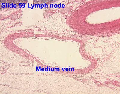
40 Medium size veins nedium size veins. Veins in these locations have a well developed muscular adventitia (muscle cells are oriented longitudinally). This is also the typical structure of large veins such as the vena cava.
41
42 Veins Large veins. Formed when venules converge Composed of 3 tunics Thin tunica media with circularly arranged smooth muscle cells. Thick tunica externa consisting of collagen fibers and elastic networks Capacitance vessels (blood reservoirs) that contain about 65% of the blood supply


43 Large Veins Large veins. Formed when venules converge Composed of 3 tunics Thin tunica media with circularly arranged smooth muscle cells. Thick tunica externa consisting of collagen fibers and elastic networks Capacitance vessels (blood reservoirs) that contain about 65% of the blood supply


44

45 * Vein 400X tunica externa tunica media tunica interna
46 Venous System: Valves Veins have much lower blood pressure and thinner walls than arteries To return blood to the heart, veins have special adaptations Large-diameter lumens, which offer little resistance to flow Valves (resembling semilunar heart valves), which prevent backflow of blood Venous sinuses specialized, flattened veins with extremely thin walls (e.g., coronary sinus of the heart and dural sinuses of the brain)


47


48

49

50 Comparison of Veins and Arteries Arteries: Veins: 50

51 Artery Vs veins
CVS HISTOLOGY. Dr. Nabil Khouri.
 CVS HISTOLOGY Dr. Nabil Khouri http://anatomy.kmu.edu.tw/blockhis/block3/slides/block4_24.html The Heart Wall Contract as a single unit Cardiac Muscle Simultaneous contraction due to depolarizing at the
UNIT 4: BLOOD VESSELS
 UNIT 4: BLOOD VESSELS Dr. Moattar Raza Rizvi NRS237, Physiology Generalized Structure of Blood Vessels 1 Tunica interna (tunica intima) Endothelial layer that lines the lumen of all vessels In vessels
Copyright 2010 Pearson Education, Inc. Blood Vessel Structure
 Blood Vessel Structure Structure of Blood Vessel Walls Arteries and veins Tunica intima, tunica media, and tunica externa Lumen Central blood-containing space Capillaries Endothelium with sparse basal
The cardiovascular system
 The cardiovascular system Components of the Cardiovascular system Heart Vessels: Arteries Capillaries Veins Functions of CVS: Transportation system where blood is the transporting vehicle Carries oxygen,
Practical Histology. Cardiovascular System. Dr Narmeen S. Ahmad
 Practical Histology Cardiovascular System Dr Narmeen S. Ahmad The Cardiovascular System A closed system of the heart and blood vessels Functions of cardiovascular system: Transport nutrients, hormones
2. capillaries - allow exchange of materials between blood and tissue fluid
 Chapter 19 - Vascular System A. categories and general functions: 1. arteries - carry blood away from heart 2. capillaries - allow exchange of materials between blood and tissue fluid 3. veins - return
Histology of the myocardium and blood vessels. Prof. Abdulameer Al-Nuaimi
 Histology of the myocardium and blood vessels Prof. Abdulameer Al-Nuaimi E-mail: a.al-nuaimi@sheffield.ac.uk E-mail: abdulameerh@yahoo.com Histology of blood vessels The walls of arteries and veins are
Blood Vessels. Types of Blood Vessels Arteries carry blood away from the heart Capillaries smallest blood vessels. Veins carry blood toward the heart
 C H A P T E R Blood Vessels 20 Types of Blood Vessels Arteries carry blood away from the heart Capillaries smallest blood vessels The site of exchange of molecules between blood and tissue fluid Veins
The Circulatory System
 The Circulatory System Dr. Sami Zaqout The circulatory system Circulatory system Blood vascular systems Lymphatic vascular systems Blood vascular systems Blood vascular systems The circulatory system Circulatory
Cardiovascular System Blood Vessels
 Cardiovascular System Blood Vessels Structure of Blood Vessels The three layers (tunics) Tunica intima composed of simple squamous epithelium Tunica media sheets of smooth muscle Contraction vasoconstriction
a) Endocardium The endocardium is the innermost layer of the heart wall. It forms the internal lining of the atria and ventricles.
 Endocardium The endocardium is the innermost layer of the heart wall. It forms the internal lining of the atria and ventricles.") Chapter 11 Circulatory System The circulatory system consists of a muscular, pulsing heart and a system of blood vessels.the blood vessels include: Arteries which will carry the blood and it's dissolved
Chapter 11 Circulatory System The circulatory system consists of a muscular, pulsing heart and a system of blood vessels.the blood vessels include: Arteries which will carry the blood and it's dissolved
Cardiovascular (Circulatory) System
 System") Cardiovascular (Circulatory) System Piryaei May 2011 Circulatory System Heart Blood Vessels Macrovasculature (More than 0.1mm) Elastic Artery Muscular (Distributing) Artery Large Arteriol Small Vein Muscular
Cardiovascular (Circulatory) System Piryaei May 2011 Circulatory System Heart Blood Vessels Macrovasculature (More than 0.1mm) Elastic Artery Muscular (Distributing) Artery Large Arteriol Small Vein Muscular
Microscopic Anatomy CARDIOVASCULAR SYSTEM
 Microscopic Anatomy CARDIOVASCULAR SYSTEM I. Introduction The cardiovascular system is a closed system consisting of a pump, the heart, and a series of tubular blood vessels that interconnect all body
Cardivascular System Module 5: Structure and Function of Blood Vessels *
 OpenStax-CNX module: m49689 1 Cardivascular System Module 5: Structure and Function of Blood Vessels * Donna Browne Based on Structure and Function of Blood Vessels by OpenStax This work is produced by
SCPA602 Cardiovascular System
 SCPA602 Cardiovascular System Associate Professor Dr. Wannee Jiraungkoorskul Department of Pathobiology, Faculty of Science, Mahidol University Tel: 02-201-5563, E-mail: wannee.jir@mahidol.ac.th 1 Objectives
Extra notes for lab- 1 histology. Slide 1 : cross section in the elastic artery ( aortic arch, ascending aorta, descending aorta )
") Extra notes for lab- 1 histology Slide 1 : cross section in the elastic artery ( aortic arch, ascending aorta, descending aorta ) - twin of ascending aorta is the pulmonary trunk. Ascending aorta represents
Extra notes for lab- 1 histology Slide 1 : cross section in the elastic artery ( aortic arch, ascending aorta, descending aorta ) - twin of ascending aorta is the pulmonary trunk. Ascending aorta represents
The Cardiovascular and Lymphatic Systems Cardiovascular System Blood Vessels Blood Vessels Arteries Arteries Arteries
 CH 12 The Cardiovascular and s The Cardiovascular and s OUTLINE: Cardiovascular System Blood Vessels Blood Pressure Cardiovascular System The cardiovascular system is composed of Blood vessels This system
Remember: the CVS system is deriving from the mesenchyma and is lined by simple squamous epithelium called the endothelium
 Lecture 10 General med_2nd semester Microscopic anatomy and embryology of cardiovascular system Microscopic structure of the heart, excitomotoric system - its structural peculiarities Blood vessels - arteries
Chapter 21 (1) An Introduction to Blood Vessels and Circulation
 An Introduction to Blood Vessels and Circulation") Chapter 21 (1) An Introduction to Blood Vessels and Circulation Lecture Objectives Compare and contrast the structure of an artery, arteriole, vein, venule, and capillary Discuss the structure and function
Chapter 21 (1) An Introduction to Blood Vessels and Circulation Lecture Objectives Compare and contrast the structure of an artery, arteriole, vein, venule, and capillary Discuss the structure and function
Anatomy Review: The Heart Graphics are used with permission of A.D.A.M. Software, Inc. and Benjamin/Cummings Publishing Co.
 Anatomy Review: The Heart Graphics are used with permission of A.D.A.M. Software, Inc. and Benjamin/Cummings Publishing Co. Anatomy Views Label the diagrams of the heart below: Interactive Physiology Study
Cardiovascular Anatomy Dr. Gary Mumaugh
 Cardiovascular Anatomy Dr. Gary Mumaugh Location of Heart Approximately the size of your fist Location o Superior surface of diaphragm o Left of the midline in mediastinum o Anterior to the vertebral column,
The Cardiovascular and Lymphatic Systems
 BIOLOGY OF HUMANS Concepts, Applications, and Issues Fifth Edition Judith Goodenough Betty McGuire 12 The Cardiovascular and Lymphatic Systems Lecture Presentation Anne Gasc Hawaii Pacific University and
Cardiovascular System
 Cardiovascular System Purpose Transport oxygen and nutrients Take waste products away from tissues & organs Things we learned Blood pressure: the force of blood pushing against the walls of blood vessels
Heart. Large lymphatic vessels Lymph node. Lymphatic. system Arteriovenous anastomosis. (exchange vessels)
") Venous system Large veins (capacitance vessels) Small veins (capacitance vessels) Postcapillary venule Thoroughfare channel Heart Large lymphatic vessels Lymph node Lymphatic system Arteriovenous anastomosis
Venous system Large veins (capacitance vessels) Small veins (capacitance vessels) Postcapillary venule Thoroughfare channel Heart Large lymphatic vessels Lymph node Lymphatic system Arteriovenous anastomosis
Approximately the size of your fist Location Superior surface of diaphragm Left of the midline in mediastinum Anterior to the vertebral column,
 Dr. Gary Mumaugh Approximately the size of your fist Location Superior surface of diaphragm Left of the midline in mediastinum Anterior to the vertebral column, posterior to the sternum Posteriorly the
Lab Activity 25. Blood Vessels & Circulation. Portland Community College BI 232
 Lab Activity 25 Blood Vessels & Circulation Portland Community College BI 232 Artery and Vein Histology Walls have 3 layers: Tunica intima Tunica media Tunica externa 2 Tunica Intima Is the innermost layer
The Cardiovascular System: Vessels and Routes. Pulmonary Circulation H E A R T. Systemic Circulation
 The Cardiovascular System: Vessels and Routes 1. Overview of Blood Circulation A. Pulmonary Circulation Lung Arterioles Pulmonary Artery Capillaries Pulmonary Circulation Venules Pulmonary Veins H E A
Chapter 14. The Cardiovascular System
 Chapter 14 The Cardiovascular System Introduction Cardiovascular system - heart, blood and blood vessels Cardiac muscle makes up bulk of heart provides force to pump blood Function - transports blood 2
Ch. 12 The Circulatory System. The heart. The heart is a double pump. A quick note on arteries vs. veins. = the muscular pump of the CV system
 Ch. 12 The Circulatory System The heart A.k.a. the cardiovascular system Blood was discussed in Ch. 11 Focus of Ch. 12: heart and blood vessels = the muscular pump of the CV system ~ 100,000 heartbeats/day!
Derived copy of Structure and Function of Blood Vessels *
 OpenStax-CNX module: m56696 1 Derived copy of Structure and Function of Blood Vessels * Stephanie Fretham Based on Structure and Function of Blood Vessels by OpenStax This work is produced by OpenStax-CNX
組織學 Historlogy 台北醫學大學 / 解剖學科教授 : 邱瑞珍分機號碼 :3261. 電子郵件信箱
 組織學 Historlogy 台北醫學大學 / 解剖學科教授 : 邱瑞珍分機號碼 :3261 電子郵件信箱 :rueijen@tmu.edu.tw 1 The Circulatory System 台北醫學大學 / 解剖學科教授 : 邱瑞珍分機號碼 :3261 電子郵件信箱 :rueijen@tmu.edu.tw 2 學習目的 The structure of the arteries The structure
Major Function of the Cardiovascular System. Transportation. Structures of the Cardiovascular System. Heart - muscular pump
 Structures of the Cardiovascular System Heart - muscular pump Blood vessels - network of tubes Blood - liquid transport vehicle brachiocephalic trunk superior vena cava right pulmonary arteries right pulmonary
Cardiac Conduction System
 Cardiac Conduction System What causes the Heart to Beat? Heart contracts by electrical signals! Cardiac muscle tissue contracts on its own an electrical signal is sent out by the heart so that all cells
Human Anatomy, First Edition
 Human Anatomy, First Edition McKinley & O'Loughlin Chapter 23 : Vessels and Circulation 23-1 Blood Vessels An efficient style of transport for oxygen, nutrients, and waste products to and from body tissues.
STRUCTURES OF THE CARDIOVASCULAR SYSTEM
 STRUCTURES OF THE CARDIOVASCULAR SYSTEM CARDIOVASCULAR SYSTEM Also called the circulatory system Consists of the heart, arteries, veins, and capillaries Main function is to pump/circulate oxygenated blood
Lecture name: blood 2 & The Circulatory System Edited by: Buthainah Al masaeed & Yousef Qandeel
 Lecture name: blood 2 & The Circulatory System Edited by: Buthainah Al masaeed & Yousef Qandeel Now we will take about A granulocytes : Lymphocyte Monocytes 1- Lymphocyte - The second major type of presence
2402 : Anatomy/Physiology
 Dr. Chris Doumen Lecture 1 2402 : Anatomy/Physiology Hemo Dynamics and Blood Vessels I nt r oduc t i on TextBook Readings Pages 721 through 734. Make use of the figures in your textbook ; a picture is
Heart. Heart 2-Tunica media: middle layer (media ='middle') muscle fibers (smooth or cardiac).
 muscle fibers (smooth or cardiac).") t. innermost lumenal General Circulatory system heart and blood vessels walls have 3 layers (inside to outside) 1-Tunica interna: aka tunica intima layer--lumenal layer epithelium--endothelium simple squamous
t. innermost lumenal General Circulatory system heart and blood vessels walls have 3 layers (inside to outside) 1-Tunica interna: aka tunica intima layer--lumenal layer epithelium--endothelium simple squamous
10. Thick deposits of lipids on the walls of blood vessels, called, can lead to serious circulatory issues. A. aneurysm B. atherosclerosis C.
 Heart Student: 1. carry blood away from the heart. A. Arteries B. Veins C. Capillaries 2. What is the leading cause of heart attack and stroke in North America? A. alcohol B. smoking C. arteriosclerosis
The Circulatory System
 The Circulatory System This system comprises both the blood and lymphatic vascular system. Blood vascular system is composed from; The heart: an organ whose function is to pump the blood. The arteries:
Any of these questions could be asked as open question or lab question, thus study them well
 Any of these questions could be asked as open question or lab question, thus study them well describe the factors which regulate cardiac output describe the sympathetic and parasympathetic control of heart
Blood Vessels. Over view. We have about 60,000 miles of blood vessels!
 Blood Vessels Over view 3 types of blood vessels arteries - carry blood away from heart "branch", "diverge", and "fork" veins - carry blood toward heart "join", "merge", and "converge" capillaries - site
The Cardiovascular System
 The Cardiovascular System The Cardiovascular System A closed system of the heart and blood vessels The heart pumps blood Blood vessels allow blood to circulate to all parts of the body The function of
Cardiovascular Physiology
 Cardiovascular Physiology Lecture 1 objectives Explain the basic anatomy of the heart and its arrangement into 4 chambers. Appreciate that blood flows in series through the systemic and pulmonary circulations.
1. What kind of blood is found in the rt. atrium? (oxygenated or deoxygenated)
") Carl Christennsen, PhD Chap. 19, 20, & 21 - Circulatory System Bio. 2304 Human Anatomy HEART 1. What kind of blood is found in the rt. atrium? (oxygenated or deoxygenated) Where does this blood come from?
Carl Christennsen, PhD Chap. 19, 20, & 21 - Circulatory System Bio. 2304 Human Anatomy HEART 1. What kind of blood is found in the rt. atrium? (oxygenated or deoxygenated) Where does this blood come from?
Cardiovascular System. Blood Vessel anatomy Physiology & regulation
 Cardiovascular System Blood Vessel anatomy Physiology & regulation Path of blood flow Aorta Arteries Arterioles Capillaries Venules Veins Vena cava Vessel anatomy: 3 layers Tunica externa (adventitia):
Chapter 21 Peripheral circulation and Regulation
 Chapter 21 Peripheral circulation and Regulation I. Blood vessel structure A. Blood flows from large arteries to small capillaries 1. Large arteries contain large amounts of elastic tissue and little smooth
BIOH122 Human Biological Science 2
 BIOH122 Human Biological Science 2 Session 5 Cardiovascular System 3 Vasculature and Capillary Exchange Bioscience Department Endeavour College of Natural Health endeavour.edu.au Session Plan o Structure
Collin County Community College
 Collin County Community College BIOL. 2402 Anatomy & Physiology WEEK 6 Blood Vessels 1 Anatomy of Blood Vessels Walls of blood vessels contain 3 distinct layers : Tunica intima innermost layer includes
Cardiovascular System. I. Structures of the heart A. : Pericardium sack that surrounds the heart
 Cardiovascular System I. Structures of the heart A. : Pericardium sack that surrounds the heart 1. : Pericardial Cavity serous fluid filled space between the heart and the pericardium B. Heart Wall 1.
Types of Blood Vessels
 Chapter 21 Peripheral Circulation and Regulation 21-1 Types of Blood Vessels Capillaries: site of exchange with tissue Arteries in dif. Types & sizes Elastic Muscular Arterioles Veins: thinner walls than
Biology 232 Final. 7. Which of the following lists the elements of the heart s conduction system in the correct order? Name
 Biology 232 Final Name 1. The heart is located within the: a) mediastinum b) pleural cavity c) pericardial cavity 2. Which of the following is not part of cardiac muscle histology: a) striations b) intercalated
Blood flows away from the heart in arteries, to the capillaries and back to the heart in the veins
 Cardiovascular System Summary Notes The cardiovascular system includes: The heart, a muscular pump The blood, a fluid connective tissue The blood vessels, arteries, veins and capillaries Blood flows away
Function: Transportation of. Oxygen Nutrients Waste Hormones gases
 Function: Transportation of Oxygen Nutrients Waste Hormones gases Pericardium: double sac of serous membrane filled with fluid (pericardial fluid to be exact) that surrounds the heart. Parietal pericardium:
Cardiovascular system
 BIO 301 Human Physiology Cardiovascular system The Cardiovascular System: consists of the heart plus all the blood vessels transports blood to all parts of the body in two 'circulations': pulmonary (lungs)
Sinusoids and venous sinuses
 LYMPHOID SYSTEM General aspects Consists of organs that are made of lymphoid tissue; Immune defense Breakdown of red blood cells. 1 Sinusoids In place of capillaries Endothelium; often fenestrated More
Microcirculation. Lecture Block 11 (contributions from Brett Burton)
") Lecture Block 11 (contributions from Brett Burton) Elements of Arterioles, capillaries, venules Structure and function: transport Fluid balance Lymph system Vessels of the Circulatory System Diameter Aorta
Lecture Block 11 (contributions from Brett Burton) Elements of Arterioles, capillaries, venules Structure and function: transport Fluid balance Lymph system Vessels of the Circulatory System Diameter Aorta
Chapter 21. Blood Vessels and Circulation
 Chapter 21 Openstax: Chapter 20 Blood Vessels and Circulation Chapter 21 Learning Outcomes After completing Chapter 21, you will be able to: 1. Distinguish among the types of blood vessels based on their
Structure and organization of blood vessels
 The cardiovascular system Structure of the heart The cardiac cycle Structure and organization of blood vessels What is the cardiovascular system? The heart is a double pump heart arteries arterioles veins
Circulation. Sinoatrial (SA) Node. Atrioventricular (AV) Node. Cardiac Conduction System. Cardiac Conduction System. Linked to the nervous system
 Node. Atrioventricular (AV) Node. Cardiac Conduction System. Cardiac Conduction System. Linked to the nervous system") Circulation Cardiac Conduction System AHS A H S Your body resembles a large roadmap. There are routes or arteries that take you downtown to the heart of the city and veins that take you to the outskirts
Circulation Cardiac Conduction System AHS A H S Your body resembles a large roadmap. There are routes or arteries that take you downtown to the heart of the city and veins that take you to the outskirts
Chapter 19: Blood Vessels. 63 slides
 Chapter 19: Blood Vessels 63 slides 1 Blood Vessels The Blood Vessels are essentially a series of tubes. Three types of blood vessel tubes: Arteries (carry blood away from heart) Arterioles are the smallest
Cardiovascular System
 Cardiovascular System I. Structure of the Heart A. Average adult heart is 14 cm long and 9 cm wide. B. Lies in the mediastinum. C. Enclosed in the pericardium. 1. Fibrous pericardium- Outer, tough connective
Vascular System Part One
 Vascular System Part One Objectives Trace the route taken by blood as it leaves, and then returns to the heart. Describe the structure of the walls of arteries and veins. Discuss the structure and function
Tissues 10/21/2016. Epithelial Tissue
 Tissues This is a generalized cell diagram. It shows the anatomy of a cell, but most cells do not actually look like this. Cells can have a wide variety of shapes and sizes, depending on their function.
2. right heart = pulmonary pump takes blood to lungs to pick up oxygen and get rid of carbon dioxide
 A. location in thorax, in inferior mediastinum posterior to sternum medial to lungs superior to diaphragm anterior to vertebrae orientation - oblique apex points down and to the left 2/3 of mass on left
Blood Vessels. Chapter 20
 Blood Vessels Chapter 20 Summary of the Characteristics of Arteries and Veins Characteristic Artery Vein Wall thickness thick thin Shape in cross section round flattened Thickest tunic media externa Collagen
Anatomy and Physiology, Spring 2015 Exam II: Form A April 9, Name Student Number
 Anatomy and Physiology, Spring 2015 Exam II: Form A April 9, 2015 Name Student Number For Questions 1 2 refer to the following table. 1 Ventricular pressure is greater than aortic 6 AV valve is open 2
Chp. 5 The cardiovascular system. What are the function of the cardiovascular system? Arteries and arterioles:
 5.1 Overview of the cardiovascular system Chp. 5 The cardiovascular system Includes the heart and blood vessels Brings nutrients to cells and helps get rid of wastes Blood is refreshed in the lung, kidneys,
Collin County Community College. ! BIOL Anatomy & Physiology! WEEK 5. The Heart
 Collin County Community College! BIOL. 2402 Anatomy & Physiology! WEEK 5 The Heart 1 (1578-1657) A groundbreaking work in the history of medicine, English physician William Harvey s Anatomical Essay on
The Cardiovascular System
 Essentials of Human Anatomy & Physiology Elaine N. Marieb Seventh Edition Chapter 11 The Cardiovascular System Slides 11.1 11.19 Lecture Slides in PowerPoint by Jerry L. Cook The Cardiovascular System
Peripheral Circulation and Regulation
 Peripheral Circulation and Regulation Functions of Peripheral Circulation 1. Contain the blood 2. Exchange nutrients, waste products, and gases with tissues 3. Transport 4. Regulate blood pressure, along
Structure. Arteries. 21_01d 4/18/12. The Cardiovascular System: Blood Vessels and Hemodynamics. Dr Badri Paudel GMC
 Goal of the Cardiovascular System: deliver blood to all parts of the body The Cardiovascular System: Blood Vessels and Hemodynamics Dr Badri Paudel GMC Does so by using different types of tubing, attached
Warm Up- Monday -AND- Setup Cornell Notes.
 Warm Up- Monday Brainstorm in your notebook: If the heart sends blood to all organs, how and where does the heart get blood to provide oxygen for its muscles? -AND- Setup Cornell Notes. Announcements Unit
Human Anatomy, First Edition
 Human Anatomy, First Edition McKinley & O'Loughlin Chapter 22 : Heart 1 Functions of the Heart Center of the cardiovascular system, the heart. Connects to blood vessels that transport blood between the
Ch 19: Cardiovascular System - The Heart -
 Ch 19: Cardiovascular System - The Heart - Give a detailed description of the superficial and internal anatomy of the heart, including the pericardium, the myocardium, and the cardiac muscle. Trace the
CIRCULATION. Cardiovascular & lymphatic systems Functions. Transport Defense / immunity Homeostasis
 CIRCULATION CIRCULATION Cardiovascular & lymphatic systems Functions Transport Defense / immunity Homeostasis 2 Types of Circulatory Systems Open circulatory system Contains vascular elements Mixing of
Cardiovascular System
 Cardiovascular System The Heart Cardiovascular System The Heart Overview What does the heart do? By timed muscular contractions creates pressure gradients blood moves then from high pressure to low pressure
Figure ) The specific chamber of the heart that is indicated by letter A is called the. Diff: 1 Page Ref: 364
 The specific chamber of the heart that is indicated by letter A is called the. Diff: 1 Page Ref: 364") Essentials of Anatomy and Physiology, 9e (Marieb) Chapter 11 The Cardiovascular System Short Answer Figure 11.1 Using Figure 11.1, identify the following: 1) The Purkinje fibers are indicated by label.
Essentials of Anatomy and Physiology, 9e (Marieb) Chapter 11 The Cardiovascular System Short Answer Figure 11.1 Using Figure 11.1, identify the following: 1) The Purkinje fibers are indicated by label.
Anatomy & Physiology of Cardiovascular System. Chapter 18 & 19
 Anatomy & Physiology of Cardiovascular System Chapter 18 & 19 Objectives..cont 1. Discuss the physiological stages of cardiac muscle contraction. 2. Trace a typical ECG and label each wave or complex 3.
Rheological, mechanical and failure properties of biological soft tissues at high strains and rates of deformation
 Rheological, mechanical and failure properties of biological soft tissues at high strains and rates of deformation Society of Rheology Conference Salt Lake City, Utah October 10, 2007 Martin L. Sentmanat,
The Circulatory System. The Heart, Blood Vessels, Blood Types
 The Circulatory System The Heart, Blood Vessels, Blood Types The Closed Circulatory System Humans have a closed circulatory system, typical of all vertebrates, in which blood is confined to vessels and
IB TOPIC 6.2 THE BLOOD SYSTEM
 IB TOPIC 6.2 THE BLOOD SYSTEM TERMS TO KNOW circulation ventricle artery vein THE BLOOD SYSTEM 6.2.U1 - Arteries convey blood at high pressure from the ventricles to the tissues of the body Circulation
The ancient Babylonians, Egyptians, Indians and Chinese believed the heart was the centre of thinking and emotions
 The Concept of Mind The ancient Babylonians, Egyptians, Indians and Chinese believed the heart was the centre of thinking and emotions Hippocrates 460 BC 370 BC - Thoughts, ideas, and feelings come from
Muscle tissues. Dr. Hersh Abdul Ham-Karim BVM&S, PG Dip, MSc and PhD
 Muscle tissues Dr. Hersh Abdul Ham-Karim BVM&S, PG Dip, MSc and PhD Muscle tissue is a soft tissue that composes muscles in animal bodies, and gives rise to muscles' ability to contract. Muscle tissue
Cardiovascular System Notes: Physiology of the Heart
 Cardiovascular System Notes: Physiology of the Heart Interesting Heart Fact Capillaries are so small it takes ten of them to equal the thickness of a human hair. Review What are the 3 parts of the cardiovascular
The Heart. Size, Form, and Location of the Heart. 1. Blunt, rounded point; most inferior part of the heart.
 12 The Heart FOCUS: The heart is composed of cardiac muscle cells, which are elongated, branching cells that appear striated. Cardiac muscle cells behave as a single electrical unit, and the highly coordinated
IB TOPIC 6.2 THE BLOOD SYSTEM
 IB TOPIC 6.2 THE BLOOD SYSTEM THE BLOOD SYSTEM TERMS TO KNOW circulation ventricle artery vein 6.2.U1 - Arteries convey blood at high pressure from the ventricles to the tissues of the body Circulation
Circulatory System 10.1
 1 Circulatory System 10.1 2 ARTERIES Arteries-blood vessels that carry blood away from the heart Thick walls Inner & Outer layers: connective tissue Middle layers are muscle and elastic connective tissue
Chapter 20: Cardiovascular System: The Heart
 Chapter 20: Cardiovascular System: The Heart I. Functions of the Heart A. List and describe the four functions of the heart: 1. 2. 3. 4. II. Size, Shape, and Location of the Heart A. Size and Shape 1.
The Cardiovascular System: The Heart
 The Cardiovascular System: The Heart 1 2 3 4 5 6 7 8 9 10 11 12 Across 1. The spontaneously changing pacemaker membranes. 7. The distinguishing feature of heart muscle. 9. What allows heart cells to beat
The Cardiovascular System
 11 PART A The Cardiovascular System PowerPoint Lecture Slide Presentation by Jerry L. Cook, Sam Houston University ESSENTIALS OF HUMAN ANATOMY & PHYSIOLOGY EIGHTH EDITION ELAINE N. MARIEB The Cardiovascular
d) Cardiovascular System Higher Human Biology
 Cardiovascular System Higher Human Biology") d) Cardiovascular System Higher Human Biology What can your remember about the heart and blood vessels? What is the Cardiovascular System? The cardiovascular system, also known as the circulatory system,
d) Cardiovascular System Higher Human Biology What can your remember about the heart and blood vessels? What is the Cardiovascular System? The cardiovascular system, also known as the circulatory system,
Cardiovascular System, Blood, and Blood Cell Formation
 Cardiovascular System, Blood, and Blood Cell Formation 7 CONTENTS OVERVIEW OF THE CARDIOVASCULAR SYSTEM GENERAL STRUCTURE OF THE HEART Heart Valves HEART AS A PUMP Contractile Properties of Cardiac Myocytes
Cardiovascular System: The Heart
 Cardiovascular System: The Heart I. Anatomy of the Heart (See lab handout for terms list) A. Describe the size, shape and location of the heart B. Describe the structure and function of the pericardium
Circula/on and Gas Exchange
 Chapter 42 Circula/on and Gas Exchange BIOL 223 Simple animals such as cnidarians Body wall only two cells thick Flatworms Gastrovascular Cavi/es Encloses gastrovascular cavity diges/on and distribu/on
AN ATOMY OF THE CARDIOVASCULAR SYSTEM
 Student Name CHAPTER 18 AN ATOMY OF THE CARDIOVASCULAR SYSTEM T he heart is actually two pumps one moves blood to the lungs, the other pushes it out into the body. These two functions seem rather elementary
The Heart. The Heart A muscular double pump. The Pulmonary and Systemic Circuits
 C H A P T E R 19 The Heart The Heart A muscular double pump circuit takes blood to and from the lungs Systemic circuit vessels transport blood to and from body tissues Atria receive blood from the pulmonary
Sample Exam Biology 2050 Circulatory and Lymphatic Systems
 Sample Exam Biology 2050 Circulatory and Lymphatic Systems Note: Not all of the lymphatic system will be on the actual exam next Monday so disregard any questions that deal with something that wasn t covered
Vessels by Design: Basic Vessel Anatomy. Student Information Page 3A
 Vessels by Design: Basic Vessel Anatomy Student Information Page 3A Activity Introduction: Once you get home from running around all day, your throat is probably a little dry. You go to your kitchen, get
Cardiovascular System. Biology 105 Lecture 15 Chapter 12
 Cardiovascular System Biology 105 Lecture 15 Chapter 12 Outline I. Functions of cardiovascular system II. Components of the cardiovascular system: I. Blood vessels II. Heart III. Regulation of the heartbeat