CARDIOVASCULAR MONITORING. Prof. Yasser Mostafa Kadah
|
|
|
- Jeffery Newton
- 5 years ago
- Views:
Transcription

1 CARDIOVASCULAR MONITORING Prof. Yasser Mostafa Kadah
2 Introduction Cardiovascular monitoring covers monitoring of heart and circulatory functions It makes it possible to commence interventions quickly in the event of any impairment Measurements are used to assess the condition of the patient, reach a diagnosis, decide on therapy, and monitor therapy Covers cardiac function in the form of electrical phenomenon (ECG) and its mechanical effects including pressure build-up and volume delivery, contractility, preload, and afterload

3 Electrocardiogram (ECG) ECG provides information about heart rate and rhythm, excitation, conduction, and repolarization and disturbances in these functions ECG does NOT provide any direct information about the pumping capacity of the heart (i.e. mechanical cardiac function) Lead Wire Color Code: IEC2 (US): RA: white, LA: black, LL: red, RL: green, V: brown, V+: gray/brown IEC1 (Europe): R: red, L: yellow, F: green, N: black, V: white, V+: gray/white
4 Heart Rate (HR) Heart rate (typical measuring range of beats/min) is determined as a moving average over a specific time (e.g. 10s), or a specified number of QRS complexes An alternative to the ECG that can be used when there are interferences in the ECG, e.g. during cauterization in the OT) Can be computed in the form of pulse rate from the arterial blood pressure or pulse curve from the SpO2 signal Basis of heart rate measurement is safe detection of QRS complexes in ECG and assessment of RR intervals
5 Hemodynamics Hemodynamics is the study of the flow of blood in the circulatory system This flow is driven by the pressure generated by the heart. Since the pressure in the vascular system is highly dependent on activity and the position of the body (hydrostatic pressure), blood pressure measurements are always taken at rest and are based on the height of the heart (right atrium) Vascular system is functionally divided into low-pressure system (small, pulmonary circuit) and high-pressure system (large, systemic circuit), connected by the heart as driving element
6 Hemodynamics Heart generates pressure in its contraction phase (systole), by means of which stroke volume (SV) is expelled from the ventricle into arterial vascular system Every stroke volume conveyed generates a pulse wave Peak pressure during the expulsion of stroke volume from ventricle is systolic blood pressure (highest point of pressure curve) Pressure at end of relaxation phase (diastole) is referred to as diastolic blood pressure (lowest point of pressure curve) Difference between systolic and diastolic pressure is blood pressure amplitude
7 Hemodynamics Pressure that maintains blood flow in vascular system and act as driving force of perfusion is mean pressure In systemic circuit mean arterial pressure is termed APm (also MAP) In pulmonary circuit mean pulmonary artery pressure is termed PAPm Stroke volume depends on preload, contractility of myocardium and afterload Preload is stretching of myocardium brought about by passive filling of ventricles at end of diastole and is best described by end-diastolic volume Afterload is force exerted by cardiac muscles to overcome resistance in outflow tract of left ventricle and peripheral circuit Mean arterial pressure and vascular resistance are measures of afterload
8 Hemodynamics Factors that also determine blood pressure behavior include elasticity of vascular system components, circulating blood volume and, in particular, peripheral vascular resistance, which is influenced by wall tension of vessels (vessel tone) controlled by sympathetic nervous system Delivery volume of blood per minute is known as the cardiac output (CO) and is the product of stroke volume and heart rate Control mechanisms in the body regulate circulation with aim of adjusting cardiac output to circulation required to supply oxygen to the organism and eliminate CO 2, keeping the blood pressure largely constant and adjusting the circulation in the individual organs and tissues to the functional state in each case
9 Hemodynamics
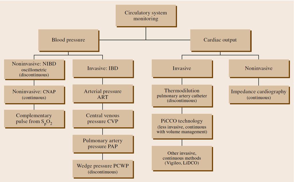
10 Hemodynamic Monitoring


11 Pulse Monitoring Pulse monitoring is performed either invasively from the arterial pressure curve or noninvasively from the plethysmogram of pulse oximetry Pulse monitoring has particular importance as a safety measure in such condition as monitoring pacemaker patients

: systolic blood pressure is read off from the manometer Sounds change until sound can no longer be heard (phase")
12 Discontinuous Noninvasive Blood Pressure (NIBP): Auscultatory Method Sphygmomanometer Riva Rocci: Systolic blood pressure measurement Korotkoff: Diastolic blood pressure Auscultatory Method Cuff placed on exposed upper arm at the level of heart with middle of rubber bladder positioned over brachial artery After palpating the brachial artery, the cuff is inflated to 30mmHg above the pressure at which the pulse can no longer be detected Stethoscope is placed against the brachial artery and the cuff pressure is slowly released. Pulsation that then begins causes knocking noises (Korotkoff sounds phase 1): systolic blood pressure is read off from the manometer Sounds change until sound can no longer be heard (phase 5) and is measured as the diastolic pressure

13 Discontinuous Noninvasive Blood Pressure (NIBP): Oscillometric Method When cuff pressure is released once the systolic pressure has been reached, the vessel walls begin to oscillate and maintain this behavior until the vessel is no longer occluded Oscillations are transmitted to air in cuff and are read on the manometer Today, oscillations are measured electronically using pressure sensors
14 Continuous Noninvasive Arterial Blood Pressure Measurement (CNAP) Blood pressure is not always constant but can change within a matter of seconds In particular, during anesthesia and its induction, variations in blood pressure can arise and require immediate medical attention Noninvasive technique of relaxed arterial wall (vascular unloading technique or volume clamp method) uses optical sensor in small cuff around finger to measure volume pulses due to each heart beat Pressure in the cuff is regulated by means of feedback such that the optical measuring path always remains constant When a pulse occurs, cuff pressure is increased accordingly, and when the pulse subsides the cuff pressure is reduced. Cuff pressure reflects the pressure occurring in the enclosed finger artery with high degree of accuracy


15 Continuous Noninvasive Arterial Blood Pressure Measurement (CNAP) CNAP uses pairs of sensor cuffs, which are placed on adjacent fingers Only one cuff is used for measurement at any time, and after no longer than half an hour continuous measurement is automatically switched Venous stasis that naturally occurs during measurement on finger very quickly decreases once more after the switch Great advantage of this method is calibration of of continuous measurement with the normal NIBP measurement, so that correct values are displayed even when the fingers are not level with the heart Advantages of both NIBP and CNAP
 and pressure")


16 Invasive Pressure Measurement in High- Pressure System Continuous availability of measurement signal (pressure curve) and pressure values Provides possibility of triggering an alarm if predefined limit values violated and of further signal processing of measurement data Connection is set up by means of an intra-arterial catheter between intravasal blood column and liquid-coupled pressure transducer
17 Invasive Pressure Measurement in Low- Pressure System Aim is to obtain information about right ventricular function, pulmonary circuit, and filling of vascular system Central Venous Pressure (CVP) Pulmonary Artery Pressure (PAP) Pulmonary Capillary Wedge Pressure (PCWP)
18 Central Venous Pressure (CVP) Measurement of the central venous pressure is by means of a liquid manometer or a pressure sensor using central venous catheter (CVC) placed in superior vena cava at the entrance to the right atrium Pressure transducers have the advantage that measurement information is available continuously and certain signal characteristics of the CVP curve are additionally available Because of the small pressure values, it is important that pressure measurement system is situated level with the heart (correct zero point positioning) in order to avoid errors Hydrostatic pressure difference can cause incorrect measurements
")
 Influenced by capacity")

19 Central Venous Pressure (CVP) Progression of CVP curve shows: Atrial contraction (a-wave) Beginning of the ventricular contraction (c-wave) Relaxation phase (v-wave) Influenced by capacity of vascular system, cardiac output, blood volume, compliance of myocardium, and afterload of right ventricle
 and")


20 Pulmonary Artery Pressure (PAP) and Pulmonary Capillary Wedge Pressure (PCWP) To monitor hemodynamics of right ventricle, balloon catheter is pushed through venous system into right atrium, right ventricle, and then through pulmonary valve into arteria pulmonalis Path of catheter is followed from different typical pressure curves
, this causes damping of pressure curves when balloon is filled and there is continuous rise in")
21 Pulmonary Artery Pressure (PAP) and Pulmonary Capillary Wedge Pressure (PCWP) Correct catheter position is reached once in wedge position That is, once the inflated balloon of the catheter blocks off the pulmonary artery branch If tip of catheter rests against wall of pulmonary artery (pseudo-wedge), this causes damping of pressure curves when balloon is filled and there is continuous rise in pressure Although catheter is in right ventricle, in the wedge position pressure in left atrium can be inferred via the distal lumen PCWP value corresponds in first approximation to the left atrial pressure (LAP) and thus to the end-diastolic filling pressure in the left ventricle Left atrium, pulmonary capillaries, and pulmonary artery under normal conditions form a common pressure connection during diastole.
22 Balloon Catheters Using balloon catheters (so-called flow-directed catheters, pulmonary artery catheters, or Swan Ganz catheters) with different length and thickness, number of lumina, position of lumen exit sites, and other characteristics; CVP, PAP, and core body temperature can be measured simultaneously, and the PCWP and CO can be measured intermittently Specialized balloon catheters provide additional possibilities such as intracardial ECG measurement, supraventricular and ventricular stimulation, measurement of mixed venous oxygen saturation SvO 2 with integration of fiber optics, transluminal stimulation probe, or additional infusion lumina Balloon catheters are not free of risks and can cause complications such as: Supraventricular and ventricular arrhythmias Ventricular tachycardia or ventricular fibrillation (rarely) Venous thrombosis (particularly with a low CO) Sepsis (risk rises as the duration of catheterization increases) Pulmonary infarction (due to catheter occlusion of peripheral pulmonary artery) Pulmonary artery rupture (by balloon inflation or the catheter tip).
23 Determining the Cardiac Output (CO) Cardiac output is volume of blood conveyed per minute (l/min) Classical way of determining CO is by Fick s principle Calculation is based quite simply on the law of conservation of mass CO is the quotient from oxygen consumption (VO 2 ) in the body and difference in oxygen content (avdo 2 ) between arterial blood flowing to the body and mixed venous blood returning from the body: CO = VO 2 /avdo 2 Unfortunately, under routine conditions oxygen consumption cannot be measured with sufficient accuracy in the clinical environment
 made thermodilution by means of a pulmonary artery catheter established as leading method for clinical")
24 Dilution Methods to Measure CO Development of fundamental work of Stewart and Hamilton to determine CO by means of dye dilution (1920s) Implemented with dye, cold, ions, radioisotopes dilution methods Introduction of thermistor catheter by Swan and Ganz (1970s) made thermodilution by means of a pulmonary artery catheter established as leading method for clinical use
25 Thermodilution Method Defined amount of saline solution at a temperature of 0 25 C (the lower the temperature, the more accurate the measurement) is injected into the right atrium via proximal port of the multilumen pulmonary artery catheter Because injected fluid is mixed with warm flowing blood (37 C) and is therefore diluted, change in temperature in blood stream can be measured by thermistor situated close to tip of catheter Shape and area of dilution curve change with cardiac output With known temperature of injected fluid and blood as well as known volume of injected fluid, measuring system determines CO from the area of thermodilution curve
26 Thermodilution Disadvantages Need for the pulmonary artery catheter, the indication of which is viewed particularly critically Connors et al. (JAMA, 1996): RHC is associated with increased mortality and increased utilization of resources! Discontinuity of measurements This was overcome by emitting heat pulses to the blood using a special pulmonary artery catheter and by evaluating their dilution curve Since heat pulses can be applied at very short intervals (30 60 s), this virtually provides continuous measurement


27 PiCCO Technology Thermodilution can, in principle, be done transpulmonarily Cold bolus passes through lungs with thermistor placed in arterial system Cold bolus is injected into right atrium as in normal thermodilution except with normal central venous catheter (more common) Temperature profile is measured in arteria femoralis Advantage of this method is that it is less invasive Omission of pulmonary artery catheter and its risks
 Offers several advantages Completely noninvasive and low risk Continuous beat-to-beat measurement Easy to apply Cannot be")
28 Impedance Cardiography It has long been known that blood volume expelled with each heart beat (stroke volume) leads to measurable variations in the thoracic impedance Attempts to determine the stroke volume and CO from the variations in thoracic electrical bioimpedance (TEB) Offers several advantages Completely noninvasive and low risk Continuous beat-to-beat measurement Easy to apply Cannot be used in some cases Example: septic shock patients PiCCO is used more due to that
 is passed through thorax by means of external ring electrodes (or special double electrodes) arranged on neck and thorax Current seeks path of least resistance,")
29 Impedance Cardiography Weak high-frequency constant current (e.g. 2.5mA, 70 khz) is passed through thorax by means of external ring electrodes (or special double electrodes) arranged on neck and thorax Current seeks path of least resistance, which is essentially blood-conducting aorta and voltage drop is measured by inner measuring electrodes ICG
30 Calculation of Hemodynamic Variables Total Peripheral Resistance (TPR), also called Systemic Vascular Resistance (SVR), is resistance of systemic circuit, computed as quotient of propulsive pressure difference (Mean Arterial Pressure APm - Central Venous Pressure CVP) and flow (CO) Pressure difference in small circuit is Mean Pulmonary Pressure minus Wedge Pressure PCWP (as a measure of the left atrial pressure). The Pulmonary Vascular Resistance (PVR) is given as:
31 Reading Assignment Read Chapter 48 of Springer Handbook of Medical Technology
Impedance Cardiography (ICG) Method, Technology and Validity
 Method, Technology and Validity") Method, Technology and Validity Hemodynamic Basics Cardiovascular System Cardiac Output (CO) Mean arterial pressure (MAP) Variable resistance (SVR) Aortic valve Left ventricle Elastic arteries / Aorta
Method, Technology and Validity Hemodynamic Basics Cardiovascular System Cardiac Output (CO) Mean arterial pressure (MAP) Variable resistance (SVR) Aortic valve Left ventricle Elastic arteries / Aorta
Hemodynamic Monitoring
 Perform Procedure And Interpret Results Hemodynamic Monitoring Tracheal Tube Cuff Pressure Dean R. Hess PhD RRT FAARC Hemodynamic Monitoring Cardiac Rate and Rhythm Arterial Blood Pressure Central Venous
Hemodynamic Monitoring and Circulatory Assist Devices
 Hemodynamic Monitoring and Circulatory Assist Devices Speaker: Jana Ogden Learning Unit 2: Hemodynamic Monitoring and Circulatory Assist Devices Hemodynamic monitoring refers to the measurement of pressure,
Topics to be Covered. Cardiac Measurements. Distribution of Blood Volume. Distribution of Pulmonary Ventilation & Blood Flow
 Topics to be Covered MODULE F HEMODYNAMIC MONITORING Cardiac Output Determinants of Stroke Volume Hemodynamic Measurements Pulmonary Artery Catheterization Control of Blood Pressure Heart Failure Cardiac
IB TOPIC 6.2 THE BLOOD SYSTEM
 IB TOPIC 6.2 THE BLOOD SYSTEM TERMS TO KNOW circulation ventricle artery vein THE BLOOD SYSTEM 6.2.U1 - Arteries convey blood at high pressure from the ventricles to the tissues of the body Circulation
IB TOPIC 6.2 THE BLOOD SYSTEM
 IB TOPIC 6.2 THE BLOOD SYSTEM THE BLOOD SYSTEM TERMS TO KNOW circulation ventricle artery vein 6.2.U1 - Arteries convey blood at high pressure from the ventricles to the tissues of the body Circulation
Chapter 13 The Cardiovascular System: Cardiac Function
 Chapter 13 The Cardiovascular System: Cardiac Function Overview of the Cardiovascular System The Path of Blood Flow through the Heart and Vasculature Anatomy of the Heart Electrical Activity of the Heart
Chapter 9, Part 2. Cardiocirculatory Adjustments to Exercise
 Chapter 9, Part 2 Cardiocirculatory Adjustments to Exercise Electrical Activity of the Heart Contraction of the heart depends on electrical stimulation of the myocardium Impulse is initiated in the right
FUNDAMENTALS OF HEMODYNAMICS, VASOACTIVE DRUGS AND IABP IN THE FAILING HEART
 FUNDAMENTALS OF HEMODYNAMICS, VASOACTIVE DRUGS AND IABP IN THE FAILING HEART CINDY BITHER, MSN, ANP, ANP, AACC, CHFN CHIEF NP, ADV HF PROGRAM MEDSTAR WASHINGTON HOSPITAL CENTER CONFLICTS OF INTEREST NONE
Technique. Technique. Technique. Monitoring 1. Local anesthetic? Aseptic technique Hyper-extend (if radial)
") Critical Care Monitoring Hemodynamic Monitoring Arterial Blood Pressure Cannulate artery Uses 2 Technique Sites Locate artery, prep 3 1 Technique Local anesthetic? Aseptic technique Hyper-extend (if radial)
Critical Care Monitoring Hemodynamic Monitoring Arterial Blood Pressure Cannulate artery Uses 2 Technique Sites Locate artery, prep 3 1 Technique Local anesthetic? Aseptic technique Hyper-extend (if radial)
4. The two inferior chambers of the heart are known as the atria. the superior and inferior vena cava, which empty into the left atrium.
 Answer each statement true or false. If the statement is false, change the underlined word to make it true. 1. The heart is located approximately between the second and fifth ribs and posterior to the
Blood Pressure Laboratory
 Introduction The blood that circulates throughout the body maintains a flow and pressure. The nervous system can change the flow and pressure based on the particular needs at a given time. For example,
12.2 Monitoring the Human Circulatory System
 12.2 Monitoring the Human Circulatory System Video 1: 3D Animation of Heart Pumping Blood blood flow through the heart... Video 2: Hank Reviews Everything on the Heart https://www.youtube.com/watch?v=x9zz6tcxari
Cardiovascular Physiology. Heart Physiology. Introduction. The heart. Electrophysiology of the heart
 Cardiovascular Physiology Heart Physiology Introduction The cardiovascular system consists of the heart and two vascular systems, the systemic and pulmonary circulations. The heart pumps blood through
Principles of Anatomy and Physiology
 Principles of Anatomy and Physiology 14 th Edition CHAPTER 20 The Cardiovascular System: The Heart Introduction The purpose of the chapter is to: 1. Learn about the components of the cardiovascular system
Cath Lab Essentials: Basic Hemodynamics for the Cath Lab and ICU
 Cath Lab Essentials: Basic Hemodynamics for the Cath Lab and ICU Ailin Barseghian El-Farra, MD, FACC Assistant Professor, Interventional Cardiology University of California, Irvine Department of Cardiology
The Cardiac Cycle Clive M. Baumgarten, Ph.D.
 The Cardiac Cycle Clive M. Baumgarten, Ph.D. OBJECTIVES: 1. Describe periods comprising cardiac cycle and events within each period 2. Describe the temporal relationships between pressure, blood flow,
The Cardiovascular System
 The Cardiovascular System The Cardiovascular System A closed system of the heart and blood vessels The heart pumps blood Blood vessels allow blood to circulate to all parts of the body The function of
Revision of 10/27/2017 Form #280 Page 1 of 12 PVDOMICS STUDY Clinical Center Right Heart Catheterization (RHC) Results Form #280
 Results Form #280") Revision of 10/27/2017 Form #280 Page 1 of 12 PVDOMICS STUDY Clinical Center Right Heart Catheterization (RHC) Results Form #280 Instructions: Review PVDOMICS MOP Chapter 100 prior to completing right
Revision of 10/27/2017 Form #280 Page 1 of 12 PVDOMICS STUDY Clinical Center Right Heart Catheterization (RHC) Results Form #280 Instructions: Review PVDOMICS MOP Chapter 100 prior to completing right
THE CARDIOVASCULAR SYSTEM. Heart 2
 THE CARDIOVASCULAR SYSTEM Heart 2 PROPERTIES OF CARDIAC MUSCLE Cardiac muscle Striated Short Wide Branched Interconnected Skeletal muscle Striated Long Narrow Cylindrical PROPERTIES OF CARDIAC MUSCLE Intercalated
Principles of Biomedical Systems & Devices. Lecture 8: Cardiovascular Dynamics Dr. Maria Tahamont
 Principles of Biomedical Systems & Devices Lecture 8: Cardiovascular Dynamics Dr. Maria Tahamont Review of Cardiac Anatomy Four chambers Two atria-receive blood from the vena cave and pulmonary veins Two
Lab #3: Electrocardiogram (ECG / EKG)
") Lab #3: Electrocardiogram (ECG / EKG) An introduction to the recording and analysis of cardiac activity Introduction The beating of the heart is triggered by an electrical signal from the pacemaker. The
Lab #3: Electrocardiogram (ECG / EKG) An introduction to the recording and analysis of cardiac activity Introduction The beating of the heart is triggered by an electrical signal from the pacemaker. The
CATCH A WAVE.. INTRODUCTION NONINVASIVE HEMODYNAMIC MONITORING 4/12/2018
 WAVES CATCH A WAVE.. W I S C O N S I N P A R A M E D I C S E M I N A R A P R I L 2 0 1 8 K E R I W Y D N E R K R A U S E R N, C C R N, E M T - P Have you considered that if you don't make waves, nobody
Cardiovascular Physiology
 Cardiovascular Physiology The mammalian heart is a pump that pushes blood around the body and is made of four chambers: right and left atria and right and left ventricles. The two atria act as collecting
Biomedical Instrumentation E. Blood Pressure
 Biomedical Instrumentation E. Blood Pressure Dr Gari Clifford Adapted from slides by Prof. Lionel Tarassenko Blood pressure Blood is pumped around the body by the heart. It makes its way around the body
Cardiac Output Monitoring - 6
 Cardiac Output Monitoring - 6 How to use Wrexham s Cardiac Output Monitors. Wrexham Maelor Critical Care Version 02.05.16 Introduction Types of Devices: NICOM - Cheetah Oesophageal Doppler +/- Pulse Contour
BUSINESS. Articles? Grades Midterm Review session
 BUSINESS Articles? Grades Midterm Review session REVIEW Cardiac cells Myogenic cells Properties of contractile cells CONDUCTION SYSTEM OF THE HEART Conduction pathway SA node (pacemaker) atrial depolarization
d) Cardiovascular System Higher Human Biology
 Cardiovascular System Higher Human Biology") d) Cardiovascular System Higher Human Biology What can your remember about the heart and blood vessels? What is the Cardiovascular System? The cardiovascular system, also known as the circulatory system,
d) Cardiovascular System Higher Human Biology What can your remember about the heart and blood vessels? What is the Cardiovascular System? The cardiovascular system, also known as the circulatory system,
Relax and Learn At the Farm 2012
 Relax and Learn At the Farm Session 9: Invasive Hemodynamic Assessment and What to Do with the Data Carol Jacobson RN, MN Cardiovascular Nursing Education Associates Function of CV system is to deliver
The Vigileo monitor by Edwards Lifesciences supports both the FloTrac Sensor for continuous cardiac output and the PreSep oximetry catheter for
 1 2 The Vigileo monitor by Edwards Lifesciences supports both the FloTrac Sensor for continuous cardiac output and the PreSep oximetry catheter for continuous central venous oximetry (ScvO 2 ) 3 The Vigileo
The Vigileo monitor by Edwards Lifesciences supports both the FloTrac Sensor for continuous cardiac output and the PreSep oximetry catheter for
 1 2 The Vigileo monitor by Edwards Lifesciences supports both the FloTrac Sensor for continuous cardiac output and the PreSep oximetry catheter for continuous central venous oximetry (ScvO2) 3 The Vigileo
The Circulatory System. Lesson Overview. Lesson Overview The Circulatory System
 33.1 THINK ABOUT IT More than one-third of the 1.2 million Americans who suffer a heart attack each year die. This grim evidence shows that the heart and the circulatory system it powers are vital to life.
37 1 The Circulatory System
 H T H E E A R T 37 1 The Circulatory System The circulatory system and respiratory system work together to supply cells with the nutrients and oxygen they need to stay alive. a) The respiratory system:
FloTrac Sensor and Edwards PreSep Central Venous Oximetry Catheter Case Presentations
 Edwards FloTrac Sensor & Edwards Vigileo Monitor FloTrac Sensor and Edwards PreSep Central Venous Oximetry Catheter Case Presentations 1 Topics System Configuration FloTrac Sensor and PreSep Catheter Thoracotomy
The Circulatory System. The Heart, Blood Vessels, Blood Types
 The Circulatory System The Heart, Blood Vessels, Blood Types The Closed Circulatory System Humans have a closed circulatory system, typical of all vertebrates, in which blood is confined to vessels and
Cardiac Cycle. Each heartbeat is called a cardiac cycle. First the two atria contract at the same time.
 The Heartbeat Cardiac Cycle Each heartbeat is called a cardiac cycle. First the two atria contract at the same time. Next the two ventricles contract at the same time. Then all the chambers relax. http://www.youtube.com/watch?v=frd3k6lkhws
Introduction. Invasive Hemodynamic Monitoring. Determinants of Cardiovascular Function. Cardiovascular System. Hemodynamic Monitoring
 Introduction Invasive Hemodynamic Monitoring Audis Bethea, Pharm.D. Assistant Professor Therapeutics IV January 21, 2004 Hemodynamic monitoring is necessary to assess and manage shock Information obtained
Outline. Electrical Activity of the Human Heart. What is the Heart? The Heart as a Pump. Anatomy of the Heart. The Hard Work
 Electrical Activity of the Human Heart Oguz Poroy, PhD Assistant Professor Department of Biomedical Engineering The University of Iowa Outline Basic Facts about the Heart Heart Chambers and Heart s The
JAMES R. KNIGHT
 JAMES R. KNIGHT knightjr@ah.org James is currently the supervisor of clinical engineering at Sonora Regional Medical Center. He is the chairman of the CMIA Training & Education Committee. He also develops
Health Science 20 Circulatory System Notes
 Health Science 20 Circulatory System Notes Functions of the Circulatory System The circulatory system functions mainly as the body s transport system. It transports: o Oxygen o Nutrients o Cell waste o
Goal-directed vs Flow-guidedresponsive
 Goal-directed vs Flow-guidedresponsive therapy S Magder Department of Critical Care, McGill University Health Centre Flow-directed vs goal directed strategy for management of hemodynamics S Magder Curr
The circulatory system
 Introduction to Physiology (Course # 72336) 1 הלב עקרונות בסיסיים (הכנה למעבדת לב) Adi Mizrahi mizrahia@cc.huji.ac.il Textbook Chapter 12 2 The circulatory system To the heart Away from the heart 3 L 2.5
Topic 6: Human Physiology
 Topic 6: Human Physiology 6.2 The Blood System D.4 The Heart Essential Questions: 6.2 The blood system continuously transports substances to cells and simultaneously collects waste products. D.3 The chemical
Introduction to Physiology (Course # 72336) 1. Adi Mizrahi Textbook Chapter 12
 1. Adi Mizrahi Textbook Chapter 12") Introduction to Physiology (Course # 72336) 1 עקרונות בסיסיים (הכנה למעבדת לב) הלב Adi Mizrahi mizrahia@cc.huji.ac.il Textbook Chapter 12 2 The circulatory system To the heart Away from the heart 3 L 2.5
Introduction to Physiology (Course # 72336) 1 עקרונות בסיסיים (הכנה למעבדת לב) הלב Adi Mizrahi mizrahia@cc.huji.ac.il Textbook Chapter 12 2 The circulatory system To the heart Away from the heart 3 L 2.5
Anatomy Review: The Heart Graphics are used with permission of A.D.A.M. Software, Inc. and Benjamin/Cummings Publishing Co.
 Anatomy Review: The Heart Graphics are used with permission of A.D.A.M. Software, Inc. and Benjamin/Cummings Publishing Co. Anatomy Views Label the diagrams of the heart below: Interactive Physiology Study
Electrical Conduction
 Sinoatrial (SA) node Electrical Conduction Sets the pace of the heartbeat at 70 bpm AV node (50 bpm) and Purkinje fibers (25 40 bpm) can act as pacemakers under some conditions Internodal pathway from
Pearson's Comprehensive Medical Assisting Administrative and Clinical Competencies
 Pearson's Comprehensive Medical Assisting Administrative and Clinical Competencies THIRD EDITION CHAPTER 27 The Cardiovascular System Lesson 1: Overview of the Cardiovascular System Lesson Objectives Upon
The Heart and Cardiovascular System
 The Heart and Cardiovascular System What you will learn The location of the heart 3 layers and covering of the heart Explain the function of the heart as 2 separate pumps Identify the 4 chambers of the
*Generating blood pressure *Routing blood: separates. *Ensuring one-way blood. *Regulating blood supply *Changes in contraction
 *Generating blood pressure *Routing blood: separates pulmonary and systemic circulations *Ensuring one-way blood flow: valves *Regulating blood supply *Changes in contraction rate and force match blood
Unit 1: Human Systems. The Circulatory System
 Unit 1: Human Systems The Circulatory System nourish all cells with oxygen, glucose, amino acids and other nutrients and carry away carbon dioxide, urea and other wastes Purposes Transport chemical messengers
Cardiovascular Physiology
 Cardiovascular Physiology Introduction The cardiovascular system consists of the heart and two vascular systems, the systemic and pulmonary circulations. The heart pumps blood through two vascular systems
Definition- study of blood flow Haemodynamic monitoring refers to monitoring of blood in the cardiovascular system Uses Is NB in the critically ill
 By Craig Definition- study of blood flow Haemodynamic monitoring refers to monitoring of blood in the cardiovascular system Uses Is NB in the critically ill pt Can assist diagnosis and decision making
Vital Signs. Vital Signs. Pulse. Temperature. Respiration. Blood Pressure
 Vital Signs Jarvis, Chapter 9 Vital Signs Classic Vital Signs TPR/BP Temperature Pulse Respirations Blood Pressure Additional Vital Signs Height Weight BMI (Kg/m2) or (702Xlbs/in2) Supine, orthostatic
Cardiovascular System- Heart. Miss Wheeler Unit 8
 Cardiovascular System- Heart Miss Wheeler Unit 8 Overview CARDIOVASCULAR SYSTEM heart vessels Made up of heart, blood vessels, and blood Functions Heart- pump blood Vessels- (veins, arteries, capillaries)
BIOL 219 Spring Chapters 14&15 Cardiovascular System
 1 BIOL 219 Spring 2013 Chapters 14&15 Cardiovascular System Outline: Components of the CV system Heart anatomy Layers of the heart wall Pericardium Heart chambers, valves, blood vessels, septum Atrioventricular
Objectives of the Heart
 Objectives of the Heart Electrical activity of the heart Action potential EKG Cardiac cycle Heart sounds Heart Rate The heart s beat separated into 2 phases Relaxed phase diastole (filling of the chambers)
Georgios C. Bompotis Cardiologist, Director of Cardiological Department, Papageorgiou Hospital,
 Georgios C. Bompotis Cardiologist, Director of Cardiological Department, Papageorgiou Hospital, Disclosure Statement of Financial Interest I, Georgios Bompotis DO NOT have a financial interest/arrangement
1 Non-invasive measurement of arterial pressure
 Non-invasive measurement of arterial pressure I. Background A. Circulatory systems Human circulation takes place in a closed system that consists of two subsystems, pulmonary circulation and systemic circulation,
Καθετηριασμός δεξιάς κοιλίας. Σ. Χατζημιλτιάδης Καθηγητής Καρδιολογίας ΑΠΘ
 Καθετηριασμός δεξιάς κοιλίας Σ. Χατζημιλτιάδης Καθηγητής Καρδιολογίας ΑΠΘ The increasing interest in pulmonary arterial hypertension (PAH), the increasing interest in implantation of LVADs, and the evolution
Edwards FloTrac Sensor & Performance Assessments of the FloTrac Sensor and Vigileo Monitor
 Edwards FloTrac Sensor & Edwards Vigileo Monitor Performance Assessments of the FloTrac Sensor and Vigileo Monitor 1 Topics System Configuration Performance and Validation Dr. William T. McGee, Validation
Vital Signs. Vital Signs. Vital Signs
 Vital Signs Vital Signs Why do vital signs? Determine relative status of vital organs Establish baseline Monitor response to Rx, meds Observe trends Determine need for further evaluation, Rx, intervention
Collin County Community College
 Collin County Community College BIOL. 2402 Anatomy & Physiology WEEK 5 The Heart 1 The Heart Beat and the EKG 2 1 The Heart Beat and the EKG P-wave = Atrial depolarization QRS-wave = Ventricular depolarization
IP: Regulation of Cardiac Output
 ANP 1105D Winter 2013 Assignment 9: The Heart, part 2: Chap... Assignment 9: The Heart, part 2: Chapter 18 Signed in as Alex Sokolowski Help Close Resources Due: 11:59pm on Monday, March 25, 2013 Note:
Cardiovascular system
 BIO 301 Human Physiology Cardiovascular system The Cardiovascular System: consists of the heart plus all the blood vessels transports blood to all parts of the body in two 'circulations': pulmonary (lungs)
The Circulatory System (p )
") The Circulatory System (p. 268-281) How Does Gravity Affect Blood Circulation? As with all land animals, the giraffe and the corn snake are constantly subject to the force of gravity The circulatory system
The Circulatory System (p. 268-281) How Does Gravity Affect Blood Circulation? As with all land animals, the giraffe and the corn snake are constantly subject to the force of gravity The circulatory system
Heart Pump and Cardiac Cycle. Faisal I. Mohammed, MD, PhD
 Heart Pump and Cardiac Cycle Faisal I. Mohammed, MD, PhD 1 Objectives To understand the volume, mechanical, pressure and electrical changes during the cardiac cycle To understand the inter-relationship
Circulation: Chapter 25. Cardiac Output. The Mammalian Heart Fig Right side of the heart
 Circulation: Chapter 25 1. Limits of Diffusion A. Small organisms use diffusion B. rapid over small distances 2. Most animals have circulatory systems A. Blood B. Pump (Heart) or propulsive structures
Chapter 27 The Heart and Blood Vessels
 Chapter 27 The Heart and Blood Vessels Most animals have a closed blood system. The blood flows continuously in vessels back to the heart. In an open system the blood is pumped into open ended tubes and
THE CARDIOVASCULAR SYSTEM
 THE CARDIOVASCULAR SYSTEM AND RESPONSES TO EXERCISE Mr. S. Kelly PSK 4U North Grenville DHS THE HEART: A REVIEW Cardiac muscle = myocardium Heart divided into two sides, 4 chambers (L & R) RS: pulmonary
The Cardiovascular System
 Essentials of Human Anatomy & Physiology Elaine N. Marieb Seventh Edition Chapter 11 The Cardiovascular System Slides 11.1 11.19 Lecture Slides in PowerPoint by Jerry L. Cook The Cardiovascular System
Swans and Pressors. Vanderbilt Surgery Summer School Ricky Shinall
 Swans and Pressors Vanderbilt Surgery Summer School Ricky Shinall Shock, Swans, Pressors in 15 minutes 4 Reasons for Shock 4 Swan numbers to know 7 Pressors =15 things to know 4 Reasons for Shock Not enough
Large Arteries of Heart
 Cardiovascular System (Part A-2) Module 5 -Chapter 8 Overview Arteries Capillaries Veins Heart Anatomy Conduction System Blood pressure Fetal circulation Susie Turner, M.D. 1/5/13 Large Arteries of Heart
Swans and Pressors. Vanderbilt Surgery Summer School Ricky Shinall
 Swans and Pressors Vanderbilt Surgery Summer School Ricky Shinall SHOCK Hypotension SHOCK Hypotension SHOCK=Reduction of systemic tissue perfusion, resulting in decreased oxygen delivery to the tissues.
Chapter 18 - Heart. I. Heart Anatomy: size of your fist; located in mediastinum (medial cavity)
") Chapter 18 - Heart I. Heart Anatomy: size of your fist; located in mediastinum (medial cavity) A. Coverings: heart enclosed in double walled sac called the pericardium 1. Fibrous pericardium: dense connective
Chapter 18 - Heart I. Heart Anatomy: size of your fist; located in mediastinum (medial cavity) A. Coverings: heart enclosed in double walled sac called the pericardium 1. Fibrous pericardium: dense connective
Impedance Cardiography (ICG) Application of ICG for Hypertension Management
 Application of ICG for Hypertension Management") Application of ICG for Hypertension Management 1mA @ 100 khz Impedance Cardiography (ICG) Non-invasive Beat-to-beat Hemodynamic Monitoring Diastole Systole Aortic valve is closed No blood flow in the aorta
Application of ICG for Hypertension Management 1mA @ 100 khz Impedance Cardiography (ICG) Non-invasive Beat-to-beat Hemodynamic Monitoring Diastole Systole Aortic valve is closed No blood flow in the aorta
The cardiovascular system is composed of the heart and blood vessels that carry blood to and from the body s organs. There are 2 major circuits:
 1 The cardiovascular system is composed of the heart and blood vessels that carry blood to and from the body s organs. There are 2 major circuits: pulmonary and systemic. The pulmonary goes out to the
Cardiac output and Venous Return. Faisal I. Mohammed, MD, PhD
 Cardiac output and Venous Return Faisal I. Mohammed, MD, PhD 1 Objectives Define cardiac output and venous return Describe the methods of measurement of CO Outline the factors that regulate cardiac output
External Oscillatory Blood Pressure - EOBPTM
 External Oscillatory Blood Pressure - EOBPTM Development of Novel Principle To Measure Blood Pressure Mindaugas Pranevicius, M.D., Osvaldas Pranevicius, M.D., Ph.D. Pranevicius Biotech Inc., Forest Hills,
Admission of patient CVICU and hemodynamic monitoring
 Admission of patient CVICU and hemodynamic monitoring Prepared by: Rami AL-Khatib King Fahad Medical City Pi Prince Salman Heart tcentre CVICU-RN Admission patient to CVICU Introduction All the patients
Cardiovascular System Notes: Physiology of the Heart
 Cardiovascular System Notes: Physiology of the Heart Interesting Heart Fact Capillaries are so small it takes ten of them to equal the thickness of a human hair. Review What are the 3 parts of the cardiovascular
10. Thick deposits of lipids on the walls of blood vessels, called, can lead to serious circulatory issues. A. aneurysm B. atherosclerosis C.
 Heart Student: 1. carry blood away from the heart. A. Arteries B. Veins C. Capillaries 2. What is the leading cause of heart attack and stroke in North America? A. alcohol B. smoking C. arteriosclerosis
Cardiovascular System
 Cardiovascular System The Heart Cardiovascular System The Heart Overview What does the heart do? By timed muscular contractions creates pressure gradients blood moves then from high pressure to low pressure
MR Advance Techniques. Cardiac Imaging. Class IV
 MR Advance Techniques Cardiac Imaging Class IV Heart The heart is a muscular organ responsible for pumping blood through the blood vessels by repeated, rhythmic contractions. Layers of the heart Endocardium
CIRCULATION. Cardiovascular & lymphatic systems Functions. Transport Defense / immunity Homeostasis
 CIRCULATION CIRCULATION Cardiovascular & lymphatic systems Functions Transport Defense / immunity Homeostasis 2 Types of Circulatory Systems Open circulatory system Contains vascular elements Mixing of
The Mammalian Circulatory System
 The Mammalian Heart The Mammalian Circulatory System Recall: What are the 3 cycles of the mammalian circulatory system? What are their functions? What are the three main vessel types in the mammalian circulatory
A. Incorrect! The left ventricle receives oxygenated blood from the lungs via the left atrium.
 Anatomy and Physiology - Problem Drill 16: The Cardiovascular System No. 1 of 10 Instruction: (1) Read the problem statement and answer choices carefully (2) Work the problems on paper as needed (3) Pick
Practice Exercises for the Cardiovascular System
 Practice Exercises for the Cardiovascular System On the diagram below, color the oxygen-rich blood red and the oxygen-poor blood blue. Label the parts: Continued on the next page... Label the parts on
Lectures on Medical Biophysics Department of Biophysics, Medical Faculty, Masaryk University in Brno. Biophysics of cardiovascular system
 Lectures on Medical Biophysics Department of Biophysics, Medical Faculty, Masaryk University in Brno Biophysics of cardiovascular system 1 Lecture outline Mechanical properties of blood vessels Reynolds
Blood flows away from the heart in arteries, to the capillaries and back to the heart in the veins
 Cardiovascular System Summary Notes The cardiovascular system includes: The heart, a muscular pump The blood, a fluid connective tissue The blood vessels, arteries, veins and capillaries Blood flows away
Human Cardiovascular Physiology: Blood Pressure and Pulse Determinations
 ighapmlre33apg269_274 5/12/04 3:10 PM Page 269 impos03 302:bjighapmL:ighapmLrevshts:layouts: NAME Human Cardiovascular Physiology: Blood Pressure and Pulse Determinations LAB TIME/DATE REVIEW SHEET exercise
HUMAN ANATOMY AND PHYSIOLOGY
 HUMAN ANATOMY AND PHYSIOLOGY NAME Detection of heart sounds. Clean the ear pieces of the stethoscope before using. The ear pieces should be pointing slightly forward when inserted into the ears because
The Electrocardiogram
 The Electrocardiogram Chapters 11 and 13 AUTUMN WEDAN AND NATASHA MCDOUGAL The Normal Electrocardiogram P-wave Generated when the atria depolarizes QRS-Complex Ventricles depolarizing before a contraction
PVDOMICS: Right Heart Catheterization Training
 PVDOMICS: Right Heart Catheterization Training Cardiovascular Physiology Core Cleveland Clinic, Cleveland OH November 7, 2016 NHLBI Pulmonary Vascular Disease Phenomics Program Funded by the National Heart,
The Cardiovascular and Lymphatic Systems Cardiovascular System Blood Vessels Blood Vessels Arteries Arteries Arteries
 CH 12 The Cardiovascular and s The Cardiovascular and s OUTLINE: Cardiovascular System Blood Vessels Blood Pressure Cardiovascular System The cardiovascular system is composed of Blood vessels This system
2.6 Cardiovascular Computer Simulation
 2.6 Cardiovascular Computer Simulation ROOM 23G22 Contents 1. INTRODUCTION... 4 1.1. GENERAL REMARKS... 4 1.2. LEARNING GOALS... 4 1.3. PHYSIOLOGICAL PARAMETERS... 5 1.4. GLOSSARY... 5 2. USING THE COMPUTER
Do Now. Get out work from last class to be checked
 Do Now Get out work from last class to be checked Heart Actions Cardiac Cycle: One complete heartbeat. The contraction of a heart chamber is called systole and the relaxation of a chamber is called diastole.
Ch.15 Cardiovascular System Pgs {15-12} {15-13}
 Ch.15 Cardiovascular System Pgs {15-12} {15-13} E. Skeleton of the Heart 1. The skeleton of the heart is composed of rings of dense connective tissue and other masses of connective tissue in the interventricular