Congenital Heart Disease Systematic Interpretation of CT Suhny Abbara, MD
|
|
|
- Alexandra Mathews
- 5 years ago
- Views:
Transcription
1 Congenital Heart Disease Systematic Interpretation of CT Suhny Abbara, MD Chief, Cardiothoracic Imaging Division Professor of Radiology UT Southwestern Medical Center, Dallas, TX
2 Disclosures Medical Advisory Board Member Partners Imaging Consultant / Editing / Authoring (honoraria): Perceptive Informatics Amirsys, Inc., Elsevier Research Funding NIH Bayer Guerbet Contrast not FDA approved for coronary CTA or MRA
3 CT Approach to Congenital Heart Disease Acquisition: ALARA Principle Can CT sufficiently answer the clinical question? Will CT results affect management? Protocol depends on specific question May require right heart injection protocols Interpretation: Segmental Approach Footsteps of a Surgeon Know surgical procedures and their complications Inglessis I, Abbara S, de Moor M. Which diagnostic modality to choose for Adult Congenital Heart Disease? JCCT 2008; 2(1):23-25
4 Segmental Approach 1. Visceroatrial situs ( 1, 2, 3 ) S = Solitus, I = Inversus, A = Ambiguus 2. Ventricular loop D = dextro, L = levo 3. Position of Great vessels S = solitus, I = inversus, D-TGV, L-TGV D-MGV = D-malposition L-MGV = L -malposition Van Praagh R. The segmental approach to diagno-sis in congenital heart disease. In: Bergsma D. Birth defects: original article series, VIII, no. 5. Williams & Wilkins, 1972; 4 23.
5 Visceroatrial Situs ( 1, 2, 3 ) Situs solitus S Normal (<1%) Situs inversus I Abdominalis, Thoracalis, Totalis Usually no cardiac abnormality (3-5%) Situs ambiguous A AKA Heterotaxy Syndromes Right isomerism (99-100%, severe) Left isomerism (90%, mild) Lapierre et al. RadioGraphics 2010; 30:
6 Heterotaxy Syndromes Hetero = different ; -taxy = arrangement Disturbance of normal left-right asymmetry Paired organs often mirror images of one another Lungs, kidneys, atria Malrotation volvulus Midline liver and biliary atresia MSK, CNS, and urinary tract Spleen may be dysfunctional infections
7 Heterotaxy Syndromes Right (atrial) isomerism Asplenia = Ivemarks syndrome Bilateral rightsidedness Two morphological right atria Midline liver, malrotation Bilateral eparterial bronchi and tri-lobed lungs Associated with severe congenital defects: TAPVR, TGA, pulmonary atresia, single ventricle Often cyanotic at birth or soon thereafter
8 Heterotaxy Syndromes Left (atrial) isomerism Polysplenia syndrome Bilateral leftsidedness Bilateral hyparterial bronchi Bilateral bi-lobed lungs Intestinal malrotation two morphologic left atria ASD, VSD, ECD, PAPVR, double outlet RV, PS


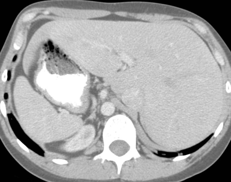
9 Transverse Midline Liver - Polysplenia Westra SJ. In: Abbara S, Walker TG. DI Cardiovascular


10 Normal Bronchial Anatomy Situs Solitus Ao Normal Bronchial Anatomy LPA


11 Situs Inversus RPA Ao Normal Bronchial Anatomy


12 Situs Inversus Totalis Kartagener s Syndrome Rt Ao Arch RV LV IVC Bronchiectasis L Liver Rt Spleen
13 Bilateral Eparterial Bronchi Bilateral Rightsidedness Asplenia Ao Normal Bronchial Anatomy NO Pulmonary artery arching over bronchi Bilateral eparterial bronchi Right isomerism Asplenia

 RPA Ao LPA Normal")


14 Bilateral Hyparterial Bronchi Azygos arch (Azygos continuation of IVC) RPA Ao LPA Normal Bronchial Anatomy Pulmonary arteries arching over both bronchi bilateral hyparterial bronchi Left isomerism Polysplenia
 = L-Loop L L-loop Rt Lt Caudal Van Praagh R. Semin Roentgenol 1985;20(3):254 271.")
15 Cardiac Loop D-loop Cranial ( 1, 2, 3 ) RV rotates either to right (dextro) = D-Loop D or to left (levo) = L-Loop L L-loop Rt Lt Caudal Van Praagh R. Semin Roentgenol 1985;20(3):
16 Identify Morphologic Right & Left Ventricles Systemic ventricle = connected to aorta Morphologic LV Fibrous continuity of AV- and arterial valves No septal trabeculations Morphologic RV Conus = complete muscular ring separating AV valve and arterial valve Septal leaflet closer to apex Moderator band, heavier trabeculated

17 Morphologic Right Ventricle Conus (complete muscular ring) separates AV-valve from arterial valve Moderator Band Septal leaflet closer to apex Bilateral conus = double outlet right ventricle Bilateral absence of conus = double outlet left ventriocle Subaortic conus - transposition
18 Morphologic Right Ventricle


19 Morphologic Left Ventricle Direct communication of AV-Valve with Arterial Valve via Intervalvular Fibrosa (No conus) No septal Trabeculations Papillary muscles attached to free wall Abbara. Anatomy of the heart. In: Abbara S. DI-Cardiovascular. Amirsys LWW 2014
20 Loop Rule Assumption used if determination of morphologic ventricles is difficult: In presence of a right-sided aortic valve, the RV is to the right of the LV (d-loop) In presence of a left-sided aortic valve, the RV is to the left of the LV (l-loop)
21 Position of Great Arteries ( 1, 2, 3 ) S = Solitus normal I = inversus mirrored D-TGV dextro transposition L-TGV levo transposition PA anterior and to the left PA anterior and to the right Ao anterior and to the right Ao anterior and to the left


22 Normally Related Great Arteries S = Solitus, NORMAL I = Inverted
23 Transposition of Great Arteries D-TGA or D-MGA L-TGA or L-MGA

24 Apply Segmental Approach


25 Step 1: Visceroatrial Situs ( S1, 2, 3 ) Right liver, left single spleen, left stomach Left sided left atrium Cardiac apex leftsided Left hyparterial bronchus Right eparterial bronchus Situs Solitus L St Spl


26 Step 2 - Cardiac Loop ( S, L2, 3 ) Moderator Band, heavy trabeculations & Complete muscular ring of systemic ventricle AV-valve hinge-point more apical IVC and SVC to RA, PAs to LV Atrio-Ventricular Discordance L-Loop


27 Step 3 - Great Arteries ( S, L, L-TGV 3 ) Ao arises from systemic morphologic RV PA arises from morphological LV Aorta anterior & left of Pulmonary artery Ventriculo-Arterial Discordance L-TGV

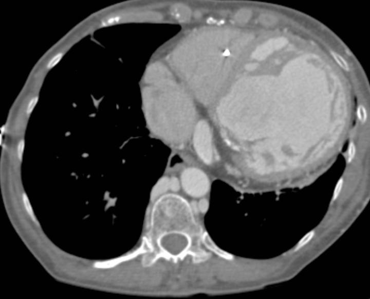
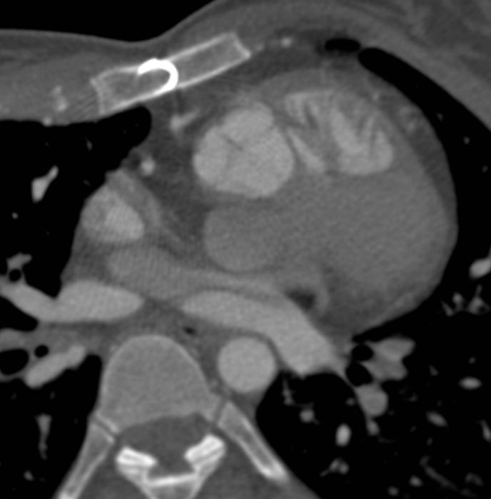
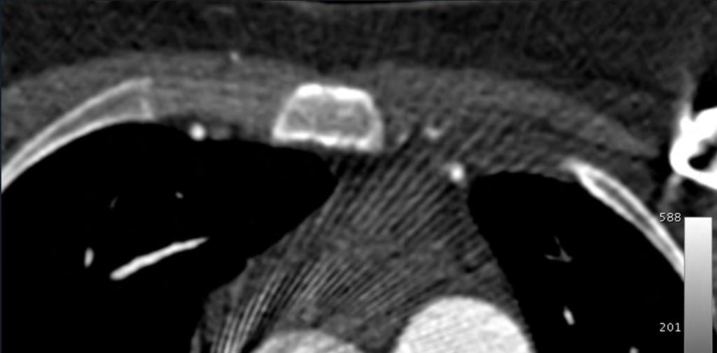
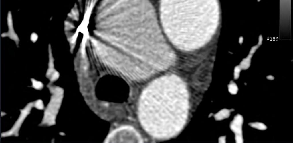
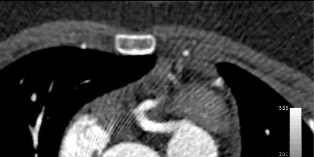
28 Footsteps of a Surgeon 44yom w. SOB. Remote heart surgery for unknown condition. ECHO: PR, dilated RVOT, RPA stenosis, gradient 27-49mmhg, LPA not seen

29 Footsteps of a Surgeon 44yom w. SOB. Remote heart surgery for unknown condition. ECHO: PR, dilated RVOT, RPA stenosis, gradient 27-49mmhg, LPA not seen


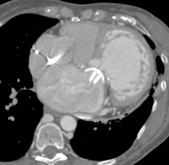
30 Footsteps of a Surgeon L Subclavian BT Shunt, surgically ligated Membranous VSD Patch Repair & RV Hypertrophy & Overriding Aorta Abbara S. Tetralogy of Fallot. In: Abbara, Walker. DI-Cardiovascular. Amirsys, Elsevier Saunders 2008


31 Footsteps of a Surgeon RVOT/PA Patch Repair of Pulmonic Stenosis RV Abbara S. Tetralogy of Fallot. In: Abbara, Walker. DI-Cardiovascular. Amirsys, Elsevier Saunders 2008


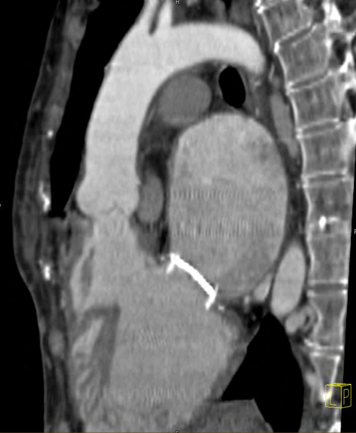
32 Know Complications Branch Pulmonary Artery Stenosis Abbara S. Tetralogy of Fallot. In: Abbara, Walker. DI-Cardiovascular. Amirsys, Elsevier Saunders 2008
 Abbara S.")

33 Know Complications Aorto-Pulmonary Systemic Collaterals (MAPCAs) Abbara S. Tetralogy of Fallot. In: Abbara, Walker. DI-Cardiovascular. Amirsys, Elsevier Saunders 2008
34 Thank you!
Congenital Heart Disease: a Pictorial Illustration of Putting Segmental Approach into Practice
 pissn 2384-1095 eissn 2384-1109 imri 2015;19:205-211 http://dx.doi.org/10.13104/imri.2015.19.4.205 Congenital Heart Disease: a Pictorial Illustration of Putting Segmental Approach into Practice Tse Hang
Segmental approach to normal and abnormal situs arrangement - Echocardiography -
 Segmental approach to normal and abnormal situs arrangement - Echocardiography - Jan Marek Great Ormond Street Hospital & Institute of Cardiovascular Sciences, University College London No disclosures
Segmental Analysis. Gautam K. Singh, M.D. Washington University School of Medicine St. Louis
 Segmental Analysis Gautam K. Singh, M.D. Washington University School of Medicine St. Louis Segmental Analysis Segmental Analysis: From Veins to Ventricles Segmental Approach to Evaluation of Congenital
Cardiopulmonary Syndromes: Conditions With Concomitant Cardiac and Pulmonary Abnormalities
 Cardiopulmonary Syndromes: Conditions With Concomitant Cardiac and Pulmonary Abnormalities Carlos S. Restrepo M.D. Professor of Radiology The University of Texas HSC at San Antonio Cardiopulmonary Syndromes
INNOVATIVE JOURNAL OF MEDICAL AND HEALTH SCIENCE
 Innovative Journal Of Medical And Health Science 8:9(2018) Contents lists available at www.innovativejournal.in INNOVATIVE JOURNAL OF MEDICAL AND HEALTH SCIENCE Available online at http://www.innovativejournal.in/index.php/ijmhs
CMR for Congenital Heart Disease
 CMR for Congenital Heart Disease * Second-line tool after TTE * Strengths of CMR : tissue characterisation, comprehensive access and coverage, relatively accurate measurements of biventricular function/
Adult Congenital Heart Disease: What All Echocardiographers Should Know Sharon L. Roble, MD, FACC Echo Hawaii 2016
 1 Adult Congenital Heart Disease: What All Echocardiographers Should Know Sharon L. Roble, MD, FACC Echo Hawaii 2016 DISCLOSURES I have no disclosures relevant to today s talk 2 Why should all echocardiographers
Heart and Soul Evaluation of the Fetal Heart
 Heart and Soul Evaluation of the Fetal Heart Ivana M. Vettraino, M.D., M.B.A. Clinical Associate Professor, Michigan State University College of Human Medicine Objectives Review the embryology of the formation
ISUOG Basic Training. Obtaining & Interpreting Heart Views Correctly Alfred Abuhamad, USA. Basic training. Editable text here
 ISUOG Basic Training Obtaining & Interpreting Heart Views Correctly Alfred Abuhamad, USA Learning Objectives 6, 7 & 8 At the end of the lecture you will be able to: describe how to assess cardiac situs
Comprehensive evaluation of complex congenital heart disease using the Van Praagh notation: step by step in MDCT
 Comprehensive evaluation of complex congenital heart disease using the Van Praagh notation: step by step in MDCT Poster No.: C-2050 Congress: ECR 2014 Type: Educational Exhibit Authors: Y.-P. Chang, Y.-T.
The Chest X-ray for Cardiologists
 Mayo Clinic & British Cardiovascular Society at the Royal College of Physicians, London : 21-23-October 2013 Cases-Controversies-Updates 2013 The Chest X-ray for Cardiologists Michael Rubens Royal Brompton
Situs at the mirror: from situs inversus to situs ambiguus
 Situs at the mirror: from situs inversus to situs ambiguus Poster No.: C-0605 Congress: ECR 2013 Type: Educational Exhibit Authors: C. Maciel, J. Maciel, A. Silva, A. F. L. Carneiro ; Porto/PT, 1 2 1 1
Common Defects With Expected Adult Survival:
 Common Defects With Expected Adult Survival: Bicuspid aortic valve :Acyanotic Mitral valve prolapse Coarctation of aorta Pulmonary valve stenosis Atrial septal defect Patent ductus arteriosus (V.S.D.)
This is the left, right?
 This is the left, right? Poster No.: C-1214 Congress: ECR 2013 Type: Educational Exhibit Authors: L.-L. Huang, L. Mitchell, S. Andronikou, F. Suleman, Z. I. Lockhat; Pretoria/ZA Keywords: Congenital, Diagnostic
NASCI 2012 Segmental Analysis
 NASCI 2012 Segmental Analysis Frandics Chan, M.D., Ph.D. Stanford University Medical Center Lucile Packard Department Children s of Radiology Hospital Menagerie of Congenital Cardiac Lesions 1. Absent
"Lecture Index. 1) Heart Progenitors. 2) Cardiac Tube Formation. 3) Valvulogenesis and Chamber Formation. 4) Epicardium Development.
 Heart Progenitors. 2) Cardiac Tube Formation. 3) Valvulogenesis and Chamber Formation. 4) Epicardium Development.") "Lecture Index 1) Heart Progenitors. 2) Cardiac Tube Formation. 3) Valvulogenesis and Chamber Formation. 4) Epicardium Development. 5) Septation and Maturation. 6) Changes in Blood Flow during Development.
"Lecture Index 1) Heart Progenitors. 2) Cardiac Tube Formation. 3) Valvulogenesis and Chamber Formation. 4) Epicardium Development. 5) Septation and Maturation. 6) Changes in Blood Flow during Development.
ADULT CONGENITAL HEART DISEASE. Stuart Lilley
 ADULT CONGENITAL HEART DISEASE Stuart Lilley More adults than children have congenital heart disease Huge variety of congenital lesions from minor to major Heart failure, re-operation and arrhythmia are
All You Need to Know About Situs and Looping Disorders: Embryology, Anatomy, and Echocardiography
 All You Need to Know About Situs and Looping Disorders: Embryology, Anatomy, and Echocardiography Helena Gardiner Co-Director of Fetal Cardiology, The Fetal Center, University of Texas at Houston Situs
ULTRASOUND OF THE FETAL HEART
 ULTRASOUND OF THE FETAL HEART Cameron A. Manbeian, MD Disclosure Statement Today s faculty: Cameron Manbeian, MD does not have any relevant financial relationships with commercial interests or affiliations
Anomalous Systemic Venous Connection Systemic venous anomaly
 World Database for Pediatric and Congenital Heart Surgery Appendix B: Diagnosis (International Paediatric and Congenital Cardiac Codes (IPCCC) and definitions) Anomalous Systemic Venous Connection Systemic
Giovanni Di Salvo MD, PhD, FESC Second University of Naples Monaldi Hospital
 Giovanni Di Salvo MD, PhD, FESC Second University of Naples Monaldi Hospital VSD is one of the most common congenital cardiac abnormalities in the newborn. It can occur as an isolated finding or in combination
Preoperative Echocardiographic Assessment of Uni-ventricular Repair
 Preoperative Echocardiographic Assessment of Uni-ventricular Repair Salem Deraz, MD Pediatric Cardiologist, Aswan Heart Centre Magdi Yacoub Heart Foundation Uni-ventricular repair A single or series of
Situs inversus. Dr praveena pulmonology- final year post graduate
 Situs inversus Dr praveena pulmonology- final year post graduate Definiton History Types Cause Clinical features Diagnosis Treatment Definition The term situs inversus is a short form of the latin phrase
Complex Congenital Heart Disease in Adults
 Complex Congenital Heart Disease in Adults Linda B. Haramati, MD Disclosures Complex Congenital Heart Disease in Adults Linda B. Haramati MD, MS Jeffrey M. Levsky MD, PhD Meir Scheinfeld MD, PhD Department
UPDATE FETAL ECHO REVIEW
 UPDATE 1 FETAL ECHO REVIEW Study Alert for RDCS Candidates D A V I E S P U B L I S H I N G I N C. Fetal Echo Review Study Alert U P D A T E D A U G U S T 1, 2 0 1 2 Nikki Stahl, RT(R)(M)(CT), RDMS, RVT
Low-dose prospective ECG-triggering dual-source CT angiography in infants and children with complex congenital heart disease: first experience
 Low-dose prospective ECG-triggering dual-source CT angiography in infants and children with complex congenital heart disease: first experience Ximing Wang, M.D., Zhaoping Cheng, M.D., Dawei Wu, M.D., Lebin
Echocardiography in Adult Congenital Heart Disease
 Echocardiography in Adult Congenital Heart Disease Michael Vogel Kinderherz-Praxis München CHD missed in childhood Subsequent lesions after repaired CHD Follow-up of cyanotic heart disease CHD missed in
Cardiac Catheterization Cases Primary Cardiac Diagnoses Facility 12 month period from to PRIMARY DIAGNOSES (one per patient)
") PRIMARY DIAGNOSES (one per patient) Septal Defects ASD (Atrial Septal Defect) PFO (Patent Foramen Ovale) ASD, Secundum ASD, Sinus venosus ASD, Coronary sinus ASD, Common atrium (single atrium) VSD (Ventricular
PRIMARY DIAGNOSES (one per patient) Septal Defects ASD (Atrial Septal Defect) PFO (Patent Foramen Ovale) ASD, Secundum ASD, Sinus venosus ASD, Coronary sinus ASD, Common atrium (single atrium) VSD (Ventricular
Congenital Heart Defects
 Normal Heart Congenital Heart Defects 1. Patent Ductus Arteriosus The ductus arteriosus connects the main pulmonary artery to the aorta. In utero, it allows the blood leaving the right ventricle to bypass
Heart and Lungs. LUNG Coronal section demonstrates relationship of pulmonary parenchyma to heart and chest wall.
 Heart and Lungs Normal Sonographic Anatomy THORAX Axial and coronal sections demonstrate integrity of thorax, fetal breathing movements, and overall size and shape. LUNG Coronal section demonstrates relationship
What do we know about Heterotaxy Syndrome? - An illustrated guide.
 What do we know about Heterotaxy Syndrome? - An illustrated guide. Poster No.: C-2369 Congress: ECR 2015 Type: Educational Exhibit Authors: M. C. Ageitos Casais, A. X. Martínez de Alegría Alonso, 1 1 2
ECHOCARDIOGRAPHIC APPROACH TO CONGENITAL HEART DISEASE: THE UNOPERATED ADULT
 ECHOCARDIOGRAPHIC APPROACH TO CONGENITAL HEART DISEASE: THE UNOPERATED ADULT Karen Stout, MD, FACC Divisions of Cardiology University of Washington Medical Center Seattle Children s Hospital NO DISCLOSURES
Disclosures. Outline. Learning Objectives. Introduction. Introduction. Sonographic Screening Examination of the Fetal Heart
 Sonographic Screening Examination of the Fetal Heart Lami Yeo, MD Director of Fetal Cardiology Perinatology Research Branch of NICHD / NIH / DHHS Bethesda, MD and Detroit, Michigan, USA Professor, Division
Congenital Heart Disease An Approach for Simple and Complex Anomalies
 Congenital Heart Disease An Approach for Simple and Complex Anomalies Michael D. Pettersen, MD Director, Echocardiography Rocky Mountain Hospital for Children Denver, CO None Disclosures 1 ASCeXAM Contains
Right isomerism with complex cardiac anomalies presenting with dysphagia - A case report
 Right isomerism with complex cardiac anomalies presenting with dysphagia - A case report Himanshu Agarwal 1, Shireesh Kumar Mittal 1*, Chaitanya D Kulkarni 1, Ashok Kumar Verma 1, Saurabh Kumar Srivastava
CONGENITAL HEART DISEASE (CHD)
") CONGENITAL HEART DISEASE (CHD) DEFINITION It is the result of a structural or functional abnormality of the cardiovascular system at birth GENERAL FEATURES OF CHD Structural defects due to specific disturbance
CONGENITAL HEART DISEASE (CHD) DEFINITION It is the result of a structural or functional abnormality of the cardiovascular system at birth GENERAL FEATURES OF CHD Structural defects due to specific disturbance
Making Sense of Cardiac Views and Imaging Characteristics for 13 Congenital Heart Defects (CHDs)
") Making Sense of Cardiac Views and Imaging Characteristics for 13 Congenital Heart Defects (CHDs) Manny Gaziano, MD, FACOG obimages.net obimages.net@gmail.com Acknowledgements: Krista Wald, RDMS, sonographer,
Making Sense of Cardiac Views and Imaging Characteristics for 13 Congenital Heart Defects (CHDs) Manny Gaziano, MD, FACOG obimages.net obimages.net@gmail.com Acknowledgements: Krista Wald, RDMS, sonographer,
Anatomy of Atrioventricular Septal Defect (AVSD)
") Surgical challenges in atrio-ventricular septal defect in grown-up congenital heart disease Anatomy of Atrioventricular Septal Defect (AVSD) S. Yen Ho Professor of Cardiac Morphology Royal Brompton and
Surgical challenges in atrio-ventricular septal defect in grown-up congenital heart disease Anatomy of Atrioventricular Septal Defect (AVSD) S. Yen Ho Professor of Cardiac Morphology Royal Brompton and
Slide 1. Slide 2. Slide 3 CONGENITAL HEART DISEASE. Papworth Hospital NHS Trust INTRODUCTION. Jakub Kadlec/Catherine Sudarshan INTRODUCTION
 Slide 1 CONGENITAL HEART DISEASE Jakub Kadlec/Catherine Sudarshan NHS Trust Slide 2 INTRODUCTION Most common congenital illness in the newborn Affects about 4 9 / 1000 full-term live births in the UK 1.5
Journal of Radiology Case Reports
 Situs Ambiguous, Levocardia, Right Sided Stomach, Obstructing Duodenal Web, and Intestinal Tomas Mujo 1*, Tess Finnegan 2, Jonathan Joshi 1, Kathirene A. Wilcoxen 3, James C. Reed 1 1. Department of Radiology,
CASE OF HETEROTAXY SYNDROME WITH POLYSPLENIA AND INTESTINAL MALROTATION
 CASE OF HETEROTAXY SYNDROME WITH POLYSPLENIA AND INTESTINAL MALROTATION *Sagar H S, Basanta Manjari Swain, Jayashree Mohanty and Sasmita Parida Department of Radio diagnosis, S.C.B. Medical College, Cuttack
Absent Pulmonary Valve Syndrome
 Absent Pulmonary Valve Syndrome Fact sheet on Absent Pulmonary Valve Syndrome In this condition, which has some similarities to Fallot's Tetralogy, there is a VSD with narrowing at the pulmonary valve.
Approach to Dextrocardia in Adults: Review
 AJR Integrative Imaging LIFELONG LEARNING FOR RADIOLOGY Approach to Dextrocardia in Adults: Review Pierre D. Maldjian 1 and Muhamed Saric 2 OBJECTIVE The educational objectives of this article are to describe
Chest radiographic findings in children with asplenia syndrome
 Asian Biomedicine Vol. 4 No. 4 August 2010; 585-594 Original article Chest radiographic findings in children with asplenia syndrome Panruethai Trinavarat a, Kullana Tantiprawan a, Apichai Khongphatthanayothin
Congenital Heart Disease. Disharmonious Patterns of Heterotaxy and Isomerism How Often Are the Classic Patterns Breached?
 Congenital Heart Disease Disharmonious Patterns of Heterotaxy and Isomerism How Often Are the Classic Patterns Breached? Deane Yim, MBchB; Hazumu Nagata, MD; Christopher Z. Lam, MD; Lars Grosse-Wortmann,
Cases in Adult Congenital Heart Disease
 Cases in Adult Congenital Heart Disease Sabrina Phillips, MD FACC FASE Associate Professor of Medicine The University of Oklahoma Health Sciences Center No Disclosures I Have Palpitations 18 Year old Man
DEVELOPMENT OF THE CIRCULATORY SYSTEM L E C T U R E 5
 DEVELOPMENT OF THE CIRCULATORY SYSTEM L E C T U R E 5 REVIEW OF CARDIAC ANATOMY Heart 4 chambers Base and apex Valves Pericardial sac 3 layers: epi, myo, endo cardium Major blood vessels Aorta and its
Notes: 1)Membranous part contribute in the formation of small portion in the septal cusp.
Membranous part contribute in the formation of small portion in the septal cusp.") Embryology 9 : Slide 16 : There is a sulcus between primitive ventricular and bulbis cordis that will disappear gradually and lead to the formation of one chamber which is called bulboventricular chamber.
Embryology 9 : Slide 16 : There is a sulcus between primitive ventricular and bulbis cordis that will disappear gradually and lead to the formation of one chamber which is called bulboventricular chamber.
9/8/2009 < 1 1,2 3,4 5,6 7,8 9,10 11,12 13,14 15,16 17,18 > 18. Tetralogy of Fallot. Complex Congenital Heart Disease.
 Current Indications for Pediatric CTA S Bruce Greenberg Professor of Radiology Arkansas Children s Hospital University of Arkansas for Medical Sciences greenbergsbruce@uams.edu 45 40 35 30 25 20 15 10
Case 47 Clinical Presentation
 93 Case 47 C Clinical Presentation 45-year-old man presents with chest pain and new onset of a murmur. Echocardiography shows severe aortic insufficiency. 94 RadCases Cardiac Imaging Imaging Findings C
Outflow Tracts Anomalies
 Diagnosis of Outflow Tract Anomalies in the Fetus General Framing D.Paladini Fetal Medicine & Surgery Unit Gasllini Children s Hospital - Genoa dariopaladini@ospedale-gaslini.ge.it Outflow Tracts Anomalies
Surgical options for tetralogy of Fallot
 Surgical options for tetralogy of Fallot Serban Stoica FRCS(CTh) MD ACHD study day, 19 September 2017 Anatomy Physiology Children Adults Complications Follow up Anatomy Etienne Fallot (1850-1911) VSD Overriding
3/14/2011 MANAGEMENT OF NEWBORNS CARDIAC INTENSIVE CARE CONFERENCE FOR HEALTH PROFESSIONALS IRVINE, CA. MARCH 7, 2011 WITH HEART DEFECTS
 CONFERENCE FOR HEALTH PROFESSIONALS IRVINE, CA. MARCH 7, 2011 MANAGEMENT OF NEWBORNS WITH HEART DEFECTS A NTHONY C. CHANG, MD, MBA, MPH M E D I C AL D I RE C T OR, HEART I N S T I T U T E C H I LDRE N
MRI (AND CT) FOR REPAIRED TETRALOGY OF FALLOT
 FOR REPAIRED TETRALOGY OF FALLOT") MRI (AND CT) FOR REPAIRED TETRALOGY OF FALLOT Linda B Haramati MD, MS Departments of Radiology and Medicine Bronx, New York OUTLINE Pathogenesis Variants Initial surgical treatments Basic MR protocols
MRI (AND CT) FOR REPAIRED TETRALOGY OF FALLOT Linda B Haramati MD, MS Departments of Radiology and Medicine Bronx, New York OUTLINE Pathogenesis Variants Initial surgical treatments Basic MR protocols
Most common fetal cardiac anomalies
 Most common fetal cardiac anomalies Common congenital heart defects CHD % of cardiac defects Chromosomal Infants Fetuses anomaly (%) 22q11 deletion (%) VSD 30 5~10 20~40 10 PS 9 5 (PA w/ VSD) HLHS 7~9
Dextrocardia and Isolated Lavocardia
 Brit. Heart J., 1966, 28, 472. Dextrocardia and Isolated Lavocardia MAURICE CAMPBELL AND D. C. DEUCHAR From the Cardiac Department, Guy's Hospital, London S.E.J, and the Institute of Cardiology, London
Cardiovascular Imaging Interactive Case Discussions
 Cardiovascular Imaging Interactive Case Discussions Satinder Singh MD, FCCP Professor of Radiology & Medicine (Division of CV Diseases) Chief Cardiopulmonary Radiology Director Cardiac CT UAB SCBT.MR September
Echocardiographic assessment in Adult Patients with Congenital Heart Diseases
 Echocardiographic assessment in Adult Patients with Congenital Heart Diseases Athanasios Koutsakis Cardiologist, Cl. Research Fellow George Giannakoulas Ass. Professor in Cardiology 1st Cardiology Department,
Functional SV with TAPVD: Contemporary Management of Right Atrial Isomerism
 Functional SV with TAPVD: Contemporary Management of Right Atrial Isomerism Yun TJ, Van Arsdell GS Asan Medical Center The Hospital for Sick Children in Toronto Functional SV, TAPVD and RAI FSV TAPVD RAI
List of Videos. Video 1.1
 Video 1.1 Video 1.2 Video 1.3 Video 1.4 Video 1.5 Video 1.6 Video 1.7 Video 1.8 The parasternal long-axis view of the left ventricle shows the left ventricular inflow and outflow tract. The left atrium
Appendix A.1: Tier 1 Surgical Procedure Terms and Definitions
 Appendix A.1: Tier 1 Surgical Procedure Terms and Definitions Tier 1 surgeries AV Canal Atrioventricular Septal Repair, Complete Repair of complete AV canal (AVSD) using one- or two-patch or other technique,
Cardiac Radiology In-Training Test Questions for Diagnostic Radiology Residents
 Cardiac Radiology In-Training Test Questions for Diagnostic Radiology Residents March, 2013 Sponsored by: Commission on Education Committee on Residency Training in Diagnostic Radiology 2013 by American
Cardiac Radiography. Jared D. Christensen, M.D.
 Cardiac Radiography Jared D. Christensen, M.D. Cardiac radiography Jared D. Christensen, M.D. Overview Basic Concepts Technique Normal anatomy Cases Technique 3 Standard Views Posterior-Anterior (PA) Anterior-Posterior
Cardiac MRI in ACHD What We. ACHD Patients
 Cardiac MRI in ACHD What We Have Learned to Apply to ACHD Patients Faris Al Mousily, MBChB, FAAC, FACC Consultant, Pediatric Cardiology, KFSH&RC/Jeddah Adjunct Faculty, Division of Pediatric Cardiology
Fetal Echocardiography and the Routine Obstetric Sonogram
 JDMS 23:143 149 May/June 2007 143 Fetal Echocardiography and the Routine Obstetric Sonogram SHELLY ZIMBELMAN, RT(R)(CT), RDMS, RDCS ASAD SHEIKH, MD, RDCS Congenital heart disease (CHD) is the most common
Basic Fetal Cardiac Evaluation
 Basic Fetal Cardiac Evaluation Mert Ozan Bahtiyar, MD Director, Fetal Care Center Division of Maternal Fetal Medicine Department of Obstetrics, Gynecology and Reproductive Sciences S L I D E 1 Background
ORIGINAL RESEARCH PAPER
 ORIGINAL RESEARCH PAPER ROLE OF CT PULMONARY ANGIOGRAPHY IN CONGENITAL HEART DISEASES IN PAEDIATRIC POPULATION Radiology KEY WORDS: Congenital heart disease, CT pulmonary angiography, pediatric heart disease,
Chapter 2 Cardiac Interpretation of Pediatric Chest X-Ray
 Chapter 2 Cardiac Interpretation of Pediatric Chest X-Ray Ra-id Abdulla and Douglas M. Luxenberg Key Facts The cardiac silhouette occupies 50 55% of the chest width on an anterior posterior chest X-ray
A Classic Case Of Polysplenia Syndrome With A Pancreatic Mass And SOLs In Liver
 ISPUB.COM The Internet Journal of Radiology Volume 13 Number 2 A Classic Case Of Polysplenia Syndrome With A Pancreatic Mass And SOLs In Liver V Gupta, N Agarwal Citation V Gupta, N Agarwal. A Classic
Congenitally Corrected Transposition of the Great Arteries (cctga or l-loop TGA)
") Congenitally Corrected Transposition of the Great Arteries (cctga or l-loop TGA) Mary Rummell, MN, RN, CPNP, CNS Clinical Nurse Specialist, Pediatric Cardiology/Cardiac Surgery Doernbecher Children s Hospital,
Congenitally Corrected Transposition of the Great Arteries (cctga or l-loop TGA) Mary Rummell, MN, RN, CPNP, CNS Clinical Nurse Specialist, Pediatric Cardiology/Cardiac Surgery Doernbecher Children s Hospital,
CARDIAC AND CORONARY ARTERY ANATOMY NO DISCLOSURES. Axial Anatomy of Heart. Axial Anatomy of Heart. Axial Anatomy of Heart
 CARDIAC AND CORONARY ARTERY ANATOMY NO DISCLOSURES NASCI MEETING, ORLANDO FLORIDA 2009 KOSTAKI G. BIS, MD, FACR DEPARTMENT OF RADIOLOGY WILLIAM BEAUMONT HOSPITAL Royal Oak, Michigan OBJECTIVES CARDIAC
Atrial Septal Defects
 Supplementary ACHD Echo Acquisition Protocol for Atrial Septal Defects The following protocol for echo in adult patients with atrial septal defects (ASDs) is a guide for performing a comprehensive assessment
An Approach to Cardiac Malposition and the Heterotaxy Syndrome Using 99mTc Sulfur Colloid Imaging
 An Approach to Cardiac Malposition and the Heterotaxy Syndrome Using 99mTc Sulfur Colloid Imaging P. M. FITZER A diagnostic approach to cardiac malposition and the heterotaxy syndrome is outlined. The
Congenital Heart Disease II: The Repaired Adult
 Congenital Heart Disease II: The Repaired Adult Doreen DeFaria Yeh, MD FACC Assistant Professor, Harvard Medical School MGH Adult Congenital Heart Disease Program Echocardiography Section, no disclosures
Cardiovascular MRI of Adult Congenital Heart Disease
 Cardiovascular MRI of Adult Congenital Heart Disease Anil K. Attili, MD Cardiovascular Magnetic Resonance imaging of Adult Congenital Heart Disease Anil Attili, M.D. Assistant Professor of Radiology /Cardiology
Figure 10.1A Transparency Master 79
 Brain Carotid arteries Jugular vein Right front leg Lungs (inflated) Cranial Right atrium To left front leg Left subclavian Bronchus capillaries Brachiocephalic vein Left atrium Dorsal aorta Right ventricle
Transposition of the Great Arteries Preoperative Diagnostic Considerations. John Simpson Evelina Children s Hospital London, UK
 Transposition of the Great Arteries Preoperative Diagnostic Considerations John Simpson Evelina Children s Hospital London, UK Areas to be covered Definitions Scope of occurrence of transposition of the
Valvular Imaging Optimizing Data Acquisition and Interpretation
 Valvular Imaging Optimizing Data Acquisition and Interpretation Suhny Abbara, MD Director Cardiovascular Imaging Section, Massachusetts General Hospital Assistant Professor, Harvard Medical School Sabbara@Partners.org
Cardiac CT in Infants with Congenital heart disease Sunrise Session. LaDonna Malone, MD May 17, 2018
 Cardiac CT in Infants with Congenital heart disease Sunrise Session LaDonna Malone, MD May 17, 2018 None Disclosures Objectives Describe cardiac CT techniques used in infants with congenital heart disease.
Index. cardiology.theclinics.com. Note: Page numbers of article titles are in boldface type.
 Index Note: Page numbers of article titles are in boldface type. A ACHD. See Adult congenital heart disease (ACHD) Adult congenital heart disease (ACHD), 503 512 across life span prevalence of, 504 506
The Triply Twisted Heart: Cyanosis in an Adult With Situs Inversus, Levocardia, Double Outlet Right Ventricle, and Malposition of the Great Arteries
 Elmer ress Case Report Cardiol Res. 2015;6(6):362-366 The Triply Twisted Heart: Cyanosis in an Adult With Situs Inversus, Levocardia, Double Outlet Right Ventricle, and Malposition of the Great Arteries
"Giancarlo Rastelli Lecture"
 "Giancarlo Rastelli Lecture" Surgical treatment of Malpositions of the Great Arteries Pascal Vouhé Giancarlo Rastelli (1933 1970) Cliquez pour modifier les styles du texte du masque Deuxième niveau Troisième
RVOTO adult and post-op
 Right ventricular outflow tract obstruction in the adult: native and post-op Helmut Baumgartner Westfälische Wilhelms-Universität Münster Adult Congenital and Valvular Heart Disease Center University of
Abnormalities of the spleen in relation to
 122 Department of Pediatric Cardiology, Children's Hospital of Pennsylvania, USA C Anderson R H Anderson J R Zuberbuhler Department of Pathology, Children's Hospital of Pennsylvania, USA W A Devine D E
Pediatric Echocardiography Examination Content Outline
 Pediatric Echocardiography Examination Content Outline (Outline Summary) # Domain Subdomain Percentage 1 Anatomy and Physiology Normal Anatomy and Physiology 10% 2 Abnormal Pathology and Pathophysiology
HISTORY. Question: What type of heart disease is suggested by this history? CHIEF COMPLAINT: Decreasing exercise tolerance.
 HISTORY 15-year-old male. CHIEF COMPLAINT: Decreasing exercise tolerance. PRESENT ILLNESS: A heart murmur was noted in childhood, but subsequent medical care was sporadic. Easy fatigability and slight
Transesophageal Echocardiographic Diagnosis and Imaging of Cardiac Situs and Malposition
 M Ganesh Kumar et al REVIEW RTICLE 10.5005/jp-journals-10034-1072 Transesophageal Echocardiographic Diagnosis and Imaging of Cardiac Situs and Malposition 1 M Ganesh Kumar, 2 Rajarajan Ganesan, 3 Goverdhan
By Dickens ATURWANAHO & ORIBA DAN LANGOYA MAKchs, MBchB CONGENTAL HEART DISEASE
 By Dickens ATURWANAHO & ORIBA DAN LANGOYA MAKchs, MBchB CONGENTAL HEART DISEASE Introduction CHDs are abnormalities of the heart or great vessels that are present at birth. Common type of heart disease
Using the Coronary Chronic Total Occlusion (CTO) Technique to Recanulate Totally Occluded Vessels in the Congenital Heart Disease Patients
 Technique to Recanulate Totally Occluded Vessels in the Congenital Heart Disease Patients") 5th Asia Pacific Congenital & Structural Heart Intervention Symposium 2014 10 12 October 2014, Hong Kong Convention and Exhibition Centre Organizer: Hong Kong Society of Congenital & Structural Heart Disease
5th Asia Pacific Congenital & Structural Heart Intervention Symposium 2014 10 12 October 2014, Hong Kong Convention and Exhibition Centre Organizer: Hong Kong Society of Congenital & Structural Heart Disease
Devendra V. Kulkarni, Rahul G. Hegde, Ankit Balani, and Anagha R. Joshi. 2. Case Report. 1. Introduction
 Case Reports in Radiology, Article ID 614647, 4 pages http://dx.doi.org/10.1155/2014/614647 Case Report A Rare Case of Pulmonary Atresia with Ventricular Septal Defect with a Right Sided Aortic Arch and
TGA Surgical techniques: tips & tricks (Arterial switch operation)
") TGA Surgical techniques: tips & tricks (Arterial switch operation) Seoul National University Children s Hospital Woong-Han Kim Surgical History 1951 Blalock and Hanlon, atrial septectomy 1954 Mustard et
TGA Surgical techniques: tips & tricks (Arterial switch operation) Seoul National University Children s Hospital Woong-Han Kim Surgical History 1951 Blalock and Hanlon, atrial septectomy 1954 Mustard et
September 26, 2012 Philip Stockwell, MD Lifespan CVI Assistant Professor of Medicine (Clinical)
") September 26, 2012 Philip Stockwell, MD Lifespan CVI Assistant Professor of Medicine (Clinical) Advances in cardiac surgery have created a new population of adult patients with repaired congenital heart
September 26, 2012 Philip Stockwell, MD Lifespan CVI Assistant Professor of Medicine (Clinical) Advances in cardiac surgery have created a new population of adult patients with repaired congenital heart
Congenital heart disease: When to act and what to do?
 Leading Article Congenital heart disease: When to act and what to do? Duminda Samarasinghe 1 Sri Lanka Journal of Child Health, 2010; 39: 39-43 (Key words: Congenital heart disease) Congenital heart disease
Children with Single Ventricle Physiology: The Possibilities
 Children with Single Ventricle Physiology: The Possibilities William I. Douglas, M.D. Pediatric Cardiovascular Surgery Children s Memorial Hermann Hospital The University of Texas Health Science Center
Fetal Tetralogy of Fallot
 36 Fetal Tetralogy of Fallot E.D. Bespalova, R.M. Gasanova, O.A.Pitirimova National Scientific and Practical Center of Cardiovascular Surgery, Moscow Elena D. Bespalova, MD Professor, Director Rena M,
Anomalies of Visceroatrial Situs
 ardiopulmonary Imaging Pictorial Essay Ghosh et al. Visceroatrial Situs nomalies ardiopulmonary Imaging Pictorial Essay Downloaded from www.ajronline.org by 46.3.194.217 on 11/22/17 from IP address 46.3.194.217.
J Somerville and V Grech. The chest x-ray in congenital heart disease 2. Images Paediatr Cardiol Jan-Mar; 12(1): 1 8.
: 1 8.") IMAGES in PAEDIATRIC CARDIOLOGY Images Paediatr Cardiol. 2010 PMCID: PMC3228330 The chest x-ray in congenital heart disease 2 J Somerville and V Grech Paediatric Department, Mater Dei Hospital, Malta Corresponding
IMAGES in PAEDIATRIC CARDIOLOGY Images Paediatr Cardiol. 2010 PMCID: PMC3228330 The chest x-ray in congenital heart disease 2 J Somerville and V Grech Paediatric Department, Mater Dei Hospital, Malta Corresponding
Anatomy & Physiology
 1 Anatomy & Physiology Heart is divided into four chambers, two atrias & two ventricles. Atrioventricular valves (tricuspid & mitral) separate the atria from ventricles. they open & close to control flow
5.8 Congenital Heart Disease
 5.8 Congenital Heart Disease Congenital heart diseases (CHD) refer to structural or functional heart diseases, which are present at birth. Some of these lesions may be discovered later. prevalence of Chd
Transposition of the great arteries
 EuroEcho 2010 - Teaching course on CHD Transposition of the great arteries - Follow-up after the arterial switch Gertjan Tj. Sieswerda, MD PhD Nothing to disclose Interuniversitary Institute for Congenital