Hemodynamic derangement. Komson Wannasai, M.D.,FRCPath. Department of Pathology Faculty of Medicine Chiang Mai University
|
|
|
- Anis Fowler
- 5 years ago
- Views:
Transcription
1 Hemodynamic derangement Komson Wannasai, M.D.,FRCPath. Department of Pathology Faculty of Medicine Chiang Mai University
2 Objective The students should be able to Explain normal body fluid homeostasis Explain definition of edema, congestion, hyperemia, hemorrhage, thrombosis, embolism and infarction Explain pathophysiology of edema, congestion, hyperemia, hemorrhage, thrombosis, embolism and infarction Explain pathological finding of edema, congestion, hyperemia, hemorrhage, thrombosis, embolism and infarction Apply basic knowledge to clinical application
3 Contents Body fluid compartments Edema Hyperemia and congestion Hemorrhage Thrombosis Embolism Infarction
4 Body fluid compartments Total body water (TBW) 60% of the body weight in kg 2/3 intracellular fluid 1/3 extracellular fluid Sodium (Na + ) Major extracellular fluid (ECF) cation Potassium (K + ) Major intracellular fluid (ICF) cation ECF is subdivided into the interstitial and vascular compartments
5 Normal water homeostasis Hydrostatic pressure Plasma oncotic pressure The exit of fluid into the interstitium is nearly balanced by inflow at the venular end A small residuum of excess interstitial fluid is drained by the lymphatics
6 Robbins 9 th edition
7 Edema Presence of increased fluid in the interstitial space of the ECF compartment Causes of edema Increased hydrostatic pressure Reduced plasma osmotic pressure Lymphatic obstruction Sodium and water retention
8 Types of edema fluid Transudate Protein poor (<3 g/dl) and cell poor fluid Produces dependent pitting edema and body cavity effusions Exudate Protein rich (>3 g/dl) and cell rich (neutrophils) fluid Produces swelling of tissue but no pitting edema Lymphedema Protein rich fluid Nonpitting edema
9 Related trems Hydrothorax (pleural effusion) Effusions involving the pleural cavity Hydropericardium (pericardial effusion) Effusions involving the pericardial cavity Hydroperitoneum (ascites) Effusions involving the peritoneal cavity

10 Pleural effusion library.med.utah.edu
11 Pathophysiology of edema (1) Alteration in Starling pressure Produces a transudate Increased vascular hydrostatic pressure Pulmonary edema in left sided heart failure Peripheral pitting edema in right sided heart failure Portal hypertension in cirrhosis producing ascites
12 Pathophysiology of edema (2) Alteration in Starling pressure Decreased vascular plasma oncotic pressure (hypoalbuminemia) Malnutrition with decreased protein intake Cirrhosis with decreased synthesis of albumin Nephrotic syndrome with increased loss of protein in urine Malabsorption with decreased reabsorption of protein Renal retention of sodium and water Increases hydrostatic pressure (increased plasma volume) Decreases oncotic pressure (dilutional effect on albumin) Examples acute renal failure, glomerulonephritis
13 Pathophysiology of edema (3) Increased vascular permeability Produces an exudate Example acute inflammation (e.g., tissue swelling following a bee sting) Lymphatic obstruction Produces lymphedema Examples Lymphedema following modified radical mastectomy and radiation Filariasis due to Wuchereria bancrofti
14 Morphology of edema Most easily recognized grossly Microscopically Cell swelling with clearing and separation of the extracellular matrix elements Edema is most commonly encountered in subcutaneous tissues, the lungs, and the brain.

15 Pitting edema cirrhosistreatment reviews.com

16 Pulmonary Edema (1) Increased fluid in the alveolar spaces and interstitium of the lung. Most common cause: left ventricular failure Increase perfusion pressure in the pulmonary capillaries and block effective lymphatic drainage
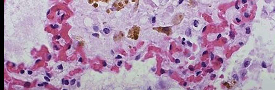
17 Pulmonary Edema (2) Gross morphology: lung Two or three times of their normal weight Tense, firm consistency Microscopic findings Congested capillaries and damaged alveoli with debris containing and extravasated RBC Proteinaceous fluid within alveolar spaces

18


19

20 secure.health.utas.edu.au

21
22 Edema in Cirrhosis of the Liver Cirrhosis Chronic disease in which liver parenchyma deteriorates; the lobules become infiltrated with fat and dense connective tissue. Decreased albumin production Decreased plasma osmotic pressure Increased portal hydrostatic pressure Increased sodium and water reabsorption

23 Severe ascites due to cirrhosis
24 Edema in the nephrotic syndrome Decreased plasma albumin Decreased plasma osmotic pressure Decreased blood volume Increased aldosterone and ADH Increased sodium and water reabsorption
25 Localized edema Obstruction of venous return Lymphatic obstruction Increased vascular permeability Inflammation Urticaria External pressure
 www.dvtvictims.")
26 Deep vein thrombosis (more explanation in vascular diseases II)
27 Brain edema (1) Causes Cerebrovascular accident Trauma Venous obstruction Inflammation; meningitis, abscess, encephalitis Complications Increased intracranial pressure Cerebral herniation


28 Brain edema (2) Morphology Grossly swollen Narrow sulci and distended, flat gyri state.edu

29 Hyperemia and congestion Both indicated a local increased volume of blood in tissues. Hyperemia Active process Increase tissue inflow due to arteriolar dilatation Red tissues Exercise, inflammation


30 Hyperemia and congestion Congestion Passive process Outflow tract obstruction Blue red tissue Both hyperemia and congestion occur together
31 Hyperemia and congestion In long standing congestion Called CHRONIC PASSIVE CONGESTION Stasis of deoxygenated blood cause hypoxia and cell death Capillary rupture: hemorrhage Hemosiderin laden macrophage
32 Hyperemia and congestion Morphology The cut surface is hemorrhagic and wet Acute pulmonary congestion Engorged capillary vessels Alveolar septal edema Focal intra alveolar hemorrhage Chronic pulmonary congestion Thickening of septa Numerous hemosiderin laden macrophage


33 Acute pulmonary congestion Robbins 7 th edition


34 chronic pulmonary congestion Robbins 7 th edition web.squ.edu.om
35 Hyperemia and congestion Morphology Acute hepatic congestion Distended central vein and sinusoids filled with blood Chronic hepatic congestion (more details in pathology of HF)
36 Normal hemostasis 1. Integrity of small blood vessels 2. Adequate numbers of platelets 3. Normal amounts of coagulation factors 4. Normal amounts of coagulation inhibitors 5. Adequate amounts of calcium ions in the blood


37 The vessels constituents and coagulation Endothelial cells are antithrombotic Antiplatelet effects Anticoagulant effects Fibrinolytic effects


38 Hemostasis and platelets Platelets Cytoplasmic fragments from megakaryocytes After vascular injury Platelet adhesion Platelet aggregation Degranulation Platelet plug
39 Robbins 9 th edition
40 Robbins 9 th edition
41 Robbins 9 th edition


42 The coagulation cascades A series of enzyme reactions ultimately resulting in the formation of thrombin which converts fibrinogen into fibrin Robbins 7 th edition
43 Robbins 9 th edition
44 How do we stop this? Fibrinolytic system Predominantly by action of plasmin Plasmin formed by action of tissue plasminogen activator and urokinase Lead to breakdown of fibrin into fibrin degradation products
45 How do we stop this? Fibrinolytic system Predominantly by action of plasmin Plasmin formed by action of tissue plasminogen activator and urokinase Lead to breakdown of fibrin into fibrin degradation products Robbins 9 th edition
46 Hemorrhage Extravasation of blood due to vessel rupture Hemorrhagic diatheses Capillary bleeding can occur under conditions of chronic congestion Rupture of a large artery or vein is almost always due to vascular injury Manifested in a variety of patterns (size, extent, and location of bleeding)
47 Hemorrhage External or may be enclosed within a tissue Hematoma accumulation of blood within tissue Petechiae minute 1 to 2 mm hemorrhages into skin, mucous membranes, or serosal surfaces Locally increased intravascular pressure Low platelet counts (thrombocytopenia) Defective platelet function
48 Hemorrhage Purpura : Slightly larger ( 3 mm) hemorrhages Associated with many of the same disorders that cause petechiae Occur secondary to trauma, vascular inflammation, or increased vascular fragility
49 Hemorrhage Ecchymoses : Larger (>1 to 2 cm) subcutaneous hematomas (i.e., bruises) Red blue color bilirubin (blue green color) hemosiderin (gold brown color) Hemothorax, hemopericardium, hemoperitoneum, or hemarthrosis Patients with extensive hemorrhage occasionally develop jaundice.


50 library.med.utah.edu healthfixit.com


51 Hemorrhage Clinical significance Volume Rate Site Patient status Robbins 9 th edition
52 Thrombosis Inappropriate activation of the hemostatic process in uninjured vasculature OR Formation of thrombus in the setting of relatively minimal vascular injury


53 Virchow s triad Robbins 9 th edition
54 Factors predisposing to thrombosis (1) Hypercoagulability Any alteration of the coagulation pathway that predisposes to thrombosis Primary (genetics) Secondary (acquired) Bed rest immobilization, obesity, cancer, atrial fibrillation, myocardial infarction, tissue damage (surgery, burns)
55 Factors predisposing to thrombosis (2) Abnormalities of blood flow Turbulance Endothelial injury Local areas of stasis Disrupt laminar flow Moves platelets from center of flow to the vessel wall Prevent dilution of activated clotting factors by flowing blood Slow down the inflow of clotting factor inhibitors Promotes endothelial cell activation
56 Thrombi Morphology Venous thrombi Usually occlusive Red (because they form in stasis syndrome and have more associated enmeshed RBCs) Long forming a cast of vein with markings on them from venous valves Red blood cells alternating with peripheral areas of fibrin


57 Venous thrombi: Clinical Robbins 7 th edition


58 Venous thrombi: Fates Organization Ingrowth of cells into thrombus with incorporation into wall Resolution It goes away Embolization Travels from its site of origin to a distal part of circulation Robbins 7 th edition

59


60 Arterial Thrombi : Morphology Adherent masses of blood that demonstrate areas of pale alternating with areas of red Lines of Zahn Mural thrombi Robbins 9 th edition


61 pathhsw5m54.ucsf.edu library.med.utah.edu
62 Arterial Thrombi Outcome Similar to venous thrombi Resolution Organization/Incorporation/ Recanalization Embolization (arterial) Propagation
63 Embolism A detached intravascular solid, liquid or gaseous mass that is carried by the blood to a site distant from its point of origin. Related terms Embolus/Emboli Thromboembolis Saddle embolism
64 Embolism Pulmonary embolism Clinically silent (most) Sudden death, right heart failure (cor pulmonale), or cardiovascular collapse Embolic obstruction of medium sized arteries may result in pulmonary hemorrhage
65 Embolism An embolus in the setting of left sided cardiac failure may result in a large infarct Embolic obstruction of small end arteriolar pulmonary branches usually does result in associated infarction Multiple emboli over time may cause pulmonary hypertension with right heart failure


66 Robbins 7 th edition library.med.utah.edu
67 Embolism Fat embolism After fractures of long bones microscopic fat globules in the circulation Fat embolism syndrome pulmonary insufficiency, neurologic symptoms, anemia, and thrombocytopenia Sudden onset of tachypnea, dyspnea, and tachycardia Neurologic symptoms irritability and restlessness, progression to delirium or coma

68

69
70 Embolism Air embolism Decompression sickness Gas emboli may also induce focal ischemia in a number of tissues A more chronic form of decompression sickness is called Caisson disease
71 Embolism Persistence of gas emboli in the skeletal system leads to multiple foci of ischemic necrosis The more common sites are the heads of the femurs, tibia, and humeri
72 Embolism Amniotic fluid embolism In complicated labor Sudden severe dyspnea, cyanosis, and hypotensive shock, followed by seizures and coma Pulmonary edema and DIC

73 library.med.utah.edu
74 Infarction An area of ischemic necrosis caused by occlusion of either the arterial supply or the venous drainage in a particular tissue Causes Thrombotic or embolic event Arterial occlusion Local vasospasm Compression of vessels Entrapment

75 Infarction Morphology Red (hemorrhagic infarct) Venous occlusion Loose tissue Tissue with dual circulation Tissue previously congested Established flow
76 Infarction White (anemic) infarct Arterial occlusion Solid organs with end arterial circulation Wedge shaped Ischemic coagulative necrosis
77 Infarction Factor that influence development of an infarct Nature of the vascular supply Rate of development of occlusion Vulnerability to hypoxia Oxygen content of blood


78 medicinembbs.blogspot.com
HEMODYNAMIC DISORDERS
 HEMODYNAMIC DISORDERS Normal fluid homeostasis requires vessel wall integrity as well as maintenance of intravascular pressure and osmolarity within certain physiologic ranges. Increases in vascular volume
Hemodynamic Disorders, Thrombosis, and Shock. Richard A. McPherson, M.D.
 Hemodynamic Disorders, Thrombosis, and Shock Richard A. McPherson, M.D. Edema The accumulation of abnormal amounts of fluid in intercellular spaces of body cavities. Inflammation and release of mediators
According to the etiology, edema may be:
 What is edema? Edema : It refers to the accumulation of excess liquid in the interstitial (extracellular) spaces of a tissue or in pre-existing cavities. It may affect any organ, but most often it appears
Hemodynamic Disorders Thrombosis and Shock. 1. Interstitial, between the cells, but outside of the vascular system. - water making up the blood and
 Hemodynamic Disorders Thrombosis and Shock I. Body water, where is it and what keeps it there? A. Intracellular B. Extracellular (intercellular) 1. Interstitial, between the cells, but outside of the vascular
Hemodynamic Disorders Thrombosis and Shock
 Hemodynamic Disorders Thrombosis and Shock SCPA 202 Basic Pathology Somphong Narkpinit, M.D. Department of Pathobiology, Faculty of Science, Mahidol University Email : somphong.nar@mahidol.ac.th Hemodynamic
Hyperemia, Congestion, and Edema
 Hyperemia, Congestion, and Edema Hyperemia Acute, actively increased blood flow Tissues look red (erythema) Congestion Chronic, passively reduced outflow Tissues look pale or blue (cyanosis) Edema Water
Disturbance of Circulation Hemodynamic Disorder
 Disturbance of Circulation Hemodynamic Disorder 2/17/2017 By Dr. Hemn Hassan Othman PhD, Pathology Fall 2016 1 Thrombosis Definition: Thrombosis is the formation of solid or semisolid blood clot within
Pathophysiology. Tutorial 3 Hemodynamic Disorders
 Pathophysiology Tutorial 3 Hemodynamic Disorders ILOs Recall different causes of thrombosis. Explain different types of embolism and their predisposing factors. Differentiate between hemorrhage types.
Shock, Hemorrhage and Thrombosis
 Shock, Hemorrhage and Thrombosis 1 Shock Systemic hypoperfusion due to: Reduction in cardiac output Reduction in effective circulating blood volume Hypotension Impaired tissue perfusion Cellular hypoxia
Thrombosis and emboli. Peter Nagy
 Thrombosis and emboli Peter Nagy A thrombus is any solid object developing from the blood in vivo within the vascular system or heart. Thrombosis is hemostasis in the wrong place. Major components, forms:
Pathology of pulmonary vascular disease. Dr.Ashraf Abdelfatah Deyab. Assistant Professor of Pathology Faculty of Medicine Almajma ah University
 Pathology of pulmonary vascular disease Dr.Ashraf Abdelfatah Deyab Assistant Professor of Pathology Faculty of Medicine Almajma ah University Pulmonary vascular disease Type of pulmonary circulation: Types
EDEMA. Learning Objectives
 EDEMA Learning Objectives Define edema Recognize and be able to describe the gross and microscopic appearance of edema Know the four pathophysiological mechanisms by which edema develops Understand the
Circulatory Disturbances 5: Thrombosis, Embolism, Infarction, Shock
 Circulatory Disturbances 5: Thrombosis, Embolism, Infarction, Shock Shannon Martinson, Feb 2016 http://people.upei.ca/smartinson/ VPM 152 General Pathology Thrombosis, Embolism, Infarction, Shock Learning
Chapter 3 Disorder of Local Blood Circulation
 Chapter 3 Disorder of Local Blood Circulation Disorder of Circulation Disorder of vascular flow may be divided into general and local categories. The local disorders contain: 1Derangement of local blood
HYPEREMIA AND CONGESTION
 HYPEREMIA AND CONGESTION Learning Objectives Define congestion and hyperemia Differentiate between the two with regard to: Mechanisms / underlying causes Appearance (gross and histologic) Effects Differentiate
Disorder of Local Blood Circulation. Pathology Department, Zhejiang University School of Medicine,
 Disorder of Local Blood Circulation Pathology Department, Zhejiang University School of Medicine, maliqin198@zju.edu.cn Hyperemia Hemorrhage Thrombosis Embolism Infarction Edema Conception: Disorder of
Circulatory Disturbances 1: Introduction and Edema
 Circulatory Disturbances 1: Introduction and Edema Shannon Martinson, January 2016 http://people.upei.ca/smartinson/ VPM 152 General Pathology INTRODUCTION NORMAL CIRCULATORY SYSTEM Important concepts
5 DISTURBANCES IN CIRCULATION. Congestion / Hyperemia Haemorrhage Thrombosis Embolism Ischemia Infarction Oedema Shock Sludged blood Model Questions
 5 DISTURBANCES IN CIRCULATION Congestion / Hyperemia Haemorrhage Thrombosis Embolism Ischemia Infarction Oedema Shock Sludged blood Model Questions CONGESTION/ HYPEREMIA Hyperemia is increased amount of
Circulatory disorders
 Circulatory disorders Edema = fluid in interstitium transudate s.w.1.012, exudate 1.020) generalized x local prominent cavities hydrothorax, hydropericardium, ascites subcutaneous tissue (pitting edema)
Hemodynamic Disorders, Thromboembolic Disease, and Shock
 Hemodynamic Disorders, Thromboembolic Disease, and Shock Kumar et al: Robbins & Cotran Pathologic Basis of Disease 7E Figure 4-1 Factors affecting fluid balance across capillary walls. Capillary hydrostatic
THROMBOSIS. Dr. Nisreen Abu Shahin Assistant Professor of Pathology Pathology Department University of Jordan
 THROMBOSIS Dr. Nisreen Abu Shahin Assistant Professor of Pathology Pathology Department University of Jordan NORMAL BLOOD VESSEL HISTOLOGY THROMBOSIS Pathogenesis (called Virchow's triad): 1. Endothelial*
General Pathology. Hemorrhage (Web)
") General Pathology Hemorrhage (Web) Paul Hanna Feb 2015 Hemorrhage escape of blood from the cardiovascular system may be external or internal Hemorrhage Causes Trauma Sepsis, viruses or toxins Coagulation
General Pathology Hemorrhage (Web) Paul Hanna Feb 2015 Hemorrhage escape of blood from the cardiovascular system may be external or internal Hemorrhage Causes Trauma Sepsis, viruses or toxins Coagulation
HEMODYNAMIC DISORDERS. J v = ([Pc Pi] σ[πc πi])
![HEMODYNAMIC DISORDERS. J v = ([Pc Pi] σ[πc πi])](/thumbs/89/98227455.jpg "HEMODYNAMIC DISORDERS. J v = ([Pc Pi] σ[πc πi])") HEMODYNAMIC DISORDERS J v = ([Pc Pi] σ[πc πi]) Hemodynamic Disorders Thromboembolic Disease Shock Overview Edema Hyperemia Congestion Hemorrhage Hemostasis Thrombosis Embolism Infarction Shock EDEMA ONLY
HEMODYNAMIC DISORDERS J v = ([Pc Pi] σ[πc πi]) Hemodynamic Disorders Thromboembolic Disease Shock Overview Edema Hyperemia Congestion Hemorrhage Hemostasis Thrombosis Embolism Infarction Shock EDEMA ONLY
Wheater: Part 1: Thrombosis, embolism and infarction. Laboratory assignment: C601/C602 Histopathology manual, hemodynamic unit.
 Pathology C 601 Hemodynamic Derangements Assignment page. Reading: Robbins: Chapter 4 Clinical Lab Source: - Protime (PT) Know about INR - Activated partial thrmboplastin time (APTT) - Activated coagulation
What are blood clots?
 What are blood clots? Dr Matthew Fay GP Principal The Willows Medical Practice- Queensbury GPwSI and Co-Founder Westcliffe Cardiology Service GP Partner Westcliffe Medical Group Created 5/31/18 Dr. Matthew
Ischaemia It means local anemia, it is characterized by a decrease amount of blood in an organ or region. Causes of Ischemia: *1.
 المرحلة الثالثة م. هالة عباس ناجي Ischaemia It means local anemia, it is characterized by a decrease amount of blood in an organ or region. Causes of Ischemia: *1.External pressure upon an artery e.g:
Lung diseases of Vascular Origin. By: Shefaa Qa qqa
 Lung diseases of Vascular Origin By: Shefaa Qa qqa Pulmonary Hypertension Pulmonary hypertension is defined as a mean pulmonary artery pressure greater than or equal to 25 mm Hg at rest. Based on underlying
Thromboembolismand Shock 血管栓塞和休克
 Thromboembolismand Shock 血管栓塞和休克 Major Hemodynamic Disorders Edema Hypermia and Congestion 充血 Haemorrhage Hemostasis 止血 and Blood Coagulation 血液凝固 Thrombosis 血栓形成 Embolism 栓塞 Infarction 梗死 Disseminated
Thrombosis (lec#1) * Pathogenesis (called Virchow's triad ): Endothelial Injury Stasis Blood Hypercoagulability. stimulated Stasis:
 * Pathogenesis (called Virchow's triad ): Endothelial Injury Stasis Blood Hypercoagulability. stimulated Stasis:") Thrombosis (lec#1) * Pathogenesis (called Virchow's triad): Endothelial Injury ( Heart, Arteries), Stasis (abnormal blood flow),blood Hypercoagulability. * Endothelial cells can be stimulated by direct
Thrombosis (lec#1) * Pathogenesis (called Virchow's triad): Endothelial Injury ( Heart, Arteries), Stasis (abnormal blood flow),blood Hypercoagulability. * Endothelial cells can be stimulated by direct
1- Thromboembolism. 2- fat embolism. 3- air embolism. 4- amniotic fluid embolism.
 Embolism Definition:- An embolus is a detached intravascular solid, liquid or gaseous mass that is carried by blood to sites distant from its point of origin. After traveling via the blood, the embolus
Hemodynamic Disorders, Thromboembolic Disease and Shock (part 1)
") Hemodynamic Disorders, Thromboembolic Disease and Shock (part 1) Lilla Madaras Semmelweis University 2 nd Department of Pathology 17 th September 2018 1 Normal fluid homeostasis Vessel wall integrity Intravascular
Hemodynamic Disorders, Thromboembolic Disease and Shock (part 1) Lilla Madaras Semmelweis University 2 nd Department of Pathology 17 th September 2018 1 Normal fluid homeostasis Vessel wall integrity Intravascular
ATHEROSCLEROSIS. Secondary changes are found in other coats of the vessel wall.
 ATHEROSCLEROSIS Atherosclerosis Atherosclerosis is a disease process affecting the intima of the aorta and large and medium arteries, taking the form of focal thickening or plaques of fibrous tissue and
Fourth Practical Pathology. Circulatory disturbances
 Fourth Practical Pathology Circulatory disturbances 12.12.2018 1 Organ: Lung (40X, low power) 1) The blood capillaries within the alveolar septa are engorged with blood 2) Pinkish proteinaceous fluid,
Cardiovascular Module
 Cardiovascular Module Cardiovascular Physiology Lect. Six Microcirculation & Lymphatics (Edema formation) Prof. Dr. Najeeb Hassan Mohammed The microcirculation and the lymphatic system The microcirculation
Aneurysms & a Brief Discussion on Embolism
 Aneurysms & a Brief Discussion on Embolism Aneurysms, overview = congenital or acquired dilations of blood vessels or the heart True aneurysms -involve all three layers of the artery (intima, media, and
CIRCULATORY DISTURBANCES
 CIRCULATORY DISTURBANCES Shannon Martinson, January 2017 Office: 418N Email: smartinson@upei.ca All lecture notes and slide shows are available online: http://people.upei.ca/smartinson REFERENCE TEXTS:
CIRCULATORY DISTURBANCES
 CIRCULATORY DISTURBANCES Shannon Martinson, January 2016 Email: smartinson@upei.ca All lecture notes and slide shows are available online: http://people.upei.ca/smartinson Office: 418N REFERENCE TEXTS:
EDEMA. Basic Course of Diagnosis
 EDEMA Basic Course of Diagnosis Definition A clinical apparent increase in the interstitial fluid volume. Distribution: local general Special form: ascites hydrothorax Pathogenesis Total body water(tbw):
CEREBROVASCULAR DISEASES. By: Shifaa AlQa qa
 CEREBROVASCULAR DISEASES By: Shifaa AlQa qa Cerebrovascular diseases Brain disorders caused by pathologic processes involving blood vessels 3 pathogenic mechanisms (1) thrombotic occlusion, (2) embolic
Disturbances of Circulation, Lab 1: Edema and Congestion/Hyperemia. Shannon Martinson, Feb
 Disturbances of Circulation, Lab 1: Edema and Congestion/Hyperemia Shannon Martinson, Feb 2017 http://people.upei.ca/smartinson/ Case #1 Signalment and History: 6-month old feeder lamb found dead on pasture
HEMODYNAMIC DISORDERS
 CHAPTER 3 HEMODYNAMIC DISORDERS Ivan Damjanov, MD, PhD EDEMA 1. How is body water distributed? Body water is divided into two main compartments: & Intracellular, comprising two thirds of total body fluid.
Bachelor of Chinese Medicine Shock
 BCM Year 2 Dr. Irene Ng Jan 28, 2003 9:30 am 1:00 pm Rm 004 UPB Bachelor of Chinese Medicine 2002 2003 Shock Learning objectives Be able to: know the definition of shock know the classification and causes
Thrombosis, Embolism and Infarction
 Thrombosis, Embolism and Infarction THROMBOSIS Thrombus formation (called Virchow's triad): (1) endothelial injury, (2) stasis or turbulent blood flow (3) hypercoagulability of the blood Endothelial Injury
HEME 10 Bleeding Disorders
 HEME 10 Bleeding Disorders When injury occurs, three mechanisms occur Blood vessels Primary hemostasis Secondary hemostasis Diseases of the blood vessels Platelet disorders Thrombocytopenia Functional
Chapter 4: Haemodynamic disorders, shock
 Chapter 4: Haemodynamic disorders, shock 1. Regarding platelets (2006) (a) They are the main source of thrombin (b) they number 150-300 x10 3 per microlitre (c) They contain a nucleus (d) They are biconcave
Blood Vessels. Chapter 20
 Blood Vessels Chapter 20 Summary of the Characteristics of Arteries and Veins Characteristic Artery Vein Wall thickness thick thin Shape in cross section round flattened Thickest tunic media externa Collagen
Pathophysiology of Cardiovascular System. Dr. Hemn Hassan Othman, PhD
 Pathophysiology of Cardiovascular System Dr. Hemn Hassan Othman, PhD hemn.othman@univsul.edu.iq What is the circulatory system? The circulatory system carries blood and dissolved substances to and from
Hematology. The Study of blood
 Hematology The Study of blood Average adult = 8-10 pints of blood Composition: PLASMA liquid portion of blood without cellular components Serum plasma after a blood clot is formed Cellular elements are
Causes of Edema That Result From an Increased Capillary Pressure. Student Name. Institution Affiliation
 Running Head: CAUSES OF EDEMA 1 Causes of Edema That Result From an Increased Capillary Pressure Student Name Institution Affiliation CAUSES OF EDEMA 2 Causes of Edema That Result From an Increased Capillary
Disturbances of Circulation. Histopathology Lab #2 (Web)
") Disturbances of Circulation Histopathology Lab #2 (Web) Paul Hanna Winter 2015 Slide #96 History: pig was fine in the morning & found dead in the afternoon there was ~100 mls of clear fluid in the pericardial
Disturbances of Circulation Histopathology Lab #2 (Web) Paul Hanna Winter 2015 Slide #96 History: pig was fine in the morning & found dead in the afternoon there was ~100 mls of clear fluid in the pericardial
Online Supplementary Data. Country Number of centers Number of patients randomized
 A Randomized, Double-Blind, -Controlled, Phase-2B Study to Evaluate the Safety and Efficacy of Recombinant Human Soluble Thrombomodulin, ART-123, in Patients with Sepsis and Suspected Disseminated Intravascular
 Hemorrhage http://www.docstoc.com/docs/106382628/hematocrit-plasma-andserum http://www.cdha.nshealth.ca/system/files/hemolysis.jpg http://www.pxbiovision.com/science/science/science/preanalyticalphase_files/
PE and DVT. Dr Anzo William Adiga WatsApp or Call Medical Officer/RHEMA MEDICAL GROUP
 PE and DVT Dr Anzo William Adiga WatsApp or Call +256777363201 Medical Officer/RHEMA MEDICAL GROUP OBJECTIVES DEFINE DVT AND P.E PATHOPHYSIOLOGY OF DVT CLINICAL PRESENTATION OF DVT/PE INVESTIGATE DVT MANAGEMENT
Jessica Bryan, Natalia Evans, Karlyn Henderson, & Whitney Parks
 Jessica Bryan, Natalia Evans, Karlyn Henderson, & Whitney Parks 1. What is the most common cause of death in hospitalized patients? 1. Hospital-acquired infection 2. Pulmonary embolism 3. Myocardial infarction
Body Fluids and Fluid Compartments
 Body Fluids and Fluid Compartments Bởi: OpenStaxCollege The chemical reactions of life take place in aqueous solutions. The dissolved substances in a solution are called solutes. In the human body, solutes
Coagulation Disorders. Dr. Muhammad Shamim Assistant Professor, BMU
 Coagulation Disorders Dr. Muhammad Shamim Assistant Professor, BMU 1 Introduction Local Vs. General Hematoma & Joint bleed Coagulation Skin/Mucosal Petechiae & Purpura PLT wound / surgical bleeding Immediate
Haemodynamic Disorders
 Haemodynamic Disorders ZHANG WEI 张伟 Ph.D., A.P. Institute of Pathology & Forensic Medicine Department of Pathology & Patho-physiology Zhejiang University School of Medicine Email:zwei72@zju.edu.cn Thrombosis
Body fluids. Lecture 13:
 Lecture 13: Body fluids Body fluids are distributed in compartments: A. Intracellular compartment: inside the cells of the body (two thirds) B. Extracellular compartment: (one third) it is divided into
Intended Learning Outcomes
 2011 Acute Limb Ischemia Definition, Etiology & Pathophysiology Clinical Evaluation Management Ali SABBOUR Prof. of Vascular Surgery, Ain Shams University Acute Limb Ischemia Intended Learning Outcomes
Thrombosis. Dr. László Terézia
 Thrombosis Dr. László Terézia HYPERCOAGULABILITY THROMBOSIS BLOODFLOW ENDOTHEL VIRCHOW ENDOTHEL INJURY L. ventricle: Arteries: surgery infection prosthetic valve hypertension irradiation chemical: cigarette
What would be the response of the sympathetic system to this patient s decrease in arterial pressure?
 CASE 51 A 62-year-old man undergoes surgery to correct a herniated disc in his spine. The patient is thought to have an uncomplicated surgery until he complains of extreme abdominal distention and pain
Average adult = 8-10 pints of blood. Functions:
 Average adult = 8-10 pints of blood Functions: Transports nutrients, oxygen, cellular waste products, and hormones Aids in distribution of heat Regulates acid-base balance Helps protect against infection
DISTURBANCES OF CIRCULATION Lisa Miller VPM 152 General Pathology
 DISTURBANCES OF CIRCULATION Lisa Miller VPM 152 PREFACE: In this portion of normal hemostasis will be reviewed. The pathological processes which may be associated with abnormal hemostasis will be discussed.
HEART HEALTH WEEK 2 SUPPLEMENT. A Beginner s Guide to Cardiovascular Disease ATHEROSCLEROSIS. Fatty deposits can narrow and harden the artery
 WEEK 2 SUPPLEMENT HEART HEALTH A Beginner s Guide to Cardiovascular Disease ATHEROSCLEROSIS FIGURE 1 Atherosclerosis is an inflammatory process where cholesterol is deposited in the wall of arteries and
Imaging abdominal vascular emergencies. V.Stoynova
 Imaging abdominal vascular emergencies V.Stoynova Abdominal vessels V. Stoynova 2 Acute liver bleeding trauma anticoagulant therapy liver disease : HCC, adenoma, meta, FNH, Hemangioma Diagnosis :CT angiography
SESSION IV: MECHANISMS OF HUMAN DISEASE: LABORATORY SESSIONS PULMONARY PATHOLOGY I. December 5, 2012
 SESSION IV: MECHANISMS OF HUMAN DISEASE: LABORATORY SESSIONS PULMONARY PATHOLOGY I December 5, 2012 FACULTY COPY GOAL: Describe the basic morphologic and pathophysiologic changes in various conditions
Hemodynamic Disorders, Thrombosis, Shock
 Hemodynamic Disorders, Thrombosis, Shock 2/2/07 11:33 AM Anirudha Halder, M.D Assistant Professor University of Missouri Kansas City School of Medicine 1. Edema increased fluid in the interstitial tissue
Thrombosis. Jeffrey Jhang, M.D.
 Thrombosis Jeffrey Jhang, M.D. Introduction The human hemostatic system has evolved to maintain blood flow under normal physiologic conditions while remaining primed to rapidly respond to vascular injury
Ischemic heart disease
 Ischemic heart disease Introduction In > 90% of cases: the cause is: reduced coronary blood flow secondary to: obstructive atherosclerotic vascular disease so most of the time it is called: coronary artery
Serous fluids. Dr. Mohamed Saad Daoud
 Serous fluids 1 Reference Books: Urinanalysis and body fluids (Susan King Strasinger- Marjorie Schaub De Lorenzo) Fifth edition 2 The closed cavities of the body namely, the pleural, pericardial, and peritoneal
Acute arterial embolism
 Acute arterial embolism Definition Thrombus come from heart or blood vessel or other embolus such as tumor,air gas or fat flow with blood stream and occlude distal limb or visceral arteries which causes
Biology 1442 Supplemental Instruction Worksheet Cardiovascular System Jacaruso - 1 -
 Biology 1442 Supplemental Instruction Worksheet Cardiovascular System Jacaruso - 1-2. Organs of a closed circulatory system: A. Have valves a. Arteriole B. Regulate blood flow b. Artery C. Lead to heart
IV. Cerebrovascular diseases
 IV. Cerebrovascular diseases - Cerebrovascular disease denotes brain disorders caused by pathologic processes involving the blood vessels. - The three main pathogenic mechanisms are: 1. Thrombotic occlusion
Heart disease remains the leading cause of morbidity and mortality in industrialized nations. It accounts for nearly 40% of all deaths in the United
 Heart disease remains the leading cause of morbidity and mortality in industrialized nations. It accounts for nearly 40% of all deaths in the United States, totaling about 750,000 individuals annually
SPECIAL PATHOPHYSIOLOGY CARDIO-VASCULAR SYSTEM
 1. Myocardia lischemia is mainly a result of: 1.Coronary hypoxemia. 2. Coronary artery disease (CAD). 3. Acute coronaritis. 4. Coronary anemia. 5. Heart remodelling. SPECIAL PATHOPHYSIOLOGY CARDIO-VASCULAR
Myocardial Infarction
 Myocardial Infarction MI = heart attack Defined as necrosis of heart muscle resulting from ischemia. A very significant cause of death worldwide. of these deaths, 33% -50% die before they can reach the
Pathology of blood circulation. (Edema, hyperemia, ischemia, hemorrhage, thrombosis, embolism).
.") Lecture # 4 General medicine Faculty Pathology of blood circulation. (Edema, hyperemia, ischemia, hemorrhage, thrombosis, embolism). Prepared by: Associate Professor, Ph.D. R. Deev Kazan, 2018 M.Mavlikeev.
Lecture # 4 General medicine Faculty Pathology of blood circulation. (Edema, hyperemia, ischemia, hemorrhage, thrombosis, embolism). Prepared by: Associate Professor, Ph.D. R. Deev Kazan, 2018 M.Mavlikeev.
IV Fluids. I.V. Fluid Osmolarity Composition 0.9% NaCL (Normal Saline Solution, NSS) Uses/Clinical Considerations
 Uses/Clinical Considerations") IV Fluids When administering IV fluids, the type and amount of fluid may influence patient outcomes. Make sure to understand the differences between fluid products and their effects. Crystalloids Crystalloid
IV Fluids When administering IV fluids, the type and amount of fluid may influence patient outcomes. Make sure to understand the differences between fluid products and their effects. Crystalloids Crystalloid
2. Subarachnoid Hemorrhage
 Causes: 2. Subarachnoid Hemorrhage A. Saccular (berry) aneurysm - Is the most frequent cause of clinically significant subarachnoid hemorrhage is rupture of a saccular (berry) aneurysm. B. Vascular malformation
Fluid and Electrolytes P A R T 2
 Fluid and Electrolytes P A R T 2 Fluid Shifts Extracellular fluid distribution is dynamic Interstitial fluid formation is continuous Venous system Large veins (capacitance vessels) Small veins (capacitance
MESENTERIC ISCHEMIA THE FORGOTTEN DIAGNOSIS. Richard M. Gore, MD North Shore University Health System University of Chicago Evanston, Illinois
 MESENTERIC ISCHEMIA THE FORGOTTEN DIAGNOSIS Richard M. Gore, MD North Shore University Health System University of Chicago Evanston, Illinois SCBT/MR 2010 San Diego, California March 8, 2010 16:00-16:10
Proceeding of the LAVECCS
 Close this window to return to IVIS Proceeding of the LAVECCS Congreso Latinoamericano de Emergencia y Cuidados Intensivos Ju1. 28-30, 2011 Santiago de Chile, Chile www.laveccs.org Reprinted in IVIS with
CNS pathology Third year medical students. Dr Heyam Awad 2018 Lecture 5: disturbed fluid balance and increased intracranial pressure
 CNS pathology Third year medical students Dr Heyam Awad 2018 Lecture 5: disturbed fluid balance and increased intracranial pressure ILOs Understand causes and symptoms of increased intracranial pressure.
1) Severe, crushing substernal chest pain 2) radiate to the neck, jaw, epigastrium, or left arm. 3- rapid and weak pulse 4- nausea (posterior MI).
 Severe, crushing substernal chest pain 2) radiate to the neck, jaw, epigastrium, or left arm. 3- rapid and weak pulse 4- nausea (posterior MI).") 1) Severe, crushing substernal chest pain 2) radiate to the neck, jaw, epigastrium, or left arm. 3- rapid and weak pulse 4- nausea (posterior MI). 5- cardiogenic shock (massive MIs >40% of the left ventricle)
1) Severe, crushing substernal chest pain 2) radiate to the neck, jaw, epigastrium, or left arm. 3- rapid and weak pulse 4- nausea (posterior MI). 5- cardiogenic shock (massive MIs >40% of the left ventricle)
Moath Darweesh. Omar Sami. Saleem Khreisha. 1 P a g e
 7 Moath Darweesh Omar Sami Saleem Khreisha 1 P a g e -First of all, I want to give a quick revision to simplify the whole hemostasis mechanism, it will be much easier here with me. Enjoy (you can skip
DEPARTMENT OF HEALTH & HUMAN SERVICES Public Health Service
 M AY. 6. 2011 10:37 A M F D A - C D R H - O D E - P M O N O. 4147 P. 1 DEPARTMENT OF HEALTH & HUMAN SERVICES Public Health Service Food and Drug Administration 10903 New Hampshire Avenue Document Control
Heart failure congestive heart failure, or CHF
 Heart failure Heart failure (also called congestive heart failure, or CHF) is a frequent end point of many of the conditions In the United States alone, CHF affects nearly 5 million individuals annually,
INFLAMMATION. 5. Which are the main phases of inflammation in their "sequence": 1. Initiation, promotion, progression.
 INFLAMMATION 1. What is inflammation: 1. Selective anti-infective pathological reaction. 2. Pathological process, typical for vascularized tissues. 3. Self-sustained pathological condition. 4. Disease
Bumex vs lasix in hypoalbuminemia
 Bumex vs lasix in hypoalbuminemia What other foods in the diet cause edema?. The veins in the legs are responsible for transporting blood up to the veins of the torso, where it is then returned to the
Vascular Lung Diseases
 Vascular Lung Diseases SESSION SPECIFIC OBJECTIVES List the major types of vascular lung disease Recognize and describe the pathology of vascular lung disease: Pulmonary embolism, thrombosis, hypertension,
DIC. Disseminated intravascular coagulation, is a life threatening pathological process in which clotting factors are abnormally activated.
 Miss. kamlah 1 DIC Disseminated intravascular coagulation, is a life threatening pathological process in which clotting factors are abnormally activated. Resulting in wide spread of clot formation in the
Acute Heart Failure. Dr. Khaled M. Al-Qudah. 4/24/2013 Dr. Khaled Al-Qudah 1
 Acute Heart Failure Dr. Khaled M. Al-Qudah 4/24/2013 Dr. Khaled Al-Qudah 1 Acute heart failure is one of the major causes of sudden death in animals, especially when death is associated with exertion or
Physiology of. The Blood hemostasis. By prof. Israa f. jaafar
 Physiology of The Blood hemostasis By prof. Israa f. jaafar Learning objectives Understand the Platelet structure and function Explane the Platelet production Understand the phases of hemostasis: vascular
Blood. Plasma. The liquid part of blood is called plasma. 1. Pale yellow fluid; forms more than half the blood volume.
 11 Blood FOCUS: Blood consists of plasma and formed elements. The plasma is 91% water with dissolved or suspended molecules, including albumin, globulins, and fibrinogen. The formed elements include erythrocytes,
Ischemic Heart Diseases. Dr. Nabila Hamdi MD, PhD
 Ischemic Heart Diseases Dr. Nabila Hamdi MD, PhD ILOs Compare and contrast the different types of angina regarding their pathogenesis, clinical manifestations and evolution. Discuss myocardial infarct,
Disseminated Intravascular Coagulation. M.Bahmanpour MD Assistant professor IUMS
 به نام خدا Disseminated Intravascular Coagulation M.Bahmanpour MD Assistant professor IUMS Algorithm for Diagnosis of DIC DIC Score factor score Presence of known underlying disorder No= 0 yes=2 Coagolation
Coagulative Necrosis of Myocardium. Dr Rodney Itaki Division of Pathology
 Coagulative Necrosis of Myocardium Dr Rodney Itaki Division of Pathology Coagulative Necrosis Gross pathology: 3 day old infarct: Yellow necrosis surrounded by hyperemic borders. Arrow points to a transmural
HAEMORRHAGIA Bleeding
 HAEMORRHAGIA Bleeding Cassification Size Location pathomechanism Hematoma: external or may be enclosed within a tissue petechiae : 1-2 mm hemorrhages into skin, mucous membranes, or serosal surfaces increased