3.5 mm LCP Olecranon Plates
|
|
|
- Loraine Carter
- 5 years ago
- Views:
Transcription


1 Part of the DePuy Synthes Locking Compression Plate (LCP ) System 3.5 mm LCP Olecranon Plates Surgical Technique
2 Table of Contents Introduction 3.5 mm LCP Olecranon Plates 2 AO Principles 3 Indications 3 Clinical Cases 4 Surgical Technique Preparation 5 Implantation 7 Insert Distal Screws 12 Implant Removal 14 Product Information Implants 15 Instruments 17 Set List 20 MR Information The 3.5 mm LCP Olecranon Plates System has not been evaluated for safety and compatibility in the MR environment. It has not been tested for heating, migration or image artifact in the MR environment. The safety of the 3.5 mm LCP Olecranon Plates System in the MR environment is unknown. Scanning a patient who has this device may result in patient injury. Image intensifier control 3.5 mm LCP Olecranon Plates Surgical Technique DePuy Synthes 1


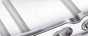
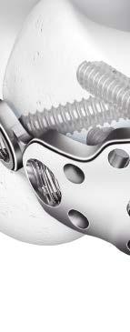
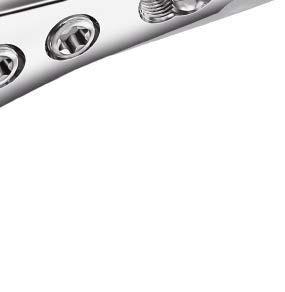
3 3.5 mm LCP Olecranon Plates. Part of the DePuy Synthes locking compression plate (LCP) System. Features Anatomically precontoured Left and right plates Head holes accept 3.5 mm locking screws, and 2.7 mm and 3.5 mm cortex screws Shaft holes accept 3.5 mm locking screws and 3.5 mm cortex screws Choice of six lengths with 2, 4, 6, 8, 10 or 12 Combi holes in the shaft Guide block for easy and correct insertion of proximal screws Combi holes: combination locking and compression holes The dual-purpose Combi hole allows placement of conventional cortex screws on one side, or threaded conical locking screws on the opposite side of the same hole. Fixed-angle stability The locking screws mate with the threaded plate holes to form a fixed-angle construct. Unicortical fixation option Unicortical locking screws provide stability and load transfer only at the near cortex due to the threaded connection between the plate and the screw. Because the screw is locked to the plate, fixation does not rely solely on the pullout strength of the screw or on the frictional force between the plate and the bone. 1 DePuy Synthes 3.5 mm LCP Olecranon Plates Surgical Technique

4 AO Principles and Indications AO PRINCIPLES In 1958, the AO formulated four basic principles, which have become the guidelines for internal fixation. 1,2 In 1958, the AO formulated four basic principles, which have become the guidelines for internal fixation 1, 2. 4_Priciples_03.pdf :08 Anatomic Anatomic reduction reduction Fracture Fracture reduction reduction and and fixation fixation to to restore restore anatomical anatomical relationships. relationships. 1 2 Stable Stable fixation fixation Fracture Fracture fixation fixation providing providing absolute absolute or relative or relative stability, stability, as required as by the required patient, by the the injury, patient, and the the injury, personality and the personality of the fracture. of the fracture. Early, Early, active mobilization Early Early and and safe safe mobilization and rehabilitation of of the injured part and and the the patient as as a whole. 4 3 Preservation Preservation of of blood blood supply supply Preservation Preservation of of the the blood blood supply supply to to soft soft tissues tissues and and bone bone by by gentle reduction gentle reduction techniques techniques and and careful careful handling. handling. Indications The 3.5 mm LCP Olecranon Plates are indicated for fixation of fractures, osteotomies and nonunions of the olecranon, particularly in osteopenic bone. 1. Müller ME, Allgöwer M, Schneider R, Willenegger H. Manual of Internal Fixation. 3rd ed. Berlin, Heidelberg, New York: Springer-Verlag; Müller Rüedi ME, TP, RE M Buckley, Allgöwer, CG R Moran. Schneider, AO Principles H Willenegger. of Fracture Manual Management. of Internal Fixation. 2nd ed. 3rd Stuttgart ed. Berlin New Heidelberg York: Thieme; New York: Springer Rüedi TP, RE Buckley, CG Moran. AO Principles of Fracture Management. 2nd ed. Stuttgart, New York: Thieme mm LCP Olecranon Plates Surgical Technique DePuy Synthes 3


, PA view Case 2 Male")

,")

5 Clinical Cases Case 1 Male patient, 79 years old Olecranon fracture: 21-B1, right arm Implant: LCP olecranon plate with 8 holes Preoperative, lateral view Image intensifier during surgery, lateral view Postoperative (one day after surgery), PA view Case 2 Male patient, 41 years old Olecranon fracture: 21-C2, right arm Implant: LCP Olecranon Plate with 4 holes Preoperative, lateral view Postoperative (ten days after surgery), lateral view Postoperative (ten days after surgery), PA view 4 DePuy Synthes 3.5 mm LCP Olecranon Plates Surgical Technique


6 Preparation 1 Position patient Place the patient either in the lateral or the prone position with the elbow flexed over a side rest. Depending on the fracture, use a posterior access up to approximately 5 cm distal from the supracondylar region. The supine position with the forearm placed across the chest is also an acceptable option, especially with extended approaches to the lateral pillar or column. 3.5 mm LCP Olecranon Plates Surgical Technique DePuy Synthes 5


7 Preparation 2 Surgical approach The incision runs posterior from the supracondylar area to a point 4 cm 5 cm distal to the fracture. It can be slightly curved to the radial side to protect the ulnar nerve. Ulnar nerve Triceps Olecranon Chevron Anconeus Common extensor tendon Ulna 3 Reduce fracture and provide temporary fixation Reduce the fracture directly or indirectly depending on the type of fracture. Ensure that the coronoid is properly reduced before fixation. Examine the reduction of the coronoid process to determine if it is correct before fixation. Use Kirschner wires for temporary fixation. 6 DePuy Synthes 3.5 mm LCP Olecranon Plates Surgical Technique


8 Implantation 1 Determine plate length and adapt plate Required set Small Fragment LCP Instrument and Implant Set, with self-tapping screws or Small Fragment Titanium LCP Instrument and Implant Set, with self-tapping screws Instruments , Bending Irons * Bending Pliers, for 2.7 mm and 3.5 mm Reconstruction Plates Select a plate length appropriate for the fracture. Due to varying patient anatomy, slight bending might be necessary. Using bending irons, contour plates as needed. The triceps tendon may have to be split in order to apply the plate. Evaluate whether or not the hole in the proximal tab should be used. If necessary, remove the tab. Precaution: If bending the tab, take care that the screw in the tab does not collide with the other proximal screws. * Also available 3.5 mm LCP Olecranon Plates Surgical Technique DePuy Synthes 7

9 Implantation 2 Position plate Instruments mm Drill Bit Guiding Block for 3.5 mm LCP Olecranon Plate, right Guiding Block for 3.5 mm LCP Olecranon Plate, left Small Hexagonal Screwdriver with Holding Sleeve Small Hexagonal Screwdriver Shaft mm Locking Screw Sleeve mm Drill Sleeve mm Wire Sleeve mm Universal Drill Guide Push-Pull Reduction Device After adapting the plate, attach the guiding block, drill sleeves and wire sleeve on the proximal part of the plate. Position the plate on the reduced bone, and attach it temporarily with a 3.5 mm cortex screw or push-pull reduction device. Note: Determine the combination of screws to be used for fixation. If a combination of locking and cortex screws will be used, cortex screws should be inserted first to pull the plate to the bone. 8 DePuy Synthes 3.5 mm LCP Olecranon Plates Surgical Technique


10 3 Proximal locking screw insertion Instruments mm Kirschner Wire with thread mm Drill Bit, quick coupling mm Threaded Drill Guide Guiding Block for 3.5 mm LCP Olecranon or Plate, right or left StarDrive Screwdriver, T StarDrive Screwdriver Shaft, T Depth Gauge Direct Measuring Device mm Locking Screw Sleeve mm Drill Sleeve mm Wire Sleeve * Torque Limiting Attachment, 1.5 Nm or Torque Limiting Attachment, 1.5 Nm, quick coupling max. 30 mm To determine screw length needed, insert a Kirschner wire through the sleeve assembly. Stop when increased resistance from the subchondral bone is felt. Since it may not always be possible to feel resistance, the use of image intensification is recommended. Slide the direct measuring device over the K-wire to determine the length of screw. Precaution: All three sleeves must be present. The direct measuring device provides an approximate screw length. Screws longer than 30 mm in the proximal part will collide with the shaft screws. * Also available 3.5 mm LCP Olecranon Plates Surgical Technique DePuy Synthes 9

11 Implantation 3. Proximal locking screw insertion continued Remove the K-wire and K-wire centering sleeve. Drill through the 2.8 mm drill sleeve and then remove it. Insert an appropriate length locking screw through the 3.5 mm locking screw sleeve using the StarDrive TM Screwdriver shaft. Whenever possible, locking screws should be inserted under power using the torque limiting attachment. The audible click will notify the surgeon that the maximum torque value has been reached and the power insertion is completed. After screw insertion using the torque limiting attachment, always check that the screws are fully inserted by hand tightening them. Precaution: Always use the torque limiter to restrict the maximum torque. 11 DePuy Synthes 3.5 mm LCP Olecranon Plates Surgical Technique

12 Alternative technique Insert the 2.8 mm threaded drill guide into a locking hole or Combi hole until fully seated. Drill to the desired depth. Remove the drill guide and use the depth gauge to determine screw length. Insert the appropriate screw. 3.5 mm LCP Olecranon Plates Surgical Technique DePuy Synthes 11


13 Insert Distal Screws 1 Fixation with 3.5 mm cortex screws Instruments mm Drill Bit, quick coupling mm Drill Bit, quick coupling Handle, with quick coupling Small Hexagonal Screwdriver with Holding Sleeve or Small Hexagonal Screwdriver Shaft Depth Gauge mm Universal Drill Guide For lag screw fixation, use the 2.5 mm drill bit through the 3.5 mm universal drill guide to drill for the threaded hole and the 3.5 mm drill bit to drill for the gliding hole. Determine the required length of the cortex screw with the depth gauge. Insert the self-tapping 3.5 mm cortex screw using the small hexagonal screwdriver shaft connected to a power tool or use the small hexagonal screwdriver with holding sleeve. 11 DePuy Synthes 3.5 mm LCP Olecranon Plates Surgical Technique


14 2 Fixation with 3.5 mm locking screws Instruments mm Drill Bit, quick coupling mm Threaded Drill Guide StarDrive Screwdriver, T StarDrive Screwdriver Shaft, T Depth Gauge * Torque Limiting Attachment, 1.5 Nm or Torque Limiting Attachment, 1.5 Nm, quick coupling Insert the 2.8 mm threaded drill guide into a locking hole or Combi hole until fully seated. Use the 2.8 mm drill bit to drill to the desired depth. Remove the drill guide and use the depth gauge to determine screw length. Insert the self-tapping 3.5 mm locking screw using the StarDrive Screwdriver shaft. Whenever possible, locking screws should be inserted under power using the torque limiting attachment. The audible click will notify the surgeon that the maximum torque value has been reached and the power insertion is completed. After screw insertion using the torque limiting attachment, always check that the screws are fully inserted by hand tightening them. Precaution: Always use the torque limiter to restrict the maximum torque. * Also available 3.5 mm LCP Olecranon Plates Surgical Technique DePuy Synthes 11
15 Implant Removal Implant removal Optional set Screw Removal Set Instruments Conical Extraction Screw Handle, with quick coupling To remove locking screws, unlock all screws from the plate, then remove the screws completely from the bone. This prevents simultaneous rotation of the plate when unlocking the last locking screw. If the screws cannot be removed with the screwdriver (e.g. if the hexagonal or StarDrive Recess of the locking screw is damaged or if the screws are stuck in the plate), insert the conical extraction screw with left-handed thread into the screwhead, using the handle with quick coupling, and loosen the locking screw by turning it counterclockwise. 11 DePuy Synthes 3.5 mm LCP Olecranon Plates Surgical Technique
16 Screws Used with the 3.5 mm LCP Olecranon Plate Stainless Steel and Titanium 3.5 mm Locking Screws Create a locked, fixed-angle screw-plate construct Self-tapping tip Used in the locking portion of the Combi holes or in round locking holes 3.5 mm Cortex Screws* May be used in the DCU portion of the Combi holes in the plate shaft or in round locking holes Compress the plate to the bone or create axial compression 2.7 mm Cortex Screws* May be used in the proximal locking holes Compress the plate to the bone Screws made of implant quality 316L stainless steel or titanium alloy (Ti-6Al-7Nb) * Screws made of implant quality 316L stainless steel or commercially pure (CP) titanium 3.5 mm LCP Olecranon Plates Surgical Technique DePuy Synthes 15
 236.502 436.502 2 86 236.")
 236.503 436.")

17 3.5 mm LCP Olecranon Plates Implants 3.5 mm LCP Olecranon Plates, right* Stainless Steel Titanium Holes Length (mm) mm LCP Olecranon Plates, left* Stainless Steel Titanium Holes Length (mm) * Plates are made of implant quality 316L stainless steel or commercially pure (CP) titanium 11 DePuy Synthes 3.5 mm LCP Olecranon Plates Surgical Technique
 312.")

 292.71 1.")

18 Instruments Instruments from the 3.5 mm LCP Olecranon Plate Set ( / ) Guiding Block for 3.5 mm LCP Olecranon Plate, right Guiding Block for 3.5 mm LCP Olecranon Plate, left Selected Instruments from the Small Fragment LCP Instrument and Implant Set ( / ) mm Kirschner Wire with thread, 150 mm, trocar point mm Drill Bit mm Drill Bit mm Drill Bit Handle, with quick coupling 3.5 mm LCP Olecranon Plates Surgical Technique DePuy Synthes 17
19 Selected Instruments from the Small Fragment LCP Instrument and Implant Set ( / ) mm Threaded Drill Guide Small Hexagonal Screwdriver with Holding Sleeve Small Hexagonal Screwdriver Shaft StarDrive Screwdriver, T StarDrive Screwdriver Shaft, T15, quick coupling Depth Gauge mm Wire Sleeve, 55 mm 18 DePuy Synthes 3.5 mm LCP Olecranon Plates Surgical Technique

20 Direct Measuring Device mm Drill Sleeve mm Wire Sleeve mm Universal Drill Guide Bending Iron, for 2.7 mm and 3.5 mm plates, 150 mm length Used with Bending Iron, for 2.7 mm and 3.5 mm plates, 150 mm length Used with * Torque Limiting Attachment, 1.5 Nm or Torque Limiting Attachment, 1.5 Nm, quick coupling * Also available 3.5 mm LCP Olecranon Plates Surgical Technique DePuy Synthes 19

21 3.5 mm LCP Olecranon Plate Set Stainless Steel ( ) and Titanium ( ) Graphic Case mm LCP Olecranon Plate Graphic Case Instruments Guiding Block for 3.5 mm LCP Olecranon Plate, right Guiding Block for 3.5 mm LCP Olecranon Plate, left Implants 3.5 mm LCP Hook Plates, 2 ea. Stainless Steel Titanium Holes mm LCP Olecranon Plates, right Stainless Steel Titanium Holes Length (mm) mm LCP Olecranon Plates, left Stainless Steel Titanium Holes Length (mm) Required Set Small Fragment LCP Instrument and Implant Set, with self-tapping screws or Small Fragment Titanium LCP Instrument and Implant Set, with self-tapping screws Note: For additional information, please refer to package insert. For detailed cleaning and sterilization instructions, please refer to or sterilization instructions, if provided. 22 DePuy Synthes 3.5 mm LCP Olecranon Plates Surgical Technique


22 Also Available mm LCP Elbow System mm Titanium LCP Elbow System Screw Removal Set mm LCP Olecranon Graphic Case Tray Bending Pliers, for 2.7 mm and 3.5 mm Reconstruction Plates Torque Limiting Attachment, 1.5 Nm Retrofit Kit for 3.5 mm LCP Elbow System Graphic Case Retrofit Kit for 3.5 mm Titanium LCP Elbow System Graphic Case 3.5 mm LCP Olecranon Plates Surgical Technique DePuy Synthes 22
23 Limited Warranty and Disclaimer: DePuy Synthes products are sold with a limited warranty to the original purchaser against defects in workmanship and materials. Any other express or implied warranties, including warranties of merchantability or fitness, are hereby disclaimed. Please also refer to the package insert(s) or other labeling associated with the devices identified in this surgical technique for additional information. CAUTION: Federal Law restricts these devices to sale by or on the order of a physician. Some devices listed in this surgical technique may not have been licensed in accordance with Canadian law and may not be for sale in Canada. Please contact your sales consultant for items approved for sale in Canada. Not all products may currently be available in all markets. Manufactured or distributed by: Synthes USA Products, LLC 1302 Wrights Lane East West Chester, PA Synthes USA, LLC 1101 Synthes Avenue Monument, CO To order (USA): To order (Canada): Note: For recognized manufacturer, refer to the product label. DePuy Synthes All rights reserved. DSUS/TRM/0916/1096 4/17 DV
Technique Guide. 3.5 mm LCP Olecranon Plates. Part of the Synthes locking compression plate (LCP) system.
 system.") Technique Guide 3.5 mm LCP Olecranon Plates. Part of the Synthes locking compression plate (LCP) system. Table of Contents Introduction 3.5 mm LCP Olecranon Plates 2 AO Principles 3 Indications 3 Clinical
Technique Guide 3.5 mm LCP Olecranon Plates. Part of the Synthes locking compression plate (LCP) system. Table of Contents Introduction 3.5 mm LCP Olecranon Plates 2 AO Principles 3 Indications 3 Clinical
3.5 mm LCP Extra-articular Distal Humerus Plate
 Part of the DePuy Synthes Locking Compression Plate (LCP ) System 3.5 mm LCP Extra-articular Distal Humerus Plate Surgical Technique Table of Contents Introduction 3.5 mm LCP Extra-articular Distal Humerus
Low Bend Distal Tibia Plates
 Part of the DePuy Synthes Locking Compression Plate (LCP ) System 3.5 mm LCP Low Bend Medial Distal Tibia Plates Surgical Technique Table of Contents Introduction 3.5 mm LCP Low Bend Medial Distal Tibia
3.5 mm LCP Hook Plate
 Part of the DePuy Synthes Locking Compression Plate (LCP ) System 3.5 mm LCP Hook Plate Surgical Technique Table of Contents Introduction 3.5 mm LCP Hook Plate 2 AO Principles 4 Indications 5 Clinical
3.5 mm LCP Distal Humerus Plates
 Part of the DePuy Synthes Locking Compression Plate (LCP ) System 3.5 mm LCP Distal Humerus Plates Surgical Technique Table of Contents Introduction 3.5 mm LCP Distal Humerus Plates 2 AO Principles 4 Indications
Long Volar Plates for Diaphyseal-Metaphyseal Radius Fractures LCP. Dia-Meta Volar Distal Radius Plates. Surgical Technique
 Long Volar Plates for Diaphyseal-Metaphyseal Radius Fractures LCP Dia-Meta Volar Distal Radius Plates Surgical Technique Table of Contents Introduction LCP Dia-Meta Volar Distal Radius Plates 2 AO Principles
2.7 mm/3.5 mm LCP Distal Fibula Plate
 Part of the DePuy Synthes Locking Compression Plate (LCP ) System 2.7 mm/3.5 mm LCP Distal Fibula Plate Surgical Technique Table of Contents Introduction 2.7 mm/3.5 mm LCP Distal Fibula Plates 2 AO Principles
3.5 mm LCP Low Bend Medial Distal Tibia Plate Aiming Instruments
 Part of the 3.5 mm LCP 3.5 mm LCP Low Bend Medial Distal Tibia Plate Aiming Instruments Surgical Technique TABLE OF CONTENTS INTRODUCTION 3.5 mm LCP Low Bend Medial Distal Tibia Plate 2 Aiming Instruments
3.5 mm LCP Clavicle Hook Plates
 Part of the Synthes Locking Compression Plate (LCP ) System 3.5 mm LCP Clavicle Hook Plates Surgical Technique Table of Contents Introduction 3.5 mm LCP Clavicle Hook Plates 2 AO Principles 4 Indications
3.5 mm Clavicle Hook Plates
 A Single Solution for Lateral Clavicle Fractures and Acromioclavicular Joint Dislocations 3.5 mm Clavicle Hook Plates Surgical Technique Discontinued December 2017 DSUS/TRM/1016/1126(1) Table of Contents
Part of the DePuy Synthes Locking Compression Plate (LCP ) System. 3.5 mm LCP Medial Proximal Tibia Plates
 System. 3.5 mm LCP Medial Proximal Tibia Plates") Part of the DePuy Synthes Locking Compression Plate (LCP ) System 3.5 mm LCP Medial Proximal Tibia Plates Surgical Technique Table of Contents Introduction 3.5 mm LCP Medial Proximal Tibia Plates 2 AO
Part of the DePuy Synthes Locking Compression Plate (LCP ) System 3.5 mm LCP Medial Proximal Tibia Plates Surgical Technique Table of Contents Introduction 3.5 mm LCP Medial Proximal Tibia Plates 2 AO
3.5 mm Locking Attachment Plate
 For Treatment of Periprosthetic Fractures 3.5 mm Locking Attachment Plate Surgical Technique Table of Contents Introduction 3.5 mm Locking Attachment Plate 2 Indications 4 Surgical Technique Preparation
The Locking Calcaneal Plate Instrument and Implant Sets
 Part of the DePuy Synthes Locking Compression Plate (LCP ) System The Locking Calcaneal Plate Instrument and Implant Sets Surgical Technique Table of Contents Introduction Locking Calcaneal Plate 2 AO
3.5 mm LCP Anterolateral Distal Tibia Plates
 Part of the DePuy Synthes Locking Compression Plate (LCP ) System 3.5 mm LCP Anterolateral Distal Tibia Plates Surgical Technique Table of Contents Introduction 3.5 mm LCP Anterolateral Distal Tibia Plates
Technique Guide. 3.5 mm LCP Low Bend Medial Distal Tibia Plates. Part of the Synthes locking compression plate (LCP) system.
 system.") Technique Guide 3.5 mm LCP Low Bend Medial Distal Tibia Plates. Part of the Synthes locking compression plate (LCP) system. Table of Contents Introduction 3.5 mm LCP Low Bend Medial Distal Tibia Plates
Technique Guide 3.5 mm LCP Low Bend Medial Distal Tibia Plates. Part of the Synthes locking compression plate (LCP) system. Table of Contents Introduction 3.5 mm LCP Low Bend Medial Distal Tibia Plates
LCP Low Bend Medial Distal Tibia Plates 3.5 mm. Anatomic plates with low profile head for intra- and extraarticular fractures.
 LCP Low Bend Medial Distal Tibia Plates 3.5 mm. Anatomic plates with low profile head for intra- and extraarticular fractures. Surgical Technique This publication is not intended for distribution in the
3.5 mm LCP Superior Anterior Clavicle Plates
 Part of the DePuy Synthes Locking Compression Plate (LCP ) System 3.5 mm LCP Superior Anterior Clavicle Plates Surgical Technique Table of Contents Introduction 3.5 mm LCP Superior Anterior Clavicle Plates
3.5 mm LCP Distal Tibia T-Plates
 Part of the DePuy Synthes Locking Compression Plate (LCP ) System 3.5 mm LCP Distal Tibia T-Plates Surgical Technique Table of Contents Introduction 3.5 mm LCP Distal Tibia T-Plates 2 AO Principles 4 Indications
4.5 mm LCP Medial Proximal Tibia Plates
 Part of the DePuy Synthes LCP Periarticular Plating System 4.5 mm LCP Medial Proximal Tibia Plates Surgical Technique Table of Contents Introduction 4.5 mm LCP Medial Proximal Tibia Plates 2 AO Principles
2.4 mm Variable Angle LCP Volar Rim Distal Radius Plates
 For Fragment-Specific Fracture Fixation With Variable Angle Locking Technology 2.4 mm Variable Angle LCP Volar Rim Distal Radius Plates Surgical Technique Table of Contents Introduction 2.4 mm Variable
LCP Proximal Radius Plates 2.4. Plates for radial head rim and for radial head neck address individual fracture patterns of the proximal radius.
 Technique Guide LCP Proximal Radius Plates 2.4. Plates for radial head rim and for radial head neck address individual fracture patterns of the proximal radius. Table of Contents Introduction LCP Proximal
2.4 mm LCP Radial Head Plates. Part of the Synthes LCP Distal Radius Plate System.
 2.4 mm LCP Radial Head Plates. Part of the Synthes LCP Distal Radius Plate System. Technique Guide Instruments and Implants approved by the AO Foundation Table of Contents Introduction 2.4 mm LCP Radial
LCP Proximal Radius Plates 2.4. Plates for radial head rim and for radial head neck address individual fracture patterns of the proximal radius.
 LCP Proximal Radius Plates 2.4. Plates for radial head rim and for radial head neck address individual fracture patterns of the proximal radius. Surgical Technique This publication is not intended for
Technique Guide. 3.5 mm LCP Low Bend Medial Distal Tibia Plate Aiming Instruments. Part of the 3.5 mm LCP Percutaneous Instrument System.
 Technique Guide 3.5 mm LCP Low Bend Medial Distal Tibia Plate Aiming Instruments. Part of the 3.5 mm LCP Percutaneous Instrument System. Table of Contents Introduction 3.5 mm LCP Low Bend Medial Distal
Distal Radius Plate Instrument and Implant Set. Discontinued December 2017 DSUS/TRM/0916/1063(1)
") Distal Radius Plate Instrument and Implant Set Surgical Technique Discontinued December 2017 DSUS/TRM/0916/1063(1) The Distal Radius Plates Indications For fixation of fractures and osteotomies, including
Distal Radius Plate Instrument and Implant Set Surgical Technique Discontinued December 2017 DSUS/TRM/0916/1063(1) The Distal Radius Plates Indications For fixation of fractures and osteotomies, including
LCP DISTAL TIBIA PLATE
 LCP DISTAL TIBIA PLATE Instruments and implants approved by the AO Foundation. This publication is not intended for distribution in the USA. SURGICAL TECHNIQUE Image intensifier control This description
Small Fragment Locking Compression Plate (LCP ) System
 System") Stainless Steel and Titanium Small Fragment Locking Compression Plate (LCP ) System Surgical Technique Table of Contents Introduction Small Fragment Locking Compression Plate (LCP) System 2 AO Principles
Stainless Steel and Titanium Small Fragment Locking Compression Plate (LCP ) System Surgical Technique Table of Contents Introduction Small Fragment Locking Compression Plate (LCP) System 2 AO Principles
LCP Medial Distal Tibia Plate, without Tab. The Low Profile Anatomic Fixation System with Angular Stability and Optimal Screw Orientation.
 LCP Medial Distal Tibia Plate, without Tab. The Low Profile Anatomic Fixation System with Angular Stability and Optimal Screw Orientation. Technique Guide LCP Small Fragment System Table of Contents Introduction
2.4 mm Variable Angle LCP Volar Extra-Articular Distal Radius System. For fragment-specific fracture fixation with variable angle locking technology.
 2.4 mm Variable Angle LCP Volar Extra-Articular Distal Radius System. For fragment-specific fracture fixation with variable angle locking technology. Surgical Technique This publication is not intended
Technique Guide. 2.7 mm/3.5 mm LCP Distal Fibula Plates. Part of the Synthes locking compression plate (LCP) system.
 system.") Technique Guide 2.7 mm/3.5 mm LCP Distal Fibula Plates. Part of the Synthes locking compression plate (LCP) system. Table of Contents Introduction 2.7 mm/3.5 mm LCP Distal Fibula Plates 2 AO Principles
Technique Guide 2.7 mm/3.5 mm LCP Distal Fibula Plates. Part of the Synthes locking compression plate (LCP) system. Table of Contents Introduction 2.7 mm/3.5 mm LCP Distal Fibula Plates 2 AO Principles
Technique Guide. 3.5 mm LCP Periarticular Proximal Humerus Plate. Part of the Synthes locking compression plate (LCP) system.
 system.") Technique Guide 3.5 mm LCP Periarticular Proximal Humerus Plate. Part of the Synthes locking compression plate (LCP) system. Table of Contents Introduction 3.5 mm LCP Proximal Humerus Plate 2 AO Principles
Technique Guide 3.5 mm LCP Periarticular Proximal Humerus Plate. Part of the Synthes locking compression plate (LCP) system. Table of Contents Introduction 3.5 mm LCP Proximal Humerus Plate 2 AO Principles
Cannulated Pediatric Osteotomy System (CAPOS)
") A Single System of Osteotomy Blade Plates and Cannulated Instrumentation Cannulated Pediatric Osteotomy System (CAPOS) Surgical Technique Table of Contents Introduction Cannulated Pediatric Osteotomy System
A Single System of Osteotomy Blade Plates and Cannulated Instrumentation Cannulated Pediatric Osteotomy System (CAPOS) Surgical Technique Table of Contents Introduction Cannulated Pediatric Osteotomy System
2.4 mm Variable Angle LCP Dorsal Distal Radius Plate
 For Fragment-Specific Fracture Fixation With Variable Angle (VA) Locking Technology 2.4 mm Variable Angle LCP Dorsal Distal Radius Plate Surgical Technique Table of Contents Introduction 2.4 mm VA LCP
Technique Guide. 2.4 mm Variable Angle LCP Distal Radius System. For fragment-specific fracture fixation with variable angle locking technology.
 Technique Guide 2.4 mm Variable Angle LCP Distal Radius System. For fragment-specific fracture fixation with variable angle locking technology. Table of Contents Introduction 2.4 mm Variable Angle LCP
Large Fragment LCP Instrument and Implant Set
 Part of the DePuy Synthes Locking Compression Plate (LCP ) System Large Fragment LCP Instrument and Implant Set Surgical Technique Table of Contents Introduction Large Fragment LCP Instrument and Implant
Olecranon Locking Plate II
 INDEX Indications Patient Position Fracture Reduction and Fixation Surgical Technique Step 1 Surgical Approach Step 2 Implantation Step 3 Proximal Locking Screw Insertion Step 4 Distal Screw Insertion
LCP Proximal Radius Plates 2.4. Plates for radial head rim and for radial head neck address individual fracture patterns of the proximal radius.
 LCP Proximal Radius Plates 2.4. Plates for radial head rim and for radial head neck address individual fracture patterns of the proximal radius. Surgical Technique This publication is not intended for
LCP Distal Fibula Plates. Part of the Synthes locking compression plate (LCP) system.
 system.") LCP Distal Fibula Plates. Part of the Synthes locking compression plate (LCP) system. Surgical Technique This publication is not intended for distribution in the USA. Instruments and implants approved
LCP Distal Fibula Plates. Part of the Synthes locking compression plate (LCP) system. Surgical Technique This publication is not intended for distribution in the USA. Instruments and implants approved
2.4 mm Variable Angle Locking Intercarpal Fusion System
 For Partial Wrist Arthrodesis With Variable Angle Locking Technology 2.4 mm Variable Angle Locking Intercarpal Fusion System Surgical Technique Table of Contents Introduction 2.4 mm Variable Angle Locking
Low Profile Neuro Plating System. Surgical Technique
 Low Profile Neuro Plating System Surgical Technique TABLE OF CONTENTS INTRODUCTION Low Profile Neuro Plating System 2 SURGICAL TECHNIQUE Technique 5 PRODUCT INFORMATION Low Profile Neuro Plates 10 Low
OBSOLETED. LCP Medial Distal Tibia Plate, without Tab. The Low Profile Anatomic Fixation System with Angular Stability and Optimal Screw Orientation.
 LCP Medial Distal Tibia Plate, without Tab. The Low Profile Anatomic Fixation System with Angular Stability and Optimal Screw Orientation. Surgical Technique LCP Small Fragment System This publication
For Distal Femur Fractures. 95º Condylar Plate. Quick Reference Chart
 For Distal Femur Fractures 95º Condylar Plate Quick Reference Chart 95 Condylar Plate. Quick reference chart for distal femur fractures. Insert guide wires Fix condylar fragments with 6.5 mm cancellous
Surgical Technique. This publication is not intended for distribution in the USA. Instruments and implants approved by the AO Foundation.
 LCP Extra-articular Distal Humerus Plate. The anatomically shaped and angular stable fixation system for extraarticular fractures of the distal humerus. Surgical Technique This publication is not intended
Trochanter Stabilization Plate for DHS Implants
 Extends DHS Plate Construct to Help Stabilize Greater Trochanter Trochanter Stabilization Plate for DHS Implants Surgical Technique Table of Contents Introduction Trochanter Stabilization Plate for DHS
Technique Guide. LCP Distal Fibula Plates. Part of the Synthes locking compression plate (LCP) system.
 system.") Technique Guide LCP Distal Fibula Plates. Part of the Synthes locking compression plate (LCP) system. Table of Contents Introduction LCP Distal Fibula Plates 2 AO Principles 4 Indications 5 Surgical Technique
Technique Guide LCP Distal Fibula Plates. Part of the Synthes locking compression plate (LCP) system. Table of Contents Introduction LCP Distal Fibula Plates 2 AO Principles 4 Indications 5 Surgical Technique
4.5 mm LCP Condylar Plate Aiming Instruments
 Part of the LCP Periarticular Aiming Instrument System (Large) 4.5 mm LCP Condylar Plate Aiming Instruments Surgical Technique Table of Contents Introduction 4.5 mm LCP Condylar Plate Aiming Instruments
VA-LCP Anterior Clavicle Plate. The anatomically precontoured fixation system with angular stability for clavicle shaft and lateral clavicle.
 Technique Guide VA-LCP Anterior Clavicle Plate. The anatomically precontoured fixation system with angular stability for clavicle shaft and lateral clavicle. Table of Contents Introduction VA-LCP Anterior
Variable Angle LCP Locking Technology
 BUILDING ON SUCCESS Variable Angle LCP Locking Technology The Evolution of Plating Technology Round Screw Hole Dynamic Compression Plate Limited-Contact Dynamic Compression Less Invasive Stabilization
Technique Guide. Small Fragment Locking Compression Plate (LCP) System. Stainless steel and titanium.
 System. Stainless steel and titanium.") Technique Guide Small Fragment Locking Compression Plate (LCP) System. Stainless steel and titanium. Table of Contents Introduction Small Fragment Locking Compression Plate (LCP) System 2 AO Principles
Technique Guide Small Fragment Locking Compression Plate (LCP) System. Stainless steel and titanium. Table of Contents Introduction Small Fragment Locking Compression Plate (LCP) System 2 AO Principles
Distal Radius Sterile Kit. Optimization And Efficiency You Can Rely On
 Distal Radius Sterile Kit Optimization And Efficiency You Can Rely On Introducing The Distal Radius Sterile Kit Improved OR Workflow The Distal Radius Sterile Kit provides high-quality single-use implants
2.4 mm LCP Distal Radius System
 A Comprehensive Plating System to Address a Variety of Fracture Patterns 2.4 mm LCP Distal Radius System Surgical Technique Table of Contents Introduction 2.4 mm LCP Distal Radius System 2 AO Principles
VA-LCP Olecranon Plates 2.7/3.5. The fracture-specific low-profile fixation system with variable angle locking technology.
 VA-LCP Olecranon Plates 2.7/3.5. The fracture-specific low-profile fixation system with variable angle locking technology. Surgical Technique This publication is not intended for distribution in the USA.
LCP Distal Fibula Plates. Part of the Synthes locking compression plate (LCP) system.
 system.") LCP Distal Fibula Plates. Part of the Synthes locking compression plate (LCP) system. Surgical Technique This publication is not intended for distribution in the USA. Instruments and implants approved
LCP Distal Fibula Plates. Part of the Synthes locking compression plate (LCP) system. Surgical Technique This publication is not intended for distribution in the USA. Instruments and implants approved
2.7 mm/3.5 mm LCP Distal Fibula Plate System. Part of the Synthes locking compression plate (LCP) system.
 system.") 2.7 mm/3.5 mm LCP Distal Fibula Plate System. Part of the locking compression plate (LCP) system. Anatomically contoured Multiple screw options for fixedangle support Coaxial holes minimize screw head
2.7 mm/3.5 mm LCP Distal Fibula Plate System. Part of the locking compression plate (LCP) system. Anatomically contoured Multiple screw options for fixedangle support Coaxial holes minimize screw head
TOMOFIX Medial Distal Femur Plate
 For Closed-Wedge Varus Femoral Osteotomies TOMOFIX Medial Distal Femur Plate Surgical Techniquee TABLE OF CONTENTS INTRODUCTION TOMOFIX Medial Distal Femur Plate 2 AO Principles 4 Indications 5 SURGICAL
LCP Anterolateral Distal Tibia Plate 3.5. The low profile anatomic fixation system with optimal plate placement and angular stability.
 LCP Anterolateral Distal Tibia Plate 3.5. The low profile anatomic fixation system with optimal plate placement and angular stability. Technique Guide LCP Small Fragment System Table of Contents Introduction
Technique Guide. TomoFix Osteotomy System. A comprehensive plating system for stable fixation of osteotomies around the knee.
 Technique Guide TomoFix Osteotomy System. A comprehensive plating system for stable fixation of osteotomies around the knee. Table of Contents Introduction TomoFix Osteotomy System 2 AO Principles 4 Indications
Technique Guide. PHILOS and PHILOS Long. The anatomic fixation system for the proximal humerus.
 Technique Guide PHILOS and PHILOS Long. The anatomic fixation system for the proximal humerus. Table of Contents Introduction PHILOS and PHILOS Long 2 AO Principles 4 Indications 5 Surgical Technique
LCP Anterolateral Distal Tibia Plate 3.5. The low profile anatomic fixation system with optimal plate placement and angular stability.
 LCP Anterolateral Distal Tibia Plate 3.5. The low profile anatomic fixation system with optimal plate placement and angular stability. Technique Guide LCP Small Fragment System Table of Contents Introduction
LCP Distal Humerus Plates
 The anatomic fixation system for the distal humerus with angular stability Surgical technique LCP Locking Compression Plate Contents Indications and contraindications 2 Implants 3 Instruments 5 Preparation
Olecranon Osteotomy Nail. For simple fractures and osteotomies of the olecranon.
 Olecranon Osteotomy Nail. For simple fractures and osteotomies of the olecranon. Technique Guide Discontinued June 2016; AVAILABLE FOR IMPLANT REMOVAL PURPOSES ONLY DSEM/TRM/0517/0843 Table of Contents
modular ClaVICle PlaTe system
 modular ClaVICle PlaTe system 3.7 mm/3.3 mm VA LCP Anterior Clavicle Plates and 3.3 mm Superior Anterior Clavicle Plates surgical TeChnIque Table of Contents Introduction Modular Clavicle Plate System
2.4 mm Variable Angle LCP Volar Extra-Articular Distal Radius System. For fragment-specific fracture fixation with variable angle locking technology.
 Technique Guide 2.4 mm Variable Angle LCP Volar Extra-Articular Distal Radius System. For fragment-specific fracture fixation with variable angle locking technology. Table of Contents Introduction 2.4
LCP Medial Proximal Tibial Plate 4.5/5.0. Part of the Synthes LCP periarticular plating system.
 LCP Medial Proximal Tibial Plate 4.5/5.0. Part of the Synthes LCP periarticular plating system. Technique Guide This publication is not intended for distribution in the USA. Instruments and implants approved
Technique Guide. LCP Proximal Femoral Hook Plate 4.5/5.0. Part of the LCP Periarticular Plating System.
 Technique Guide LCP Proximal Femoral Hook Plate 4.5/5.0. Part of the LCP Periarticular Plating System. Table of Contents Introduction Features and Benefits 2 AO ASIF Principles 4 Indications 5 Surgical
low ProfIle neuro PlaTIng system
 low ProfIle neuro PlaTIng system surgical TeChnIque Table of Contents Introduction Low Profile Neuro Cranial Plating System 2 Surgical Technique Technique 5 Product Information Low Profile Neuro Plates
LCP Distal Tibia Plate
 Surgical Technique LCP Locking Compression Plate Original Instruments and Implants of the Association for the Study of Internal Fixation AO/ASIF Table of contents Indications 3 Implants/Instruments 5 Surgical
LCP Extra-articular Distal Humerus Plate.
 Technique Guide LCP Extra-articular Distal Humerus Plate. The anatomically shaped and angular stable fixation system for extraarticular fractures of the distal humerus. Table of Contents Introduction
LCP Periarticular Proximal Humerus Plate 3.5. The anatomic fixation system with anterolateral shaft placement.
 LCP Periarticular Proximal Humerus Plate 3.5. The anatomic fixation system with anterolateral shaft placement. Surgical Technique This publication is not intended for distribution in the USA. Instruments
LCP Medial Proximal Tibial Plate 3.5. Part of the Synthes small fragment Locking Compression Plate (LCP) system.
 system.") LCP Medial Proximal Tibial Plate 3.5. Part of the Synthes small fragment Locking Compression Plate (LCP) system. Technique Guide This publication is not intended for distribution in the USA. Instruments
LCP Medial Proximal Tibial Plate 3.5. Part of the Synthes small fragment Locking Compression Plate (LCP) system. Technique Guide This publication is not intended for distribution in the USA. Instruments
LCP Superior Clavicle Plate. The anatomically precontoured fixation system with angular stability for clavicle shaft and lateral clavicle.
 Technique Guide LCP Superior Clavicle Plate. The anatomically precontoured fixation system with angular stability for clavicle shaft and lateral clavicle. Table of Contents Introduction LCP Superior Clavicle
3.5 mm LCP Superior and Superior Anterior Clavicle Plates. Part of the Synthes modular clavicle plate system.
 3.5 mm LCP Superior and Superior Anterior Clavicle Plates. Part of the Synthes modular clavicle plate system. Technique Guide Instruments and implants approved by the AO Foundation Table of Contents Introduction
LCP Metaphyseal Plates. For extra-articular fractures.
 LCP Metaphyseal Plates. For extra-articular fractures. Surgical Technique This publication is not intended for distribution in the USA. Instruments and implants approved by the AO Foundation. Image intensifier
2.4 mm LCP Volar Column Distal Radius Plates. Part of the 2.4 mm LCP Distal Radius System.
 2.4 mm LCP Volar Column Distal Radius Plates. Part of the 2.4 mm LCP Distal Radius System. 12 anatomically shaped volar plates Multiple screw options for fixedangle support to articular surface Combi holes
LCP Condylar Plate 4.5/5.0. Part of the LCP Periarticular Plating System.
 LCP Condylar Plate 4.5/5.0. Part of the LCP Periarticular Plating System. Surgical Technique This publication is not intended for distribution in the USA. Instruments and implants approved by the AO Foundation.
Variable Angle LCP Volar Rim Distal Radius Plate 2.4. For fragment-specific fracture fixation with variable angle locking technology.
 Technique Guide Variable Angle LCP Volar Rim Distal Radius Plate 2.4. For fragment-specific fracture fixation with variable angle locking technology. Image intensifier control Warning This description
LCP Superior Clavicle Plate. The anatomically precontoured fixation system with angular stability for clavicle shaft and lateral clavicle.
 LCP Superior Clavicle Plate. The anatomically precontoured fixation system with angular stability for clavicle shaft and lateral clavicle. Surgical Technique This publication is not intended for distribution
2.7 mm/3.5 mm VA-LCP Anterior Clavicle Plates. Part of the Synthes modular clavicle plate system.
 2.7 mm/3.5 mm VA-LCP Anterior Clavicle Plates. Part of the Synthes modular clavicle plate system. Technique Guide Instruments and implants approved by the AO Foundation Table of Contents Introduction 2.7
DOUBLE/TRIPLE PELVIC OSTEOTOMY PLATES For Treating Coxofemoral Joint Instability and Subluxation in Immature Dogs
 DOUBLE/TRIPLE PELVIC OSTEOTOMY PLATES For Treating Coxofemoral Joint Instability and Subluxation in Immature Dogs Instruments and implants approved by the AO Foundation. This publication is not intended
Technique Guide. Locking Attachment Plate. For treatment of periprosthetic fractures.
 Technique Guide Locking Attachment Plate. For treatment of periprosthetic fractures. Table of Contents Introduction Locking Attachment Plate 2 Indications 4 Surgical Technique Patient Positioning 5 Preparation
Mini External Fixator
 Stabilize the Phalanges and Metacarpals Mini External Fixator Surgical Technique Table of Contents Introduction Mini External Fixator 2 Indications 4 Surgical Technique Technique Overview 5 Product Information
Technique Guide. LCP Posterior Medial Proximal Tibial Plate 3.5. Part of the Synthes small fragment LCP system.
 Technique Guide LCP Posterior Medial Proximal Tibial Plate 3.5. Part of the Synthes small fragment LCP system. Table of Contents Introduction LCP Posterior Medial Proximal Tibial Plate 3.5 2 AO Principles
Technique Guide. 4.5 mm LCP Proximal Tibia Plates. Part of the Synthes LCP Periarticular Plating System.
 Technique Guide 4.5 mm LCP Proximal Tibia Plates. Part of the Synthes LCP Periarticular Plating System. Table of Contents Introduction 4.5 mm LCP Proximal Tibia Plates 2 AO Principles 4 Indications 5 Surgical
2.7 mm/3.5 mm Variable Angle LCP. Ankle Trauma System
 Part of the DePuy Synthes Variable Angle Locking Compression Plate (VA LCP ) System 2.7 mm/3.5 mm Variable Angle LCP Ankle Trauma System Surgical Technique Table of Contents Introduction 2.7 mm/3.5 mm
VA-LCP Distal Humerus Plates 2.7/3.5. The low-profile fixation system with variable angle locking technology.
 VA-LCP Distal Humerus Plates 2.7/3.5. The low-profile fixation system with variable angle locking technology. Technique Guide This publication is not intended for distribution in the USA. Instruments and
VA-LCP Anterior Clavicle Plate. The anatomically precontoured fixation system with angular stability for clavicle shaft and lateral clavicle.
 VA-LCP Anterior Clavicle Plate. The anatomically precontoured fixation system with angular stability for clavicle shaft and lateral clavicle. Surgical Technique This publication is not intended for distribution
COMPACT ANKLE FRACTURE SYSTEM
 COMPACT ANKLE FRACTURE SYSTEM Consolidated ankle fracture solution for surgery centers SURGICAL TECHNIQUE TABLE OF CONTENTS INTRODUCTION Compact Ankle Fracture System 2 AO Principles 8 Indications 9 Clinical
Compact Distal Radius System. Consolidated solution for Distal Radius Plating Systems
 Compact Distal Radius System. Consolidated solution for Distal Radius Plating Systems Designed to allow customized system configurations Variable Angle (VA) and Locking Compression Plate (LCP ) options
Technique Guide. VA-LCP Distal Humerus Plates 2.7/3.5. The low-profile fixation system with variable angle locking technology.
 Technique Guide VA-LCP Distal Humerus Plates 2.7/3.5. The low-profile fixation system with variable angle locking technology. Image intensifier control Warning This description alone does not provide sufficient
LCP Superior Anterior Clavicle Plate. The anatomically precontoured fixation system with angular stability for clavicle shaft and lateral clavicle.
 LCP Superior Anterior Clavicle Plate. The anatomically precontoured fixation system with angular stability for clavicle shaft and lateral clavicle. Surgical Technique This publication is not intended for
PHILOS and PHILOS Long. The anatomic fixation system for the proximal humerus.
 PHILOS and PHILOS Long. The anatomic fixation system for the proximal humerus. Surgical Technique This publication is not intended for distribution in the USA. Instruments and implants approved by the
VA-LCP Distal Humerus Plates 2.7/3.5. The low-profile fixation system with variable angle locking technology.
 VA-LCP Distal Humerus Plates 2.7/3.5. The low-profile fixation system with variable angle locking technology. Surgical Technique This publication is not intended for distribution in the USA. Instruments
3.5 MM VA-LCP PROXIMAL TIBIA PLATE SYSTEM
 3.5 MM VA-LCP PROXIMAL TIBIA PLATE SYSTEM Part of the DePuy Synthes Variable Angle Periarticular Plating System SURGICAL TECHNIQUE TABLE OF CONTENTS INTRODUCTION 3.5 mm VA-LCP Proximal Tibial Plate 2 AO
VA LOCKING CALCANEAL PLATES 2.7
 VA LOCKING CALCANEAL PLATES 2.7 Instruments and Implants approved by the AO Foundation. This publication is not intended for distribution in the USA. SURGICAL TECHNIQUE Image intensifier control Warning
2.4 mm Variable Angle LCP Intercarpal Fusion System. Variable angle locking technology for mediocarpal partial arthrodesis.
 2.4 mm Variable Angle LCP Intercarpal Fusion System. Variable angle locking technology for mediocarpal partial arthrodesis. Technique Guide Instruments and implants approved by the AO Foundation Table
Titanium Distal Femoral Nail System
 For Retrograde Insertion Titanium Distal Femoral Nail System Surgical Technique Table of Contents Introduction Titanium Distal Femoral Nail System 2 AO Principles 4 Indications 5 Clinical Cases 6 Surgical
Lag Screw Device Intended for symphyseal fracture fixation of the mandible
 Lag Screw Device Intended for symphyseal fracture fixation of the mandible SUrgicaL TecHNiqUe Lag Screw Device Intended for symphyseal fracture fixation of the mandible Simplifies the lag screw fixation
LCP Medial Proximal Tibial Plate 3.5. Part of the Synthes small fragment Locking Compression Plate (LCP) system.
 system.") LCP Medial Proximal Tibial Plate 3.5. Part of the Synthes small fragment Locking Compression Plate (LCP) system. Surgical Technique This publication is not intended for distribution in the USA. Instruments
LCP Medial Proximal Tibial Plate 3.5. Part of the Synthes small fragment Locking Compression Plate (LCP) system. Surgical Technique This publication is not intended for distribution in the USA. Instruments