VA LOCKING CALCANEAL PLATES 2.7
|
|
|
- Joshua Clarke
- 5 years ago
- Views:
Transcription


1 VA LOCKING CALCANEAL PLATES 2.7 Instruments and Implants approved by the AO Foundation. This publication is not intended for distribution in the USA. SURGICAL TECHNIQUE
2 Image intensifier control Warning This description alone does not provide sufficient background for direct use of DePuy Synthes products. Instruction by a surgeon experienced in handling these products is highly recommended. Processing, Reprocessing, Care and Maintenance For general guidelines, function control and dismantling of multi-part instruments, as well as processing guidelines for implants, please contact your local sales representative or refer to: For general information about reprocessing, care and maintenance of Synthes reusable devices, instrument trays and cases, as well as processing of Synthes non-sterile implants, please consult the Important Information leaflet (SE_023827) or refer to:
3 TABLE OF CONTENTS AO PRINCIPLES 2 INTRODUCTION Indications 3 SURGICAL TECHNIQUE VA Locking Calcaneal Plates Reduction joystick 8 Variable Angle Locking Technique B 2.7 mm 9 Preparation 13 Surgical Technique 15 Implant Removal 24 PRODUCT INFORMATION Implants 25 Screws 26 Instruments 28 Sets 33 MRI INFORMATION 36 VA Locking Calcaneal Plates 2.7 Surgical Technique DePuy Synthes 1

4 AO PRINCIPLES In 1958, the AO formulated four basic principles, which have become the guidelines for internal fixation.¹, ² 4_Priciples_03.pdf :08 Anatomic reduction Fracture reduction and fixation to restore anatomical relationships. 1 2 Stable fixation Fracture fixation providing absolute or relative stability, as required by the patient, the injury, and the personality of the fracture. Early, active mobilization Early and safe mobilization and rehabilitation of the injured part and the patient as a whole. 4 3 Preservation of blood supply Preservation of the blood supply to soft tissues and bone by gentle reduction techniques and careful handling. Copyright 2007 by AO Foundation ¹ Müller ME, M Allgöwer, R Schneider, H Willenegger. Manual of Internal Fixation. 3rd ed. Berlin, Heidelberg, New York: Springer ² Rüedi TP, RE Buckley, CG Moran. AO Principles of Fracture Management. 2nd ed. Stuttgart, New York: Thieme DePuy Synthes VA Locking Calcaneal Plates 2.7 Surgical Technique

5 INDICATIONS VA Locking Calcaneal Plate 2.7 The Synthes Variable Angle Locking Calcaneal plates 2.7 are indicated for intra- and extra-articular fractures of the calcaneus, as well as deformities and malunions. VA Locking Calcaneal Plates 2.7 Surgical Technique DePuy Synthes 3


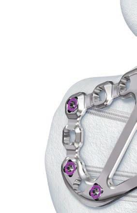
6 VA LOCKING CALCANEAL PLATES 2.7 Anatomic, low-profile plates designed specifically for the calcaneus. Pre-contoured to fit the anterior process, posterior facet, and calcaneal tuberosity Variable Angle Locking screws are designed to sit flush within the plate* to reduce the likelihood of screw prominence creating a low profile construct when used at nominal angle. Variable Angle Locking Technology Offers a variety of options for fixation of calcaneal fractures including: Ability to adapt screw trajectory to match calcaneal anatomy and fracture pattern Ability to angulate screws towards specific fragments or areas of cortical bone Variable Angle Locking screw B 2.7 mm holes: Accept VA Locking B 2.7 mm, Metaphyseal, Locking **, and Cortex screws * Variable Angle Locking screws sit flush within the plate when inserted at nominal angle. ** Locking screws inserted only at nominal angle. 1 DePuy Synthes VA Locking Calcaneal Plates 2.7 Surgical Technique
 angle.")


7 Variable Angle Locking screw holes target dense cortical bone around the perimeter of the calcaneus when the screws are inserted at nominal (fixed) angle. Variable Angle Locking screws are targeted to buttress the posterior and middle facet and converge in the hard bone of the sustentaculum. Anterior process screws are targeted to buttress the anterior facet and are angled in line with the calcaneal-cuboid joint. Five screw holes in this area to provide multiple fixation options Tuberosity screws are angled inferior and posterior to target hard cortical bone around the perimeter of the tuberosity. VA Locking Calcaneal Plates 2.7 Surgical Technique DePuy Synthes 1


8 VA Locking Calcaneal Plates 2.7 Strut Plate profile and screw holes targeting the inferior portion of the tuberosity are located superior to the incision line to reduce the likelihood of the implant causing stress on the incision area. Strut down the center of the plate is designed to provide additional construct strength and support for any lateral wall comminution. Plate is designed to allow for additional contouring in the tuberosity and anterior process portion of the plate to improve fit among varying patient anatomy. Scalloped edge along the posterior facet portion of the plate provides clearance for independent screw fixation. Scallops accommodate screws size B 2.7 mm, B 3.5 mm, and B 4.0 mm 6 DePuy Synthes VA Locking Calcaneal Plates 2.7 Surgical Technique
, Medium (64 mm), and Large (70 mm) sizes Left and right plate designs Stainless steel and titanium VA Locking Calcaneal Plates 2.")

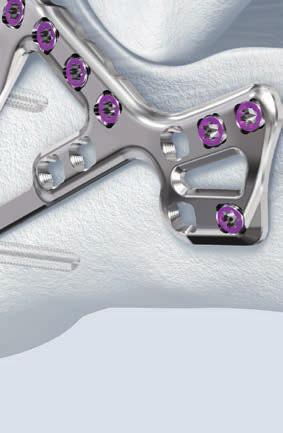
9 Plate designed with no screw holes in the neutral triangle area of the calcaneus in order to increase plate strength where the fracture line typically lies. VA Locking Calcaneal Plates 2.7 Available in Small (58 mm), Medium (64 mm), and Large (70 mm) sizes Left and right plate designs Stainless steel and titanium VA Locking Calcaneal Plates 2.7, with tabs Plates with tabs are available to provide additional support of fracture fragments, especially in the case of severely comminuted fractures where screw fixation alone is insufficient or not possible. Tabs provide additional support of anterior process and plantar fracture fragments Additional superior screw hole is contourable, and allows for supplemental fixation through calcaneal body Available in Medium (64 mm) and Large (70 mm) sizes VA Locking Calcaneal Plates 2.7 Surgical Technique DePuy Synthes 7


10 REDUCTION JOYSTICK Reduction tool to aid in fracture manipulation. The threaded buttress sleeve provides a large surface area to reduce the likelihood of cut-out in cancellous bone. Available in sizes 5.0 mm and 6.5 mm. Refer to page for specific technique information. Threaded buttress sleeve 8 DePuy Synthes VA Locking Calcaneal Plates 2.7 Surgical Technique


11 VARIABLE ANGLE LOCKING TECHNIQUE B 2.7 MM 1 Drill for variable angle locking screws A. Conical (off nominal-axis) insertion Instruments VA LCP Drill Sleeve 2.7, for Drill Bits B 2.0 mm Drill Bit B 2.0 mm, with double marking, length 140/115 mm, 3-flute, for Quick Coupling Optional VA LCP Drill Sleeve 2.7, conical, for Drill Bits B 2.0 mm VA Drill Guide 2.7 with cone point To insert the Variable Angle Locking screw off the nominal axis, insert the cone-shaped side of drill guide in the desired variable angle locking screw hole in the plate. The funnel of the drill guide allows a drilling angle within a 30 cone. When drilling off-axis, the drill guide should remain in place and the drill bit may be aimed in any direction within the cone. Verify the drill bit angle and depth under radiographic imaging to ensure the desired angle has been achieved. If necessary, drill at a different angle and verify again under imaging. Precaution: Avoid excessive re-drilling, especially in poor bone quality. VA Locking Calcaneal Plates 2.7 Surgical Technique DePuy Synthes 9


12 Variable Angle Locking Technique B 2.7 mm B. Coaxial (fixed angle) insertion Instruments VA LCP Drill Sleeve 2.7, for Drill Bits B 2.0 mm Drill Bit B 2.0 mm, with double marking, length 140/115 mm, 3-flute, for Quick Coupling Optional VA LCP Drill Sleeve 2.7, coaxial, for Drill Bits B 2.0 mm To insert VA locking screws into the plate in line with the predefined screw trajectory insert the coaxial side of the drill sleeve into the desired hole in the plate. Drill to the desired depth. Verify the drill bit depth under radiographic imaging. Use the depth gauge to measure for the correct screw length Instrument Depth Gauge, percutaneous Optional Depth Gauge for Screws B 2.0 to 2.7 mm, measuring range up to 40 mm Use the depth gauge to measure for the correct screw length. 11 DePuy Synthes VA Locking Calcaneal Plates 2.7 Surgical Technique

13 2 Insert Variable Angle Locking screws Instrument Screwdriver Shaft, Stardrive, T8, self-holding Optional Silicone Handle with AO/ASIF Quick Coupling Insert the correct length Variable Angle Locking screw. The Variable Angle Locking screws can be inserted manually or with power. For manual insertion, use the Stardrive Screwdriver Shaft and handle with quick coupling. Initial insertion of Variable Angle Locking screws may be done using power equipment. Do not lock the screws with power tools. Confirm screw position and length prior to final tightening. Final tightening must be done manually with the torque limiter. Precautions: Do not engage the screw head with the plate hole while inserting under power. Screw engagement and final locking must be done manually with the torque limiter. Do not use the torque limiting handle for screw removal. VA Locking Calcaneal Plates 2.7 Surgical Technique DePuy Synthes 11


14 Variable Angle Locking Technique B 2.7 mm 3 Lock Variable Angle Locking screws Instruments Torque Limiter, 1.2 Nm, with AO/ASIF Quick Coupling Handle for Torque Limiters 0.4/0.8/1.2 Nm Screwdriver Shaft, Stardrive, T8, self-holding Use the torque limiter for final tightening of Variable Angle Locking screws. The use of the torque limiter is mandatory when engaging the screws into Variable Angle Locking holes to ensure the appropriate amount of torque is applied. Confirm screw position and length prior to final tightening. Precaution: Do not lock the screws to the plate under power. Screw engagement and final tightening must be done manually with the torque limiter and handle: 1.2 Nm torque limiter for B 2.7 mm Do not use the torque limiters for screw removal 11 DePuy Synthes VA Locking Calcaneal Plates 2.7 Surgical Technique
15 PREPARATION Required set(s) Plates, Stainless steel VA-Locking Calcaneal Plate 2.7 and VA-Locking Anterolateral Calcaneal Plate 2.7, Stainless Steel, in Modular Tray, Vario Case System Screws, Stainless steel Screw Rack Module (metal) for VA Locking Screws 2.7 and Cortex Screws 2.7 (Stainless Steel) or Plates, Titanium VA-Locking Calcaneal Plate 2.7 and VA-Locking Anterolateral Calcaneal Plate 2.7, (Titanium), in Modular Tray, Vario Case System Screws, Titanium Screw Rack Module (metal) for VA Locking Screws 2.7 and Cortex Screws 2.7 (Titanium) Instruments Instruments for VA-Locking and Cortex Screw Insertion 2.7, in Modular Tray, Vario Case System Reduction Joystick 5.0 and 6.5 mm, in Modular Tray, Vario Case System VA Locking Calcaneal Plates 2.7 Surgical Technique DePuy Synthes 11
16 Preparation Optional set(s) Compression/Distraction Device Set for Orthopaedic Foot Instruments, with Lid, with Contents Set for Bending Pliers for VA Locking Plates 2.4/ Compression System VA 2.4/2.7, in Modular Tray, Vario Case System Special Instrument Set for Hindfoot 11 DePuy Synthes VA Locking Calcaneal Plates 2.7 Surgical Technique


17 SURGICAL TECHNIQUE 1 Approach Place the patient in lateral decubitus position. Make an extensile, L-shaped, right-angled lateral incision. The vertical portion of the incision should be just anterior to the heel cord and extend down to the plantar and lateral skin junction. Continue the incision forward, horizontally, exposing the calcaneocuboid joint. The incision is carried straight down to bone at its angle and then developed to allow a single, thick flap to be lifted from the periosteal surface. This approach allows raising a single flap consisting of skin and soft tissue which includes the peroneal tendons, sural nerve and the detached calcaneofibular ligament. Warning: Care should be taken to avoid the sural nerve when dissecting. VA Locking Calcaneal Plates 2.7 Surgical Technique DePuy Synthes 11


18 Surgical Technique 2 Reduce fracture Instruments Kirschner Wire B 1.6 mm with trocar tip, length 150 mm, Stainless Steel Kirschner Wire B 1.6 mm with trocar tip, length 150 mm, Titanium Alloy (TAV) or Compression Wire B 1.6 mm, length 150 mm, thread length mm Reduce the fracture fragments and hold fragments in place with Kirschner wires. The Kirschner wires should be placed to avoid interference with final plate placement. Optional Technique: Reduction Joystick The Reduction Joystick and Small Universal Chuck with T-handle can be used to aid in the reduction of the tuberosity as required. Instruments Reduction Joystick 5.0 mm or Reduction Joystick 6.5 mm Universal Chuck, small, with T-Handle Assemble the Threaded Buttress Sleeve onto the Reduction Joystick. Thread the Buttress sleeve onto the shaft of the Reduction Joystick to expose all of the bone threads. Insert the proper size centering pin into the cannulation of the Reduction Joystick. Fully seat the hexagonal head of the Centering Pin into the recess of the Reduction Joystick. Precaution: The proper size centering pin must be inserted through the cannulation of the Reduction Joystick prior to insertion (Reduction Joystick, 5.0 mm takes 1.6 mm Centering Pin; Reduction Joystick, 6.0 mm takes 2.8 mm Centering Pin). Bone Threads Threaded Buttress Sleeve 11 DePuy Synthes VA Locking Calcaneal Plates 2.7 Surgical Technique


19 Insert the Reduction Joystick under power by connecting to the quick coupling on the centering pin, or manually with the Small Universal Chuck with T-handle. Remove the centering pin and attach the Small Universal Chuck with T-handle to the Reduction Joystick. Turn the Threaded buttress sleeve down to the bone so that the surface of the device contacts the bone for added stability. VA Locking Calcaneal Plates 2.7 Surgical Technique DePuy Synthes 11

20 Surgical Technique Pull the tuberosity posterior, inferior, and out of varus. Optional Compression/Distraction Device Set for Orthopaedic Foot Instruments, with Lid, with Contents Use the compression/distraction device to distract the calcaneus and restore length. Place one pin in the cuboid or anterior process and one pin in the tuberosity and distract. Note: Reference the Orthopaedic Foot Instrument Technique Guide for the Compression Distraction technique steps and product assembly. 11 DePuy Synthes VA Locking Calcaneal Plates 2.7 Surgical Technique

21 3 Contour plate (optional) Instrument Bending Pliers for VA Locking Plates Plates may require additional contouring depending on patient anatomy. Assess the plate fit and contour further if needed after fracture reduction. The 2.4 mm/2.7 mm VA LCP Bending Pliers can be used to contour the plate further. The 2.4 mm/2.7 mm VA LCP Bending Pliers should be used to protect the variable angle locking screw holes. Warning: Extensive contouring may weaken the plate. VA Locking Calcaneal Plates 2.7 Surgical Technique DePuy Synthes 11

22 Surgical Technique 4 Position plate Instruments Kirschner Wire B 1.6 mm with trocar tip, length 150 mm, Stainless Steel Kirschner Wire B 1.6 mm with trocar tip, length 150 mm, Titanium Alloy (TAV) or Compression Wire B 1.6 mm, length 150 mm, thread length mm Position plate and provisionally fix the plate to bone with Kirschner wires or 1.6 mm Compression Wires. The plate should sit just under the ridge of the subchondral bone located below the subtalar joint. Note: If enlarged peroneal tubercle prevents ideal plate placement, the tubercle may need to be removed. 22 DePuy Synthes VA Locking Calcaneal Plates 2.7 Surgical Technique


23 Optional Technique: VA Locking Calcaneal Plate 2.7 with tabs Instrument Bending Pliers for VA Locking Plates Optional Bender for Tabs of Calcaneal Locking Plate Universal Bending Pliers, length 165 mm If using the VA Locking Calcaneal Plate with tabs, use the 2.4/2.7 VA LCP Bending Pliers to contour the superior screw hole. 2.4 mm/2.7 mm VA LCP Bending Pliers Use the 2.4/2.7 VA LCP Bending pliers to pre-bend the tabs, while simultaneously protecting the Variable Angle Locking screw hole with a second 2.4/2.7 VA LCP Bending Plier. Universal Bending Pliers Optional Once the plate is positioned on the calcaneus, the Locking Calcaneal Plate Tab Bending Pliers may be used to bend the tabs further if needed. Locking Calcaneal Plate Tab Bending Pliers Insert the square portion of the Tab Bending Pliers into the hole adjacent to the tab, and bend the tab. VA Locking Calcaneal Plates 2.7 Surgical Technique DePuy Synthes 22


24 Surgical Technique 5 Insert screws For final fixation, insert the Variable Angle Locking screws B 2.7 mm as described in the Variable Angle Locking Technique on page Precautions: A minimum of four Variable Angle Locking or Locking screws should be inserted in the posterior facet portion of the plate to provide adequate fixation.* Only screws B 2.7 mm should be inserted in the calcaneal plates. Use caution when inserting screws targeting the sustentaculum. Confirm screw positioning under fluoroscopic imaging. Optional Technique Metaphyseal screws B 2.7 mm can also be inserted prior to Variable Angle locking screws to ensure appropriate bone contact with the plate. The Metaphyseal screws B 2.7 mm can be inserted under power or manually following the 2.7 mm Variable Angle Locking Technique on page Precaution: Final metaphyseal screw insertion, similar to a cortex screw, should be completed manually using the T8 Stardrive screwdriver shaft and handle with quick coupling. * Testing on file at DePuy Synthes. 22 DePuy Synthes VA Locking Calcaneal Plates 2.7 Surgical Technique


25 6 Lock Variable Angle Locking screws Lock the Variable Angle Locking screws B 2.7 mm manually with the 1.2 Nm Torque Limiting Attachment and handle as described in the Variable Angle Locking Technique on page Confirm reduction and fixation under fluoroscopic imaging. Take lateral, axial heel, and Broden s view images. VA Locking Calcaneal Plates 2.7 Surgical Technique DePuy Synthes 22

26 IMPLANT REMOVAL Instruments Screwdriver Shaft, Stardrive, T8, self-holding Silicone Handle with AO/ASIF Quick Coupling Optional Handle with Quick Coupling If implant removal is desired, unlock all screws manually from the plate using the proper screwdriver shaft and handle. Then remove the screws completely from the bone. Precaution: Do not use the torque limiters for screw removal. 22 DePuy Synthes VA Locking Calcaneal Plates 2.7 Surgical Technique

 02.211.406 04.211.406 Right Medium 64 02.211.407 04.211.407 Left Medium 64 02.")

27 IMPLANTS VA Locking Calcaneal Plates 2.7* Stainless Steel Titanium Right/Left Size Length (mm) Right Small Left Small Right Medium Left Medium Right Large Left Large 70 Right Left VA Locking Calcaneal Plates 2.7, with tabs* Stainless Steel Titanium Right/Left Size Length (mm) Right Medium Left Medium Right Large Left Large 70 Right Left * Available nonsterile or sterile-packed. Add S to catalog number to order sterile product. VA Locking Calcaneal Plates 2.7 Surgical Technique DePuy Synthes 22
28 SCREWS The VA Locking Calcaneal plates 2.7 accept the following screws: Variable Angle Locking Screws B 2.7 mm Threaded, rounded head locks securely into the variable angle locking holes Locked screws allow unicortical screw fixation and load transfer to near cortex Used with drill bit B 2.0 mm T8 Stardrive recess Self-tapping tip Color coded for easier identification Screws in set: 16 mm to 56 mm lengths (0X X ) Additionally available: 8 mm to 60 mm lengths (0X X ) Note: VA locking screws B 2.7 mm must be tightened to 1.2 Nm. Metaphyseal Screws B 2.7 mm For use in locking, non-locking, or combi-holes Used to provide compression of plate to the bone Feature locking screw thread in screw shaft Low-profile head Used with drill bit B 2.0 mm T8 Stardrive recess Self-tapping tip Screws in set: 16 mm to 56 mm lengths (0X X ) Additionally available: 10 mm to 70 mm lengths (0X X ) X = 2 (Stainless Steel), 4 (Titanium) 22 DePuy Synthes VA Locking Calcaneal Plates 2.7 Surgical Technique
29 Cortex Screws* B 2.7 mm For use in locking, non-locking, or combi-holes Used to provide compression or neutral fixation Used with drill bit B 2.0 mm T8 Stardrive recess Self-tapping tip Additionally available: 10 mm to 60 mm (X X02.969) Locking Screws* B 2.7 mm Threaded, conical head locks securely into the variable angle locking holes Only for axial insertion in the variable angle locking holes Used with drill bit B 2.0 mm T8 Stardrive recess Self-tapping tip Additionally available: 8 mm to 60 mm (X X02.260) * Also available. X = 2 (Stainless Steel), 4 (Titanium) VA Locking Calcaneal Plates 2.7 Surgical Technique DePuy Synthes 22


30 INSTRUMENTS Drill Bit B 2.0 mm, with double marking, length 140/115 mm, 3-flute, for Quick Coupling Drill Bit B 2.7 mm, length 125/100 mm, 3-flute, for Quick Coupling VA LCP Drill Sleeve 2.7, for Drill Bits B 2.0 mm Torque Limiter, 1.2 Nm, with AO/ASIF Quick Coupling Handle for Torque Limiters 0.4/0.8/1.2 Nm Also Available VA LCP Drill Sleeve 2.7, conical, for Drill Bits B 2.0 mm VA LCP Drill Sleeve 2.7, coaxial, for Drill Bits B 2.0 mm 28 DePuy Synthes VA Locking Calcaneal Plates 2.7 Surgical Technique
31 Depth Gauge, percutaneous Screwdriver Shaft, Stardrive, T8, self-holding Universal Drill Guide Drill Sleeve 2.7, for Aiming Arm No , for DHP Holding Pin for VA Locking Plates 2.4/ Silicone Handle with AO/ASIF Quick Coupling Compression/Distraction Rod for VA Locking Hole B 2.7 mm VA Locking Calcaneal Plates 2.7 Surgical Technique DePuy Synthes 29


32 Instruments Optional VA Drill Guide 2.7 with cone point Depth Gauge for Screws B 2.0 to 2.7 mm, measuring range up to 40 mm Compression Forceps for use with Compression Wire Compression Wire B 1.6 mm, length 150 mm, thread Length (mm) Kirschner Wire B 1.6 mm with trocar tip, length 150 mm Stainless Steel Titanium Alloy (TAV) 31 DePuy Synthes VA Locking Calcaneal Plates 2.7 Surgical Technique


33 Bending Pliers and Reduction Instruments Reduction Joystick mm mm Universal Chuck, small, with T-Handle Optional Bending Pliers for VA Locking Plates Universal Bending Pliers, length 165 mm Also Available Bender for Tabs of Calcaneal Locking Plate Wire Cutter, long, length 230 mm Centering Pin for Reduction Joystick B 1.6 mm for B 5.0 mm or B 2.8 mm for B 6.5 mm VA Locking Calcaneal Plates 2.7 Surgical Technique DePuy Synthes 33

34 Instruments Drill Bit B 2.9 mm, length 150 mm, 2-flute, for Quick Coupling Drill Bit B 4.0 mm, length 160 mm, 2-flute, for Quick Coupling Double Drill Guide 4.0/ Screwdriver Shaft, hexagonal, small, B 2.5 mm 33 DePuy Synthes VA Locking Calcaneal Plates 2.7 Surgical Technique
, in Modular Tray, Vario Case System Contents: Modular tray for VA Locking Calcaneal Plates 2.7 (68.211.")
 Reduction Joystick 5.0 mm with Centering Pin 1.6 mm and sleeve Reduction Joystick 6.")
35 SETS VA-Locking Calcaneal Plate 2.7 and VA-Locking Anterolateral Calcaneal Plate 2.7, Stainless Steel, in Modular Tray, Vario Case System VA-Locking Calcaneal Plate 2.7 and VA-Locking Anterolateral Calcaneal Plate 2.7, (Titanium), in Modular Tray, Vario Case System Contents: Modular tray for VA Locking Calcaneal Plates 2.7 ( ) One plate per size small, medium and large, with and without tabs, left and right The lid for this tray can be ordered separately ( ) Reduction Joystick 5.0 and 6.5 mm, in Modular Tray, Vario Case System Contents: Modular Tray for Reduction Joystick 5.0/6.5 mm ( ) Reduction Joystick 5.0 mm with Centering Pin 1.6 mm and sleeve Reduction Joystick 6.5 mm with Centering Pin 2.8 mm and sleeve Universal Chuck, small, with T-Handle The lid for this tray can be ordered separately ( ) VA Locking Calcaneal Plates 2.7 Surgical Technique DePuy Synthes 33

 01.211.005 Set for Bending Pliers for VA Locking Plates 2.4/2.7 Contents: Modular Tray for Bending Pliers for VA Locking Plates 2.4/2.7 Bending Pliers for VA Locking Plates 2.")
36 Sets Instruments for VA-Locking and Cortex Screw Insertion 2.7, in Modular Tray, Vario Case System Contents: Modular tray for VA Locking and Cortex Screw Insertion 2.7 ( ) Instruments needed for the insertion of VA Locking and Cortex Screws B 2.7 mm The lid for this tray can be ordered separately ( ) Set for Bending Pliers for VA Locking Plates 2.4/2.7 Contents: Modular Tray for Bending Pliers for VA Locking Plates 2.4/2.7 Bending Pliers for VA Locking Plates 2.4/ DePuy Synthes VA Locking Calcaneal Plates 2.7 Surgical Technique
, including lid VA Locking Screw Stardrive B 2.7 mm, length 10 60 mm Cortex Screw Stardrive B 2.7 mm, length 10 60 mm Optional: Cortex Screw B 3.")
 for VA Locking Screws 2.7 and Cortex Screws 2.")

37 Screw Rack Module (metal) for VA Locking Screws 2.7 and Cortex Screws 2.7 (Stainless Steel) Contents: Screw Rack, metal, size 1/2, ( ), including lid VA Locking Screw Stardrive B 2.7 mm, length mm Cortex Screw Stardrive B 2.7 mm, length mm Optional: Cortex Screw B 3.5 mm, length mm can be ordered if supplemental fixation is done with 3.5 mm screws Cortex Screw B 4.0 mm, length mm can be ordered if supplemental fixation is done with screws B 4.0 mm Screw Rack Module (metal) for VA Locking Screws 2.7 and Cortex Screws 2.7 (Titanium) Contents: Screw Rack, metal, size 1/2, ( ), including lid VA Locking Screw Stardrive B 2.7 mm, length mm Cortex Screw Stardrive B 2.7 mm, length mm Optional: Cortex Screw B 3.5 mm, length mm can be ordered if supplemental fixation is done with screws B 3.5 mm Cortex Screw B 4.0 mm, length mm can be ordered if supplemental fixation is done with screws B 4.0 mm VA Locking Calcaneal Plates 2.7 Surgical Technique DePuy Synthes 31
38 MRI INFORMATION Torque, Displacement and Image Artifacts according to ASTM F , ASTM F e1 and ASTM F Non-clinical testing of worst case scenario in a 3 T MRI system did not reveal any relevant torque or displacement of the construct for an experimentally measured local spatial gradient of the magnetic field of 3.69 T/m. The largest image artifact extended approximately 169 mm from the construct when scanned using the Gradient Echo (GE). Testing was conducted on a 3 T MRI system. Radio-Frequency-(RF-)induced heating according to ASTM F a Non-clinical electromagnetic and thermal testing of worst case scenario lead to peak temperature rise of 9.5 C with an average temperature rise of 6.6 C (1.5 T) and a peak temperature rise of 5.9 C (3 T) under MRI Conditions using RF Coils [whole body averaged specific absorption rate (SAR) of 2 W/kg for 6 minutes (1.5 T) and for 15 minutes (3 T)]. Precautions: The above mentioned test relies on non-clinical testing. The actual temperature rise in the patient will depend on a variety of factors beyond the SAR and time of RF application. Thus, it is recommended to pay particular attention to the following points: It is recommended to thoroughly monitor patients undergoing MR scanning for perceived temperature and/or pain sensations. Patients with impaired thermo regulation or temperature sensation should be excluded from MR scanning procedures. Generally it is recommended to use a MR system with low field strength in the presence of conductive implants. The employed specific absorption rate (SAR) should be reduced as far as possible. Using the ventilation system may further contribute to reduce temperature increase in the body. 33 DePuy Synthes VA Locking Calcaneal Plates 2.7 Surgical Technique
39
40
41
42 Synthes GmbH Eimattstrasse Oberdorf Switzerland Tel: Fax: This publication is not intended for distribution in the USA. All surgical techniques are available as PDF files at DePuy Synthes Trauma, a division of Synthes GmbH All rights reserved. DSEM/TRM/0415/0362(1) 09/15
The Calcaneal Plate. The Synthes non-locking solution for the Calcaneus.
 The Calcaneal Plate. The Synthes non-locking solution for the Calcaneus. Surgical Technique This publication is not intended for distribution in the USA. Instruments and implants approved by the AO Foundation.
2.4 mm Variable Angle LCP Volar Extra-Articular Distal Radius System. For fragment-specific fracture fixation with variable angle locking technology.
 2.4 mm Variable Angle LCP Volar Extra-Articular Distal Radius System. For fragment-specific fracture fixation with variable angle locking technology. Surgical Technique This publication is not intended
LCP Metaphyseal Plates. For extra-articular fractures.
 LCP Metaphyseal Plates. For extra-articular fractures. Surgical Technique This publication is not intended for distribution in the USA. Instruments and implants approved by the AO Foundation. Image intensifier
LCP Low Bend Medial Distal Tibia Plates 3.5 mm. Anatomic plates with low profile head for intra- and extraarticular fractures.
 LCP Low Bend Medial Distal Tibia Plates 3.5 mm. Anatomic plates with low profile head for intra- and extraarticular fractures. Surgical Technique This publication is not intended for distribution in the
OBSOLETED. LCP Medial Distal Tibia Plate, without Tab. The Low Profile Anatomic Fixation System with Angular Stability and Optimal Screw Orientation.
 LCP Medial Distal Tibia Plate, without Tab. The Low Profile Anatomic Fixation System with Angular Stability and Optimal Screw Orientation. Surgical Technique LCP Small Fragment System This publication
LCP DISTAL TIBIA PLATE
 LCP DISTAL TIBIA PLATE Instruments and implants approved by the AO Foundation. This publication is not intended for distribution in the USA. SURGICAL TECHNIQUE Image intensifier control This description
LCP Proximal Radius Plates 2.4. Plates for radial head rim and for radial head neck address individual fracture patterns of the proximal radius.
 LCP Proximal Radius Plates 2.4. Plates for radial head rim and for radial head neck address individual fracture patterns of the proximal radius. Surgical Technique This publication is not intended for
The Locking Calcaneal Plate Instrument and Implant Sets
 Part of the DePuy Synthes Locking Compression Plate (LCP ) System The Locking Calcaneal Plate Instrument and Implant Sets Surgical Technique Table of Contents Introduction Locking Calcaneal Plate 2 AO
LCP Proximal Radius Plates 2.4. Plates for radial head rim and for radial head neck address individual fracture patterns of the proximal radius.
 LCP Proximal Radius Plates 2.4. Plates for radial head rim and for radial head neck address individual fracture patterns of the proximal radius. Surgical Technique This publication is not intended for
LCP Distal Fibula Plates. Part of the Synthes locking compression plate (LCP) system.
 system.") LCP Distal Fibula Plates. Part of the Synthes locking compression plate (LCP) system. Surgical Technique This publication is not intended for distribution in the USA. Instruments and implants approved
LCP Distal Fibula Plates. Part of the Synthes locking compression plate (LCP) system. Surgical Technique This publication is not intended for distribution in the USA. Instruments and implants approved
VA LCP MEDIAL COLUMN FUSION PLATES 3.5
 VA LCP MEDIAL COLUMN FUSION PLATES 3.5 Instruments and Implants approved by the AO Foundation. This publication is not intended for distribution in the USA. SURGICAL TECHNIQUE Image intensifier control
LCP Distal Fibula Plates. Part of the Synthes locking compression plate (LCP) system.
 system.") LCP Distal Fibula Plates. Part of the Synthes locking compression plate (LCP) system. Surgical Technique This publication is not intended for distribution in the USA. Instruments and implants approved
LCP Distal Fibula Plates. Part of the Synthes locking compression plate (LCP) system. Surgical Technique This publication is not intended for distribution in the USA. Instruments and implants approved
Distal Radius Plate 2.4/2.7 dorsal and volar
 Distal Radius Plate 2.4/2.7 dorsal and volar Surgical Technique This publication is not intended for distribution in the USA. Instruments and implants approved by the AO Foundation. Distal Radius Plate
VA-LCP Anterior Clavicle Plate. The anatomically precontoured fixation system with angular stability for clavicle shaft and lateral clavicle.
 VA-LCP Anterior Clavicle Plate. The anatomically precontoured fixation system with angular stability for clavicle shaft and lateral clavicle. Surgical Technique This publication is not intended for distribution
Low Bend Distal Tibia Plates
 Part of the DePuy Synthes Locking Compression Plate (LCP ) System 3.5 mm LCP Low Bend Medial Distal Tibia Plates Surgical Technique Table of Contents Introduction 3.5 mm LCP Low Bend Medial Distal Tibia
LCP Medial Proximal Tibial Plate 3.5. Part of the Synthes small fragment Locking Compression Plate (LCP) system.
 system.") LCP Medial Proximal Tibial Plate 3.5. Part of the Synthes small fragment Locking Compression Plate (LCP) system. Surgical Technique This publication is not intended for distribution in the USA. Instruments
LCP Medial Proximal Tibial Plate 3.5. Part of the Synthes small fragment Locking Compression Plate (LCP) system. Surgical Technique This publication is not intended for distribution in the USA. Instruments
VA-LCP Ankle Trauma System 2.7/3.5. Our most comprehensive ankle plating system.
 VA-LCP Ankle Trauma System 2.7/3.5. Our most comprehensive ankle plating system. Surgical Technique This publication is not intended for distribution in the USA. Instruments and implants approved by the
VA-LCP Anterior Clavicle Plate. The anatomically precontoured fixation system with angular stability for clavicle shaft and lateral clavicle.
 Technique Guide VA-LCP Anterior Clavicle Plate. The anatomically precontoured fixation system with angular stability for clavicle shaft and lateral clavicle. Table of Contents Introduction VA-LCP Anterior
Surgical Technique. This publication is not intended for distribution in the USA. Instruments and implants approved by the AO Foundation.
 LCP Extra-articular Distal Humerus Plate. The anatomically shaped and angular stable fixation system for extraarticular fractures of the distal humerus. Surgical Technique This publication is not intended
VA-LCP Olecranon Plates 2.7/3.5. The fracture-specific low-profile fixation system with variable angle locking technology.
 VA-LCP Olecranon Plates 2.7/3.5. The fracture-specific low-profile fixation system with variable angle locking technology. Surgical Technique This publication is not intended for distribution in the USA.
LCP Superior Anterior Clavicle Plate. The anatomically precontoured fixation system with angular stability for clavicle shaft and lateral clavicle.
 LCP Superior Anterior Clavicle Plate. The anatomically precontoured fixation system with angular stability for clavicle shaft and lateral clavicle. Surgical Technique This publication is not intended for
3.5 mm LCP Extra-articular Distal Humerus Plate
 Part of the DePuy Synthes Locking Compression Plate (LCP ) System 3.5 mm LCP Extra-articular Distal Humerus Plate Surgical Technique Table of Contents Introduction 3.5 mm LCP Extra-articular Distal Humerus
LCP Periarticular Proximal Humerus Plate 3.5. The anatomic fixation system with anterolateral shaft placement.
 LCP Periarticular Proximal Humerus Plate 3.5. The anatomic fixation system with anterolateral shaft placement. Surgical Technique This publication is not intended for distribution in the USA. Instruments
3.5 mm LCP Olecranon Plates
 Part of the DePuy Synthes Locking Compression Plate (LCP ) System 3.5 mm LCP Olecranon Plates Surgical Technique Table of Contents Introduction 3.5 mm LCP Olecranon Plates 2 AO Principles 3 Indications
Long Volar Plates for Diaphyseal-Metaphyseal Radius Fractures LCP. Dia-Meta Volar Distal Radius Plates. Surgical Technique
 Long Volar Plates for Diaphyseal-Metaphyseal Radius Fractures LCP Dia-Meta Volar Distal Radius Plates Surgical Technique Table of Contents Introduction LCP Dia-Meta Volar Distal Radius Plates 2 AO Principles
Technique Guide. LCP Distal Fibula Plates. Part of the Synthes locking compression plate (LCP) system.
 system.") Technique Guide LCP Distal Fibula Plates. Part of the Synthes locking compression plate (LCP) system. Table of Contents Introduction LCP Distal Fibula Plates 2 AO Principles 4 Indications 5 Surgical Technique
Technique Guide LCP Distal Fibula Plates. Part of the Synthes locking compression plate (LCP) system. Table of Contents Introduction LCP Distal Fibula Plates 2 AO Principles 4 Indications 5 Surgical Technique
LCP Condylar Plate 4.5/5.0. Part of the LCP Periarticular Plating System.
 LCP Condylar Plate 4.5/5.0. Part of the LCP Periarticular Plating System. Surgical Technique This publication is not intended for distribution in the USA. Instruments and implants approved by the AO Foundation.
VA-LCP Ankle Trauma System 2.7/3.5. Our most comprehensive ankle plating system.
 VA-LCP Ankle Trauma System 2.7/3.5. Our most comprehensive ankle plating system. Surgical Technique This publication is not intended for distribution in the USA. Instruments and implants approved by the
Variable Angle LCP Volar Rim Distal Radius Plate 2.4. For fragment-specific fracture fixation with variable angle locking technology.
 Technique Guide Variable Angle LCP Volar Rim Distal Radius Plate 2.4. For fragment-specific fracture fixation with variable angle locking technology. Image intensifier control Warning This description
LCP Medial Distal Tibia Plate, without Tab. The Low Profile Anatomic Fixation System with Angular Stability and Optimal Screw Orientation.
 LCP Medial Distal Tibia Plate, without Tab. The Low Profile Anatomic Fixation System with Angular Stability and Optimal Screw Orientation. Technique Guide LCP Small Fragment System Table of Contents Introduction
PHILOS and PHILOS Long. The anatomic fixation system for the proximal humerus.
 PHILOS and PHILOS Long. The anatomic fixation system for the proximal humerus. Surgical Technique This publication is not intended for distribution in the USA. Instruments and implants approved by the
VA-Locking Intercarpal Fusion System. Variable angle locking technology for mediocarpal partial arthrodesis.
 VA-Locking Intercarpal Fusion System. Variable angle locking technology for mediocarpal partial arthrodesis. Surgical Technique This publication is not intended for distribution in the USA. Image intensifier
LCP Medial Proximal Tibial Plate 3.5. Part of the Synthes small fragment Locking Compression Plate (LCP) system.
 system.") LCP Medial Proximal Tibial Plate 3.5. Part of the Synthes small fragment Locking Compression Plate (LCP) system. Technique Guide This publication is not intended for distribution in the USA. Instruments
LCP Medial Proximal Tibial Plate 3.5. Part of the Synthes small fragment Locking Compression Plate (LCP) system. Technique Guide This publication is not intended for distribution in the USA. Instruments
2.7 mm/3.5 mm LCP Distal Fibula Plate
 Part of the DePuy Synthes Locking Compression Plate (LCP ) System 2.7 mm/3.5 mm LCP Distal Fibula Plate Surgical Technique Table of Contents Introduction 2.7 mm/3.5 mm LCP Distal Fibula Plates 2 AO Principles
Technique Guide. 3.5 mm LCP Low Bend Medial Distal Tibia Plates. Part of the Synthes locking compression plate (LCP) system.
 system.") Technique Guide 3.5 mm LCP Low Bend Medial Distal Tibia Plates. Part of the Synthes locking compression plate (LCP) system. Table of Contents Introduction 3.5 mm LCP Low Bend Medial Distal Tibia Plates
Technique Guide 3.5 mm LCP Low Bend Medial Distal Tibia Plates. Part of the Synthes locking compression plate (LCP) system. Table of Contents Introduction 3.5 mm LCP Low Bend Medial Distal Tibia Plates
LCP Superior Clavicle Plate. The anatomically precontoured fixation system with angular stability for clavicle shaft and lateral clavicle.
 LCP Superior Clavicle Plate. The anatomically precontoured fixation system with angular stability for clavicle shaft and lateral clavicle. Surgical Technique This publication is not intended for distribution
Part of the DePuy Synthes Locking Compression Plate (LCP ) System. 3.5 mm LCP Medial Proximal Tibia Plates
 System. 3.5 mm LCP Medial Proximal Tibia Plates") Part of the DePuy Synthes Locking Compression Plate (LCP ) System 3.5 mm LCP Medial Proximal Tibia Plates Surgical Technique Table of Contents Introduction 3.5 mm LCP Medial Proximal Tibia Plates 2 AO
Part of the DePuy Synthes Locking Compression Plate (LCP ) System 3.5 mm LCP Medial Proximal Tibia Plates Surgical Technique Table of Contents Introduction 3.5 mm LCP Medial Proximal Tibia Plates 2 AO
LCP Proximal Tibial Plate 4.5/5.0 with Periarticular Aiming Arm Instruments
 LCP Proximal Tibial Plate 4.5/5.0 with Periarticular Aiming Arm Instruments Surgical Technique This publication is not intended for distribution in the USA. Instruments and implants approved by the AO
LCP Proximal Femoral Hook Plate 4.5/5.0. Part of the LCP Periarticular Plating System.
 LCP Proximal Femoral Hook Plate 4.5/5.0. Part of the LCP Periarticular Plating System. Surgical Technique This publication is not intended for distribution in the USA. Instruments and implants approved
Button Plate. Reinforcement for transosseous fixations.
 Button Plate. Reinforcement for transosseous fixations. Product Information This publication is not intended for distribution in the USA. Instruments and implants approved by the AO Foundation. Image intensifier
3.5 mm LCP Distal Humerus Plates
 Part of the DePuy Synthes Locking Compression Plate (LCP ) System 3.5 mm LCP Distal Humerus Plates Surgical Technique Table of Contents Introduction 3.5 mm LCP Distal Humerus Plates 2 AO Principles 4 Indications
LCP Condylar Plate 4.5/5.0. Part of the LCP Periarticular Plating System.
 LCP Condylar Plate 4.5/5.0. Part of the LCP Periarticular Plating System. Surgical Technique This publication is not intended for distribution in the USA. Instruments and implants approved by the AO Foundation.
Technique Guide. LCP Posterior Medial Proximal Tibial Plate 3.5. Part of the Synthes small fragment LCP system.
 Technique Guide LCP Posterior Medial Proximal Tibial Plate 3.5. Part of the Synthes small fragment LCP system. Table of Contents Introduction LCP Posterior Medial Proximal Tibial Plate 3.5 2 AO Principles
LCP Superior Clavicle Plate. The anatomically precontoured fixation system with angular stability for clavicle shaft and lateral clavicle.
 Technique Guide LCP Superior Clavicle Plate. The anatomically precontoured fixation system with angular stability for clavicle shaft and lateral clavicle. Table of Contents Introduction LCP Superior Clavicle
LCP Medial Proximal Tibial Plate 4.5/5.0. Part of the Synthes LCP periarticular plating system.
 LCP Medial Proximal Tibial Plate 4.5/5.0. Part of the Synthes LCP periarticular plating system. Technique Guide This publication is not intended for distribution in the USA. Instruments and implants approved
2.4 mm Variable Angle LCP Volar Extra-Articular Distal Radius System. For fragment-specific fracture fixation with variable angle locking technology.
 Technique Guide 2.4 mm Variable Angle LCP Volar Extra-Articular Distal Radius System. For fragment-specific fracture fixation with variable angle locking technology. Table of Contents Introduction 2.4
3.5 mm LCP Low Bend Medial Distal Tibia Plate Aiming Instruments
 Part of the 3.5 mm LCP 3.5 mm LCP Low Bend Medial Distal Tibia Plate Aiming Instruments Surgical Technique TABLE OF CONTENTS INTRODUCTION 3.5 mm LCP Low Bend Medial Distal Tibia Plate 2 Aiming Instruments
Wrist Fusion Instrument and Implant Set.
 Wrist Fusion Instrument and Implant Set. Surgical Technique Discontinued December 2016 DSEM/TRM/0815/0479(2) This publication is not intended for distribution in the USA. Instruments and implants approved
2.4 mm Variable Angle LCP Volar Rim Distal Radius Plates
 For Fragment-Specific Fracture Fixation With Variable Angle Locking Technology 2.4 mm Variable Angle LCP Volar Rim Distal Radius Plates Surgical Technique Table of Contents Introduction 2.4 mm Variable
Technique Guide. 3.5 mm LCP Periarticular Proximal Humerus Plate. Part of the Synthes locking compression plate (LCP) system.
 system.") Technique Guide 3.5 mm LCP Periarticular Proximal Humerus Plate. Part of the Synthes locking compression plate (LCP) system. Table of Contents Introduction 3.5 mm LCP Proximal Humerus Plate 2 AO Principles
Technique Guide 3.5 mm LCP Periarticular Proximal Humerus Plate. Part of the Synthes locking compression plate (LCP) system. Table of Contents Introduction 3.5 mm LCP Proximal Humerus Plate 2 AO Principles
LCP Proximal Radius Plates 2.4. Plates for radial head rim and for radial head neck address individual fracture patterns of the proximal radius.
 Technique Guide LCP Proximal Radius Plates 2.4. Plates for radial head rim and for radial head neck address individual fracture patterns of the proximal radius. Table of Contents Introduction LCP Proximal
3.5 mm LCP Clavicle Hook Plates
 Part of the Synthes Locking Compression Plate (LCP ) System 3.5 mm LCP Clavicle Hook Plates Surgical Technique Table of Contents Introduction 3.5 mm LCP Clavicle Hook Plates 2 AO Principles 4 Indications
LCP Percutaneous Aiming System 3.5 for PHILOS. For less invasive surgery at the proximal humerus.
 LCP Percutaneous Aiming System 3.5 for PHILOS. For less invasive surgery at the proximal humerus. Surgical Technique This publication is not intended for distribution in the USA. Instruments and implants
DOUBLE/TRIPLE PELVIC OSTEOTOMY PLATES For Treating Coxofemoral Joint Instability and Subluxation in Immature Dogs
 DOUBLE/TRIPLE PELVIC OSTEOTOMY PLATES For Treating Coxofemoral Joint Instability and Subluxation in Immature Dogs Instruments and implants approved by the AO Foundation. This publication is not intended
3.0/3.5/4.0/4.5/6.5/7.0/7.3. Cannulated Screws. Surgical Technique
 3.0/3.5/4.0/4.5/6.5/7.0/7.3 Cannulated Screws Surgical Technique Image intensifier control This description alone does not provide sufficient background for direct use of DePuy Synthes products. Instruction
LCP Anterolateral Distal Tibia Plate 3.5. The low profile anatomic fixation system with optimal plate placement and angular stability.
 LCP Anterolateral Distal Tibia Plate 3.5. The low profile anatomic fixation system with optimal plate placement and angular stability. Surgical Technique LCP Small Fragment System This publication is not
VA-LCP Distal Humerus Plates 2.7/3.5. The low-profile fixation system with variable angle locking technology.
 VA-LCP Distal Humerus Plates 2.7/3.5. The low-profile fixation system with variable angle locking technology. Surgical Technique This publication is not intended for distribution in the USA. Instruments
LCP Ulna Osteotomy System 2.7. Low profile angular stable fixation for ulna shortening osteotomies.
 LCP Ulna Osteotomy System 2.7. Low profile angular stable fixation for ulna shortening osteotomies. Surgical Technique This publication is not intended for distribution in the USA. Instruments and implants
4.5 mm LCP Medial Proximal Tibia Plates
 Part of the DePuy Synthes LCP Periarticular Plating System 4.5 mm LCP Medial Proximal Tibia Plates Surgical Technique Table of Contents Introduction 4.5 mm LCP Medial Proximal Tibia Plates 2 AO Principles
Technique Guide. 3.5 mm LCP Olecranon Plates. Part of the Synthes locking compression plate (LCP) system.
 system.") Technique Guide 3.5 mm LCP Olecranon Plates. Part of the Synthes locking compression plate (LCP) system. Table of Contents Introduction 3.5 mm LCP Olecranon Plates 2 AO Principles 3 Indications 3 Clinical
Technique Guide 3.5 mm LCP Olecranon Plates. Part of the Synthes locking compression plate (LCP) system. Table of Contents Introduction 3.5 mm LCP Olecranon Plates 2 AO Principles 3 Indications 3 Clinical
LCP Wrist Fusion Set. Anatomic plates for total wrist fusion.
 LCP Wrist Fusion Set. Anatomic plates for total wrist fusion. Surgical Technique This publication is not intended for distribution in the USA. Instruments and implants approved by the AO Foundation. Image
Technique Guide. LCP Proximal Femoral Hook Plate 4.5/5.0. Part of the LCP Periarticular Plating System.
 Technique Guide LCP Proximal Femoral Hook Plate 4.5/5.0. Part of the LCP Periarticular Plating System. Table of Contents Introduction Features and Benefits 2 AO ASIF Principles 4 Indications 5 Surgical
2.7 mm/3.5 mm Variable Angle LCP. Ankle Trauma System
 Part of the DePuy Synthes Variable Angle Locking Compression Plate (VA LCP ) System 2.7 mm/3.5 mm Variable Angle LCP Ankle Trauma System Surgical Technique Table of Contents Introduction 2.7 mm/3.5 mm
Technique Guide. 2.4 mm Variable Angle LCP Distal Radius System. For fragment-specific fracture fixation with variable angle locking technology.
 Technique Guide 2.4 mm Variable Angle LCP Distal Radius System. For fragment-specific fracture fixation with variable angle locking technology. Table of Contents Introduction 2.4 mm Variable Angle LCP
3.5 mm LCP Hook Plate
 Part of the DePuy Synthes Locking Compression Plate (LCP ) System 3.5 mm LCP Hook Plate Surgical Technique Table of Contents Introduction 3.5 mm LCP Hook Plate 2 AO Principles 4 Indications 5 Clinical
LOW PROFILE NEURO. This publication is not intended for distribution in the USA. SURGICAL TECHNIQUE
 LOW PROFILE NEURO This publication is not intended for distribution in the USA. SURGICAL TECHNIQUE TABLE OF CONTENTS INTRODUCTION Low Profile Neuro Plating System 2 Intended Use, Indications, Contraindications
Technique Guide. 2.7 mm/3.5 mm LCP Distal Fibula Plates. Part of the Synthes locking compression plate (LCP) system.
 system.") Technique Guide 2.7 mm/3.5 mm LCP Distal Fibula Plates. Part of the Synthes locking compression plate (LCP) system. Table of Contents Introduction 2.7 mm/3.5 mm LCP Distal Fibula Plates 2 AO Principles
Technique Guide 2.7 mm/3.5 mm LCP Distal Fibula Plates. Part of the Synthes locking compression plate (LCP) system. Table of Contents Introduction 2.7 mm/3.5 mm LCP Distal Fibula Plates 2 AO Principles
Technique Guide. PHILOS and PHILOS Long. The anatomic fixation system for the proximal humerus.
 Technique Guide PHILOS and PHILOS Long. The anatomic fixation system for the proximal humerus. Table of Contents Introduction PHILOS and PHILOS Long 2 AO Principles 4 Indications 5 Surgical Technique
ANGLED BLADE PLATES FOR ADULTS
 ANGLED BLADE PLATES FOR ADULTS Instruments and implants approved by the AO Foundation. This publication is not intended for distribution in the USA. SURGICAL TECHNIQUE Image intensifier control This description
Periarticular Aiming Arm Instruments for LCP Condylar Plate 4.5/5.0. Part of the LCP Periarticular Aiming Arm Instrument System (large).
.") Periarticular Aiming Arm Instruments for LCP Condylar Plate 4.5/5.0. Part of the LCP Periarticular Aiming Arm Instrument System (large). Surgical Technique This publication is not intended for distribution
Periarticular Aiming Arm Instruments for LCP Condylar Plate 4.5/5.0. Part of the LCP Periarticular Aiming Arm Instrument System (large). Surgical Technique This publication is not intended for distribution
2.4 mm Variable Angle LCP Dorsal Distal Radius Plate
 For Fragment-Specific Fracture Fixation With Variable Angle (VA) Locking Technology 2.4 mm Variable Angle LCP Dorsal Distal Radius Plate Surgical Technique Table of Contents Introduction 2.4 mm VA LCP
LCP Anterolateral Distal Tibia Plate 3.5. The low profile anatomic fixation system with optimal plate placement and angular stability.
 LCP Anterolateral Distal Tibia Plate 3.5. The low profile anatomic fixation system with optimal plate placement and angular stability. Technique Guide LCP Small Fragment System Table of Contents Introduction
2.4 mm LCP Radial Head Plates. Part of the Synthes LCP Distal Radius Plate System.
 2.4 mm LCP Radial Head Plates. Part of the Synthes LCP Distal Radius Plate System. Technique Guide Instruments and Implants approved by the AO Foundation Table of Contents Introduction 2.4 mm LCP Radial
3.5 mm LCP Anterolateral Distal Tibia Plates
 Part of the DePuy Synthes Locking Compression Plate (LCP ) System 3.5 mm LCP Anterolateral Distal Tibia Plates Surgical Technique Table of Contents Introduction 3.5 mm LCP Anterolateral Distal Tibia Plates
Technique Guide. 3.5 mm LCP Low Bend Medial Distal Tibia Plate Aiming Instruments. Part of the 3.5 mm LCP Percutaneous Instrument System.
 Technique Guide 3.5 mm LCP Low Bend Medial Distal Tibia Plate Aiming Instruments. Part of the 3.5 mm LCP Percutaneous Instrument System. Table of Contents Introduction 3.5 mm LCP Low Bend Medial Distal
3.5 mm Locking Attachment Plate
 For Treatment of Periprosthetic Fractures 3.5 mm Locking Attachment Plate Surgical Technique Table of Contents Introduction 3.5 mm Locking Attachment Plate 2 Indications 4 Surgical Technique Preparation
Femoral Neck System. Surgical Technique
 Femoral Neck System Surgical Technique Image intensifier control This description alone does not provide sufficient background for direct use of DePuy Synthes products. Instruction by a surgeon experienced
3.5 mm VarIable angle lcp medial column fusion PlatIng SYStem. Part of the 2.7 mm and 3.5 mm VA LCP Midfoot/Hindfoot System
 3.5 mm VarIable angle lcp medial column fusion PlatIng SYStem Part of the 2.7 mm and 3.5 mm VA LCP Midfoot/Hindfoot System SurgIcal technique table of contents Introduction AO Principles and Indications
3.5 mm LCP Superior Anterior Clavicle Plates
 Part of the DePuy Synthes Locking Compression Plate (LCP ) System 3.5 mm LCP Superior Anterior Clavicle Plates Surgical Technique Table of Contents Introduction 3.5 mm LCP Superior Anterior Clavicle Plates
Technique Guide. Compact 2.0 LOCK Mandible. The locking system for the mandible.
 Technique Guide Compact 2.0 LOCK Mandible. The locking system for the mandible. Table of Contents Introduction Compact 2.0 LOCK Mandible 2 AO Principles 4 Indications and Contraindications 5 Surgical
2.4 mm Variable Angle Locking Intercarpal Fusion System
 For Partial Wrist Arthrodesis With Variable Angle Locking Technology 2.4 mm Variable Angle Locking Intercarpal Fusion System Surgical Technique Table of Contents Introduction 2.4 mm Variable Angle Locking
LCP Anterolateral Distal Tibia Plate 3.5. The low profile anatomic fixation system with optimal plate placement and angular stability.
 LCP Anterolateral Distal Tibia Plate 3.5. The low profile anatomic fixation system with optimal plate placement and angular stability. Technique Guide LCP Small Fragment System Table of Contents Introduction
Technique Guide. Locking Attachment Plate. For treatment of periprosthetic fractures.
 Technique Guide Locking Attachment Plate. For treatment of periprosthetic fractures. Table of Contents Introduction Locking Attachment Plate 2 Indications 4 Surgical Technique Patient Positioning 5 Preparation
3.5 MM VA-LCP PROXIMAL TIBIA PLATE SYSTEM
 3.5 MM VA-LCP PROXIMAL TIBIA PLATE SYSTEM Part of the DePuy Synthes Variable Angle Periarticular Plating System SURGICAL TECHNIQUE TABLE OF CONTENTS INTRODUCTION 3.5 mm VA-LCP Proximal Tibial Plate 2 AO
VA-LCP Proximal Tibial Plate 3.5
 Part of the Synthes Variable Angle Periarticular Plating System VA-LCP Proximal Tibial Plate 3.5 Surgical Technique Image intensifier control This description alone does not provide sufficient background
Cannulated Pediatric Osteotomy System (CAPOS). A single system of osteotomy blade plates and cannulated instrumentation.
. A single system of osteotomy blade plates and cannulated instrumentation.") Cannulated Pediatric Osteotomy System (CAPOS). A single system of osteotomy blade plates and cannulated instrumentation. Surgical Technique This publication is not intended for distribution in the USA.
Cannulated Pediatric Osteotomy System (CAPOS). A single system of osteotomy blade plates and cannulated instrumentation. Surgical Technique This publication is not intended for distribution in the USA.
Mandible External Fixator II. Provides treatment for fractures of the maxillofacial area.
 Mandible External Fixator II. Provides treatment for fractures of the maxillofacial area. Technique Guide This publication is not intended for distribution in the USA. Instruments and implants approved
LCP Locking Compression Plate. Surgical Technique
 LCP Locking Compression Plate Surgical Technique Image intensifier control This description alone does not provide sufficient background for direct use of DePuy Synthes products. Instruction by a surgeon
LCP Distal Tibia Plate
 Surgical Technique LCP Locking Compression Plate Original Instruments and Implants of the Association for the Study of Internal Fixation AO/ASIF Table of contents Indications 3 Implants/Instruments 5 Surgical
Periarticular Aiming Arm Instruments for LCP Proximal Tibial Plate 4.5/5.0. Part of the LCP Periarticular Aiming Arm Instrument System (large).
.") Technique Guide Periarticular Aiming Arm Instruments for LCP Proximal Tibial Plate 4.5/5.0. Part of the LCP Periarticular Aiming Arm Instrument System (large). Image intensifier control Warning This description
Technique Guide Periarticular Aiming Arm Instruments for LCP Proximal Tibial Plate 4.5/5.0. Part of the LCP Periarticular Aiming Arm Instrument System (large). Image intensifier control Warning This description
VECTRA-T SURGICAL TECHNIQUE. The Translational Anterior Cervical Palate System. This publication is not intended for distribution in the USA.
 VECTRA-T The Translational Anterior Cervical Palate System This publication is not intended for distribution in the USA. SURGICAL TECHNIQUE Image intensifier control This description alone does not provide
3.5 mm LCP Distal Tibia T-Plates
 Part of the DePuy Synthes Locking Compression Plate (LCP ) System 3.5 mm LCP Distal Tibia T-Plates Surgical Technique Table of Contents Introduction 3.5 mm LCP Distal Tibia T-Plates 2 AO Principles 4 Indications
Midfoot Fusion Bolt 6.5 mm. Intramedullary fixation of the medial column of the foot.
 Midfoot Fusion Bolt 6.5 mm. Intramedullary fixation of the medial column of the foot. Surgical Technique This publication is not intended for distribution in the USA. Instruments and implants approved
Small Fragment Locking Compression Plate (LCP ) System
 System") Stainless Steel and Titanium Small Fragment Locking Compression Plate (LCP ) System Surgical Technique Table of Contents Introduction Small Fragment Locking Compression Plate (LCP) System 2 AO Principles
Stainless Steel and Titanium Small Fragment Locking Compression Plate (LCP ) System Surgical Technique Table of Contents Introduction Small Fragment Locking Compression Plate (LCP) System 2 AO Principles
Olecranon Osteotomy Nail. For simple fractures and osteotomies of the olecranon.
 Olecranon Osteotomy Nail. For simple fractures and osteotomies of the olecranon. Technique Guide Discontinued June 2016; AVAILABLE FOR IMPLANT REMOVAL PURPOSES ONLY DSEM/TRM/0517/0843 Table of Contents
Instrument and Implant for wrist fracture
 Instrument and Implant for wrist fracture Jansri Janpanya Product specialist The Bangkok Unitrade Co,.ltd. Objectives Type of LCP for distal radius Fx. The new LCP design for distal radius Fx. Have knowledge
VA-LCP Distal Humerus Plates 2.7/3.5. The low-profile fixation system with variable angle locking technology.
 VA-LCP Distal Humerus Plates 2.7/3.5. The low-profile fixation system with variable angle locking technology. Technique Guide This publication is not intended for distribution in the USA. Instruments and
3.5 mm Clavicle Hook Plates
 A Single Solution for Lateral Clavicle Fractures and Acromioclavicular Joint Dislocations 3.5 mm Clavicle Hook Plates Surgical Technique Discontinued December 2017 DSUS/TRM/1016/1126(1) Table of Contents
Surgical Technique. Calcaneal Locking Plate
 Surgical Technique Calcaneal Locking Plate PERI-LOC Locked Plating System Calcaneal Locking Plate Surgical TechniqueCatalog Infor Table of Contents Introduction...2 Indications...3 Plate Features...3 Patient
LCP Wrist Fusion Set. Anatomic plates for total wrist fusion.
 LCP Wrist Fusion Set. Anatomic plates for total wrist fusion. Technique Guide This publication is not intended for distribution in the USA. Instruments and implants approved by the AO Foundation. Table