Lower Limb Venous Ultrasound. Colin P. Griffin MSc, BSc (Hons)
|
|
|
- Gyles Kevin Higgins
- 5 years ago
- Views:
Transcription


1 Lower Limb Venous Ultrasound Colin P. Griffin MSc, BSc (Hons)


2 Peripheral Vessels Lower Limb
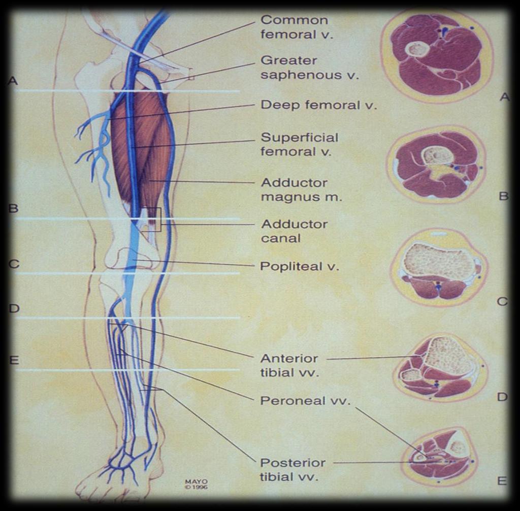
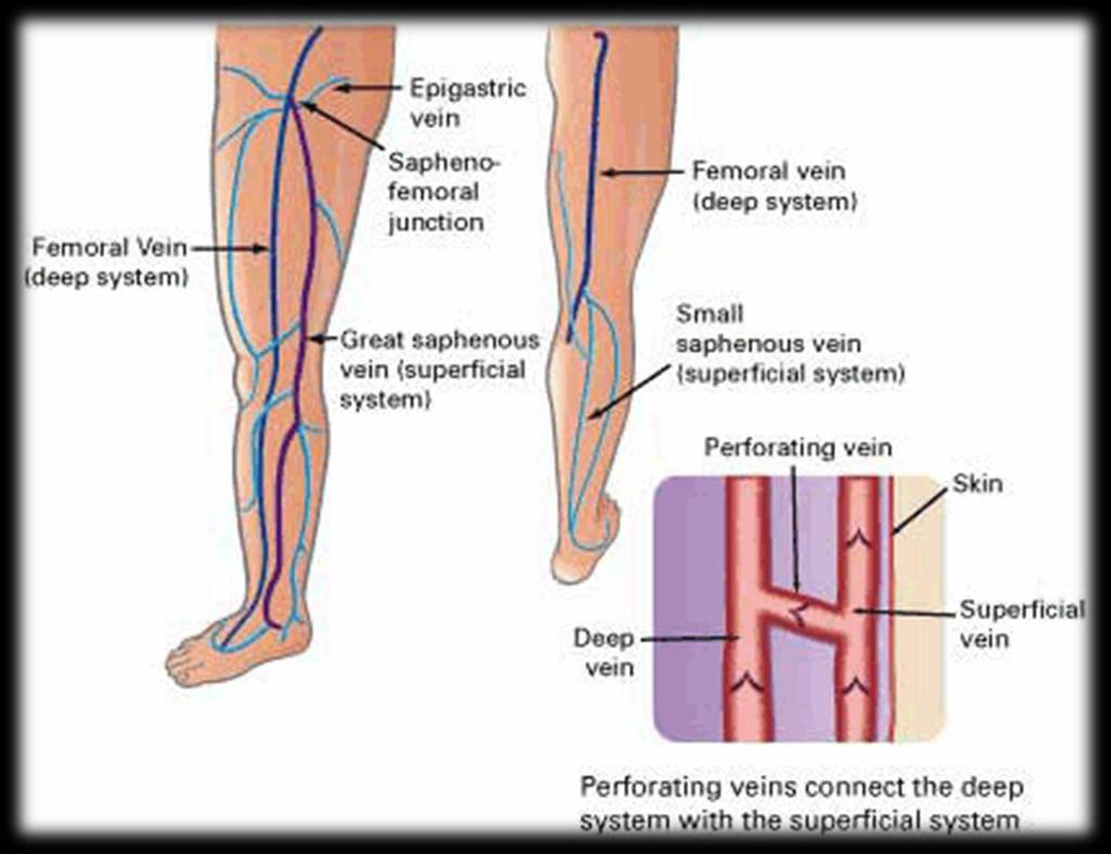
3 Peripheral Vessels Lower Limb Venous Deep System Common Iliac External/Internal Iliac Common Femoral Femoral Profunda Femoris Popliteal Anterior Tibial Posterior Tibial Peroneal Superficial System Long (Great) Saphenous Short (Small) Saphenous Perforators Giacomini Gastrocnemius Soleal Pudendal
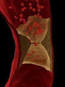
4 What is deep vein thrombosis? Formation of blood clot within a deep vein Form of thrombophlebitis Commonly affects the leg or pelvis and occasionally the arm Painful, swollen, red, warm DVT is a medical emergency
5 Mechanisms Virchow s Triad Decreased blood flow Damage to vessel wall Hypercoagulability Physical trauma Compression of vessel Cancer Infections Stroke, heart failure, nephrotic syndrome
 Factor V Lieden; Antithrombin; etc.")
6 Risk Factors Oestrogen containing oral contraception Recent long-haul travel Intravenous drug use History of miscarriage Smoker Obesity Family history (hereditary thrombophilia) Factor V Lieden; Antithrombin; etc.
7 The Wells Score Clinical examination system Ranges from -2 to +9 Active cancer treatment within 6/12 or palliative (+1) Calf swelling >3cm (+1) Collateral superficial veins (+1) Pitting oedema (+1) Swelling to entire leg (+1) Localised pain along deep vein distribution (+1) Paralysis, paresis or recent cast (+1) Recently bedridden (+1) Previous DVT (+1) Alternative diagnosis at least as likely (-2)
8 Diagnosis Physical examination Homan s sign Dorsiflexion of foot elicits pain in posterior calf Pratt s sign Squeezing of posterior calf elicits pain Wells Score Score ranges from -2 to +9 Score 2 DVT likely Score 1 DVT unlikely, consider D-dimer Blood Tests D-dimer fibrin degredation product False +ve: liver disease; rheumatoid factor; inflammation; malignancy; fracture; pregnancy; recent surgery; advanced age CBC; Coagulation studies; LFTs; U&Es


9 Diagnosis continued Imaging Impedance Plethysmography Venography Radionuclide Imaging MR Imaging Ultrasound B-mode power / colour Doppler Spectral Doppler

10 Impedance Plethysmography Advantages Cheap Portable Single Operator Non-invasive Disadvantages Non-specific

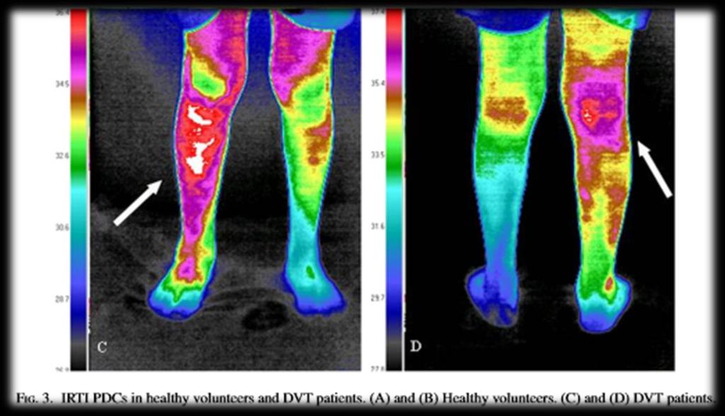
11 Liquid Crystal Thermography


12 Venography Advantages Gold Standard Accurate Disadvantages Invasive Contrast Reactions Ionising Radiation Cost

13 Radionuclide Imaging Blood pool scintigraphy Fibrinogen scanning Tc 99m pertechnetate injection T 99m HMPAO labelled platelts 111 In antifibrin antibodies Sensitivity 61% Specificity 88%


14 Computed Tomography IVC Pelvic vessels CFV FV LSV


15 MR Imaging Advantages Very Accurate Disadvantages Availability Cost


16 Ultrasound Advantages Non-invasive Accurate Relatively quick Other pathology Disadvantages Highly skilled operator required Fat Legs IVDU anatomy Ex-Fix / Frames
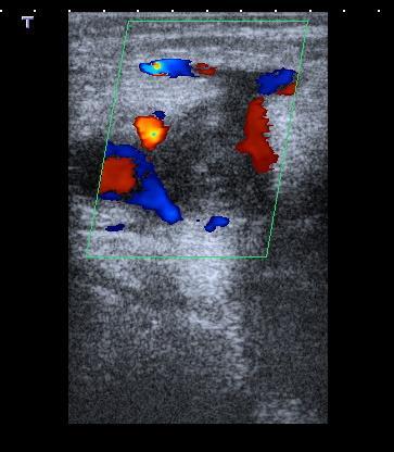
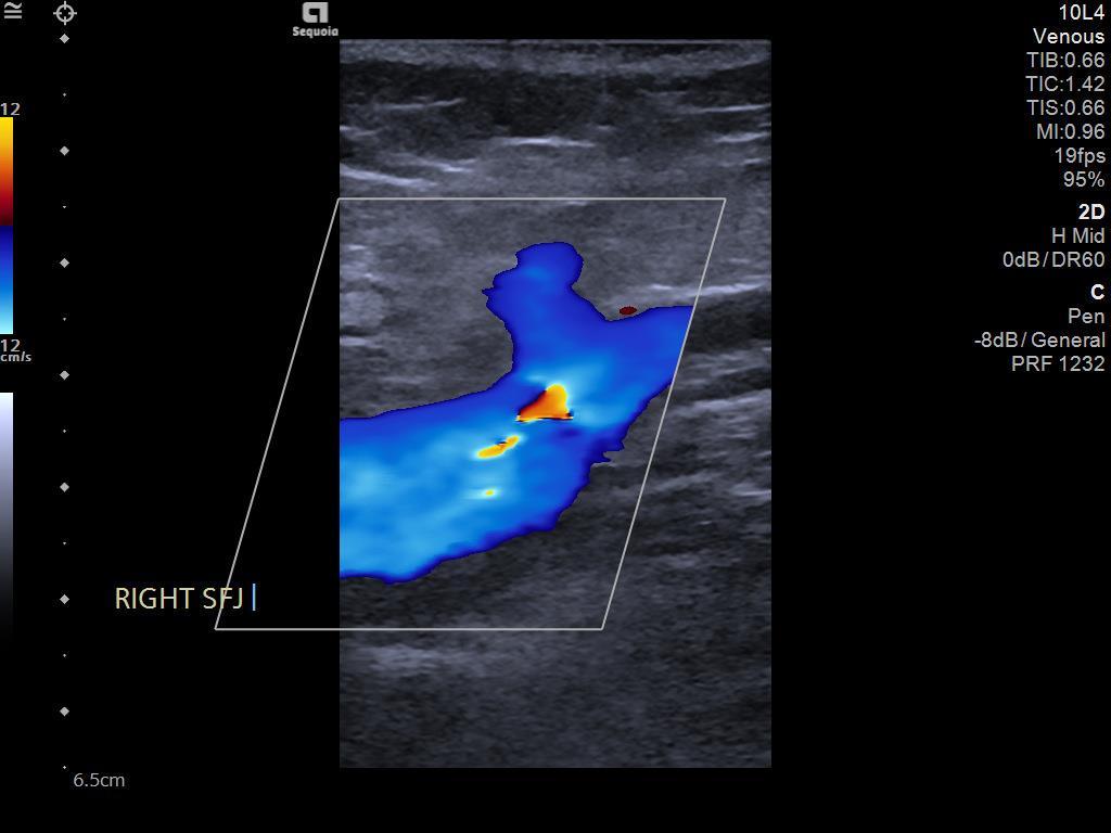
17 Saphenofemoral Junction
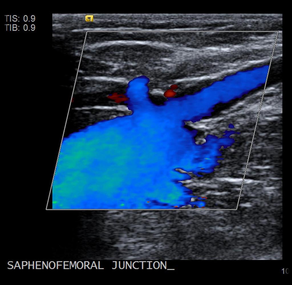
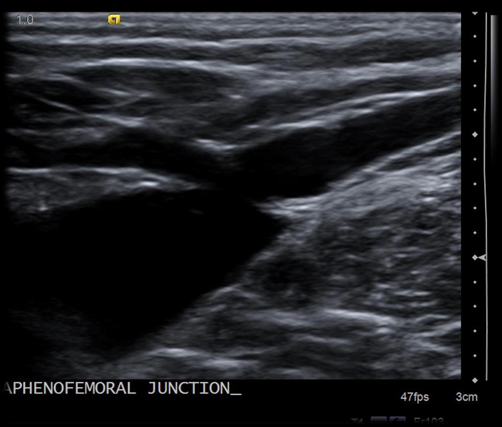
18 Saphenofemoral Junction

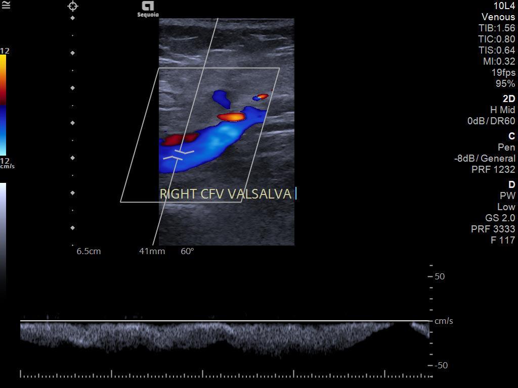
19 Common Femoral Vein

20 Femoral Vein

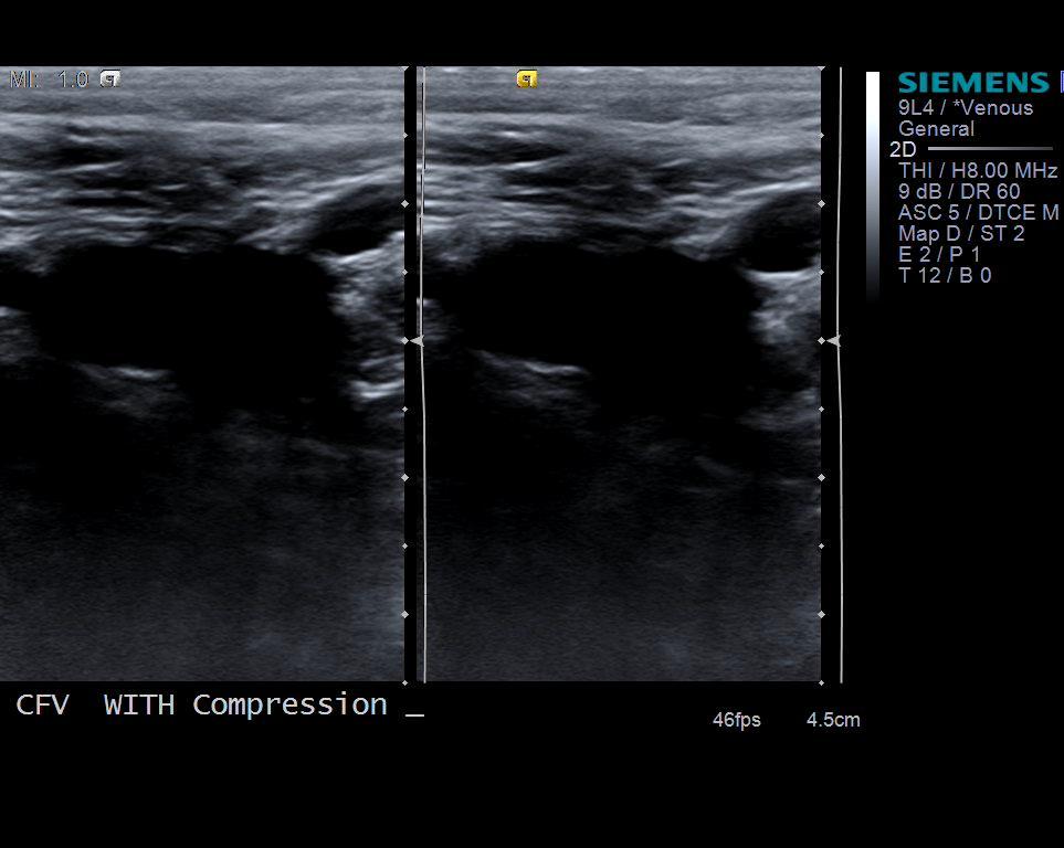
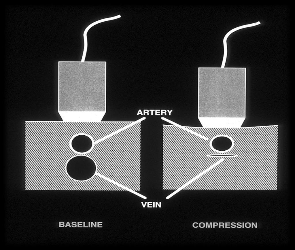
21 CFV & Femoral Vein Compressions
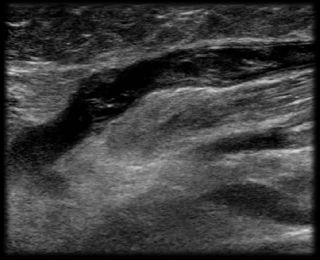
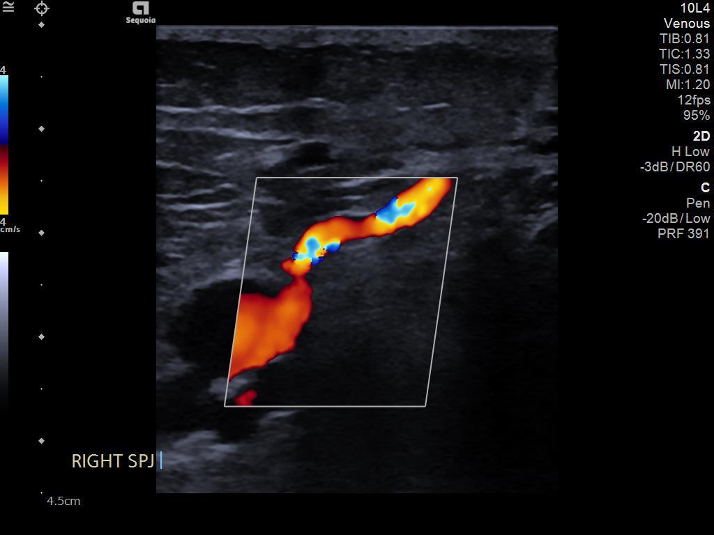
22 Saphenopopliteal Junction

23 Popliteal Vein

24 Popliteal Fossa
25 Characterisation of Thrombus Occlusive vs Partially-occlusive (mural) Acute echo-poor no flow Organisation dilated vessel with some material inside Recanalising Old clot starting to break down Chronic Occluded vein, normal size with internal echoes, collaterals Acute-on-chronic Report as cannot exclude acute-on-chronic and treat as +ve
26 Differential Diagnosis Superficial thrombophlebitis Haematoma Cellulitis Subcutaneous oedema Popliteal (Baker s) cyst Abscess Muscle tear
27 Make sure not to forget! When there is thrombus within the CFV You MUST assess the iliac vessels up to the IVC This is required for treatment planning, i.e. the need for IVC filters

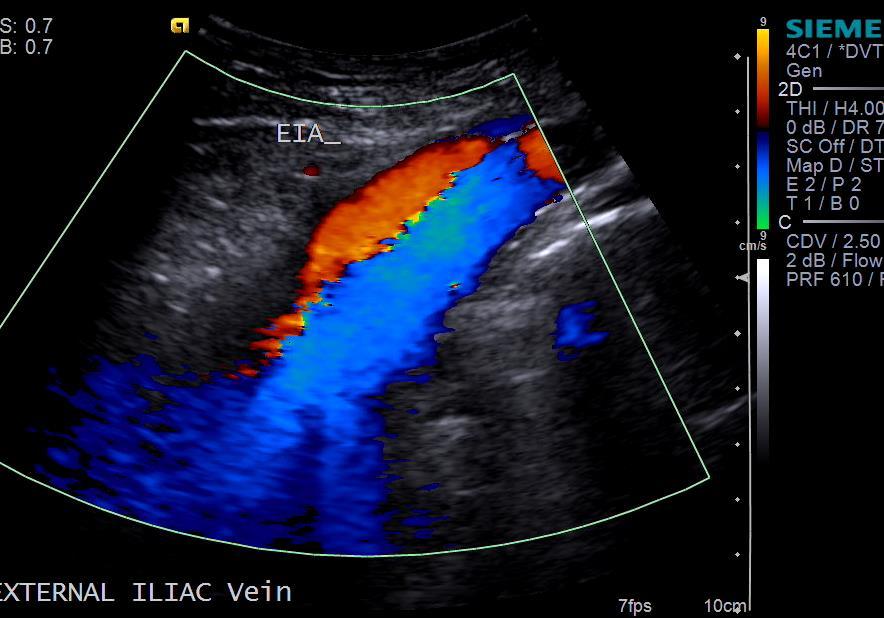
28 Iliac Veins

29 Calf Muscle Pump
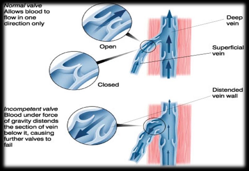
30 Venous Insufficiency
31 Venous Insufficiency Weakness in collagen fibres causes weakness in the valve causing a pressure force that cause downward flow problems Risk factors Past thrombotic syndrome Obesity Arthritis Immobility TREATMENT OPTIONS Compression Injection Surgery Laser ablation (EVLT)
 20% of people aged 20 80% of people aged 60 Most common vascular surgery 30% of presentations are recurrent 66% due to inadequate")
32 Varicose Veins Primary hereditary Secondary post DVT Recurrent post surgery or sclerotherapy Types Trunk (thick knobbly veins) Reticular (network of small red veins) 20% of people aged 20 80% of people aged 60 Most common vascular surgery 30% of presentations are recurrent 66% due to inadequate surgery
33 Examination Protocol Deep veins Patency, compressibility and reflux Saphenofemoral junction & LSV Reflux and patency Thigh perforators Competency (superficial to deep is normal) Saphenopopliteal junction & SSV Competency Calf


34 Grading of Venous Reflux Normal Reflux duration less than 0.5 s Moderate Reflux duration of s Severe Reflux duration of greater than 1s Erect position Test for valve incompetence Cough Valsalva Muscle squeeze / augmentation Most effective

35 Venous Ulcer Improper functioning venous valves Chronic wounds 48% recurrence
36 Everything Else. Non-Vascular Findings in lower limb US
37 Traumatic injuries Contusion common Range from simple haemorrhagic infiltration of fat lobules to fat necrosis, haematomas and abscesses Bloodly fat infiltrate Increased echogenicity of fatty lobules, makes separation from hyperechoic skin and connective tissue strands of subcut tissues undefined, absence of anaechoic septa distinguishes from simple oedema


38 Oedema Early hyperechoic fat lobules Progressive connective septa enlarge and anechoic distension of lymphatic channels, outline hyperechoic fat lobules, graded pressure does not collapse channels.

39 Abscess Variable appearance Superficial/deep Simple anechoic mixed echogenic Well defined ill defined Thin thick walled +/- surrounding hyperaemia +/- displacing internal echogenic material +/- gas


40 Synovial cyst Lined by synovial tissue Communicate with joint space Baker s cyst Posteromedial knee OA/inflamm arth/meniscal tears/chronic effusions Synovitis/debris/bodies Complications AC joint cyst Geyser sign

41 Haematoma Variable appearance Age Acute Fluid fluid levels Plasma, red cell, fat, thrombus, Heterogeneous Chronic Liquefaction Septations Expanding mimic mass lesion


42 Beware the spontaneous haematoma especially in patients without anti-coagulation & insignificant trauma Repeat imaging after 6-8 weeks
43 To Sum Up Ultrasound has many uses Lower Limb DVT Venous Insufficiency Trauma Soft tissue Lumps and Bumps
44 Any Questions?
DISORDERS OF VENOUS SYSTEM
 DISORDERS OF VENOUS SYSTEM Varicose Veins Any dilated, elongated and tortuous vein irrespective of size Varicose veins are common in the superficial veins of the leg which are subject to high pressure
Anatomy. Patterns of reflux. Technique. Testing Reflux time Patient position. Difficult! Learning. NOT system optimisation. Clinical Assesment
 Anatomy Patterns of reflux Awareness Technique Testing Reflux time Patient position Difficult! Learning NOT system optimisation Enlarged Clinical Assesment Twisted Where are the symptoms? Why they are
Proper Diagnosis of Venous Thromboembolism (VTE)
") Proper Diagnosis of Venous Thromboembolism (VTE) Whal Lee, M.D. Seoul National University Hospital Department of Radiology 2 nd EFORT Asia Symposium, 3 rd November 2010, Taipei DVT - Risk Factors Previous
Proper Diagnosis of Venous Thromboembolism (VTE) Whal Lee, M.D. Seoul National University Hospital Department of Radiology 2 nd EFORT Asia Symposium, 3 rd November 2010, Taipei DVT - Risk Factors Previous
Venous thrombosis is common and often occurs spontaneously, but it also frequently accompanies medical and surgical conditions, both in the community
 Venous Thrombosis Venous Thrombosis It occurs mainly in the deep veins of the leg (deep vein thrombosis, DVT), from which parts of the clot frequently embolize to the lungs (pulmonary embolism, PE). Fewer
Guidelines, Policies and Statements D20 Statement on Peripheral Venous Ultrasound
 Guidelines, Policies and Statements D20 Statement on Peripheral Venous Ultrasound Disclaimer and Copyright The ASUM Standards of Practice Board have made every effort to ensure that this Guideline/Policy/Statement
Protocols for the evaluation of lower extremity venous reflux: supine, sitting, or standing?
 Protocols for the evaluation of lower extremity venous reflux: supine, sitting, or standing? Susan Whitelaw RVT, RDMS PURPOSE Duplex imaging of the lower extremity veins is performed to assess the deep
Chronic Venous Insufficiency
 Chronic Venous Insufficiency None Disclosures Lesley Enfinger, MSN,NP-C Chronic Venous Insufficiency Over 24 Million Americans affected by Chronic Venous Insufficiency (CVI) 10 x More Americans suffer
BEDSIDE ULTRASOUND BEDSIDE ULTRASOUND. Deep Vein Thrombosis. Probe used
 BEDSIDE ULTRASOUND Part 2 Diagnosis of deep vein thrombosis Kishore Kumar Pichamuthu, Professor, Department of Critical Care, CMC, Vellore Summary: Deep vein thrombosis (DVT) is a problem encountered in
Deep Vein Thrombosis and Pulmonary Embolism: Patient Information
 Deep Vein Thrombosis and Pulmonary Embolism: Patient Information A Deep Vein Thrombosis (DVT) and a Pulmonary Embolism (PE) are both disorders of unwanted blood clotting. Unwanted blood clots can occur
High Level Overview: Venous Anatomy of Lower Extremities. Anatomy of a Vein 5/11/2015. Barbara Deusterman, RN
 High Level Overview: Venous Anatomy of Lower Extremities Barbara Deusterman, RN What does this anatomy lecture have to do with visually guided sclerotherapy (VGS)? May 11, 2015 2 Anatomy of a Vein Almeida,
DOPPLER ULTRASOUND OF DEEP VENOUS THROMBOSIS
 TOKUDA HOSPITAL SOFIA DOPPLER ULTRASOUND OF DEEP VENOUS THROMBOSIS MILENA STANEVA, MD, PhD Department of vascular surgery and angiology Venous thromboembolic disease continues to cause significant morbidity
NCVH. Ultrasongraphy: State of the Art Vein Forum 2015 A Multidisciplinary Approach to Otptimizing Venous Circulation From Wounds to WOW
 Ultrasongraphy: State of the Art 2015 NCVH New Cardiovascular Horizons Vein Forum 2015 A Multidisciplinary Approach to Otptimizing Venous Circulation From Wounds to WOW Anil K. Chagarlamudi, M.D. Cardiovascular
Venous Reflux Duplex Exam
 Venous Reflux Duplex Exam GWENDOLYN CARMEL, RVT PHYSIOLOGIST, DEPARTMENT OF VASCULAR SURGERY NEW JERSEY VETERANS HEALTHCARE CENTER EAST ORANGE, NJ PURPOSE: To identify patterns of incompetence and which
Let s Take a Look Venous Insufficiency Ultrasound Techniques
 Let s Take a Look Venous Insufficiency Ultrasound Techniques Brent Wilkinson RVT, RDMS Steve Schomaker RVT, RDCS, RDMS Let s take a look Differentiate between normal venous flow and venous insufficiency
Suspected Deep Vein Thrombosis (DVT) Pathway for Non Pregnant patients Updated November 2016, with new D-dimer reference range
 Pathway for Non Pregnant patients Updated November 2016, with new D-dimer reference range") Suspected Deep Vein Thrombosis (DVT) Pathway for Non Pregnant patients Updated November 2016, with new D-dimer reference range Suspect a DVT? Complete a Two-level DVT Wells score on ICE system (see page
Suspected Deep Vein Thrombosis (DVT) Pathway for Non Pregnant patients Updated November 2016, with new D-dimer reference range Suspect a DVT? Complete a Two-level DVT Wells score on ICE system (see page
Determine the patients relative risk of thrombosis. Be confident that you have had a meaningful discussion with the patient.
 Patient Assessment :Venous History, Examination and Introduction to Doppler and PPG Dr Louis Loizou The 11 th Annual Scientific Meeting and Workshops of the Australasian College of Phlebology Tuesday 18
Priorities Forum Statement
 Priorities Forum Statement Number 9 Subject Varicose Vein Surgery Date of decision September 2014 Date refreshed March 2017 Date of review September 2018 Relevant OPCS codes: L841-46, L848-49, L851-53,
RECOGNITION AND ENDOVASCULAR TREATMENT OF CHRONIC VENOUS INSUFFICIENCY
 RECOGNITION AND ENDOVASCULAR TREATMENT OF CHRONIC VENOUS INSUFFICIENCY Paul Kramer, MD, FACC, FSCAI Liberty Cardiovascular Specialists Liberty Regional Heart and Vascular Center DISCLOSURES NONE Venous
Bedside Ultrasound for DVT. Linear Probe. Leg Veins
 Bedside Ultrasound for DVT J. Christian Fox, MD, RDMS, FAAEM, FAIUM Director of Emergency Ultrasound Fellowship University of California, Irvine Jchrsitianfox@gmail.com Linear Probe High frequency transducer
Starting with deep venous treatment
 Starting with deep venous treatment Carsten Arnoldussen, MD Interventional Radiologist Maastricht University Medical Centre, Maastricht VieCuri Medical Centre, Venlo The Netherlands Background Maastricht
Original Research Article Role of Colour Flow Duplex Sonography in Evaluation of Chronic Venous Insufficiency in Lower Limbs
 Original Research Article in Evaluation of Chronic Venous Insufficiency in Lower Limbs Mohammed Abdul Azhar 1 1 Assistant Professor, Department of Radiology, Shadan Institute of Medical Sciences, Hyderabad,
DVT and Pulmonary Embolus. Dr Piers Blombery BSc(Biomed), MBBS (Hons), FRACP, FRCPA Consultant Haematologist Peter MacCallum Cancer Centre
, MBBS (Hons), FRACP, FRCPA Consultant Haematologist Peter MacCallum Cancer Centre") DVT and Pulmonary Embolus Dr Piers Blombery BSc(Biomed), MBBS (Hons), FRACP, FRCPA Consultant Haematologist Peter MacCallum Cancer Centre Overview Structure of deep and superficial venous system of upper
DVT and Pulmonary Embolus Dr Piers Blombery BSc(Biomed), MBBS (Hons), FRACP, FRCPA Consultant Haematologist Peter MacCallum Cancer Centre Overview Structure of deep and superficial venous system of upper
Step by step ultrasound examination of varicose veins. Dr. Özgün Sensebat Vascular Surgeon Private Vascular Clinic Dorsten & Borken, Germany
 Step by step ultrasound examination of varicose Dr. Özgün Sensebat Vascular Surgeon Private Vascular Clinic Dorsten & Borken, Germany Required technical setup: B-mode vessel imaging combined with color
Clinical/Duplex Evaluation of Varicose Veins: Who to Treat?
 Clinical/Duplex Evaluation of Varicose Veins: Who to Treat? Sanjoy Kundu MD, FASA, FCIRSE, FSIR The Vein Institute of Toronto Scarborough Vascular Group Scarborough Vascular Ultrasound Scarborough Vascular
Segmental GSV reflux
 Segmental GSV reflux History of presentation A 43 year old female presented with right lower extremity varicose veins and swelling. She had symptoms of aching, heaviness and tiredness in the right leg.
LOWER EXTREMITY VENOUS COMPRESSION ULTRASOUND. CPT Stacey Good, DO Emergency Medicine Ultrasound Fellow Madigan Army Medical Center
 LOWER EXTREMITY VENOUS COMPRESSION ULTRASOUND CPT Stacey Good, DO Emergency Medicine Ultrasound Fellow Madigan Army Medical Center Learning Objectives Setup and patient positioning for optimizing success
POINT OF CARE ULTRASOUND - Venous US for DVT
 POINT OF CARE ULTRASOUND - Venous US for DVT The diagnosis of deep venous thrombosis (DVT) using ultrasound in the emergency department. DVT US is easy to perform and can be usually be completed in less
Dr. Riaz JanMohamed Consultant Haematologist The Hillingdon Hospital Foundation Trust
 MANAGEMENT OF PATIENTS WITH DEEP VEIN THROMBOSIS (DVT) IN THE COMMUNITY SETTING & ANTICOAGULATION CLINICS THE PAST, PRESENT AND THE FUTURE Dr. Riaz JanMohamed Consultant Haematologist The Hillingdon Hospital
Venous drainage of the lower limb
 Venous drainage of the lower limb INTRODUCTION It is of immense clinical and surgical importance. The venous blood against gravity. FACTORS HELPING THE VENOUS DRAINAGE OF THE LOWER LIMB The contraction
Varicose Vein Information Sheet
 Neil Goldstein, MD Joseph Hewett, MD Board- Certified Physicians in Interventional, Diagnostic, and Vascular Radiology, Surgery, Vascular Surgery and Phlebology Varicose Vein Information Sheet PREVALENCE
CHAPTER 2 VENOUS THROMBOEMBOLISM
 CHAPTER 2 VENOUS THROMBOEMBOLISM Objectives Venous Thromboembolism (VTE) Prevalence Patho-physiology Risk Factors Diagnosis Pulmonary Embolism (PE) Management of DVT/PE Prevention VTE Patho-physiology
Approach to Thrombosis
 Approach to Thrombosis Theera Ruchutrakool, M.D. Division of Hematology Department of Medicine Siriraj Hospital Faculty of Medicine Mahidol University Approach to Thrombosis Thrombosis: thrombus formation
Acute Versus Chronic DVT Imaging in the Vascular Lab Heather Gornik, MD, RVT, RPVI
 Acute Versus Chronic DVT Imaging in the Vascular Lab Heather Gornik, MD, RVT, RPVI Cleveland Clinic Heart and Vascular Institute Heather L. Gornik, MD has the following relationships to disclose: CVR Global
PE and DVT. Dr Anzo William Adiga WatsApp or Call Medical Officer/RHEMA MEDICAL GROUP
 PE and DVT Dr Anzo William Adiga WatsApp or Call +256777363201 Medical Officer/RHEMA MEDICAL GROUP OBJECTIVES DEFINE DVT AND P.E PATHOPHYSIOLOGY OF DVT CLINICAL PRESENTATION OF DVT/PE INVESTIGATE DVT MANAGEMENT
NOTE: Deep Vein Thrombosis (DVT) Risk Factors
 Risk Factors") Deep Vein Thrombosis (DVT) Deep Vein Thrombosis (DVT) is the formation of a blood clot, known as a thrombus, in the deep leg vein. It is a very serious condition that can cause permanent damage to the
Deep Vein Thrombosis (DVT) Deep Vein Thrombosis (DVT) is the formation of a blood clot, known as a thrombus, in the deep leg vein. It is a very serious condition that can cause permanent damage to the
Deep vein thrombosis (DVT) and pulmonary embolism (PE) advice for ophthalmic surgery patients
 and pulmonary embolism (PE) advice for ophthalmic surgery patients") Deep vein thrombosis (DVT) and pulmonary embolism (PE) advice for ophthalmic surgery patients What is a deep vein thrombosis (DVT)? A DVT is a blood clot that forms within a vein deep in the leg but can
Deep vein thrombosis (DVT) and pulmonary embolism (PE) advice for ophthalmic surgery patients What is a deep vein thrombosis (DVT)? A DVT is a blood clot that forms within a vein deep in the leg but can
Preventing Blood Clots in Adult Patients
 Manchester Royal Eye Hospital Surgical Services Information for Patients Preventing Blood Clots in Adult Patients This leaflet will give you information on how to reduce the risk of developing blood clots
VENOUS DRAINAGE OF THE LOWER LIMB
 Anatomy of the lower limb Superficial veins & nerve injuries Dr. Hayder VENOUS DRAINAGE OF THE LOWER LIMB The venous drainage of the lower limb is of huge clinical & surgical importance. Since the venous
MedStar Health, Inc. POLICY AND PROCEDURE MANUAL Policy Number: MP.066.MH Last Review Date: 11/08/2018 Effective Date: 01/01/2019
 MedStar Health, Inc. POLICY AND PROCEDURE MANUAL This policy applies to the following lines of business: MedStar Employee (Select) MedStar CareFirst PPO MedStar Health considers the treatment of Varicose
: A guide to Doppler US evaluation of chronic lower limb venous insufficiency
 : A guide to Doppler US evaluation of chronic lower limb venous insufficiency Poster No.: C-1781 Congress: ECR 2011 Type: Educational Exhibit Authors: T. M. O. Couto, H. Patricio, Â. Moreira, A. Estevao
Peripheral Vascular Examination. Dr. Gary Mumaugh Western Physical Assessment
 Peripheral Vascular Examination Dr. Gary Mumaugh Western Physical Assessment Competencies 1. Inspection of upper extremity for: size symmetry swelling venous pattern color Texture nail beds Competencies
Introduction. Background Evidence System of examination Diagnoses & Variants Final actions Limitation of the examination
 Rule in DVT Introduction Background Evidence System of examination Diagnoses & Variants Final actions Limitation of the examination BACKGROUND Common presentation Influence initial management NICE Guidelines
Deep Vein Thrombosis
 Deep Vein Thrombosis from NHS (UK) guidelines Introduction Deep vein thrombosis (DVT) is a blood clot in one of the deep veins in the body. Blood clots that develop in a vein are also known as venous thrombosis.
Carotid Doppler: Doppler wave forms obtained from the common, external and internal carotid arteries. As well as the vertebral and subclavian
 Competency Carotid Doppler: Doppler wave forms obtained from the common, external and internal carotid arteries. As well as the vertebral and subclavian arteries. Preferred angle is 60 degrees or less.
Lower Extremity Venous Insufficiency Evaluation
 VASCULAR TECHNOLOGY PROFESSIONAL PERFORMANCE GUIDELINES Lower Extremity Venous Insufficiency Evaluation This Protocol was prepared by members of the Society for Vascular Ultrasound (SVU) as a template
TREATMENT OPTIONS FOR CHRONIC VENOUS INSUFFICIENCY
 TREATMENT OPTIONS FOR CHRONIC VENOUS INSUFFICIENCY TL LUK Consultant Vascular Surgeon Sarawak General Hospital HKL Vascular Conference 19/06/2013 PREVALENCE OF LOWER LIMB VENOUS DISEASE Affects half of
Patient assessment and strategy making for endovenous treatment
 Patient assessment and strategy making for endovenous treatment Raghu Kolluri, MD Director Vascular Medicine OhioHealth Riverside Methodist Hospital Columbus, OH Disclosures Current Medtronic Consultant/
THROMBOPROPHYLAXIS IN ELECTIVE FOOT AND ANKLE SURGERY
 THROMBOPROPHYLAXIS IN ELECTIVE FOOT AND ANKLE SURGERY Andy Molloy University Hospital Aintree BMI Sefton Hospital 1 Pathology /signs / investigation (brief!) What is the evidence? What do people do? What
What is the real place of venous echo Doppler in aircrew member flying rehabilitation after a thromboembolism event?
 89 th ASMA ANNUAL SCIENTIFIC MEETING DALLAS- May 6-10, 2018 What is the real place of venous echo Doppler in aircrew member flying rehabilitation after a thromboembolism event? S BISCONTE (1), V MARICOURT
Chronic Venous Insufficiency Compression and Beyond
 Disclosure of Conflict of Interest Chronic Venous Insufficiency Compression and Beyond Shawn Amyot, MD, CCFP Fellow of the Canadian Society of Phlebology Ottawa Vein Centre I do not have relevant financial
Disclosures. DVT: Diagnosis and Treatment. Questions To Ask. Dr. Susanna Shin - DVT: Diagnosis and Treatment. Acute Venous Thromboembolism (VTE) None
 None") Disclosures DVT: Diagnosis and Treatment None Susanna Shin, MD, FACS Assistant Professor University of Washington Acute Venous Thromboembolism (VTE) Deep Venous Thrombosis (DVT) Pulmonary Embolism (PE)
Disclosures DVT: Diagnosis and Treatment None Susanna Shin, MD, FACS Assistant Professor University of Washington Acute Venous Thromboembolism (VTE) Deep Venous Thrombosis (DVT) Pulmonary Embolism (PE)
Endothermal Ablation for Venous Insufficiency. Dr. S. Kundu Medical Director The Vein Institute of Toronto
 Endothermal Ablation for Venous Insufficiency Dr. S. Kundu Medical Director The Vein Institute of Toronto Objective: remove the GSV from the circulation 1. Surgical - HL & stripping 2. Chemical sclerotherapy
Bedside Ultrasound for Detection of Deep Vein Thrombosis: the Two-Point Compression Method
 Bedside Ultrasound for Detection of Deep Vein Thrombosis: the Two-Point Compression Method Tom Ashar MD RDMS a, Krishnaraj Jayarama DO, Raymond Yun MD Department of Emergency Medicine, Newark Beth Israel
CURRENT & FUTURE THERAPEUTIC MANAGEMENT OF VENOUS THROMBOEMBOLISM. Gordon Lowe Professor of Vascular Medicine University of Glasgow
 CURRENT & FUTURE THERAPEUTIC MANAGEMENT OF VENOUS THROMBOEMBOLISM Gordon Lowe Professor of Vascular Medicine University of Glasgow VENOUS THROMBOEMBOLISM Common cause of death and disability 50% hospital-acquired
2017 Florida Vascular Society
 Current Management of Venous Leg Ulcers: How to Identify Patients with Correctable Venous Disease and Interventional Procedures to Heal and Prevent Recurrence 2017 Florida Vascular Society Bill Marston
Vein & Body Specialists at The Bellevue Hospital Spider Vein and Varicose Vein Treatments
 1 Vein & Body Specialists at The Bellevue Hospital Spider Vein and Varicose Vein Treatments What are spider veins? Spider veins are dilated, small blood vessels that have a red or bluish color. They appear
Deep Vein Thrombosis
 Deep Vein Thrombosis Introduction Deep vein thrombosis (DVT) is a blood clot in a vein. This condition can affect men and women of any age and race. DVT is a potentially serious condition. If not treated,
Lecture 09. Popliteal Fossa. BY Dr Farooq Khan Aurakzai
 Lecture 09 Popliteal Fossa BY Dr Farooq Khan Aurakzai Dated: 14.02.2018 What is popliteus? Introduction Anything relating to, or near the part of the leg behind the knee. From New Latin popliteus the muscle
DEEP VENOUS THROMBOSIS DIFERENTIAL DIAGNOSIS
 Department of Radiology of the Hospital de Clinicas DEEP VENOUS THROMBOSISDIFERENTIAL DIAGNOSIS 1 DEEP VENOUS THROMBOSIS DIFERENTIAL DIAGNOSIS Dra. Liliana Servente (1), Dr. Martin Rodriguez Parodi, Dr.
Inferior Vena Cava Filter for DVT
 Inferior Vena Cava Filter for DVT Deep Vein Thrombosis A deep vein thrombosis (DVT) is a blood clot that forms in a deep vein. This is a serious condition that occurs more often than you might think. If
Certificate in Clinician Performed Ultrasound (CCPU) Syllabus
 Syllabus") Certificate in Clinician Performed Ultrasound (CCPU) Syllabus Proximal Deep Vein Thrombosis (DVT) Page 1 of 6 03/17 Deep Vein Thrombosis (DVT) Syllabus Purpose: This unit is designed to cover the theoretical
Certificate in Clinician Performed Ultrasound (CCPU) Syllabus Proximal Deep Vein Thrombosis (DVT) Page 1 of 6 03/17 Deep Vein Thrombosis (DVT) Syllabus Purpose: This unit is designed to cover the theoretical
Reducing the risk of venous thrombo-embolism (VTE) in hospital and after discharge
 in hospital and after discharge") Reducing the risk of venous thrombo-embolism (VTE) in hospital and after discharge What is a venous thromboembolism (VTE)? This is a medical term that describes a blood clot that develops in a deep vein
Reducing the risk of venous thrombo-embolism (VTE) in hospital and after discharge What is a venous thromboembolism (VTE)? This is a medical term that describes a blood clot that develops in a deep vein
DVT - initial management NSCCG
 Background information Information resources for patients and carers Updates to this care map Synonyms Below knee DVT and bleeding risks Patient with confirmed DVT Scan confirms superficial thrombophlebitis
The Use of Adjunctive Venography and Endovascular Manoeuvres In The Treatment of Saphenous Vein Insufficiency. A Prospective, Multi-centre Study
 The Use of Adjunctive Venography and Endovascular Manoeuvres In The Treatment of Saphenous Vein Insufficiency A Prospective, Multi-centre Study Ramon L. Varcoe, MBBS, MS, FRACS, PhD Associate Professor
A Brief Guide Treatment and Prevention
 A Brief Guide Treatment and Prevention Deep Vein Thrombosis and Pulmonary Embolism 08/18 Dear reader, This brochure provides you with information about deep vein thrombosis and pulmonary embolism. This
Linda Antonucci, RPhS, RVT, RDCS
 Linda Antonucci, RPhS, RVT, RDCS DISCLOSURE Linda Antonucci, RPhS, RVT, RDCS I have no financial relationships to disclose relevant to this talk. SIMILARITIESBETWEEN ARTERIES AND VEINS Composed of three
Patients with suspected DVT of the lower limb how to exam the patient
 Patients with suspected DVT of the lower limb how to exam the patient Johannes Godt Dep. of Radiology and Nuclear Medicine Oslo University Hospital Ullevål NORDTER 2015, Oslo Content Anatomy and pathophysiology
The role of ultrasound duplex in endovenous procedures
 The role of ultrasound duplex in endovenous procedures Neophytos A. Zambas MD, PhD Vascular Surgeon Polyclinic Ygia, Limassol, Cyprus ΚΕΑΕΧ ΚΥΠΡΙΑΚΗ ΕΤΑΙΡΕΙΑ ΑΓΓΕΙΑΚΗΣ ΚΑΙ ΕΝΔΑΓΓΕΙΑΚΗΣ ΧΕΙΡΟΥΡΓΙΚΗΣ Pre
Bedside Emergency Ultrasound For Deep Venous Thrombosis
 Bedside Emergency Ultrasound For Deep Venous Thrombosis Michael Blaivas, MD, MBA(candidate) FACEP, FAIUM Professor of Medicine University of South Carolina School of Medicine AIUM Third Vice President
Jordan M. Garrison, MD FACS, FASMBS
 Jordan M. Garrison, MD FACS, FASMBS Peripheral Arterial Disease (PAD) Near or Complete obstruction of > 1 Peripheral Artery Peripheral Venous reflux Disease Varicose Veins Chronic Venous Stasis Ulcer Disease
Additional Information S-55
 Additional Information S-55 Network providers are encouraged, but not required to participate in the on-line American Venous Forum Registry (AVR) - The First National Registry for the Treatment of Varicose
Learning Objectives for Rotations in Vascular Surgery Year 3 Basic Clerkship
 Learning Objectives for Rotations in Vascular Surgery Year 3 Basic Clerkship CLINICAL PROBLEMS IN VASCULAR SURGERY 1. ABDOMINAL AORTIC ANEURYSM A 70 year old man presents in the emergency department with
DEEP VENOUS THROMBOSIS A PRACTICAL APPROACH TO IMPROVING CLINICAL OUTCOMES
 DEEP VENOUS THROMBOSIS A PRACTICAL APPROACH TO IMPROVING CLINICAL OUTCOMES Jose M. Borromeo M.D. Vascular Surgeon Iowa Heart Center Disclosures: AstraZeneca Pharmaceuticals Cook CVRx LeMaitre Vascular,
Controversies & updates in Vascular Surgery. Paris - february
 Controversies & updates in Vascular Surgery Paris - february 09 2019 Venous session Recurrence at the popliteal fossea Pathogenesis and duplex investigation Philippe LEMASLE Le Chesnay - France I have
Suspected Deep Vein Thrombosis (DVT) Assessment
 Assessment") CHI no... First name... DOB... /... /... Last name... Sex: c M c F Address...... Telephone... or attach addressograph label here Hospital/Location: c Hairmyres c Monklands c Wishaw Other (specify)... Ward/Base...
CHI no... First name... DOB... /... /... Last name... Sex: c M c F Address...... Telephone... or attach addressograph label here Hospital/Location: c Hairmyres c Monklands c Wishaw Other (specify)... Ward/Base...
Chronic Iliocaval Venous Occlusive Disease
 none Chronic Iliocaval Venous Occlusive Disease David Rigberg, M.D. Clinical Professor of Surgery Division of Vascular Surgery University of California Los Angeles Chronic Venous Occlusive Disease Chronic
chronic venous disorders, varicose vein, CEAP classification, lipodermatosclerosis, Klippel- Trenaunay syndrome DVT CVD
 Online publication August 27, 2009 chronic venous disorders: CVD CEAP 4 CEAP CVD J Jpn Coll Angiol, 2009, 49: 201 205 chronic venous disorders, varicose vein, CEAP classification, lipodermatosclerosis,
Incidence of Post-Operative Venous Thromboembolism Using Compression Ultrasonography Following Trauma to Spine and Long Bones of Lower Extremity
 Open Journal of Orthopedics, 2013, 3, 97-101 http://dx.doi.org/10.4236/ojo.2013.32019 Published Online June 2013 (http://www.scirp.org/journal/ojo) 97 Incidence of Post-Operative Venous Thromboembolism
VASCULAR DISEASE: THREE THINGS YOU SHOULD KNOW JAMES A.M. SMITH, D.O. KANSAS VASCULAR MEDICINE, P.A. WICHITA, KANSAS
 VASCULAR DISEASE: THREE THINGS YOU SHOULD KNOW JAMES A.M. SMITH, D.O. KANSAS VASCULAR MEDICINE, P.A. WICHITA, KANSAS KANSAS ASSOCIATION OF OSTEOPATHIC MEDICINE ANNUAL CME CONVENTION APRIL 13, 2018 THREE
PROVIDER POLICIES & PROCEDURES
 PROVIDER POLICIES & PROCEDURES TREATMENT OF VARICOSE VEINS OF THE LOWER EXTREMITIES STAB PHLEBECTOMY AND SCLEROTHERAPY TREATMENT The primary purpose of this document is to assist providers enrolled in
Clinical case. Symptomatic anterior accessory great saphenous vein (AAGSV) reflux
 reflux") Clinical case Symptomatic anterior accessory great saphenous vein (AAGSV) reflux A 70 year-old female presents with symptomatic varicose veins on left leg for more than 10 years. She complains of heaviness,
Clinical case Symptomatic anterior accessory great saphenous vein (AAGSV) reflux A 70 year-old female presents with symptomatic varicose veins on left leg for more than 10 years. She complains of heaviness,
The Peripheral Vascular System
 The Peripheral Vascular System Anatomy and Physiology Arteries Arteries contain 3 concentric layers of tissue: - the intima - the media - the adventitia The intima The endothelium of the intima has metabolic
Varicose Veins. These are abnormal veins in the legs that appear as unsightly or cause other problems.
 Varicose Veins What are varicose veins? These are abnormal veins in the legs that appear as unsightly or cause other problems. They develop due to abnormal valve function that allows the blood to travel
1-Muscles: 2-Blood supply: Branches of the profunda femoris artery. 3-Nerve supply: Sciatic nerve
 1-Muscles: B i c e p s f e m o r i s S e m i t e n d i n o s u s S e m i m e m b r a n o s u s a small part of the adductor magnus (h a m s t r i n g p a r t o r i s c h i a l p a r t ) 2-Blood supply:
Interactive Learning Session
 Chronic Venous Disease - Part I Interactive Learning Session 2011 Ali Sabbour Prof of Vascular Surgery http://mic.shams.edu.eg/moodle6 Login as a guest Surgery 2 Ali Sabbour - Chronic Venous Disease Intended
Recurrent Varicose Veins
 Recurrent Varicose Veins Part I: Evaluation Utilizing Duplex Venous Imaging PAUL KENNETH THIBAULT, MBBS WARREN ANTHONY LEWIS, DMU PHLEBOLOGY There is the need to develop a universally accepted standard
VASCULAR WOUNDS PATHOPHYSIOLOGY AND MANAGEMENT
 VASCULAR WOUNDS PATHOPHYSIOLOGY AND MANAGEMENT Lucy Stopher, A/CNS Vascular Surgery ...it is best to think of a wound not as a disease, but rather as a manifestation of disease. Joe McCulloch In order
Role of free tissue transfer in management of chronic venous ulcer
 Original Article Role of free tissue transfer in management of chronic venous ulcer K. Murali Mohan Reddy, D. Mukunda Reddy Department of Plastic Surgery, Nizams Institute of Medical Sciences, India. Address
Anatomy MCQs Week 13
 Anatomy MCQs Week 13 1. Posterior to the medial malleolus of the ankle: The neurovascular bundle lies between Tibialis Posterior and Flexor Digitorum Longus The tendon of Tibialis Posterior inserts into
Where should you palpate the pulse of different arteries in the lower limb?
 Where should you palpate the pulse of different arteries in the lower limb? The femoral artery In the femoral triangle, its pulse is easily felt just inferior to the inguinal ligament midway between the
Varicose Veins What Are They? Sclerotherapy in the Treatment of Venous Disease Zachary C. Schmittling, MD, FACS May 4, 2018
 Sclerotherapy in the Treatment of Venous Disease Zachary C. Schmittling, MD, FACS May 4, 2018 Sclerotherapy for Venous Disease: Overview 1 in 5 Americans Approximately 25% of women have some type of lower
PUT YOUR BEST FOOT FORWARD
 PUT YOUR BEST FOOT FORWARD Bala Ramanan, MBBS 1 st year vascular surgery fellow Introduction The epidemic of diabetes and ageing of our population ensures critical limb ischemia will continue to grow.
Current Management of Varicose Veins
 Current Management of Varicose Veins Michael J. Heidenreich, MD St. Joseph Mercy Hospital Ann Arbor, MI March 23, 2013 Nothing to disclose History Prevalence Anatomy Risk factors Clinical manifestations
Interventional Treatment VTE: Radiologic Approach
 Interventional Treatment VTE: Radiologic Approach Hae Giu Lee, MD Professor, Dept of Radiology Seoul St. Mary s Hospital The Catholic University of Korea Introduction Incidence High incidence: 250,000-1,000,000/year
Cover Page. The handle holds various files of this Leiden University dissertation.
 Cover Page The handle http://hdl.handle.net/1887/19768 holds various files of this Leiden University dissertation. Author: Langevelde, Kirsten van Title: Are pulmonary embolism and deep-vein thrombosis
validation study Original article Clinical examination of varicose veins - a Jong Kim, Simon Richards, Patrick J Kent
 The Royal College of Surgeons of England : 171175 Original article Clinical examination of varicose veins a validation study Jong Kim, Simon Richards, Patrick J Kent Department of Vascular and Endovascular
As with any intervention, selection of an appropriate
 DVT: ccess Decisions for Interventions ssessing the advantages and disadvantages of venous access options is crucial for safe and successful DVT intervention. Y JOHN. KUFMN, MD, MS, FSIR, FH, FCIRSE, EIR
Complete Evaluation of the Chronic Venous Patient: Recognizing deep venous obstruction. Erin H. Murphy, MD Rane Center
 Complete Evaluation of the Chronic Venous Patient: Recognizing deep venous obstruction Erin H. Murphy, MD Rane Center Disclosure Speaker name: Erin H. Murphy... I have the following potential conflicts
Varicose Veins are a Symptom of Vein Disease. Now you can treat the source of your varicose veins with non-surgical endovenous laser treatment.
 Varicose Veins are a Symptom of Vein Disease. Now you can treat the source of your varicose veins with non-surgical endovenous laser treatment. Approximately 1 in 5 adult Americans suffer from superficial