Orthopaedic Rehabilitation of Common Ankle and Foot Disorders. Cuneyt Mirzanli Istanbul Gelisim University
|
|
|
- Jeffry Wheeler
- 5 years ago
- Views:
Transcription
1 Orthopaedic Rehabilitation of Common Ankle and Foot Disorders Cuneyt Mirzanli Istanbul Gelisim University
2 Foot and Ankle Fractures Common traumatic injuries. Result in significant functional impairment and disability. ANKLE FRACTURES: The highest incidence occurs in elderly females and young adult males. Most common cause is a twisting or rotational injury of ankle.
3 The ankle joint: Consists of the articulations of the distal tibia, distal fibula and the talus. A hinge joint. In addition to the bony anatomy, ligamentous structures contribute to the stability of the ankle.
4 The deltoid ligament complex provides support to the medial aspect of the ankle. The syndesmotic ligament complex provides stability to the distal tibia-fibula articulation, and the fibular collateral ligament complex add lateral stability to the joint.
5 Ankle fractures can generally be divided into: avulsion fractures, isolated malleolus fractures, bimalleolar fractures, trimalleolar fractures, and those with syndesmosis disruption.
6 Rehabilitation: Stability plays a critical role in the rehabilitation protocols. In the case of stable isolated malleolus fractures, the patient can be treated with bracing and may weight bear as tolerated.
7 Physical therapy should initially focus on managing swelling and edema, and returning ankle range of motion (ROM) to preinjury levels. After a period of about 4 to 6 weeks, the patient may progressively pass to the full weight bearing.
8 At this time, physical therapy should focus on strengthening of the foot and ankle musculature, in particular the lateral evertors, and move on to proprioceptive training. Ankle fractures which treated in an operative manner, typically held immobilized in a splint or cast for 10 to 14 days to allow wound healing postoperatively.
9 In a patient with good bone quality and stable anatomic fixation, gentle ankle active and passive ROM exercises within pain tolerance may begin. Early motion is associated with improved early functional outcomes in these fractures.
10 After 6 weeks, patients may progressively begin to work toward restoration of full ROM and strength.

11
12 CALCANEUS FRACTURES Calcaneus fractures can have long-lasting effects on patient comfort and function. Typically involve a high-energy mechanism, such as a fall from height or MVA. The calcaneus bone has a thin cortical shell that surrounds cancellous, well vascularized bone. For this reason, healing and union of calcaneus fractures is not problematic. But malunion is common.
13 These fractures can be trated operatively or nonoperatively. ORIF Cast or braces

14

15

16

17
18 Rehabilitation Calcaneal fractures treated nonoperatively are placed into a supportive well-padded splint to allow resolution of the initial fracture edema. The patient is then converted to a fracture boot or cast within 2 weeks with the ankle in neutral flexion. Early ankle joint gentle AROM may be initiated at this point.
19 Operatively treated calcaneus fractures are placed into a short leg splint or cast in neutral position postoperatively. Immobilization is continued until the wound is well healed, typically 2 3 weeks. The patient is then converted to a fracture boot and early subtalar and ankle ROM may be initiated.
20 Isometric strengthening may begin before weight bearing, but on average these patients are kept NWB for about 8 to 10 weeks. When weight bearing begins, therapy may address restoration of full ROM strengthening, gait training and balance training in progressive fashion.
21 Ankle Sprains Common injuries in active individuals. ANATOMY: The ankle or talocrural joint is a junction of the tibia, fibula and talus. The bony congruity confers stability to ankle joint, especially in static weight bearing in the neutral position.
22 During motion, the anterior talofibular ligament (ATFL), calcaneofibular ligament (CFL) and posterior talofibular ligament (PTFL) provide support to the joint laterally, whereas the deltoid ligament complex (DLC), provides medial support. The syndesmotic complex, provide stability to the tibia-fibula articulation and thereby also support the ankle joint.
23
24
25 The ATFL is the most commonly injured ligament, followed by the CFL. The CFL is usually injured in combination with the ATFL. Sprains to both the ATFL and CFL are a result of a combined inversion and plantarflexion mechanism.

26
27 The most stable position of the ankle is dorsiflexion.
28 CLASSIFICATION OF ANKLE SPRAINS Regardless of whether the lateral or medial ligaments are injured, the severity of an ankle sprain is typically placed into one of three grades based on the amount of ligamentous damage.
29 Grade I ankle sprains result in a stretching of the ligamentous fibers and are considered minor sprains. Grade II ankle sprains result in a partial tearing of the ligamentous fibers and are considered to be moderate sprains. Grade III ankle sprains result in tearing of the ligamentous fibers and are considered severe sprains.
30 THE INJURY AND HEALING PROCESS The body s healing process occurs in a natural sequence of events and can be divided into three stages: the inflammatory or acute stage; the subacute, repair or proliferation stage; and the remodeling or maturation stage.
31 In the acute stage, the cardinal signs and symptoms of inflammation (pain, edema, erythema, warmth, decrease function) are evident. This stage begins immediately after the onset of injury and typically lasts 3 to 5 days.
32 The subacute stage, which begins at about 3 days after injury and can last up to 6 weeks, is marked by a decrease in the signs and symptoms of inflammation and the beginning of tissue repair. During this stage that weak collagen fibers begin to develop a scar at the injured site. Approximately 7 days after injury, there is a significant amount of collagen in the area.
33 During the maturation stage, the collagen tissues become stronger and more organized.
34 TREATMENT AND REHABILITATION PROTOCOL FOR ACUTE ANKLE SPRAIN The steps in treating and rehabilitating ankle sprains typically follow this progression: Step 1: Protect the area from further injury. Step 2: Decrease pain, swelling, and spasm. Step 3: Re-establish range of motion (ROM), flexibility and tissue mobility.
35 Step 4: Re-establish neuromuscular control, muscular strength, endurance and power. Step 5: Re-establish proprioception, coordination Step 6: Re-establish functional skills
36 Acute Stage: Goals and Interventions After Ankle Sprain During the acute phase the primary goals of the rehabilitation program are as follows: Protect the injured tissues from further injury. Encourage tissue healing Limit the pain, swelling and spasm associated with inflammation. Maintain function of the noninjured tissues. Maintain overall body conditioning
37 Goal A: Protect the Injured Tissues From Further Injury: Although the patient should rest the injured tissues to limit additional stress and potential injury, it is important to remember that absolute rest is seldom a wise choice.
38 Patients should be encouraged to participate in pain-free activities that do not stress the injured ligaments. Because most ankle sprains involve the lateral ligaments and are caused by plantarflexion and inversion, the patient should avoid activities that cause extremes of these motions for at least the first several days.
39 Protection with splinting, bracing, taping or wrapping the injured ankle may be necessary, especially in grade II or III sprains. Patients with grade II or III sprains also may need supportive devices such as crutches, a walking cane or a walking boot to move.
40 There has long been debate as to whether or not to immobilize sprained ankles or immediately begin a functional treatment plan. Current practices are in favor of a functional treatment plan, especially when managing grades I and II sprains.
41 A functional treatment plan limits immobilization and encourages pain-free activities. Functional rehabilitation has been shown to be associated with more frequent return to sports and higher rates of patient satisfaction than immobilization.
42 If the patient s ankle is to be immobilized, it should be noted that long periods of immobilization may lead to: prolonged joint stiffness, contractures, weakening of noninjured ligaments, and muscle atrophy.

43

44
45 Goal B: Encourage Tissue Healing: In a healthy patient, the body will go through its normal healing process as long as there is no additional trauma to the tissues. Rest and protection of the injured tissues are important to allow the body to progress through its normal healing processes. Toward the end of the acute phase, pulsed ultrasound can be used to promote tissue healing.
46 Goal C: Limit Pain, Swelling, and Spasm: The combination of rest, ice, compression and elevation (RICE) is one of the more commonly used approaches to treat the acute inflammatory response. Ice and other forms of cryotherapy help prevent swelling, decrease pain and limit spasm.
47 Both elevation and compression with elastic wraps or compression stockinet assist with minimizing swelling. Electrical stimulation can also be used to minimize pain, swelling and spasm.

48

49 Performing a joint mobilization technique to the distal tibiofibular joint often provides pain relief.
50 Goal D: Maintain Function of Noninjured Tissues: Although rest may be needed for the injured ankle ligaments, muscles, tendons and joint capsule, normal function of the noninjured tissues must be maintained with activity.
51 The patient should be encouraged to engage in activities that do not stress the injured ligaments. Because most ankle sprains involve the lateral ligaments and are caused by plantarflexion and inversion, care must be taken to minimize extreme motions in those directions, especially in grade II and III sprains.
52 With injuries that involve the deltoid ligament complex (DLC), care is taken to avoid extreme eversion. General mobility exercises are useful in preventing disuse of the noninjured tissues while minimizing stress to the injured ligaments.
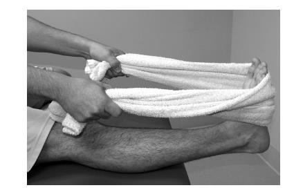
53 General mobility exercises Ankle pumps Plantarflexion and dorsiflexion ROM progressing from passive range of motion (PROM) to active-assisted range of motion (AAROM) to active range of motion (AROM) as tolerated. Heel-cord and posterior calf stretches Towel curls and/or marble pick-ups

54
55

56
57 If the patient is immobilized, placed in a walking boot or prevented from full weight bearing (FWB) ambulation for a period, the metatarsophalangeal (MTP) joints should also be treated with some form of mobilization activities (joint mobilizations, PROM, AAROM, AROM, stretches)

58
59 Goal E: Maintain Overall Body Conditioning: Although the injured ligaments may need to be rested, the rest of the body does not. Patients should be encouraged to engage in pain-free physical activities to maintain their overall body conditioning.
60 Strengthening exercises for the lower extremities, such as open kinetic chain knee flexion and extension and open kinetic chain hip flexion, extension, abduction, and adduction exercises, should also be performed.
61 These exercises also help to prevent disuse issues of the noninjured body areas while minimizing stress on the injured tissues. Patients should also continue their normal strength training exercises for the trunk and upper extremities.
62 Many of these activities put the patient s ankle in a gravity-dependent position and this cause ankle edema. The use of a compression stockinet or elastic wrap while performing these exercises can help prevent the edema of the area.
63 Subacute Stage: Goals and Interventions During the subacute phase the primary goals are as follows: Prevent further injury. Minimize pain and inflammation. Promote tissue healing. Restore ROM and flexibility. Re-establish neuromuscular control and restore muscular strength and endurance.
64 Re-establish proprioception and coordination. Maintain overall body conditioning.
65 Goal F: Prevent Further Injury: Although the initial inflammatory response has ended and the early scar tissue is beginning to develop, it is important to remember that the scar tissue is still very weak and improper activities can easily cause re-injury.
66 In the early days of this phase, extremes of plantarflexion and inversion should still be minimized to prevent damage to the newly formed scar tissue.
67 Goal G: Minimize Pain and Inflammation: As the initial signs and symptoms of acute inflammation diminish, thermotherapy techniques such as warm whirlpools and hot packs should be introduced. Thermotherapy techniques help to reduce pain, spasm and subacute inflammation. Therapeutic ultrasound may also be used at this time, progressing from pulsed to continuous duty cycles.
68 Continuous ultrasound also assists with pain relief, tissue healing and reduction of subacute edema. The continued use of electrical stimulation can assist with minimizing pain and inflammation. It is still wise to continue cryotherapy, especially after activity, to reduce pain and limit inflammation.
69 An increase in pain or inflammation, especially after the acute stage, often is a sign that the injured structures are not ready for the activity being performed.
70 If the patient experiences an increase in pain, inflammation or both, he or she should be re-evaluated to ensure there is no worsening of the injury and the rehabilitation protocol should be slowed until the pain and inflammation are under control.

71 Grades I and II joint mobilizations are also indicated at this time to assist with pain control.
72 Goal H: Promote Tissue Healing: Continuing to protect the injured ligaments from re-injury will allow the body to go through its normal healing process. The continued use of therapeutic modalities such as ultrasound and thermotherapy helps promote tissue healing. The introduction of ROM and strengthening exercises will also promote proper alignment and improved strength of the scar tissue.
73 Therapeutic massage techniques can also be used to promote blood flow and circulation and progressing to more aggressive techniques such as cross-friction massage to promote tissue alignment.
74 Goal I: Restore Range of Motion and Flexibility: The general mobility and ROM exercises that were begun in the acute stage are continued. The patient should be encouraged to perform ROM exercises and stretches several times throughout the day. Initially, dorsiflexion and limited plantarflexion should be emphasized.
75 Pedaling on a stationary bike can help with both plantarflexion and dorsiflexion. Use of a BAPS can be introduced, first in a NWB position before progressing to PWB then FWB position. BAPS (Biomechanical Ankle Platform System)is used to improve balance and proprioception in the ankle, knee and hip after injury or surgery.

76
77 Progress to circling the board while touching all sides of the board in both clockwise and counterclockwise directions is begun. Massage, myofascial release and other manual therapy techniques to treat soft tissue restrictions may also help restore ROM, flexibility and tissue mobility in this stage.
78 Goal J: Re-Establish Neuromuscular Control and Restore Muscular Strength and Endurance: Patients can begin isometric exercises in a neutral ankle position against plantarflexion, dorsiflexion, inversion, and eversion forces. Because strength gains related to isometric exercises only strengthen the muscle at that length, it is important to progress to performing isometrics in a variety of degrees within a ROM, but painful ROM should be avoided.
79 Isometric exercises should begin with submaximal contractions and progress to maximal contractions. Isometric exercises should be progressed to isotonic exercises as tolerated. Resistance can be provided manually, with cuff weights or elastic bands or cords.

80
81 Isotonic exercises should begin with a limited ROM and progress to full ROM as tolerated and should progress from submaximal resistance to maximal efforts. As weight bearing becomes tolerated, heel and toe raises can be incorporated as can walking on the heels or toes.

82
83 As the patient s pain-free ROM increases, proprioceptive neuromuscular facilitation (PNF) techniques can be used.
84 Goal K: Re-Establish Proprioception, Agility, and Coordination: In the early phase of proprioception training, the patient may need to perform unloaded exercises such as joint repositioning.
85 The patient should progress to PWB and FWB exercises as tolerated. Early exercises to encourage loading of the ankle include weight shifts in various directions. With weight shifts, the patient stands with his or her weight shifted to the noninjured leg, then progressively shifts the weight onto the injured leg before returning to the NWB position.
86 The patient should progressively shift more of his or her weight to the injured leg until equal weight is distributed on both legs. This progresses to the patient shifting more weight on the injured leg until he or she can finally bear full weight on the injured leg. The patient should perform these step-ups and step-downs in various directions.

87
88 The patient next progresses to activities with patient-controlled perturbations. The patient stands first in a two-foot stance with the weight evenly distributed, while performing upper extremity or trunk exercises such as pulling on elastic bands in various directions, moving a weighted medicine ball in various directions or bending over to pick up an object.

89
90 The patient then progresses to activities where he or she must react to perturbations provided by the clinician. These types perturbations include pushing or pulling on the patient s body, either by direct contact or with elastic tubing or a stick and playing catch with the patient.

91
92 As the patient s proprioception improves, agility and coordination exercises should be introduced. Walking, walking backward, step-ups, stepdowns. Patients can also perform lateral movement exercises on a slide board.

93
94 Goal L: Maintain Overall Body Conditioning: As the patient better tolerates weight bearing, closed kinetic chain (CKC) lower extremity strength training exercises such as squats, leg presses, and calf raises can be added to the program.

95

96
97 Maturation Stage: Goals and Interventions During the maturation phase the primary goals are as follows: Prevent re-injury. Restore ROM and flexibility. Improve muscular strength, endurance and power. Improve proprioception, agility and coordination. Improve functional (sport-specific) skills. Maintain overall body conditioning
98 RETURN TO ACTIVITY CRITERIA AFTER ACUTE ANKLE SPRAIN The patient should be pain free when performing his or her activity. The ankle should not be swollen. The ankle should have full functional range of motion. The ankle should have full functional muscle strength, endurance and power. The patient should be psychologically ready to return to activity.
99 Inferior Heel Pain (Plantar Fasciitis)
100 Plantar fasciitis is among the most common foot disorders treated by health care providers. In a survey of 500 physical therapists (2001), all 117 who responded listed plantar fasciitis as the most common foot condition seen in their clinic.
101 ANATOMY AND PATHOMECHANICS The plantar fascia is a dense, fibrous connective tissue structure originating from the medial tuberosity of the calcaneus. The plantar fascia is an important static support for the longitudinal arch of the foot.
102 The bony spur at the bottom of the heel does not cause the pain of plantar fasciitis. Rather, this is caused by the inflammation and microtears of the plantar fascia. Heel spurs have been found in approximately 50% of patients with plantar fasciitis. Spur formation is also related to progression of age.
103 Bone spurs may be associated with plantar fasciitis but are not believed to be the cause of it. Many studies show no clear association between spurs and plantar fasciitis.

104

105

106
107 ETIOLOGY Plantar fasciitis is more common in sports that involve running and long-distance walking and is also frequent in dancers, tennis players, basketball players and nonathletes whose occupations require prolonged weight bearing.
108 Direct repetitive microtrauma with heel strike to the ligamentous and nerve structures has been implicated, especially in middle-aged, overweight, nonathletic individuals who stand on hard surfaces and in long-distance runners.
109 The heel absorbs 110 % of body weight at heel strike and up to 200% with running. After age 40 the fat begins to atrophy, with loss of water collagen and elastic tissue and resultant loss of shock absorption in the heel. This is a potential contributor to some sources of inferior heel pain.
110 NATURAL HISTORY Although plantar fasciitis can seem debilitating during the acute phase, it rarely causes lifelong problems. It is estimated that 90% to 95% of patients who have true plantar fasciitis recover with conservative treatment.
111 However, it may take 6 months to 1 year and patients often require much encouragement to continue stretching, wearing appropriate and supportive shoes and avoiding high-impact activities or prolonged standing on hard surfaces.
112 BILATERAL HEEL INVOLVEMENT Bilateral plantar fasciitis symptoms require ruling out systemic disorders such as: Reiter s syndrome, ankylosing spondylitis, gouty arthropathy, and systemic lupus erythematosus. A high index of suspicion for a systemic disorder should accompany bilateral heel pain in a young male aged 15 to 35 years.
113 SIGNS AND SYMPTOMS The classic presentation of plantar fasciitis includes a gradual, insidious onset of inferomedial heel pain at the insertion of the plantar fascia. Pain and stiffness are worse with rising in the morning or after prolonged ambulation and may be exacerbated by climbing stairs or doing toe raises.

114
115 TREATMENT OF PLANTAR FASCIITIS Among the possible nonoperative treatment modalities used to treat plantar fasciitis are: rest, massage, stretching exercises,nsaids, night splints, heel cups and pads, custom and off-the-shelf orthoses, injections, casts, and physical therapy measures such as shock wave therapy.

116

117

118
119 Stretching of the plantar fascia and the Achilles tendon has traditionally been the primary treatment of plantar fasciitis. Plantar fascia specific stretching exercises aim to produce maximal tissue tension through a controlled stretch of the plantar fascia.
120 The use of a walking cast for a brief period has been advocated to unload the heel and immobilize the plantar fascia to minimize repetitive microtrauma.
121 Injections of corticosteroid appears to be short-lived, and complications such as plantar fascia rupture and plantar fat pad atrophy have been associated with this form of treatment.
122 Extracorporeal shock wave therapy (ESWT) has been shown to be effective in 60% to 80% of patients. ESWT is based on lithotripsy technology in which shock waves (acoustic impulses) are targeted to the plantar fascia origin.

123
124 Stretching Exercises for Plantar Fasciitis-Home Rehabilitation Program Stretch 1: In standing position with involved foot farthest away from the wall, lean forward while keeping your heel on the floor and knee bent. Lean forward until you feel a stretch in the calf and/or Achilles region.

125
126 Perform this exercise at home 3 times daily for 2 repetitions, holding each for 30 seconds.
127 Stretch 2: In standing position, with involved foot farthest away from the wall, lean forward while keeping your heel on the floor and the back knee straight. Lean forward until you feel a stretch in the calf and/ or Achilles region.

128
129 Perform this exercise at home 3 times daily for 2 repetitions, holding each for 30 seconds.
130 Ankle eversion self-mobilization: Stabilize your leg with your arm. Your stabilizing hand should wrap around the very end of your leg just above your ankle. Use your other hand to grasp the back part of your foot and push toward the floor.

131
132 Perform 30 times, repeat 3 times in a day.
133 Self-stretching and mobilization of plantar fascia and flexor hallucis longus: Cross the affected leg over the nonaffected leg. While placing your fingers over the base of your toes, pull the toes back toward your shin until a stretch is felt in your plantar fascia. With your other hand, mobilize the plantar fascia and flexor hallucis longus from your heel toward your toes.

134
135 Perform for 3 to 5 minutes.

136 Achilles Tendinopathy The Achilles tendon is the largest and strongest tendon in the body. The tendon has no true synovial sheath but is encased i a paratenon, a single cell layer of fatty areolar tissue.

137

138
139 The primary factors resulting in damage of the athlete s Achilles tendon (e.g., middle- and long-distance runners) are: training errors such as a sudden increase in activity, a sudden increase in training intensity, Resuming training after a long period of inactivity and running on uneven or loose terrain.
140 Achilles dysfunction can also be related to: postural problems (e.g., pronation), poor footwear (generally poor hindfoot support) and a tight gastrocsoleus complex.
141 EXAMINATION Examination is performed with the patient placed prone a the feet hanging off the edge of the table. The entire substance of the gastrocnemius soleus myotendinous complex is palpated, while the ankle is put through active and passive ROM.
142 Decreased ankle dorsiflexion (from tightness in the gastrocnemius soleus tendon complex) and hamstring tightness are commonly found in patients with Achilles tendon pathology.
143 While seated on the examination table the patient s foot should be passively dorsiflexed, first with the knee flexed and then with the knee fully extended. This will tell the examiner how tight the Achilles tendon is.
144 If Achilles rupture is suspected, the Thompson squeeze test is performed to evaluate the continuity of the Achilles tendon A positive Thompson test (no plantarflexion of the foot with squeezing of the calf muscle) usually indicates no tendon continuity and thus a complete rupture of the Achilles tendon. Calf atrophy is common in any Achilles tendon dysfunction
145
146 ACHILLES PARATENONITIS Isolated tendinitis is especially common in middle- and longdistance runners. Diffuse tenderness and swelling usually are present on both sides of the Achilles tendon, although the medial side is more frequently involved. Achilles paratenonitis most commonly occurs in mature athletes involved in running and jumping activities.
147 Clinical Signs and Symptoms Pain starts with initial morning activity. The discomfort is well localized tenderness and sharp, burning pain with activity. The discomfort is present 2 to 6 cm proximal to the insertion of the Achilles tendon into the calcaneus.
148 Pain is primarily aggravated by activity and relieved by rest. Pain is present with single-heel raise and absent on the Thompson test. Significant heel cord contracture will exacerbate symptoms.
149 Swelling, local tenderness, warmth, and tendon thickening are common. Calf atrophy and weakness and tendon nodularity can be present in chronic cases. Crepitation is rare.

150
151 ACHILLES TENDINOSIS Tendinosis is a noninflammatory condition involving intratendinous degeneration and atrophy. Most commonly due to repetitive microtrauma, aging. Achilles tendinosis most commonly occurs in mature athletes. It is associated with an increased risk of Achilles tendon rupture.
152 Obesity has been identified as an etiologic factor in tendinosis, other factors are systemic factors such as hypertension and hormone replacement therapy in women.
153 Clinical Signs and Symptoms Achilles tendinosis is often asymptomatic and remains subclinical until it presents as a rupture. It may elicit low-grade discomfort related to activities, and a palpable painless mass or nodüle may be present 2 to 6 cm proximal to the insertion of the tendon.

154
155 This can progress to gradual thickening of the entire tendon substance.
156 INSERTIONAL ACHILLES TENDINITIS Insertional Achilles tendinitis often begins as a painful inflammatory process within the tendinous insertion of the Achilles on the calcaneus. It is most frequent in patients who are obese and in older or athletes and often may be associated with prominent posterior calcaneal tuberosity (Haglund deformity) or retrocalcaneal bursitis.

157

158
159 Patients wi insertional tendinitis report morning ankle stiffness, posterior heel pain, and swelling that worsens with activity. Examination reveals tenderness at the bone tendon junction and limited ankle dorsiflexion.
160 RETROCALCANEAL BURSITIS Inflammation of the bursa between the calcaneus and the Achilles tendon. The bursa can become inflamed, hypertrophied, and adherent to the Achilles tendon, which can lead to degenerative changes within the tendon. Retrocalcaneal bursitis is characterized by pain anterior to the Achilles tendon and is frequent in athletes training with uphill running.

161

162
163 TREATMENT OF ACHILLES TENDINOPATHY The initial treatment of all forms of Achilles tendinopathy is nonoperative, aimed to: relieve symptoms, correct training errors, modify limb alignment with orthotics, and improve flexibility, Generally begins with rest, cryotherapy and physical therapy that includes stretching and strengthening exercises.
164 ECCENTRIC ACHILLES TRAINING FOR ACHILLES PATHOLOGY For Achilles tendinosis physical therapy concentrates on enhancing dorsiflexion strength (eccentric Achilles training), which is typically limited in patients with chronic tendinopathy.
165 ECCENTRIC EXERCISE REGIMEN The athlete stands with his or her forefoot only (both feet) close to the edge of a step, with the heels hanging off the step and the uninvolved leg providing the force to rise up onto the forefeet. The uninvolved leg is then lifted off the step so that full body weight is supported only by the involved leg.
166
167 The heel of the involved leg is then slowly lowered until it is well behind and below the edge of the step, moving the ankle from plantarflexion to dorsiflexion.
168
169 When the exercises can be done without pain or discomfort, weight is added by having the athlete wear a backpack.
170
171 Other forms of eccentric exercise include wall calf stretches andresistance band calf exercises.

172
Prevention and Treatment of Injuries. Anatomy. Anatomy. Tibia: the second longest bone in the body
 Prevention and Treatment of Injuries The Ankle and Lower Leg Westfield High School Houston, Texas Anatomy Tibia: the second longest bone in the body Serves as the principle weight-bearing bone of the leg.
Everything. You Should Know. About Your Ankles
 Everything You Should Know About Your Ankles How Your Ankle Works The ankle joint is a hinge type joint that participates in movement and is involved in lower limb stability. There are 2 types of motions
Review relevant anatomy of the foot and ankle. Learn the approach to examining the foot and ankle
 Objectives Review relevant anatomy of the foot and ankle Learn the approach to examining the foot and ankle Learn the basics of diagnosis and treatment of ankle sprains Overview of other common causes
Jozef Murar, M.D. TCO Edina Crosstown 4010 W 65 th St, Edina, MN Tel: Fax:
 Jozef Murar, M.D. TCO Edina Crosstown 4010 W 65 th St, Edina, MN 55435 Tel: 952-456-7000 Fax: 952-832-0477 www.tcomn.com ACHILLES TENDON REHABILITATION PROTOCOL Pre-op: Gait training Post-op: Week 2 Post-op
Plantar fasciitis occurs when the strong band of tissue that supports the arch of your foot becomes irritated and inflamed.
 Plantar Fasciitis and Bone Spurs Plantar fasciitis (fashee-eye-tiss) is the most common cause of pain on the bottom of the heel. Approximately 2 million patients are treated for this condition every year.
MEDIAL HEAD GASTROCNEMIUS TEAR (Tennis Leg)
") MEDIAL HEAD GASTROCNEMIUS TEAR (Tennis Leg) Description Expected Outcome Medial head gastrocnemius tear is a strain of the inner part (medial head) of the major calf muscle (gastrocnemius muscle). Muscle
MEDIAL HEAD GASTROCNEMIUS TEAR (Tennis Leg) Description Expected Outcome Medial head gastrocnemius tear is a strain of the inner part (medial head) of the major calf muscle (gastrocnemius muscle). Muscle
Anatomy and evaluation of the ankle.
 Anatomy and evaluation of the ankle www.fisiokinesiterapia.biz Ankle Anatomical Structures Tibia Fibular Talus Tibia This is the strongest largest bone of the lower leg. It bears weight and the bone creates
ACHILLES TENDON REPAIR REHAB GUIDELINES
 ACHILLES TENDON REPAIR REHAB GUIDELINES Typically patients are discharged on the day of the operation or the next day. The leg is usually immobilized in a cast or hinged brace, ranging from 4-8 weeks.
Caring For Your Lateral Ankle Middlebury College
 Caring For Your Lateral Ankle Sprain @ Middlebury College ** severe sprains or medial (inner side of ankle) sprains may require a different program Anatomy, Pathology, and Classification of Ankle Sprains
Copyright 2004, Yoshiyuki Shiratori. All right reserved.
 Ankle and Leg Evaluation 1. History Chief Complaint: A. What happened? B. Is it a sharp or dull pain? C. How long have you had the pain? D. Can you pinpoint the pain? E. Do you have any numbness or tingling?
Sky Ridge Medical Center, Aspen Building Ridgegate Pkwy., Suite 309 Lone Tree, Colorado Office: Fax:
 ANKLE SPRAIN What is the ATFL? The ankle joint is made up of the tibia, fibula (bones in the lower leg) and the talus (bone below the tibia and fibula). Ligaments in the ankle connect bone to bone and
Recognizing common injuries to the lower extremity
 Recognizing common injuries to the lower extremity Bones Femur Patella Tibia Tibial Tuberosity Medial Malleolus Fibula Lateral Malleolus Bones Tarsals Talus Calcaneus Metatarsals Phalanges Joints - Knee
NICHOLAS J. AVALLONE, M.D.
 NICHOLAS J. AVALLONE, M.D. www.dravallone.com ACHILLES TENDON REPAIR REHAB GUIDELINES DISCLAIMER: The intent of this protocol is to provide therapists with guidelines for rehabilitation based on a review
METATARSAL FRACTURE (Including Jones and Dancer s Fractures)
") METATARSAL FRACTURE (Including Jones and Dancer s Fractures) Description Possible Complications Metatarsal fracture is a broken bone (fracture) in the middle Nonunion (fracture does not heal, particularly
METATARSAL FRACTURE (Including Jones and Dancer s Fractures) Description Possible Complications Metatarsal fracture is a broken bone (fracture) in the middle Nonunion (fracture does not heal, particularly
Foot and Ankle Conditioning Program
 Foot and Ankle Conditioning Program Studies show, an exercise conditioning program will help you return to daily activities and enjoy a more active, healthy lifestyle. A program of this nature, focused
Tarsal Tunnel Syndrome
 43 Thames Street, St Albans, Christchurch 8013 Phone: (03) 356 1353. Website: philip-bayliss.com Tarsal Tunnel Syndrome The foot is subjected to forces hundreds of times the bodyweight, thousands of times
MEDIAL TIBIAL STRESS SYNDROME (Shin Splints)
") MEDIAL TIBIAL STRESS SYNDROME (Shin Splints) Description Expected Outcome Shin splints is a term broadly used to describe pain in the lower extremity brought on by exercise or athletic activity. Most commonly
MEDIAL TIBIAL STRESS SYNDROME (Shin Splints) Description Expected Outcome Shin splints is a term broadly used to describe pain in the lower extremity brought on by exercise or athletic activity. Most commonly
5 minutes: Attendance and Breath of Arrival. 50 minutes: Problem Solving Ankles and Feet
 5 minutes: Attendance and Breath of Arrival 50 minutes: Problem Solving Ankles and Feet Punctuality- everybody's time is precious: o o Be ready to learn by the start of class, we'll have you out of here
Foot and Ankle Conditioning Program
 Foot and Ankle Conditioning Program Purpose of Program After an injury or surgery, an exercise conditioning program will help you return to daily activities and enjoy a more active, healthy lifestyle.
Achilles Tendon Rupture
 43 Thames Street, St Albans, Christchurch 8013 Phone: (03) 356 1353 Website: philip-bayliss.com Achilles Tendon Rupture Summary Achilles tendon ruptures commonly occur in athletic individuals in their
Foot and Ankle Conditioning Program
 Prepared for: Prepared by: Purpose of Program After an injury or surgery, an exercise conditioning program will help you return to daily activities and enjoy a more active, healthy lifestyle. Following
ACHILLES TENDON RUPTURE
 ACHILLES TENDON RUPTURE Description Expected Outcome Achilles tendon rupture is a complete tear of the Achilles tendon. This tendon, sometimes called the heel cord, is the tendon attachment of the calf
LATERAL LIGAMENT SPRAIN OF THE ANKLE
 MUSCULOSKELETAL YOUR GUIDE TO LATERAL LIGAMENT SPRAIN OF THE ANKLE An IPRS Guide to provide you with exercises and advice to ease your condition Contents The ankle joint..................................................
This article is also available in Spanish: Fascitis plantar y protuberancias óseas (topic.cfm?topic=a00702).
.") 1 of 5 17 Oct 2015 11:04 AM This article is also available in Spanish: Fascitis plantar y protuberancias óseas (topic.cfm?topic=a00702). Plantar fasciitis (fashee-eye-tiss) is the most common cause of
1 of 5 17 Oct 2015 11:04 AM This article is also available in Spanish: Fascitis plantar y protuberancias óseas (topic.cfm?topic=a00702). Plantar fasciitis (fashee-eye-tiss) is the most common cause of
Introduction. Anatomy
 the patella is called the quadriceps mechanism. Though we think of it as a single device, the quadriceps mechanism has two separate tendons, the quadriceps tendon on top of the patella and the patellar
ANKLE SPRAIN, ACUTE. Description
 Description ANKLE SPRAIN, ACUTE An acute ankle sprain involves the stretching and tearing of one or more ligaments in the ankle. A two-ligament sprain causes more disability than a single-ligament sprain.
Dr Schock Achilles Tendon Repair Protocol
 Dr Schock Achilles Tendon Repair Protocol Phase 1- Maximum Protective Phase (2-4 post-op) Goals for Phase 1 Protect integrity of repair Minimize effusion ROM per guidelines listed Immobilization/Weight
ILIOTIBIAL BAND SYNDROME
 Dr. S. Matthew Hollenbeck, MD Kansas Orthopaedic Center, PA 7550 West Village Circle, Wichita, KS 67205 2450 N Woodlawn, Wichita, KS 67220 Phone: (316) 838-2020 Fax: (316) 838-7574 Description ILIOTIBIAL
Key Points for Success:
 ANKLE & FOOT 1 2 All of the stretches described in this chapter are detailed to stretch the right side. Key Points for Success: Keep your movements slow and precise. Breathe in before you move and breathe
ANTERIOR ANKLE IMPINGEMENT
 ANTERIOR ANKLE IMPINGEMENT Description Possible Complications Pinching of bone or soft tissue, including scar tissue, at the Frequent recurrence of symptoms, resulting in chronically front of the ankle
ANKLE JOINT ANATOMY 3. TALRSALS = (FOOT BONES) Fibula. Frances Daly MSc 1 CALCANEUS 2. TALUS 3. NAVICULAR 4. CUBOID 5.
 Fibula. Frances Daly MSc 1 CALCANEUS 2. TALUS 3. NAVICULAR 4. CUBOID 5.") ANKLE JOINT ANATOMY The ankle joint is a synovial joint of the hinge type. The joint is formed by the distal end of the tibia and medial malleolus, the fibula and lateral malleolus and talus bone. It is
ANKLE JOINT ANATOMY The ankle joint is a synovial joint of the hinge type. The joint is formed by the distal end of the tibia and medial malleolus, the fibula and lateral malleolus and talus bone. It is
WHAT IS PLANTAR FASCIITIS?
 WHAT IS PLANTAR FASCIITIS? If you're finding when you climb out of bed each morning that your first couple steps cause your foot and heel to hurt, this might be a sign of plantar fasciitis. A common condition
4 ACHILLES TENDONITIS
 4 ACHILLES TENDONITIS What is it? The Achilles tendon is a band of connective tissue that attaches your calf muscle (gastrocnemius and soleus) onto the back of your heel (calcaneus) and is the bodies largest
KNEE AND LEG EXERCISE PROGRAM
 KNEE AND LEG EXERCISE PROGRAM These exercises are specifically designed to rehabilitate the muscles of the hip and knee by increasing the strength and flexibility of the involved leg. This exercise program
Servers Disease (Calcaneal Apophysitis ) 101
 101") Servers Disease (Calcaneal Apophysitis ) 101 Servers Disease Causes a disturbance to the growing area at the back of the heel bone (calcaneus) where the strong Achilles tendon attaches to it. It is most
Servers Disease (Calcaneal Apophysitis ) 101 Servers Disease Causes a disturbance to the growing area at the back of the heel bone (calcaneus) where the strong Achilles tendon attaches to it. It is most
Dr. Huff Modified Brostrom Repair Rehabilitation Protocol:
 Dr. Huff Modified Brostrom Repair Rehabilitation Protocol: The intent of this protocol is to provide the clinician with a guideline of the post-operative rehabilitation course of a patient who has undergone
Dr. Abigail R. Hamilton, MD
 ACHILLES TENDINITIS Dr. Abigail R. Hamilton, MD ANATOMY The Achilles tendon is a strong tendon that connects the calf muscles to the heel. When the calf muscles contract, they pull on the Achilles tendon
Physical & Occupational Therapy
 In this section you will find our recommendations for exercises and everyday activities around your home. We hope that by following our guidelines your healing process will go faster and there will be
Main Menu. Ankle and Foot Joints click here. The Power is in Your Hands
 1 The Ankle and Foot Joints click here Main Menu Copyright HandsOn Therapy Schools 2009 K.8 http://www.handsonlineeducation.com/classes/k8/k8entry.htm[3/27/18, 1:40:03 PM] Ankle and Foot Joint 26 bones
Managing Tibialis Posterior Tendon Injuries
 Managing Tibialis Posterior Tendon Injuries by Thomas C. Michaud, DC Published April 1, 2015 by Dynamic Chiropractic Magazine Tibialis posterior is the deepest, strongest, and most central muscle of the
ANKLE SPRAINS. Explanation. Causes. Symptoms
 ANKLE SPRAINS Explanation Ankle sprains occur when ligaments in the ankle are partially or completely torn due to sudden stretching, either laterally or medially, or when the ankle is suddenly twisted
Ankle and Foot Orthopaedic Tests Orthopedics and Neurology DX 612
 Ankle and Foot Orthopaedic Tests Orthopedics and Neurology DX 612 James J. Lehman, DC, MBA, DABCO University of Bridgeport College of Chiropractic Ankle & Foot Anatomy Stability of the ankle is dependent
Dr. Gene Desepoli Anterolateral Shin Splints Summary Treatment Sheet
 Dr. Gene Desepoli Anterolateral Shin Splints Summary Treatment Sheet Pathology: Anterolateral shin splints results from strain to the tibialis anterior muscle from eccentric overuse, running on hard ground
SEMIMEMBRANOSUS TENDINITIS
 SEMIMEMBRANOSUS TENDINITIS Description Maintain appropriate conditioning: Semimembranosus tendinitis is characterized by inflammation and pain at the knee joint on the back part of the inner side of the
Common Lower Limb Pathology Related to Running. Catherine Irwin, PT, OCS January 10, 2012
 Common Lower Limb Pathology Related to Running Catherine Irwin, PT, OCS January 10, 2012 Objectives Pathology Treatment Shoe guidelines Pathology Shin Splints Posterior Tibialis Tendonitis Achilles Tendonopathy/Sever
Iliotibial (IT) Band Syndrome
 Band Syndrome") Iliotibial (IT) Band Syndrome The iliotibial band is the tendon attachment of hip muscles into the upper leg (tibia) just below the knee to the outer side of the front of the leg. Where the tendon passes
Iliotibial (IT) Band Syndrome The iliotibial band is the tendon attachment of hip muscles into the upper leg (tibia) just below the knee to the outer side of the front of the leg. Where the tendon passes
Post Operative ACL Reconstruction Protocol Brian J. White, MD
 Post Operative ACL Reconstruction Protocol Brian J. White, MD www.western-ortho.com The intent of this protocol is to provide guidelines for progression of rehabilitation. It is not intended to serve as
5 COMMON INJURIES IN THE FOOT & ANKLE
 5 COMMON INJURIES IN THE FOOT & ANKLE MICHAEL P. CLARE, MD FLORIDA ORTHOPAEDIC INSTITUTE TAMPA, FL USA MECHANISM OF INJURY HOW DID IT HAPPEN? HIGH ENERGY VS LOW ENERGY DIRECTION OF FORCES INVOLVED LIVING
Total Hip Replacement Rehabilitation: Progression and Restrictions
 Total Hip Replacement Rehabilitation: Progression and Restrictions The success of total hip replacement (THR) is a result of predictable pain relief, improvements in quality of life, and restoration of
Post Operative Total Hip Replacement Protocol Brian J. White, MD
 Post Operative Total Hip Replacement Protocol Brian J. White, MD www.western-ortho.com The intent of this protocol is to provide guidelines for progression of rehabilitation. It is not intended to serve
 P R E S E N T S Dr. Mufa T. Ghadiali is skilled in all aspects of General Surgery. His General Surgery Services include: General Surgery Advanced Laparoscopic Surgery Surgical Oncology Gastrointestinal
MENISCUS TEAR. Description
 MENISCUS TEAR Description Expected Outcome The meniscus is a C-shaped cartilage structure in the knee that sits on top of the leg bone (tibia). Each knee has two menisci, an inner and outer meniscus. The
A Patient s Guide to Ankle Sprain and Instability. Foot and Ankle Center of Massachusetts, P.C.
 A Patient s Guide to Ankle Sprain and Instability Welcome to Foot and Ankle Center of Massachusetts, where we believe in accelerating your learning curve with educational materials that are clearly written
Common%Work%Related%Foot% and%ankle%problems
 Common%Work%Related%Foot% and%ankle%problems Dr. George H. Theodore Massachusetts General Hospital Harvard Medical School Foot and Ankle Consultant Boston Red Sox New England Patriots Boston Bruins Work%Related%Foot%and%Ankle%
PLANTAR FASCIITIS. Contents What is Plantar Fasciitis?... 3
 Contents What is Plantar Fasciitis?........................................................ 3 What causes Plantar Fasciitis?.................................................. 4 YOUR GUIDE TO PLANTAR FASCIITIS
Jennifer L. Cook, MD
 Jennifer L. Cook, MD Florida Joint Replacement and Sports Medicine Center 5243 Hanff Lane New Port Richey, FL 34652 Phone: (727)848-4249 Fax: (727) 841-8934 ANTERIOR CRUCIATE LIGAMENT RECONSTRUCTION POST-OPERATIVE
Post-Operative Meniscus Repair Protocol Brian J.White, MD
 Post-Operative Meniscus Repair Protocol Brian J.White, MD www.western-ortho.com (This protocol should be used with combined a ACL Reconstruction and meniscus repair) The intent of this protocol is to provide
A Patient s Guide to Adult-Acquired Flatfoot Deformity
 A Patient s Guide to Adult-Acquired Flatfoot Deformity Glendale Adventist Medical Center 1509 Wilson Terrace Glendale, CA 91206 Phone: (818) 409-8000 DISCLAIMER: The information in this booklet is compiled
ILIOTIBIAL BAND SYNDROME
 ILIOTIBIAL BAND SYNDROME Description Maintain appropriate conditioning: The iliotibial band is the tendon attachment of hip muscles into the upper leg (tibia) just below the knee to the outer side of the
The causes of OA of the knee are multiple and include aging (wear and tear), obesity, and previous knee trauma or surgery. OA affects usually the
, obesity, and previous knee trauma or surgery. OA affects usually the") The Arthritic Knee The causes of OA of the knee are multiple and include aging (wear and tear), obesity, and previous knee trauma or surgery. OA affects usually the medial compartment of the knee, and
The Arthritic Knee The causes of OA of the knee are multiple and include aging (wear and tear), obesity, and previous knee trauma or surgery. OA affects usually the medial compartment of the knee, and
Hip Arthroscopy with CAM resection/labral Repair Protocol
 Hip Arthroscopy with CAM resection/labral Repair Protocol As tolerated should be understood to perform with safety for the reconstruction/repair. Pain, limp, swelling, or other undesirable factors are
ANKLE FRACTURES. Contents The Ankle Joint... 3
 Contents The Ankle Joint................................................. 3 What is a fractured ankle?........................................ 4 YOUR GUIDE TO ANKLE FRACTURES What treatment can I receive?.....................................
Bones = phalanges 5 metatarsals 7 tarsals
 The Foot (Bones) Bones = 26 14 phalanges 5 metatarsals 7 tarsals Toes (Phalanges) Designed to give wider base for balance and propelling the body forward. 1st toe (Hallux) Two sesamoid bones located under
A PATIENT S GUIDE TO REHABILITATION POST KNEE REPLACEMENT SURGERY
 A PATIENT S GUIDE TO REHABILITATION POST KNEE REPLACEMENT SURGERY Georgia Bouffard Student Physiotherapist Colin Walker Orthopaedic Knee Specialist Frank Gilroy BSc MSCP 1 CONTENTS Anatomy of the knee
Biokinesiology of the Ankle Complex
 Rehabilitation Considerations Following Ankle Fracture: Impact on Gait & Closed Kinetic Chain Function Disclosures David Nolan, PT, DPT, MS, OCS, SCS, CSCS I have no actual or potential conflict of interest
TIBIAL PLATEAU FRACTURE
 TIBIAL PLATEAU FRACTURE Description Preventive Measures A tibial plateau fracture is a complete or incomplete break Appropriately warm up and stretch before practice or in the larger of the two leg bones
A patient s guide to. Inferior Heel Pain
 A patient s guide to Inferior Heel Pain The Foot & Ankle Unit at the Royal National Orthopaedic Hospital is made up of a multi-disciplinary team. The team consists of four specialist orthopaedic foot and
Ankle Rehabilitation with Wakefield Sports Clinic
 Ankle Rehabilitation with Wakefield Sports Clinic With Michael Woodcock Adelaide 36ERS & Wakefield Sports Clinic Physiotherapist The ankle joint is one of the major weight bearing structures in the body.
Accelerated Rehabilitation Following ACL-PTG Reconstruction & PCL Reconstruction with Medial Collateral Ligament Repair
 Page 1 of 7 Accelerated Rehabilitation Following ACL-PTG Reconstruction & PCL Reconstruction with Medial Collateral Ligament Repair PREOPERATIVE PHASE Goals: Diminish inflammation, swelling, and pain Restore
ACL REHABILITATION PROTOCOL
 Name: ID: Date Of Surgery :DD / MM / YYYY Procedure: ACL REHABILITATION PROTOCOL Note :If another procedure like meniscus repair or OATS (Osteochondralautograft transfer) has been done along with ACL reconstruction
A Patient s Guide to Ankle Syndesmosis Injuries
 A Patient s Guide to Ankle Syndesmosis Injuries Introduction An ankle injury common to athletes is the ankle syndesmosis injury. This type of injury is sometimes called a high ankle sprain because it involves
Scar Engorged veins. Size of the foot [In clubfoot, small foot]
![Scar Engorged veins. Size of the foot [In clubfoot, small foot]](/thumbs/78/77722241.jpg "Scar Engorged veins. Size of the foot [In clubfoot, small foot]") 6. FOOT HISTORY Pain: Walking, Running Foot wear problem Swelling; tingly feeling Deformity Stiffness Disability: At work; recreation; night; walk; ADL, Sports Previous Rx Comorbidities Smoke, Sugar, Steroid
6. FOOT HISTORY Pain: Walking, Running Foot wear problem Swelling; tingly feeling Deformity Stiffness Disability: At work; recreation; night; walk; ADL, Sports Previous Rx Comorbidities Smoke, Sugar, Steroid
Calcaneal Fracture. Phase 1 Maximum Protection Phase (0-8 weeks)
") Phase 1 Maximum Protection Phase (0-8 ) Goals for Phase 1 Education of injury and surgical precautions Decrease edema Pain reduction Scar tissue mobility Increase ankle ROM Prevent muscular inhibition
Phase 1 Maximum Protection Phase (0-8 ) Goals for Phase 1 Education of injury and surgical precautions Decrease edema Pain reduction Scar tissue mobility Increase ankle ROM Prevent muscular inhibition
Acute Achilles Tendon Repair Protocol
 Acute Achilles Tendon Repair Protocol As tolerated should be understood to perform with safety for the reconstruction/repair. Pain, limp, swelling, or other undesirable factors are indicators that you
Achilles Tendonitis and Tears
 Achilles Tendonitis and Tears The Achilles tendon is an important structure for normal ankle motion and normal function, even for daily activities such as walking. Achilles tendonitis can occur in patients
ACHILLES TENDON DISORDERS
 MUSCULOSKELETAL YOUR GUIDE TO ACHILLES TENDON DISORDERS An IPRS Guide to provide you with exercises and advice to ease your condition Contents What is the Achilles Tendon?.....................................
Leg and Ankle Problems in Primary Care.
 Leg and Ankle Problems in Primary Care www.fisiokinesiterapia.biz Leg and Ankle Presentations 4Trauma 4Pain Ankle Trauma 41. Twist and Fall--Fracture or Sprain 42. Patient hears/feels a pop--tendon or
Make sure you have properly fitting running shoes and break these in gradually. Never wear new running shoes for a race or a long run.
 Common Running Injuries We are delighted that you have decided to run in the next Bath Half Marathon and very much hope that you have good running shoes, undertake a regular training programme and don
Plantar Fasciitis and Heel Pain
 PATIENT INFORMATION Plantar Fasciitis and Heel Pain What is plantar fasciitis? Heel pain and plantar fasciitis Plantar fasciitis causes pain under your heel. It usually goes in time. Treatment may speed
Index. Clin Sports Med 23 (2004) Note: Page numbers of article titles are in boldface type.
 Note: Page numbers of article titles are in boldface type.") Clin Sports Med 23 (2004) 169 173 Index Note: Page numbers of article titles are in boldface type. A Achilles enthesopathy, calcaneal spur with, 133 clinical presentation of, 135 136 definition of, 131
Clin Sports Med 23 (2004) 169 173 Index Note: Page numbers of article titles are in boldface type. A Achilles enthesopathy, calcaneal spur with, 133 clinical presentation of, 135 136 definition of, 131
Achilles Tendon Repair and Rehabilitation
 1 Achilles Tendon Repair and Rehabilitation Surgical Indications and Considerations Anatomical Considerations: The poorest blood supply to the Achilles tendon is in the central part of the tendon approximately
Knee Replacement PROGRAM. Nightingale. Home Healthcare
 Knee Replacement PROGRAM TM Nightingale Home Healthcare With the help of Nightingale s experienced and professional rehabilitation team, you will be guided through a more complete and successful recovery
Accelerated Rehabilitation Following ACL-PTG Reconstruction with Medial Collateral Ligament Repair
 Page 1 of 7 Accelerated Rehabilitation Following ACL-PTG Reconstruction with Medial Collateral Ligament Repair PREOPERATIVE PHASE Goals: Diminish inflammation, swelling, and pain Restore normal range of
Evidence-Based Examination of the Foot Presented by Alexis Wright, PT, PhD, DPT, FAAOMPT Practice Sessions/Skill Check-offs
 Evidence-Based Examination of the Foot Presented by Alexis Wright, PT, PhD, DPT, FAAOMPT Practice Sessions/Skill Check-offs Module Five: Movement Assessment of the Foot/Ankle (1 hour CEU Time) Skilled
Mr. Siva Chandrasekaran Orthopaedic Surgeon MBBS MSpMed MPhil (surg) FRACS
 FRACS") Sprained Ankle An ankle sprain occurs when the strong ligaments that support the ankle stretch beyond their limits and tear. Ankle sprains are common injuries that occur among people of all ages. They
Sprained Ankle An ankle sprain occurs when the strong ligaments that support the ankle stretch beyond their limits and tear. Ankle sprains are common injuries that occur among people of all ages. They
AMG Virtual CME Series Plantar Fasciitis Brian T. Dix, DPM, FACFAS Board Certified in Foot and Reconstructive Hindfoot & Ankle Surgery
 AMG Virtual CME Series Plantar Fasciitis 11-9-17 Brian T. Dix, DPM, FACFAS Board Certified in Foot and Reconstructive Hindfoot & Ankle Surgery Anatomy 3 bands of dense connective tissue, which originate
Anterior Cruciate Ligament (ACL)
") Anterior Cruciate Ligament (ACL) The anterior cruciate ligament (ACL) is one of the 4 major ligament stabilizers of the knee. ACL tears are among the most common major knee injuries in active people of
Anterior Cruciate Ligament (ACL) The anterior cruciate ligament (ACL) is one of the 4 major ligament stabilizers of the knee. ACL tears are among the most common major knee injuries in active people of
Rehabilitation Guidelines for Achilles Tendon Repair
 UW HEALTH SPORTS REHABILITATION Rehabilitation Guidelines for Achilles Tendon Repair The Achilles tendon is the strongest and thickest tendon in the body. It attaches the calf muscles (soleus and gastrocnemius)
9180 KATY FREEWAY, STE. 200 (713)
") OSTEOCHONDRAL AUTOGRAFT TRANSPLANTATION Patella/Trochlea Rehabilitation Guidelines PHASE I - PROTECTION PHASE (WEEKS 0-6) Goals: - Protection of healing tissue from load and shear forces - Decrease pain
OSTEOCHONDRAL AUTOGRAFT TRANSPLANTATION Patella/Trochlea Rehabilitation Guidelines PHASE I - PROTECTION PHASE (WEEKS 0-6) Goals: - Protection of healing tissue from load and shear forces - Decrease pain
Integrated Manual Therapy & Orthopedic Massage For Complicated Lower Extremity Conditions
 Integrated Manual Therapy & Orthopedic Massage For Complicated Lower Extremity Conditions Assessment Protocols Treatment Protocols Treatment Protocols Corrective Exercises Artwork and slides taken from
PHASE ONE: THE FIRST SIX WEEKS AFTER INJURY
 Exercises After Injury to the Anterior Cruciate Ligament (ACL) of the Knee Dr. Abigail R. Hamilton, M.D. PHASE ONE: THE FIRST SIX WEEKS AFTER INJURY Initially, the knee needs to be protected-use the knee
Contents The Ankle Joint What is a sprained ankle? What treatment can I receive? Exercises Introduction Please take note of the following
 Contents The Ankle Joint................................ 3 What is a sprained ankle?.................... 4 MUSCULOSKELETAL YOUR GUIDE TO ANKLE SPRAINS An IPRS Guide to provide you with exercises and advice
Patellar Tendon Debridement & Repair Rehabilitation Protocol
 Patellar Tendon Debridement & Repair Rehabilitation Protocol PREOPERATIVE PHASE Diminish inflammation, swelling, and pain Restore normal range of motion (especially knee extension) Restore voluntary muscle
ACL Reconstruction Rehabilitation Bone Patellar Tendon Bone Graft Kyle F. Chun, MD
 ACL Reconstruction Rehabilitation Bone Patellar Tendon Bone Graft Kyle F. Chun, MD [ ] Meniscus Repair (If checked, WBAT in brace in full extension, ROM 0-90 x 6 wks; WBAT 0-90, ROM 0-120 weeks 7-12; WBAT/ROMAT
Ankle Sprain Recovery and Rehabilitation Protocol:
 Ankle Sprain Recovery and Rehabilitation Protocol: ***NOTE: Depending on the severity of your injury, you may be placed into a boot, brace, or similar type of supportive device for a brief period of time
Anterior Cruciate Ligament (ACL) Reconstruction Protocol. Hamstring Autograft, Allograft, or Revision
 Reconstruction Protocol. Hamstring Autograft, Allograft, or Revision") Anterior Cruciate Ligament (ACL) Reconstruction Protocol Hamstring Autograft, Allograft, or Revision As tolerated should be understood to perform with safety for the reconstruction/repair. Pain, limp,
Anterior Cruciate Ligament (ACL) Reconstruction Protocol Hamstring Autograft, Allograft, or Revision As tolerated should be understood to perform with safety for the reconstruction/repair. Pain, limp,
Sprains. Initially the ankle is swollen, painful, and may turn eccyhmotic (bruised). The bruising, and the initial swelling, is due to ruptured
. The bruising, and the initial swelling, is due to ruptured") Sprains Introduction An ankle sprain is a common injury and usually results when the ankle is twisted, or inverted. The term sprain signifies injury to the soft tissues, usually the ligaments, of the ankle.
Sprains Introduction An ankle sprain is a common injury and usually results when the ankle is twisted, or inverted. The term sprain signifies injury to the soft tissues, usually the ligaments, of the ankle.
Mr Keith Winters MBChB, FRACS (Orth) Specialist Orthopaedic Surgeon
 Specialist Orthopaedic Surgeon") Mr Keith Winters MBChB, FRACS (Orth) Specialist Orthopaedic Surgeon Ph: (03) 9598 0691 Post op Instructions: Achilles Tendon Repair Recommended appliances for after your surgery: Crutches, walking frame
Mr Keith Winters MBChB, FRACS (Orth) Specialist Orthopaedic Surgeon Ph: (03) 9598 0691 Post op Instructions: Achilles Tendon Repair Recommended appliances for after your surgery: Crutches, walking frame