|
|
|
- Robert Booth
- 5 years ago
- Views:
Transcription
1 Ankle Injuries Ankle injuries fall into the same basic categories as do all athletic injuries: Contusions Sprains Strains Fractures
2 85% of all ankle sprains involve some plantar flexion of the ankle and inversion of the foot. The remaining 15% consist of eversion mechanisms which are often the result of an outside force such as being fallen on from the outside.
 Assessment of a syndesmosis sprain will be difficult for the initial 24 to 48 hours.")
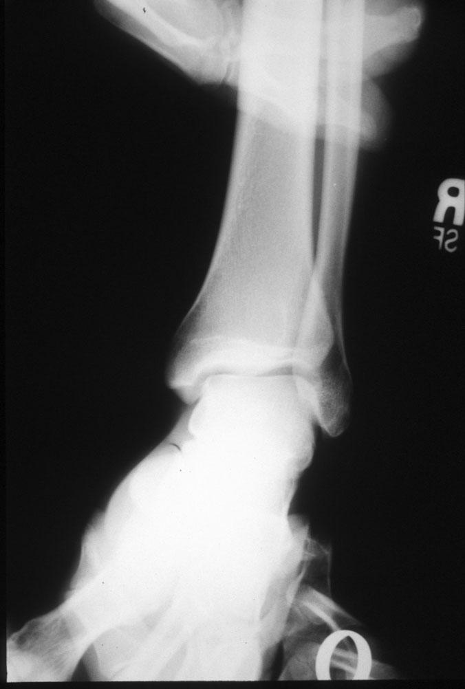
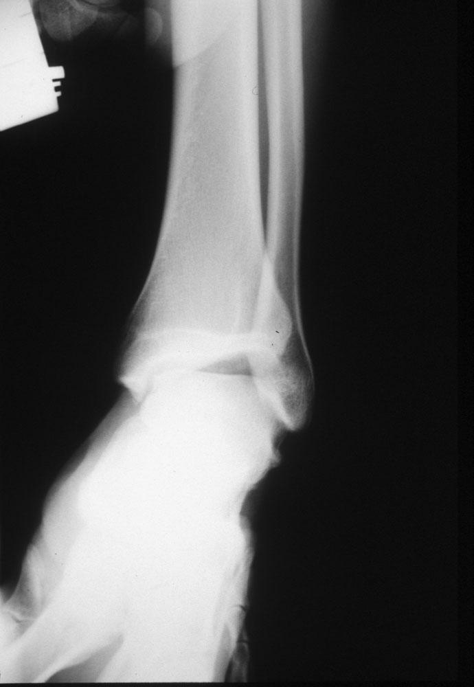
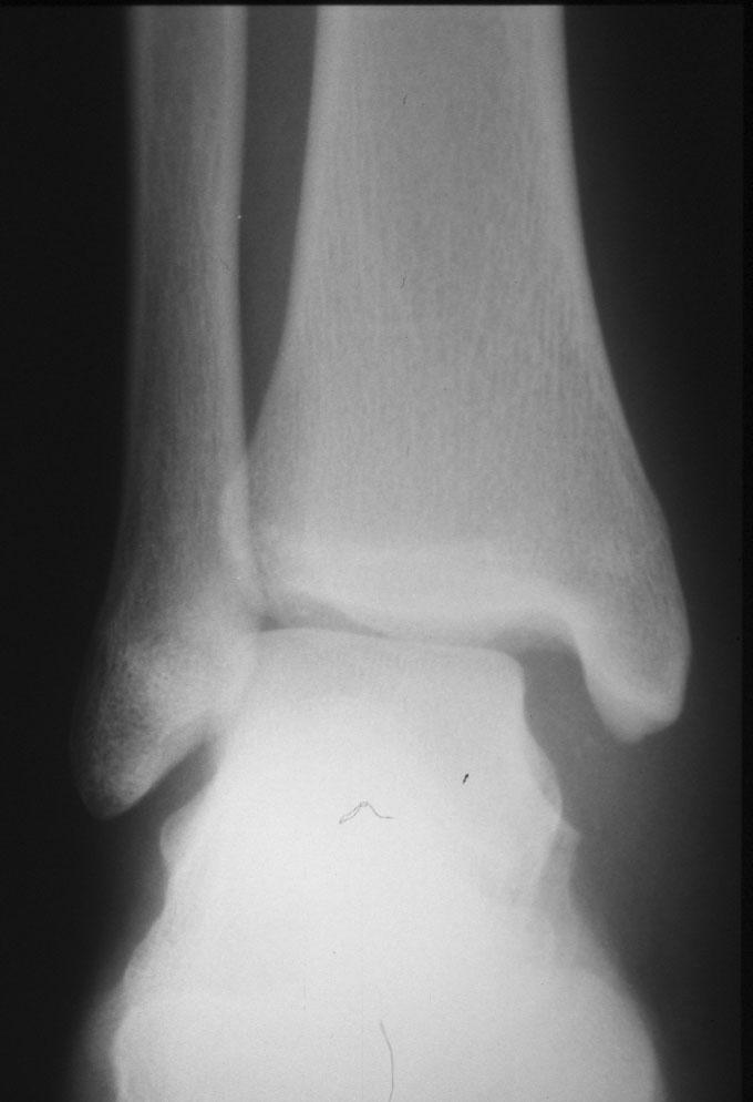
3 The syndesmosis ligament is often also injured with an eversion force. If the tibia and fibula spread on the talus, the ankle mortise is disrupted and the ankle can become very unstable. It is also not unusual to see an associated fibula fracture with an eversion mechanism. (see x-rays below) Assessment of a syndesmosis sprain will be difficult for the initial 24 to 48 hours. If the ankle is quite swollen and edematous assessment of a syndesmosis sprain may be difficult until the pain and swelling have isolated to individual areas or x-rays show some spreading of the ankle mortise.
4
5
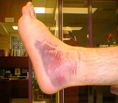
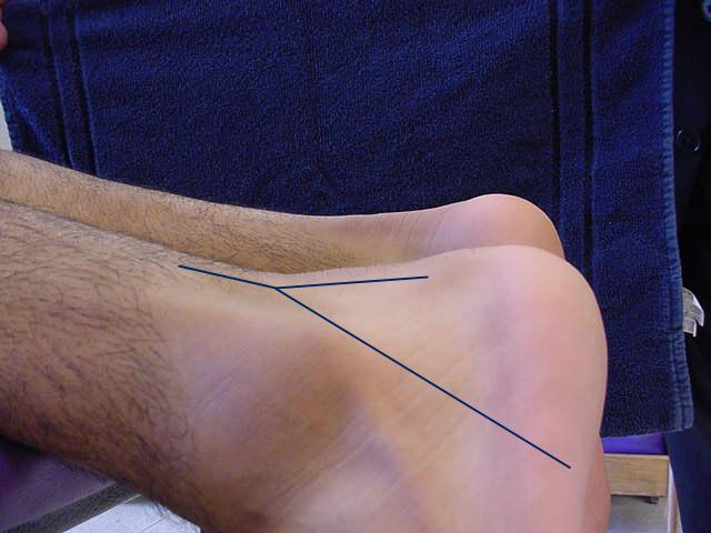
6 Ankle Ecchymosis

7

8 Maison - Neuve type fracture.
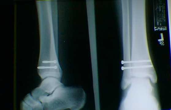
9

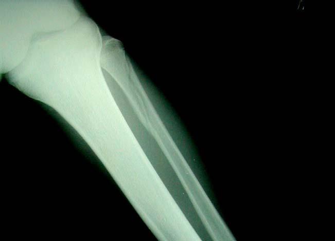
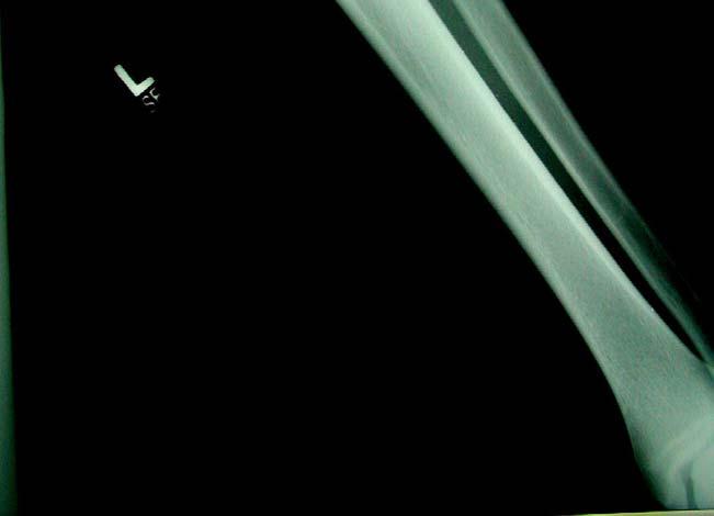
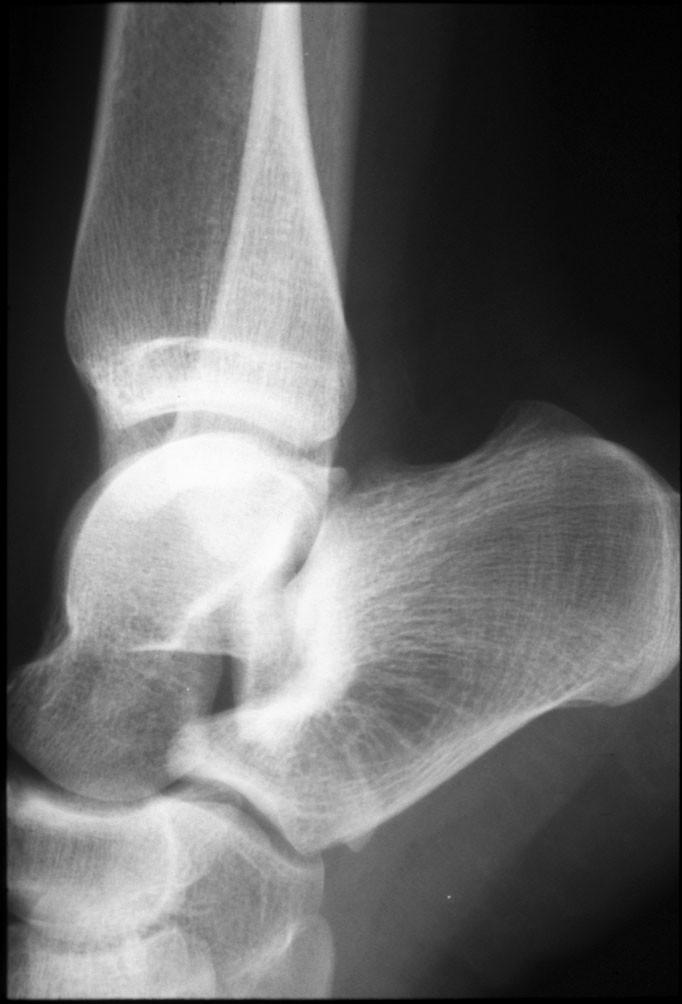
10 Distal Fibula fracture with associated medial deltoid ligament disruption. This injury is frequently the result of the foot being planted with a valgus load applied to the leg.

11 Notice the disruption of the medial deltoid ligament and the widening between the medial malleolus and the talus. This is indicative of a ruptured deltoid ligament.
12
13

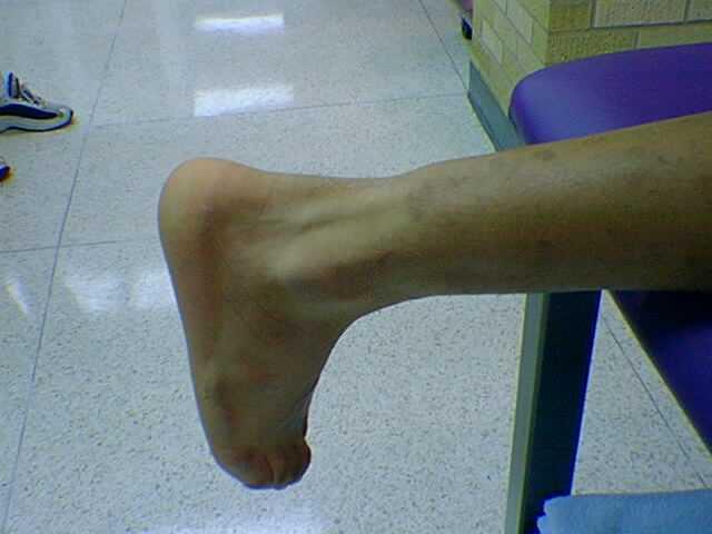
14 Os Trigonum
15
16 This fracture requires surgical fixation of the fibula using a screw and plate system. The plate should be removed prior to return to competitive athletic activity as it will cause stress areas in the bone at each end of the plate. Recovery time (return to athletic activity) for a generally healthy patient with this type of fracture will be in the 6 month range.
17 Name the Injury

18 Talar Dome - AVN

19 Talar Dome - AVN

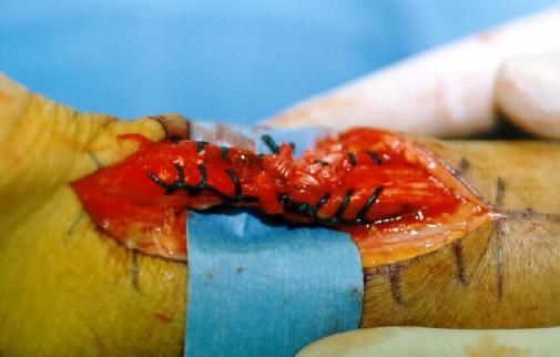
20

21 Ankle dislocation with no fractures. This takes a high degree of trauma and force. In this case this was generated as the result of a high flip off of a trampoline and impact with the ground. The ankle was in a plantar flexion and inverted position upon impact. This was an open dislocation.
22
23

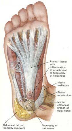
24 Negative Thompson Test

25 Positive Thompson Test
26


27 Achilles Tendon Tear and Repair
28
29 Foot Injuries
30
31 Plantar Fasciitis/Arch Strain A. Mechanism of Injury 1. strain of plantar fascia-usually at medial insertion into calcaneus 2. middle strip of plantar fascia sometimes involved 3. lateral strip almost never involved B. Possible Responsible Factors 1. shoes 2. artificial turf 3. severe pronation 4. excessive weight 5. leg length discrepancy 6. tight bed sheets
32
33 Treatment 1. stretching, Achilles, plantar fascia (night splints) 2. strapping 3. shoe padding - medial heel wedge -saddle pads -arch supports -lift type / padded heel pad -heel lift for short leg
34 4. orthotics-rigid (for heavy lineman, need more support & control) -sports orthotics (lighter in weight with more flexibility for backs and wide receivers) 5. Non Steroidal Anti Inflammatories 6. Steroid Injections - once weekly for 3 weeks 7. Surgical intervention - fasciotomy last resort, after 1 year of conservative treatment
35 Chronic plantar fasciitis can lead to formation of heel spurs. Plantar Fasciitis is the most common injury seen among long distance runners. It is very painful and can be chronic, extending over several years. The heel spur does not cause the plantar fasciitis, the fasciitis causes the heel spur.
36 Morton's Neuroma A. Mechanism of Injury 1. direct or microtrauma to an interdigital nerve 2. 90% of neuromas involve the 3rd common digital nerve approximately 10% involve the 2nd common interdigital nerve - 3 & 4 metatarsals B. Possible Responsible Factors 1. poorly cushioned and or tight shoes, high heels 2. pronation - nerve gets pinched between the heads of the 3rd and 4th metatarsals and the base of the proximal phalanx of the 3rd & 4th toes 3. hard surfaces 4. leg length discrepancy
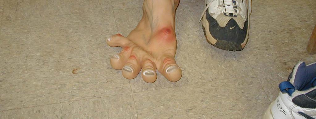
37 Calcaneal Apophysitis (Severs Disease) (pump bumps) A. Mechanism of Injury 1. direct or microtrauma to the growth center of the posterior calcaneus 2. causes avascularity to the apophysis 3. Usually 8-12 year olds
38 B. Possible Responsible Factors 1. hard playing surfaces 2. shoes - poorly padded - cleats - poor support 3. cavus type foot 4. tight Achilles and or plantar fascia
39 C. Treatment 1. get out of cleats 2. shock absorbent heel pads 3. strapping - to help support plantar fascia 4. orthotic or heel stabilizers 5. in resistant cases immobilization for 4-6 weeks may be needed
40 Sesamoiditis/Sesamoid Fractures Sesamoid fractures must be differentiated from a normal bipartite sesamoid. A. Mechanism of Injury 1. direct trauma to tibial (medial) sesamoid - most common 2. direct trauma to fibular (lateral) sesamoid - rare 3. overuse - chronic microtrauma
41 B. Possible Responsible Factors 1. hard playing surfaces 2. hallux valgus - tibial sesamoid directly under mp joint 3. lack of cushioning in shoes C. Structures involved 1. sesamoids 2. joint capsule 3. flexor brevis 4. plantar 1st metatarsal head
42 Treatment Sesamoiditis 1. shoe padding - transfer weight away from sesamoid 2. super cushion inner soles 3. ice, elevation, compression 4. possible post-op shoe 5. steroid injection Sesamoid fracture 1. cast for 3 weeks - BK 2. post-op shoe
43 Surgical excision of affected sesamoid in resistant cases very often will not heal. If hallux valgus present should correct at time of surgery, because weakening of flexor apparatus will increase deformity. This is a last resort in most cases as it changes the bio mechanical forces on the flexor tendons and if a single sesamoid is left in place, the weight bearing mechanics of the foot are greatly altered.
44 Turf Toe A. Mechanism of Injury 1. hyperextension (most common) 2. hyperflexion 3. valgus injury - usually from sudden acceleration B. Possible Responsible Factors 1. artificial turf - no give, can be like playing on hard asphalt 2. shoes - too much forefoot flexion (no turf toe plate) 3. combination of turf & shoes
45 Specific Structures Involved 1. capsular & ligamentous structures 2. flexor apparatus 3. possibly sesamoids
46 D. Treatment 1. rest, ice, elevation, compression 2. possible immobilization and non weight bearing 3. shoe modifications - spring steel splint 4. activity is resumed within the limits of pain Starting with flat foot walking, then normal gait, then jogging, then straight ahead running at full speed, next running from stance, last performing cutting maneuvers.
47 5. taping the toe to prevent injury from recurring 6. anti-inflammatories 7. surgery - for capsular repair in non responsive cases
48 Misc. Aggravations A. Hallux Valgus (bunions) 1. Possible Responsible Factors a. heredity b. shoes - irritate but don't cause c. pronation - accentuates 2. Specific Structures Involved a. 1st MP - all structures b. sesamoids c. lst metatarsal - medial cuneiform joint
49 3. Treatment a. accommodate in wider shoes b. shoe stretching c. surgical correction in off season if chronically painful (may cause some limitation of joint movement)
50 Hallux Limitus 1. Possible Responsible Factors a. heredity b. trauma to joint c. foot type - plantar flexed 1st digit 2. Specific Structures Involved a. 1st MP - degeneration of joint cartilage with osteophytic limping of 1st metatarsal head and base of proximal phalanx b. sesamoids - in advanced cases
51 3. Treatment a. rigid soled shoes which limit dorsiflexion b. taping c. injection with local and steroid when symptoms acute d. when condition becomes debilitating & conservative measures fail then surgical intervention is necessary - usually with placement of plastic implant (will weaken push off)
52 Corns (digital clavi) Calluses (tylomas) 1. Possible Responsible Factors a. Cavus foot - toes hammer - plantar flexion of forefoot causes excess pressure on metatarsal heads b. pronated foot - abnormal weight transfer c. poor fitting shoes
53 Specific Structures Involved a. interphalangeal joints of toes b. extensor & flexor tendons c. metatarsal heads 3. Treatment a. deep & wide toe box b. débride hyperkeratotic tissue regularly c. Vaseline d. padding - Spenco 2nd skin - moleskin
54 Ingrown Nails 1. Possible Responsible Factors a. improper cutting of nails b. heredity c. injury d. tight shoes 2. Specific Structures Involved a. tibial & fibular borders, usually hallux nails b. nail groove 3. Treatment a. packing cotton under affected border b. wedge resection of affected border c. partial radical nail procedure with matrix destruction (phenol method)
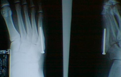
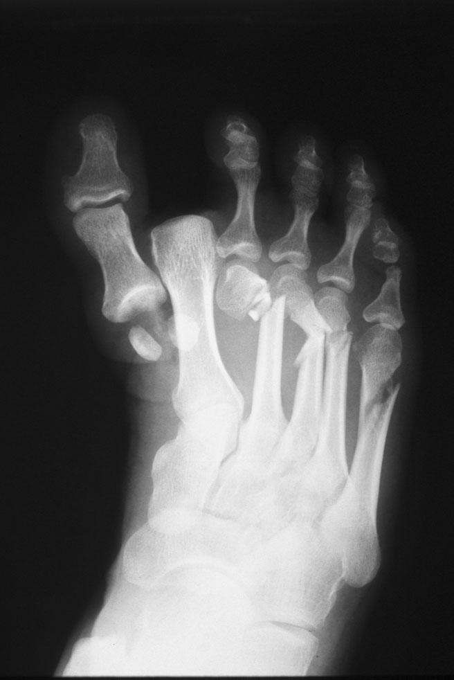
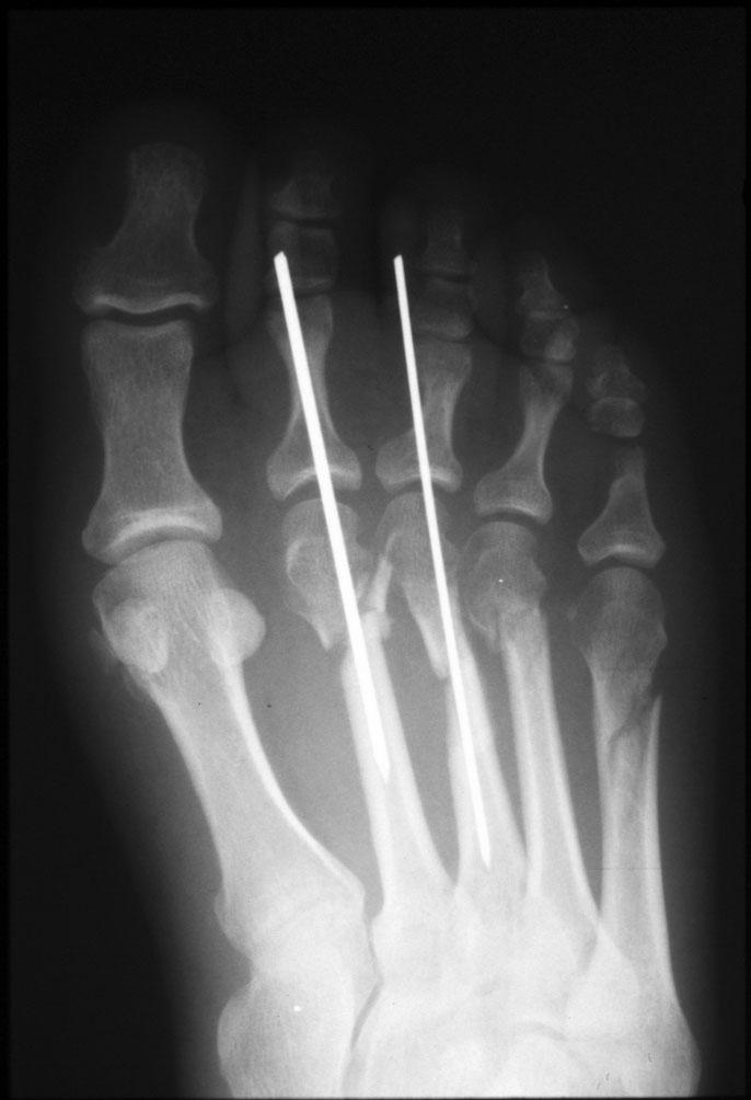
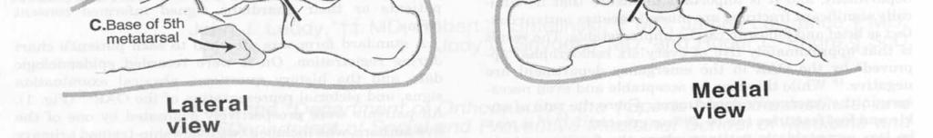
55 Black Toe (Subungual Hematoma) 1. Possible Responsible Factors a. shoes too tight b. shoes too loose c. low toe box d. long 2nd toe e. cleats f. kicking g. direct trauma
56 Specific Structures Involved a. pedal nails b. nail bed c. distal phalanx - possible formation of subungual exostosis 3. Treatment a. drain hematoma as soon as possible b. if nail partially avulsed - remove nail completely & débride the nail bed - start soaks & topical antibiotics c. if chronic, hypertrophied nail - keep nail débrided back & thinned as much as possible - complete avulsion of nail plate with destruction of matrix
57 Plantar Verruca (Warts) 1. Possible Responsible Factors a. hyperhidrosis b. abrasions to plantar surface of the foot c. exposure to verruca virus - showers- locker rooms - brothers & sisters d. age - most commonly seen in teen years
58 Specific Structure Involved a. skin - warts do not penetrate the basement membrane of the skin b. metatarsal heads and/or calcaneus- areas of most pressure in weight bearing 3. Treatment a. mechanical debridement prn. b. topical acids c. cryotherapy d. surgical removal - does not leave scar e. laser
59 Name the Injury

60 Rodeo Clown Foot
61 Foot Fractures

62 This is a ballerina type fracture of the 5th metatarsal. The etiology involves and avulsion of the proximal tip of the 5th metatarsal where the peroneus brevis muscle tendon attaches.
63 Fracture of Styloid process of 5th metatarsal A. Mechanism of injury 1. severe inversion ankle sprain causes peroneus brevis tendon to pull away the base of the 5th metatarsal (Ballerina fracture)

64 These X-Rays show a fracture of the proximal end of the 5th Metatarsal. This fracture is commonly called a "Jones Fracture".
65 Direct trauma to base of 5th (Jones fracture) A. Possible Responsible Factors 1. Cavus foot type 2. chronic ankle sprains 3. poor shoe and/or tape support B. Specific structures involved 1. peroneus brevis tendon 2. styloid process 5th metatarsal base
66 Treatment 1. ice, elevation, compression and lift under 5th metatarsal base 2. short leg walking brace 3. if severe avulsion of fragment, open reduction screw fixation
67 Treatment of Jones Fractures includes several options. Option 1 - immobilization of foot and ankle with non weight bearing for a period of 1 month to 6 plus weeks and more time may be required if the bone healing is delayed. The peroneus brevis tendon attaches at the proximal end of the 5th Metatarsal and treatment without ankle immobilization is not effective. Every time the muscle contracts and pulls on the tendon, the fracture site is disrupted. This type of fracture is known to form a non union.
68 Option 2 - insertion of a intramedullary screw into the fracture to compress the fragment and the bones back together. May or may not be used with a bone graft. Many physicians will also opt to use a bone growth stimulator on this fracture to insure that healing occurs. Option 2 is certainly preferred in the authors opinion since the fracture site is stabilized and the ends of the fracture are approximated. The screw fixation allows for earlier return to weight bearing and decreased immobilization time. (Dr. Joe Milne, Dr. Steve Brotherton)
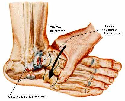
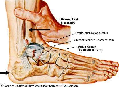
69
70
71
72

73
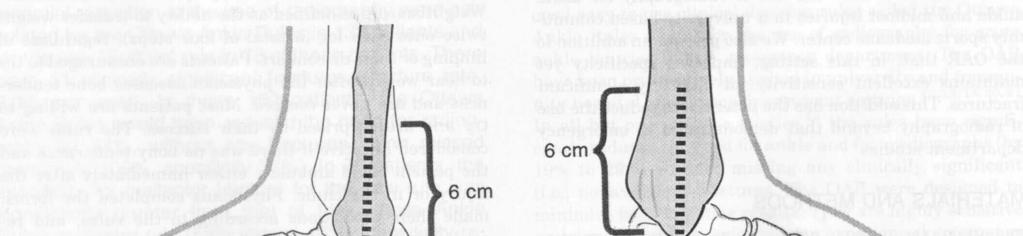
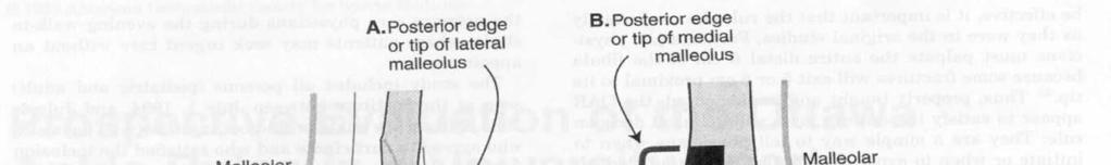
74
75 Ankle Evaluation How did it happen? (etiology) Previous history of injury to that area? Sight evaluation a) swelling? b) deformities? c) discoloration?
76 Palpation evaluation examine ligaments (1) medial: deltoid / anterior, medial, and posterior portions (2) lateral: Anterior talo-fib

77 Anterior drawer tests should always be performed with the knee bent to eliminate the Achilles and Gastrocnemius muscles from providing any stability to the ankle. A lateral talar tilt test can be conducted at the same time.

78 Talar Tilt

79 Inversion Stress Test
80 Ottawa - Buffalo Modification for Ankle Exam The incidence of ankle fractures in athletes involved in controlled sports activities is relatively low. However, the decision(s) on which ankles to study radiographically with x- rays is not always easy. Not all ankle or foot injuries require immediate x-rays. The allied health practitioner can determine to a relatively accurate degree the need for futher study through a good clinical exam and by following the Ottawa Ankle Rules and with the use of the Buffalo Modification. (for more information, consult the AJSM, Vol 26, No )
81 Research has shown that in a hospital based E.R. of every 6 ankles approved for x-ray under the O.A.R. (Ottawa Ankle Rules), 5 have no radiographic findings. Steill, I.G., JAMA, 269: , Clinical ankle exam If the patient needs x-rays, they will usually present with lateral fibular malleolus pain in the distal 6 cm, medial tibial malleolus pain in the distal 6 cm or pain to palpation over the proximal tip of the 5th metatarsal or the Navicular. The inability to bear weight may indicate a lesion to the dome of the Talus or other associated trauma to the other structures of the ankle which may require x-ray study.
82 Squeeze test - check malleolus (2) Check tibia and fibula 7. Functional tests a) walking - check gait b) toe raises 1) both feet 2) one foot c) jump and land on both feet and then on one foot 8. Refer to doctor for further evaluation and possible x-ray
83 Refer to physician for further evaluation and possible x-ray Non weight bearing x-rays Weight bearing x-rays (syndesmosis spreading) Stress x-rays for talar tilt and syndesmosis spreading Other associated ankle and lower leg tests Homan's Sign - patient is supine on the table, the knee is fully extended and the foot is dorsi flexed. Reproduction of pain with localized edema is considered a positive test for deep venous thrombophlebitis.

84
85
Review relevant anatomy of the foot and ankle. Learn the approach to examining the foot and ankle
 Objectives Review relevant anatomy of the foot and ankle Learn the approach to examining the foot and ankle Learn the basics of diagnosis and treatment of ankle sprains Overview of other common causes
Copyright 2004, Yoshiyuki Shiratori. All right reserved.
 Ankle and Leg Evaluation 1. History Chief Complaint: A. What happened? B. Is it a sharp or dull pain? C. How long have you had the pain? D. Can you pinpoint the pain? E. Do you have any numbness or tingling?
Anatomy and evaluation of the ankle.
 Anatomy and evaluation of the ankle www.fisiokinesiterapia.biz Ankle Anatomical Structures Tibia Fibular Talus Tibia This is the strongest largest bone of the lower leg. It bears weight and the bone creates
Prevention and Treatment of Injuries. Anatomy. Anatomy. Tibia: the second longest bone in the body
 Prevention and Treatment of Injuries The Ankle and Lower Leg Westfield High School Houston, Texas Anatomy Tibia: the second longest bone in the body Serves as the principle weight-bearing bone of the leg.
Financial Disclosure. Turf Toe
 Seth O Brien, CP, LP Financial Disclosure Mr. Seth O'Brien has no relevant financial relationships with commercial interests to disclose. Turf Toe Common in athletes playing on firm, artificial turf Forceful
Ankle and Foot Orthopaedic Tests Orthopedics and Neurology DX 612
 Ankle and Foot Orthopaedic Tests Orthopedics and Neurology DX 612 James J. Lehman, DC, MBA, DABCO University of Bridgeport College of Chiropractic Ankle & Foot Anatomy Stability of the ankle is dependent
Recognizing common injuries to the lower extremity
 Recognizing common injuries to the lower extremity Bones Femur Patella Tibia Tibial Tuberosity Medial Malleolus Fibula Lateral Malleolus Bones Tarsals Talus Calcaneus Metatarsals Phalanges Joints - Knee
ANKLE JOINT ANATOMY 3. TALRSALS = (FOOT BONES) Fibula. Frances Daly MSc 1 CALCANEUS 2. TALUS 3. NAVICULAR 4. CUBOID 5.
 Fibula. Frances Daly MSc 1 CALCANEUS 2. TALUS 3. NAVICULAR 4. CUBOID 5.") ANKLE JOINT ANATOMY The ankle joint is a synovial joint of the hinge type. The joint is formed by the distal end of the tibia and medial malleolus, the fibula and lateral malleolus and talus bone. It is
ANKLE JOINT ANATOMY The ankle joint is a synovial joint of the hinge type. The joint is formed by the distal end of the tibia and medial malleolus, the fibula and lateral malleolus and talus bone. It is
Injuries to the Foot. NOCROP Sports Medicine and Therapy
 Injuries to the Foot Arches of the Foot Plantar Fascia - a flat band of connective tissue that connects your heel bone to your toes. It supports the arch of your foot. Muscle of the Foot and Lower Leg
Scar Engorged veins. Size of the foot [In clubfoot, small foot]
![Scar Engorged veins. Size of the foot [In clubfoot, small foot]](/thumbs/78/77722241.jpg "Scar Engorged veins. Size of the foot [In clubfoot, small foot]") 6. FOOT HISTORY Pain: Walking, Running Foot wear problem Swelling; tingly feeling Deformity Stiffness Disability: At work; recreation; night; walk; ADL, Sports Previous Rx Comorbidities Smoke, Sugar, Steroid
6. FOOT HISTORY Pain: Walking, Running Foot wear problem Swelling; tingly feeling Deformity Stiffness Disability: At work; recreation; night; walk; ADL, Sports Previous Rx Comorbidities Smoke, Sugar, Steroid
Index. Clin Sports Med 23 (2004) Note: Page numbers of article titles are in boldface type.
 Note: Page numbers of article titles are in boldface type.") Clin Sports Med 23 (2004) 169 173 Index Note: Page numbers of article titles are in boldface type. A Achilles enthesopathy, calcaneal spur with, 133 clinical presentation of, 135 136 definition of, 131
Clin Sports Med 23 (2004) 169 173 Index Note: Page numbers of article titles are in boldface type. A Achilles enthesopathy, calcaneal spur with, 133 clinical presentation of, 135 136 definition of, 131
Outline. Ankle/Foot Anatomy Ankle Sprains Ottawa Ankle Rules DDx: The Sprain That Wasn t
 Ankle Injuries Outline Ankle/Foot Anatomy Ankle Sprains Ottawa Ankle Rules DDx: The Sprain That Wasn t Anatomy: Ankle Mortise Bony Anatomy Lateral Ligament Complex Medial Ligament Complex Ankle Sprains
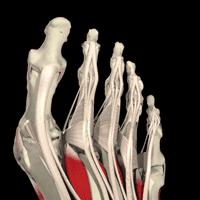
Bones = phalanges 5 metatarsals 7 tarsals
 The Foot (Bones) Bones = 26 14 phalanges 5 metatarsals 7 tarsals Toes (Phalanges) Designed to give wider base for balance and propelling the body forward. 1st toe (Hallux) Two sesamoid bones located under
Ankle Tendons in Athletes. Laura W. Bancroft, M.D.
 Ankle Tendons in Athletes Laura W. Bancroft, M.D. Outline Protocols Normal Anatomy Tendinopathy, partial and complete tears Posterior tibial, Flexor Hallucis Longus, Achilles, Peroneal and Anterior Tibial
Foot and Ankle Complaints.
 Foot and Ankle Complaints www.fisiokinesiterapia.biz INTRODUCTION Anatomy and Function Foot Ankle Common complaints Common diagnoses FOOT AND ANKLE ANATOMY 26 bones and 2 sesamoids Forefoot Metatarsals
Main Menu. Ankle and Foot Joints click here. The Power is in Your Hands
 1 The Ankle and Foot Joints click here Main Menu Copyright HandsOn Therapy Schools 2009 K.8 http://www.handsonlineeducation.com/classes/k8/k8entry.htm[3/27/18, 1:40:03 PM] Ankle and Foot Joint 26 bones
BUCKS MSK: FOOT AND ANKLE PATHWAY GP MANAGEMENT. Hallux Valgus. Assessment: Early Management. (must be attempted prior to any referral to imsk):
:") Hallux Valgus Common condition: affecting around 28% of the adult population. Prevalence increases with age and in females. Observation: Lateral deviation of the great toe. May cause secondary irritation
Hallux Valgus Common condition: affecting around 28% of the adult population. Prevalence increases with age and in females. Observation: Lateral deviation of the great toe. May cause secondary irritation
Section Three: The Leg, Ankle, and Foot Lecture: Review of Clinical Anatomy, Patterns of Dysfunction and Injury, and
 Section Three: The Leg, Ankle, and Foot Lecture: Review of Clinical Anatomy, Patterns of Dysfunction and Injury, and Treatment Implications for the Leg, Ankle, and Foot Levels I and II Demonstration and
Surgery-Ortho. Fractures of the tibia and fibula. Management. Treatment of low energy fractures. Fifth stage. Lec-6 د.
 Fifth stage Lec-6 د. مثنى Surgery-Ortho 28/4/2016 Indirect force: (low energy) Fractures of the tibia and fibula Twisting: spiral fractures of both bones Angulatory: oblique fractures with butterfly segment.
Clarification of Terms
 Clarification of Terms The plantar aspect of the foot refers to the role or its bottom The dorsal aspect refers to the top or its superior portion The ankle and foot perform three main functions: 1. shock
What Happens to the Paediatric Flat Foot? Peter J Briggs Freeman Hospital Newcastle upon Tyne
 What Happens to the Paediatric Flat Foot? Peter J Briggs Freeman Hospital Newcastle upon Tyne We don t know!! Population Studies 2300 children aged 4-13 years Shoe wearers Flat foot 8.6% Non-shoe wearers
CHRONIC FOOT PROBLEMS FOOT and ANKLE BASICS
 CHRONIC FOOT PROBLEMS FOOT and ANKLE BASICS ABC s of Comprehensive Musculoskeletal Care December 1 st, 2007 Stephen Pinney MD Chief, UCSF Foot and Ankle Service Chronic problems typically occur gradually
Leg and Ankle Problems in Primary Care.
 Leg and Ankle Problems in Primary Care www.fisiokinesiterapia.biz Leg and Ankle Presentations 4Trauma 4Pain Ankle Trauma 41. Twist and Fall--Fracture or Sprain 42. Patient hears/feels a pop--tendon or
17/10/2017. Foot and Ankle
 17/10/2017 Alicia M. Yochum RN, DC, DACBR, RMSK Foot and Ankle Plantar Fasciitis Hallux Valgus Deformity Achilles Tendinosis Posterior Tibialis Tendon tendinopathy Stress Fracture Ligamentous tearing Turf
Physical Examination of the Foot & Ankle
 Inspection Standing, feet straight forward facing toward examiner Swelling Deformity Flatfoot (pes planus and hindfoot valgus) High arch (pes cavus and hindfoot varus) Peek-a-boo heel Varus Too many toes
ANKLE PLANTAR FLEXION
 ANKLE PLANTAR FLEXION Evaluation and Measurements By Isabelle Devreux 1 Ankle Plantar Flexion: Gastrocnemius and Soleus ROM: 0 to 40-45 A. Soleus: Origin: Posterior of head of fibula and proximal1/3 of
Acute Ankle Injuries, Part 1: Office Evaluation and Management
 t June 08, 2009 Obesity [1] Each acute ankle injury commonly seen in the office has associated with it a mechanism by which it can be injured, trademark symptoms that the patient experiences during the
Ankle Sprains and Their Imitators
 Ankle Sprains and Their Imitators Mark Halstead, MD Dr. Mark Halstead is the Associate Professor of the Departments of Orthopedics and Pediatrics at Washington University School of Medicine; Director of
Servers Disease (Calcaneal Apophysitis ) 101
 101") Servers Disease (Calcaneal Apophysitis ) 101 Servers Disease Causes a disturbance to the growing area at the back of the heel bone (calcaneus) where the strong Achilles tendon attaches to it. It is most
Servers Disease (Calcaneal Apophysitis ) 101 Servers Disease Causes a disturbance to the growing area at the back of the heel bone (calcaneus) where the strong Achilles tendon attaches to it. It is most
CASE ONE CASE ONE. RADIAL HEAD FRACTURE Mason Classification. RADIAL HEAD FRACTURE Mechanism of Injury. RADIAL HEAD FRACTURE Imaging
 CASE ONE An eighteen year old female falls during a basketball game, striking her elbow on the court. She presents to your office that day with a painful, swollen elbow that she is unable to flex or extend
Feet First. Michael K. Cooper, DO FACOFP Family Practice/OMM St John Clinic - Claremore OOA 2018 Annual Convention
 Feet First Michael K. Cooper, DO FACOFP Family Practice/OMM St John Clinic - Claremore OOA 2018 Annual Convention Disclaimer I have no conflict of interest. I am not on any pharmaceutical company payroll
Sports Injuries of the Foot and Ankle. Mark McEleney, MD University of Iowa College of Medicine Refresher Course for the Family Physician 4/4/2018
 Sports Injuries of the Foot and Ankle Mark McEleney, MD University of Iowa College of Medicine Refresher Course for the Family Physician 4/4/2018 I. Objectives A. By the end of the lecture attendees will
Paul Alley MD,DPM,MS,FACS,FAAOS,BFD Eby Orthopaedics,Jasper,Indiana
 Paul Alley MD,DPM,MS,FACS,FAAOS,BFD Eby Orthopaedics,Jasper,Indiana Very common Bone=fractures Description (cracked,broke,busted,or smashed) A=anatomic area of bone eg: head,neck,shaft B=bone involved
Commonly Missed Foot and Ankle Conditions. David Miller, DPM AMG Podiatry
 Commonly Missed Foot and Ankle Conditions David Miller, DPM AMG Podiatry Lisfranc Injuries Wide spectrum of injuries High energy Subtle subluxation which could be easily missed injuries Men are 2-4x s
Foot and ankle update
 Foot and ankle update Mr Ian Garnham Consultant Foot and Ankle Surgeon Whipps Cross University Hospital Hallux Rigidus Symptoms first ray and 1st MTP pain and swelling worse with push off or forced dorsiflexion
Everything. You Should Know. About Your Ankles
 Everything You Should Know About Your Ankles How Your Ankle Works The ankle joint is a hinge type joint that participates in movement and is involved in lower limb stability. There are 2 types of motions
Dr Nabil khouri MD. MSc. Ph.D
 Dr Nabil khouri MD. MSc. Ph.D Foot Anatomy The foot consists of 26 bones: 14 phalangeal, 5 metatarsal, and 7 tarsal. Toes are used to balance the body. Metatarsal Bones gives elasticity to the foot in
Tarsal Tunnel Syndrome
 43 Thames Street, St Albans, Christchurch 8013 Phone: (03) 356 1353. Website: philip-bayliss.com Tarsal Tunnel Syndrome The foot is subjected to forces hundreds of times the bodyweight, thousands of times
PAINFUL SESAMOID OF THE GREAT TOE Dr Vasu Pai ANATOMICAL CONSIDERATION. At the big toe MTP joint: Tibial sesamoid (medial) & fibular (lateral)
 & fibular (lateral)") PAINFUL SESAMOID OF THE GREAT TOE Dr Vasu Pai ANATOMICAL CONSIDERATION At the big toe MTP joint: Tibial sesamoid (medial) & fibular (lateral) They are contained within the tendons of Flexor Hallucis Brevis
PAINFUL SESAMOID OF THE GREAT TOE Dr Vasu Pai ANATOMICAL CONSIDERATION At the big toe MTP joint: Tibial sesamoid (medial) & fibular (lateral) They are contained within the tendons of Flexor Hallucis Brevis
ii ANKLE INJURIES SPECIFIC TRAINING AFTER INJURY TO THE FOOT OR ANKLE
 40 Ankle injuries are among the most common injuries in sport. Ankle sprain (which is a mechanism rather than a diagnosis) is the most common injury in virtually all epidemiological studies. Being the
MEDIAL HEAD GASTROCNEMIUS TEAR (Tennis Leg)
") MEDIAL HEAD GASTROCNEMIUS TEAR (Tennis Leg) Description Expected Outcome Medial head gastrocnemius tear is a strain of the inner part (medial head) of the major calf muscle (gastrocnemius muscle). Muscle
MEDIAL HEAD GASTROCNEMIUS TEAR (Tennis Leg) Description Expected Outcome Medial head gastrocnemius tear is a strain of the inner part (medial head) of the major calf muscle (gastrocnemius muscle). Muscle
Aetiology: Pressure of Distal intermetatarsal ligament against common digital nerve. Lumbar radiculopathy Instability MTPJ joint or inflammatory MPJ
 MORTON S NEUROMA 80% III web space (next common is II). Never occurs in III or IV Common in females in fifties Aetiology: Pressure of Distal intermetatarsal ligament against common digital nerve Rule out
Common%Work%Related%Foot% and%ankle%problems
 Common%Work%Related%Foot% and%ankle%problems Dr. George H. Theodore Massachusetts General Hospital Harvard Medical School Foot and Ankle Consultant Boston Red Sox New England Patriots Boston Bruins Work%Related%Foot%and%Ankle%
Imaging of Ankle and Foot pain
 Imaging of Ankle and Foot pain Pramot Tanutit, M.D. Department of Radiology Faculty of Medicine, Prince of Songkla University 1 Outlines Plain film: anatomy Common causes of ankle and foot pain Exclude:
Copyright 2012 by The McGraw-Hill Companies, Inc. All rights reserved. McGraw-Hill/Irwin
 CHAPTER 8: THE LOWER EXTREMITY: KNEE, ANKLE, AND FOOT KINESIOLOGY Scientific Basis of Human Motion, 12 th edition Hamilton, Weimar & Luttgens Presentation Created by TK Koesterer, Ph.D., ATC Humboldt State
Key Points for Success:
 ANKLE & FOOT 1 2 All of the stretches described in this chapter are detailed to stretch the right side. Key Points for Success: Keep your movements slow and precise. Breathe in before you move and breathe
Index. Note: Page numbers of article titles are in boldface type.
 Index Note: Page numbers of article titles are in boldface type. A Achilles tendon injury of, pathophysiology of, 10 peritendinitis of, 119 120 rupture of, 32 35, 117 135 anatomy of, 117 118 chronic, 126
Anatomy 1% 29% 64% 6%
 Mortons Neuroma Perineural fibrosis of the plantar digital nerve Females 8-10 3 rd plantar webspace most commonly effected Burning pain Sensory changes 3&4 digits / interdigital space Etiology Excessive
Managing Tibialis Posterior Tendon Injuries
 Managing Tibialis Posterior Tendon Injuries by Thomas C. Michaud, DC Published April 1, 2015 by Dynamic Chiropractic Magazine Tibialis posterior is the deepest, strongest, and most central muscle of the
6/5/2018. Forefoot Disorders. Highgate Private Hospital (Royal Free London NHS Foundation Trust (Barnet & Chase Farm Hospitals) Hallux Rigidus
 Hallux Rigidus") Forefoot Disorders Mr Pinak Ray (MS, MCh(Orth), FRCS, FRCS(Tr&Orth)) Highgate Private Hospital (Royal Free London NHS Foundation Trust (Barnet & Chase Farm Hospitals) E: ray.secretary@uk-conslutants Our
Forefoot Disorders Mr Pinak Ray (MS, MCh(Orth), FRCS, FRCS(Tr&Orth)) Highgate Private Hospital (Royal Free London NHS Foundation Trust (Barnet & Chase Farm Hospitals) E: ray.secretary@uk-conslutants Our
Medical Practice for Sports Injuries and Disorders of the Lower Limb
 Sports-Related Injuries and Disorders Medical Practice for Sports Injuries and Disorders of the Lower Limb JMAJ 48(1): 25 29, 2005 Motonobu NATSUYAMA Chief Surgeon, Department of Orthopedic Surgery, Kantoh
PROBLEMS AND ORTHOTIC SOLUTIONS. Problem/Issue Underlying treatment goal Solution Pes Cavus foot
 PROBLEMS AND ORTHOTIC SOLUTIONS Problem/Issue Underlying treatment goal Solution Pes Cavus foot Usually also a supinated foot Rigid high arched foot with poor shock absorption and cushioning. Often roll
WHAT IS PLANTAR FASCIITIS?
 WHAT IS PLANTAR FASCIITIS? If you're finding when you climb out of bed each morning that your first couple steps cause your foot and heel to hurt, this might be a sign of plantar fasciitis. A common condition
Overuse Injuries & special skeletal injuries Dr M.Taghavi Director of sport medicine center of olympic academy
 Overuse Injuries & special skeletal injuries Dr M.Taghavi Director of sport medicine center of olympic academy Prevalence of Overuse Injuries 30 to 50% of all sport injuries are from overuse In some sports
X-Ray Rounds: (Plain) Radiographic Evaluation of the Ankle.
 Radiographic Evaluation of the Ankle.") X-Ray Rounds: (Plain) Radiographic Evaluation of the Ankle www.fisiokinesiterapia.biz Anatomy Complex hinge joint Articulations among: Fibula Tibia Talus Tibial plafond Distal tibial articular surface
X-Ray Rounds: (Plain) Radiographic Evaluation of the Ankle www.fisiokinesiterapia.biz Anatomy Complex hinge joint Articulations among: Fibula Tibia Talus Tibial plafond Distal tibial articular surface
5 minutes: Attendance and Breath of Arrival. 50 minutes: Problem Solving Ankles and Feet
 5 minutes: Attendance and Breath of Arrival 50 minutes: Problem Solving Ankles and Feet Punctuality- everybody's time is precious: o o Be ready to learn by the start of class, we'll have you out of here
بسم هللا الرحمن الرحيم
 بسم هللا الرحمن الرحيم Laboratory RHS 221 Manual Muscle Testing Theory 1 hour practical 2 hours Dr. Ali Aldali, MS, PT Department of Physical Therapy King Saud University Talocrural and Subtalar Joint
Peggers Super Summaries: Foot Injuries
 Lisfranc Injury ANATOMY Roman arch with recessed 2 nd MT base AP medial side of intermediate cuneiform to 2 nd MT base Oblique medial side of lateral cuneiform with 3 rd MT base and 4 th with medial boarder
Prevention and Management of Common Running Injuries. Presented by. Huub Habets (Sports Physiotherapist) Lynsey Ellis (Soft Tissue Therapist)
 Lynsey Ellis (Soft Tissue Therapist)") Prevention and Management of Common Running Injuries Presented by Huub Habets (Sports Physiotherapist) Lynsey Ellis (Soft Tissue Therapist) Objectives DIALOGUE AND INTERACTION We are not here to preach,
Prevention and Management of Common Running Injuries Presented by Huub Habets (Sports Physiotherapist) Lynsey Ellis (Soft Tissue Therapist) Objectives DIALOGUE AND INTERACTION We are not here to preach,
Therapeutic Foot Care Certificate Program Part I: Online Home Study Program
 Therapeutic Foot Care Certificate Program Part I: Online Home Study Program 1 Anatomy And Terminology Of The Lower Extremity Joan E. Edelstein, MA, PT, FISPO Associate Professor of Clinical Physical Therapy
Burwood Road, Concord Dora Street, Hurstville 119 Lethbridge Street, Penrith 160 Belmore Road, Randwick
 www.orthosports.com.au 47 49 Burwood Road, Concord 29 31 Dora Street, Hurstville 119 Lethbridge Street, Penrith 160 Belmore Road, Randwick Turf Toe Injury By Todd Gothelf Foot, Ankle, Shoulder History
Understanding Leg Anatomy and Function THE UPPER LEG
 Understanding Leg Anatomy and Function THE UPPER LEG The long thigh bone is the femur. It connects to the pelvis to form the hip joint and then extends down to meet the tibia (shin bone) at the knee joint.
MEDIAL TIBIAL STRESS SYNDROME (Shin Splints)
") MEDIAL TIBIAL STRESS SYNDROME (Shin Splints) Description Expected Outcome Shin splints is a term broadly used to describe pain in the lower extremity brought on by exercise or athletic activity. Most commonly
MEDIAL TIBIAL STRESS SYNDROME (Shin Splints) Description Expected Outcome Shin splints is a term broadly used to describe pain in the lower extremity brought on by exercise or athletic activity. Most commonly
Conditions Information on common problems we treat.
 Conditions Information on common problems we treat. Perthpodiatricsurgery.com 9383 3851 Perth Podiatric Surgery 01 Problems affecting the big toe 01 Bunions Bunions are a common foot problem which affect
ORTHOTIC ARCH SUPPORTS
 ORTHOTIC ARCH SUPPORTS COMMON FOOT PROBLEMS & ORTHOTIC THERAPY The foot and ankle are the foundation for the overall posture of the skeletal body. Many problems with the feet, legs, knees, hips and lower
Foot & Ankle Examination Workshop Morteza Khodaee, MD, MPH, FACSM, FAAFP Associate Professor Department of Family Medicine University of Colorado
 Foot & Ankle Examination Workshop Morteza Khodaee, MD, MPH, FACSM, FAAFP Associate Professor Department of Family Medicine University of Colorado School of Medicine July 4, 2013 Objectives Participants
Biomechanical Explanations for Selective Sport Injuries of the Lower Extremity
 Biomechanical Explanations for Selective Sport Injuries of the Lower Extremity American Osteopathic Academy of Sports Medicine Presentation April 23, 2015 Understanding Normalcy What is Normal? Rearfoot/heel
Plantar fasciitis occurs when the strong band of tissue that supports the arch of your foot becomes irritated and inflamed.
 Plantar Fasciitis and Bone Spurs Plantar fasciitis (fashee-eye-tiss) is the most common cause of pain on the bottom of the heel. Approximately 2 million patients are treated for this condition every year.
 P R E S E N T S Dr. Mufa T. Ghadiali is skilled in all aspects of General Surgery. His General Surgery Services include: General Surgery Advanced Laparoscopic Surgery Surgical Oncology Gastrointestinal
Posterior Tibialis Tendon Dysfunction & Repair
 1 Posterior Tibialis Tendon Dysfunction & Repair Surgical Indications and Considerations Anatomical Considerations: The posterior tibialis muscle arises from the interosseous membrane and the adjacent
BCCH Emergency Department LOWER LIMB INJURIES Resource pack
 1 BCCH Emergency Department LOWER LIMB INJURIES Resource pack Developed by: Rena Heathcote RN. 2 Knee Injuries The knee joint consists of a variety of structures including: 3 bones (excluding the patella)
Integrated Manual Therapy & Orthopedic Massage For Complicated Lower Extremity Conditions
 Integrated Manual Therapy & Orthopedic Massage For Complicated Lower Extremity Conditions Assessment Protocols Treatment Protocols Treatment Protocols Corrective Exercises Artwork and slides taken from
Evaluation of Gait Mechanics Using Computerized Plantar Surface Pressure Analysis and it s Relation to Common Musculoskeletal Problems
 Evaluation of Gait Mechanics Using Computerized Plantar Surface Pressure Analysis and it s Relation to Common Musculoskeletal Problems Laws of Physics effecting gait Ground Reaction Forces Friction Stored
The Lower Limb VII: The Ankle & Foot. Anatomy RHS 241 Lecture 7 Dr. Einas Al-Eisa
 The Lower Limb VII: The Ankle & Foot Anatomy RHS 241 Lecture 7 Dr. Einas Al-Eisa Ankle joint Synovial, hinge joint Allow movement of the foot in the sagittal plane only (1 degree of freedom): dorsiflexion:
Ankle Ligament Injury: Don t Worry- It s Only a Sprain Wes Jackson MD Orthopaedic Foot & Ankle
 Ankle Ligament Injury: Don t Worry- It s Only a Sprain Wes Jackson MD Orthopaedic Foot & Ankle Outline I. Epidemiology II. Classification and Types of Sprains III. Anatomy IV. Clinical Assessment and Imaging
Localized collection of pus in a cavity
 Localized collection of pus in a cavity Loss of feeling or sensation induced to permit surgery Common example: Injection given to numb up the toe prior to performing an ingrown toenail procedure Mechanical
Disclosures. Syndesmosis Injury. Syndesmosis Ligaments. Objectives. Mark M. Casillas, M.D.
 Disclosures Syndesmosis Injury No relevant disclosures Mark M. Casillas, M.D. 1 Objectives Syndesmosis Ligaments Understand the syndesmosis anatomy and function Classify syndesmosis injuries Describe treatment
Sky Ridge Medical Center, Aspen Building Ridgegate Pkwy., Suite 309 Lone Tree, Colorado Office: Fax:
 ANKLE SPRAIN What is the ATFL? The ankle joint is made up of the tibia, fibula (bones in the lower leg) and the talus (bone below the tibia and fibula). Ligaments in the ankle connect bone to bone and
Chapter 18: The Foot
 Chapter 18: The Foot Arches of the Foot Plantar Fascia Joints and ligaments of the Foot Muscle of the Foot and Lower Leg Nerve Supply and Blood Supply Functional Anatomy of the Foot and Biomechanics ATC
The Leg. Prof. Oluwadiya KS
 The Leg Prof. Oluwadiya KS www.oluwadiya.sitesled.com Compartments of the leg 4 Four Compartments: 1. Anterior compartment Deep fibular nerve Dorsiflexes the foot and toes 2. Lateral Compartment Superficial
BLUE SKY SCHOOL OF PROFESSIONAL MASSAGE AND THERAPEUTIC BODYWORK Musculoskeletal Anatomy & Kinesiology KNEE & ANKLE MUSCLES
 BLUE SKY SCHOOL OF PROFESSIONAL MASSAGE AND THERAPEUTIC BODYWORK Musculoskeletal Anatomy & Kinesiology KNEE & ANKLE MUSCLES MSAK201-I Session 3 1) REVIEW a) THIGH, LEG, ANKLE & FOOT i) Tibia Medial Malleolus
The Lower Limb VI: The Leg. Anatomy RHS 241 Lecture 6 Dr. Einas Al-Eisa
 The Lower Limb VI: The Leg Anatomy RHS 241 Lecture 6 Dr. Einas Al-Eisa Muscles of the leg Posterior compartment (superficial & deep): primary plantar flexors of the foot flexors of the toes Anterior compartment:
CHAPTER 17. The Foot, Ankle, and Lower Leg KEY TERMS OBJECTIVES
 CHAPTER 17 The Foot, Ankle, and Lower Leg KEY TERMS Achilles tendon anterior compartment compartment syndrome cramp deep posterior compartment extrinsic muscles intrinsic muscles lateral longitudinal arch
A Patient s Guide to Flatfoot Deformity (Pes Planus) in Children
 in Children") A Patient s Guide to Flatfoot Deformity (Pes Planus) in Children 2350 Royal Boulevard Suite 200 Elgin, IL 60123 Phone: 847.931.5300 Fax: 847.931.9072 DISCLAIMER: The information in this booklet is compiled
A Patient s Guide to Flatfoot Deformity (Pes Planus) in Children 2350 Royal Boulevard Suite 200 Elgin, IL 60123 Phone: 847.931.5300 Fax: 847.931.9072 DISCLAIMER: The information in this booklet is compiled
Common Injuries & Ailments
 Common Injuries & Ailments Basic Understanding Tendonitis/ Soft tissue injuries Tendonitis is an inflammation of a tendon. It typically has a pattern of pain when it s cool, improves when it warms up,
Ankle Pain After a Sprain.
 Ankle Pain After a Sprain www.fisiokinesiterapia.biz Anterior Drawer Stress Test Talar Tilt Talar Tilt (CFL) Difficult to isolate from subtalar ROM Slight plantar flexion (dorsi = relative subtalar isolation)
My Technique for Adjusting the Excessively Pronated Foot
 My Technique for Adjusting the Excessively Pronated Foot by Mark N. Charrette, DC One can think of Chiropractic in terms of science, art, and philosophy. The art or application of Chiropractic technique
Common Foot and Ankle Conditions: How Can You Find Relief?
 Common Foot and Ankle Conditions: How Can You Find Relief? Your Feet and Ankles are Workhorses They bear a lot of weight They perform various movements Common Conditions That Cause Foot/Ankle Pain Plantar
5 COMMON CONDITIONS IN THE FOOT & ANKLE
 5 COMMON CONDITIONS IN THE FOOT & ANKLE MICHAEL P. CLARE, MD FLORIDA ORTHOPAEDIC INSTITUTE TAMPA, FL USA IN A NUTSHELL ~ ALL ANATOMY & BIOMECHANICS >90% OF CONDITIONS IN FOOT & ANKLE DIAGNISED FROM GOOD
Alberta Health Care Insurance Plan. Schedule Of Anaesthetic Rates Applicable To Podiatry. Procedure List. As Of. 01 April Government of Alberta
 Alberta Health Care Insurance Plan Procedure List As Of 01 April 2017 Alberta Health Care Insurance Plan Page i Generated 2017/03/14 TABLE OF CONTENTS As of 2017/04/01 II. OPERATIONS ON THE NERVOUS SYSTEM.......................
Posterior Ankle Impingement: Don t Get Pinched
 Posterior Ankle Impingement: Don t Get Pinched 11 th Annual Sports Medicine Continuing Education Conference Gregory P Witkowski, MD Orthopaedic Trauma and Foot/Ankle Surgery Disclosures I have nothing
Are you suffering from heel pain? We can help you!
 Are you suffering from heel pain? We can help you! STOP THE PAIN! Heel pain can be effectively combated with the proven Body Armor Night Splint. Heel spurs and heel pain Why? Heel pain is among the most
Frank K. Galbraith D.P.M. Dr. Frank Galbraith
 Frank K. Galbraith D.P.M. Dr. Frank Galbraith Ingrown Toenails Paronychia (infected toenail) Onychomycosis (fungal nails) From improper trimming, leaving nail sharp corners Curved nails Thick (Hypertrophic)
1 of 5 1/8/2017 8:06 PM
 Four s: Recognizing Plantar Fasciitis Symptoms Understanding the Causes of Plantar Fasciitis Getting a Diagnosis Treating Plantar Fasciitis Plantar fasciitis is a common heel inflammatory disorder that
From Childhood to Adulthood OMT for LOWER EXTREMITY Hip, Knee, Ankle, Foot. Objectives
 From Childhood to Adulthood OMT for LOWER EXTREMITY Hip, Knee, Ankle, Foot Jan Hendryx, DO, FAAO Peek n Peak CME March 1, 2019 Objectives 1. Demonstrate knowledge of the anatomy of the lower extremity-
June 2013 Case Study. Author: T. Walker Robinson, MD, MPH, Nationwide Children s Hospital
 June 2013 Case Study Author: T. Walker Robinson, MD, MPH, Nationwide Children s Hospital Chief Complaint: Right ankle pain HPI: A 10 year old female dancer presents to the clinic with a five day history
Introduction to Anatomy. Dr. Maher Hadidi. Laith Al-Hawajreh. Mar/25 th /2013
 Introduction to Anatomy Dr. Maher Hadidi Laith Al-Hawajreh 22 Mar/25 th /2013 Lower limb - The leg The skeleton of the leg is formed by two bones: 1) Medial: Tibia 2) Lateral: Fibula The two bones are
SUBTLE CAVUS IN SPORTS INJURIES
 SUBTLE CAVUS IN SPORTS INJURIES MICHAEL P. CLARE, MD FLORIDA ORTHOPAEDIC INSTITUTE TAMPA, FL USA NON-NEUROMUSCULAR NORMAL VARIANT: 20-25% INCIDENCE LEDOUX, ET AL. FAI 24, 2003 FOREFOOT-DRIVEN / MORE SUBTLE
7/16/2014. Anatomy (bones) Chapter 18 & 19 Foot, Ankle, & Low Leg. Anatomy (bones) Lower leg anatomy. Lateral ligaments
 Chapter 18 & 19 Foot, Ankle, & Low Leg. Anatomy (bones) Lower leg anatomy. Lateral ligaments") Anatomy (bones) Chapter 18 & 19 Foot, Ankle, & Low Leg Athletic Training Spring 2014 Jihong Park 26 foot bones 14 Phalanges 5 Metatarsals 7 Tarsal 2 leg bones Tibia Fibula Anatomy (bones) 7 tarsal bones
Anatomy (bones) Chapter 18 & 19 Foot, Ankle, & Low Leg Athletic Training Spring 2014 Jihong Park 26 foot bones 14 Phalanges 5 Metatarsals 7 Tarsal 2 leg bones Tibia Fibula Anatomy (bones) 7 tarsal bones
Plantar Fasciitis. What is Plantar Fasciitis: Anatomy of the Plantar Fascia: Problems with the Plantar Fascia:
 Plantar Fasciitis What is Plantar Fasciitis: Plantar Fasciitis is one of the most common causes of heel pain in Los Angeles and globally. The Foot and Ankle Institute is a world leader in the research