JMSCR Vol 05 Issue 11 Page November 2017
|
|
|
- Jeremy Allen
- 5 years ago
- Views:
Transcription
1 Impact Factor 5.84 Index Copernicus Value: ISSN (e) x ISSN (p) DOI: Incidence of Ankle Arthritis in Syndesmotic Injuries Even after Operative Intervention Authors Nithyakumar.V.R 1, R.Neelakrishnan 2 *1 Postgraduate in Orthopaedics, Rajah Muthiah Medical College & Hospital, Annamalai University 2 Professor & HOD of Orthopaedics, Rajah Muthiah Medical College & Hospital, Annamalai University dr.nithy.2k2@gmail.com Abstract Introduction: In a predicted 1 11 percent of sprained ankles, distal tibiofibular syndesmosis got injured. 40 percent of patients have ankle instability even after six months. This is because of widening of the ankle mortise due to stretch of syndesmosis after acute sprain. In fractures of the ankle, syndesmotic damage takes place in approximately 50 percent of type B Weber and in all of type C Weber fractures. Aim: A Study to assess the incidence of ankle arthritis in syndesmotic disruption even after operative intervention. Materials & Methods: All cases of ankle injuries were assessed for syndesmotic disruption and finally taken up for surgery and Follow up achieved at regular time period for durations of 1 year. During the long term follow up few patients encounter ankle arthritis. Results: Twenty patients with disruption of syndesmotic ligament in ankle injuries had been treated with stainless steel syndesmotic screws and subsequently removed in 8-12 weeks then allowed for weight bearing have studied from june 2015 to September Few patients had persistent pain in the ankle after weight bearing and ankle arthritis was encountered. The reason for persistent ankle pain and subsequent ankle arthritis was analysed. Conclusion: It is necessary to fix the syndesmotic disruption with screws or endobutton to prevent ankle arthritis. But even after surgical management there are few patients had persistent ankle pain and subsequent arthritis. Keywords- syndesmotic disruption, syndesmotic screws, ankle arthritis. Introduction Syndesmosis is described as a fibrous joint wherein adjacent bones are related by means of a strong ligaments. This also applies for the tibiofibular syndesmosis of ankle, which is a syndesmotic joint composed of two bones and four ligaments. The distal fibula and tibia share the bony segments and are joined via the distal anterior and posterior tibiofibular ligament, transverse ligament and interosseous ligament. Although syndesmosis is a joint, within the literature the term syndesmotic injury shows injury of the syndesmotic ligaments. In an estimated 1 11 percent of all ankle sprains, injury of the distal tibiofibular syndesmosis takes place. 40 percent of patients still complaining of ankle instability six months after sprain in the ankle. This might be because of widening of the Nithyakumar.V.R et al JMSCR Volume 05 Issue 11 November 2017 Page 30632
2 ankle mortise as a result of expanded size of the syndesmotic ligaments after ankle sprain. As widening of the ankle mortise through one mm increases the space of the tibiotalar joint by 42 % (1), this will cause instability and therefore early arthritis of the tibiotalar joint. Syndesmotic disruption can occur in ankle injuries, with or without a fracture. In ankle fractures, syndesmotic get injured in about 50percent of Weber type B and in all Weber type C fractures, whereas in ankle sprains without fracture, syndesmotic disruption accounts for 1 11percent of all ankle trauma (2). However, in discussing syndesmotic injury it seems the exact proximal and distal borders of the distal tibiofibular syndesmosis are not properly described. There is no clear assertion in etiological (3), genetic (4) or topographical (5) fracture classifications, regarding the exact volume of the syndesmosis. Syndesmotic joint is now not in reality described in anatomical textual content books (6) Kelikian (7) postulate that the distal tibiofibular joint starts origin at the level of the tibiofibular ligaments from the tibia and ends where those ligaments insert into the fibular malleolus. Materials and Methodology This is a prospective, time bound hospital based study performed in Rajah Muthiah Medical College, Chidambaram between July 2015 to September This study consist of 20 patients of syndesmotic disruption along with variable ankle fracture, who were evaluated preoperatively and intervened with appropriate fracture fixation and fixed with syndesmotic screws. All patients were informed about the study and informed written consent become received. All the patients were subsequently followed up for the period of 12 to 18 months. They were evaluated for the occurrence of ankle arthritis Inclusion criteria a) Patient who have been recognized to have distal tibiofibular syndesmotic disruption based on clinical and radiological strategies. b) Age group among 18 and sixty five years. c) Patient presented with less than 1 month old trauma. d) Patient who have medical co -morbidities under control. e) Patients who were medically fit for surgical procedure. f) Patient who had been willing for the study and surgery. Exclusion criteria a) Patients less than 18 years and more than 65 years. b) Patients who had greater than 1 month old trauma c) Patients who have uncontrollable medical co-morbidities. d) Patients who were medically not fit for surgery e) Patients who had been not willing to undergo surgery or study. Data Collection and Analysis After the patient who have been identified with syndesmotic disruption was admitted after preliminary stabilisation with below knee slab. All the necessary medical information had been recorded within the profoma prepared for this study. All the cases with diagnosed syndesmotic disruption with fractures had been treated by means of syndesmotic screw (4mm cancellous screw) fixation. In doubtfull cases, to begin with the fractures have been fixed after which by means of performing cotton hook test, after confirming syndemotic disruption, it was fixed with 4mm cancellous screws. Number of cortices to be purchased and number of screws needed was decided intra-operatively depending at the need of stability of disruption. Intra-operative datas recorded in the profoma. After completing the hospital treatment, patients were discharged and called for regular follow-up at 1,3,6,12 and 18 months. Meanwhile the need of screw removal will be decided at 3 months and dealt with Nithyakumar.V.R et al JMSCR Volume 05 Issue 11 November 2017 Page 30633
3 therefore relying on the pain and restricted dorsiflexion and subsequently allowed for weight bearing. All the patients have been clinically assessed by 3 months interval. Radiological assessment for progression and time of union, fracture alignment, implant related complications and occurrence of ankle arthritis have been analysed. Data collected at the end of the study was statistically analysed. Diagnostic Criteria for Syndesmotic Disruption: As in all ankle injuries, clinical examination should be in a systematic approach. By palpation of the malleoli and other bony landmarks, consisting of the proximal fibula, fundamental ligamentous regions need to be assessed to exclude related issues. Many assessments were described. Apart from diagnostic check there are some standards which confirms syndesmotic disruption radiologically. They are 1. Fracture of posterior malleoli 2. Increase in medial clear apace 3. Disruption of ankle mortise 4. Anterior inferior tibio fibular ligament disruption proven with the aid of decrease in tibio fibular overlap Other test are as follows: In the squeeze check, pain is elicited over the ankle joint as the distal tibia and fibula separate while the mid calf of the leg or just above is compressed. The fibular translation test, the Cotton test, and the crossover leg test have been additionally described (8). In the fibular translation test, the examiner tries to move the fibula in the anterior posterior plane: An increased translation, in comparison with the opposite side indicative of a syndesmosis injury. In the Cotton hook test, the talus is moved in the mortise within the medial lateral plane: Increased movement suggest a positive test. Crossed-leg test shows, the patient is seated with the injured leg over the normal knee; the involvement of the syndesmotic injury is identified when the pain is felt (9). A tibiofibular clear space more than 6 mm located 10 mm above the plafond is indicative of syndesmosis damage, while increase in space between the medial malleolus and the talus indicates syndesmotic and deltoid ligament disruption (10). Finally all injured ankles were examined the use of intraoperative fluoroscopic external rotation stress assessments. The contralateral limb is used as a control. A external rotation force is carried out for stress examination. Stress test was repeated after both lateral and medial malleolar and syndesmotic fixation (11). Management of the Patient As soon as the patient become admitted, a detailed history was taken and a meticulous examination of the patients was achieved. The informations were recorded in the profoma organized. X-ray ankle anteroposterior view, Mortise view and lateral view have been taken. Depending at the increase in medial clear space and reduced tibiofibular overlap, similarly the syndesmotic disruption become confirmed by means of stress dorsiflexion view. Further the fracture was classified according to Lauge Hansen classification. Only the fracture with syndesmotic disruption included in the study. Below knee slab applied and the limb was kept in Bohler braun splint until the patient became taken up for surgery. Patient was advised for non-weight bearing till the screw removal or upto 3 months. Subsequently allowed for weight bearing and followed up for months and incidence of ankle arthritis was assessed. Long Term Follow-Up 20 patients with syndesmotic disruption were fixed with 4mm cancellous screws, and subsequently followed up in 3 months interval. Out of 20 patients 3 patients were complaining of persistent ankle pain. Among the three patients, screws were removed in two patients, one patient had screws insitu. These 3 patients along with other patients were followed up and progression of ankle arthritis were assessed. 3 patients had persistent ankle pain and arthritis developed in injured ankles. Nithyakumar.V.R et al JMSCR Volume 05 Issue 11 November 2017 Page 30634
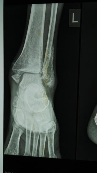
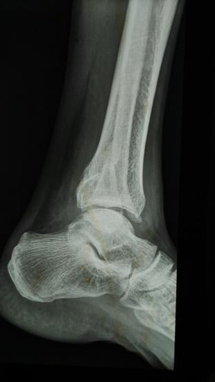
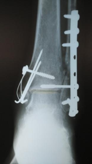
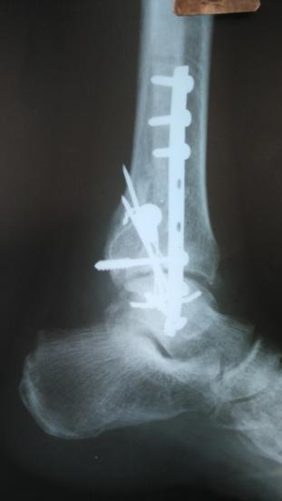
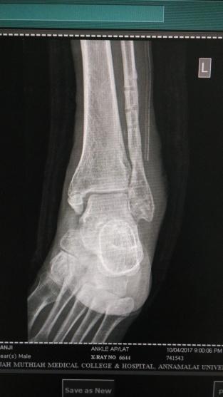
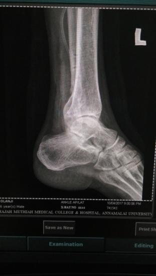
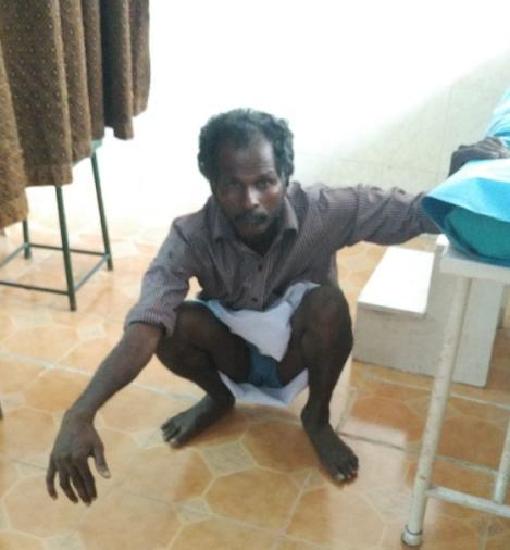
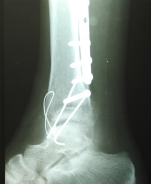
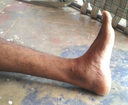
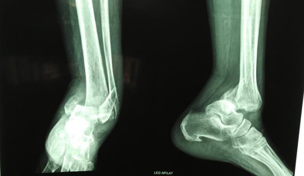
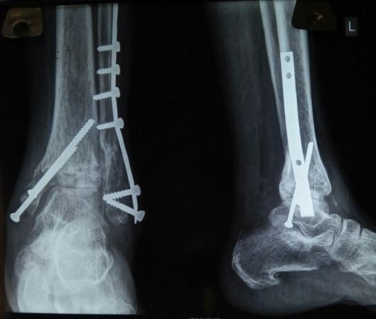
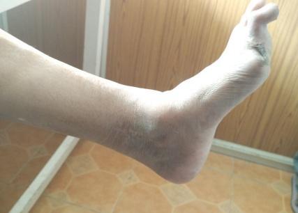
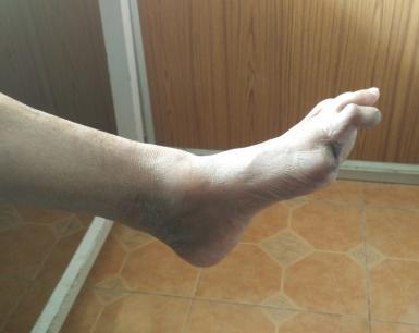
4 Case-1 Nithyakumar.V.R et al JMSCR Volume 05 Issue 11 November 2017 Page 30635

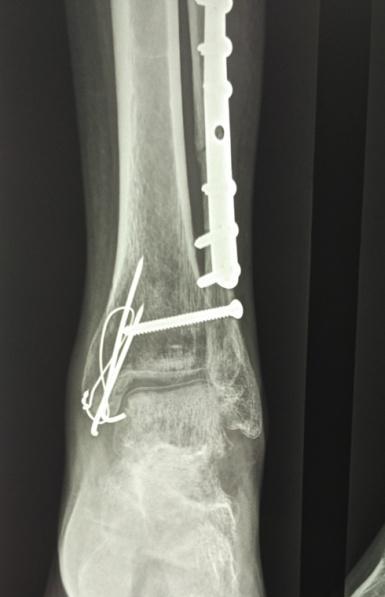
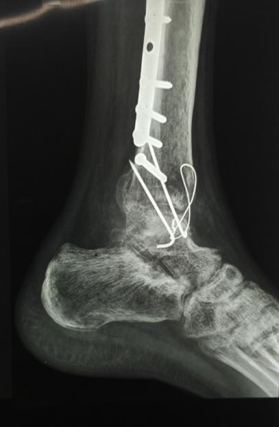


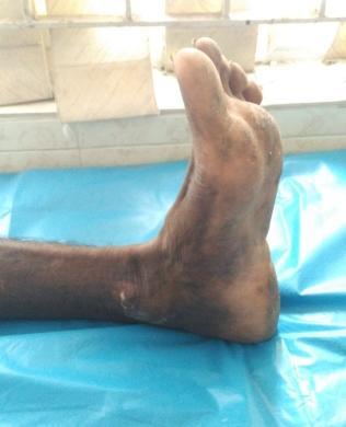
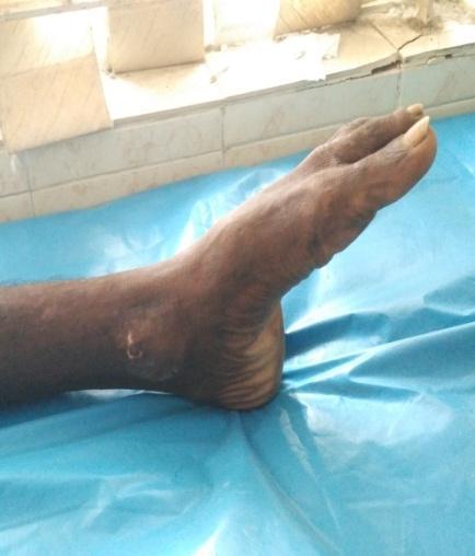
5 Case 2 Case 3 Nithyakumar.V.R et al JMSCR Volume 05 Issue 11 November 2017 Page 30636
6 Table 1: Type of Fracture Type of fracture Number of patients Trimalleolar fracture 4 Bimalleolar fracture 11 Only lateral malleolus fracture 2 Lateral malleolus fracture with deltoid ligament disruption 3 Table 2: Complications Arthritis 3 Post operative infection 1 Screw backout 1 Late diastasis 0 Tibio fibular synostosis 0 Discussion Our study consists of 20 cases of closed ankle fractures with syndesmotic disruption diagnosed by radiological views and intra-operative tests. Maximum incidence of the injury was in the fifth decade of life. Injury was more common in males- 12 (60%) and females being 8(40%). Right side was more commonly involved-12 patients (60%). Road traffic accidents contributed to 65% of injuries, followed by self fall while walking (35%). Out of 20 patients, 14 are PER TYPE III pattern, 3 patients are PER TYPE IV pattern, 1 cases of PER TYPE II pattern and 2 cases of SER TYPE IV. The most common injury pattern seen in our study was Pronation and external rotation type III. Stress radiographs are useful to assess ankle instability. Pronation external rotation injury type III and IV was very unstable and also associated syndesmotic disruption which should be diagnosed clinically and radiologically and must be fixed with syndesmotic screw from fibula to tibia, which may be stabilised by tricortical or quadricortical fixation depends on pattern of injury. Functional outcome was equal for both tricortical and quadricortical fixation supported by Macleod mark et al (12). But in fear of chance of screw breakage in quadricortical screw we usually prefer tricortical fixation. Usually we plan the whole implant removed at the end of 1 year. Till then the patients were allowed for weight bearing with screws insitu. David paul bell et al (13) study shows maximum of three years of patient with retained screw without complications In pronation external rotation injury fibular length restoration and rotation, ankle mortise and syndesmotic stability is important factor as noted by maverick et al (14). We had good to excellent results in all cases as we could maintain the syndesmotic stability and fibular length by syndesmotic screws and fibular plating. Displacement is position of talus in the mortise and depends on uninjured deltoid ligament (15). Fixing only the malleolar fragment will not restore ankle stability in case of deltoid ligament rupture. It must be repaired subsequently if the deep deltoid ligament is torn (16). Stable fractures shows no displacement in axial loading (17). Treatment depends on the stability of fracture. Prognosis is depends on energy of injury. If the deltoid ligament is torn it must be repaired. Even though Lauge-Hansen classification describes in detail about the pattern of ankle fracture it does not deal with syndesmotic injuries According to Micheal Bekorom (18), pronation injuries/ type C weber fractures are usually associated with syndesmotic injuries than supination injuries/ type B weber injuries. Miller study shows for ankle fractures syndesmotic injuries are fixed with screws and studied the functional outcome which was ranging from of olerud and molander scoring system. (19) Our study shows 20% of excellent result with score of 95 and 55% shows good result with score of 81-90, 25% shows fair results with some complications which were treated subsequently. Among 20 patients, screws were removed around 12 weeks for 6 patients, and the whole implant including fibular plate and malleolar screw was removed with syndesmotic screws in 6 patients, 6 patients were on regular follow-up waiting for screw removal along with other implant, 2 patients were on long follow up who refuse for second surgery for implant removal was on regular followup without implant breakage but complaing of ankle pain. Further these patients were Nithyakumar.V.R et al JMSCR Volume 05 Issue 11 November 2017 Page 30637
7 segregated and focused on ankle arthritis. On regular follow up there is progression of symptoms, were managed symptomatically and explained to the patients On retrospective analysis, the reason for arthritis were found out. In one patient we advised delayed weight bearing as he managed initially by native bone setters in the form of oil bandages, later he was operated for syndesmotic disruption by screw fixation, but due to early weight bearing by the patient, he developed arthritic features. In second patient, he was a manual labourer, and advised delayed return to labour activities, as he had comminuted fibula fracture, but the patient did not followed up and later he ended up in arthritis. Third patient had diabetic mellitus with osteoporotic bone. She had spontaneous screw back out at the time of 10 weeks post operative period. Initially she had no arthritis, but had persistent ankle pain. But on long term follow up (after 1 year) she was ended in arthritis. Conclusion Though pronation and supination injuries produces syndesmotic disruption, we encounter most commonly the pronation-external rotation injury. In that PER TYPE III and IV are commonly seen. Though there are many controversies regarding need to fix syndesmotic disruption or not, our study concludes there is definitive need of fixation of syndesmotic disruption that too by syndesmotic screw to allow for early mobilisation, early return activities, to prevent ankle arthritis. Though there are various methods of fixation, in our study fixing with screws shows good functional outcome. Regarding the amount of cortical purchase we prefer tricortical purchase, which will avoid the implant breakage and helps in delayed implant removal as a whole, which helps to avoid multiple surgeries for implant removal. Among the 20 patients, 3 patients had ankle arthritis which eventually started as chronic pain, then later on regular follow up ankle arthritis was noted both clinically and radiologically. Since the surgical fixation of syndesmotic injury was important to prevent ankle arthritis, three patients (15%) of 20 patients had ankle arthritis, due to various avoidable factors. References 1. Ramsey PL, Hamilton W. Changes in tibiotalar area of contact caused by lateral talar shift. J Bone Joint Surg Am. 1976;58: Hopkinson WJ, St Pierre P, Ryan JB, et al. Syndesmosis sprains of the ankle. Foot Ankle. 1990;10: Ashhurst APC, Bromer RS. Classification and mechanism of fractures of the leg bones involving the ankle: based on a study of three hundred cases from Episcopal Hospital. Arch Surg. 1922;4: Lauge-Hansen N. Fractures of the ankle. II. Combined experimental-surgical and experimental-roentgenologic investigations. Arch Surg. 1950;60: Weber BG. Die Verletzungen des oberen Sprunggelenkes. Bern: Hans Huber; pp Lanz VT, Wachsmuth W. Praktische Anatomie. Berlin: Springer-Verlag; pp Kelikian H, Kelikian S. Disorders of the Ankle. Philadelphia: W.B. Saunders Company; pp Jenkinson, Richard J MD; Sanders, David W MD, MSc, FRCS(C); Macleod, Mark D MD, FRCS(C); Domonkos, Andrea BSc; Lydestadt, Jeanette RNJournal of Orthopaedic Trauma: October Volume 19 - Issue 9 - pp doi: /01.bot McCollum G, van den Bekerom M, Kerkhoffs G, Calder J, van Dijk C. Syndesmosis and deltoid ligament injuries in the athlete. Knee Surg Sports Traumatol Arthrosc. 2013; 21: doi: /s Nithyakumar.V.R et al JMSCR Volume 05 Issue 11 November 2017 Page 30638
8 10. Beumer A, Swiestra B, Moulders P. Clinical diagnosis of syndesmosis instability: evaluation of stress tests behind the curtains. Acta Orthop Scand. 2002; 73: Cesar de Cesar P, Muller E. Comparison of MRI to physical examination for syndesmotic injuries after lateral ankle sprain. Foot Ankle Int. 2011;32: doi: /FAI Manjoo, Ajay MD; Sanders, David W MD, MSc, FRCSC; Tieszer, Christina MSc; MacLeod, Mark D MD, FRCSC Journal of Orthopaedic Traum A doi: /BOT.0b013e3181a9f7a5 13. David Paul Bell, Merng Koon Wong b Department of Orthopaedic Surgery, Changi General Hospital, 2 Simei Street 3, Singapore , Republic of Singapore, Accepted 1 February 2006, Available online 19 April Yablon I.G., Segal.D., Leanch R.E., Ankle Injuries, New york, 1983, Churchill Livingstone. 15. Yablon I.G., Heller. F.G., Shouse.L; The Key role of lateral malleolus in displaced fractures of ankle: J. Bone Joint Surg : 59 A: 169, Nithyakumar.V.R et al JMSCR Volume 05 Issue 11 November 2017 Page 30639
JMSCR Vol 05 Issue 11 Page November 2017
 www.jmscr.igmpublication.org Impact Factor 5.84 Index Copernicus Value: 71.58 ISSN (e)-2347-176x ISSN (p) 2455-0450 DOI: https://dx.doi.org/10.18535/jmscr/v5i11.30 Assesment of Outcome of Using Syndesmotic
PRONATION-ABDUCTION FRACTURES
 C H A P T E R 1 2 PRONATION-ABDUCTION FRACTURES George S. Gumann, DPM (The opinions of the author should not be considered as reflecting official policy of the US Army Medical Department.) Pronation-abduction
Deltoid and Syndesmosis Ligament Injury of the Ankle Without Fracture
 Deltoid and Syndesmosis Ligament Injury of the Ankle Without Fracture Chris D. Miller, MD, Walter R. Shelton,* MD, Gene R. Barrett, MD, F. H. Savoie, MD, and Andrea D. Dukes, MS From the Mississippi Sports
CURRENT TREATMENT OPTIONS
 CURRENT TREATMENT OPTIONS Fix single column or both: Always fix both. A study by Svend-Hansen corroborated the poor results associated with isolated medial malleolar fixation in bimalleolar ankle fractures.
Clinical evaluation where no obvious fracture a. Squeeze test
 7:43 am The Syndesmotic Injury: From Subtle to Severe Robert B. Anderson, MD Chief, Foot and Ankle Carolinas Medical Center OrthoCarolina (Charlotte, North Carolina) 7:30-8:25 am Symposium 1: Management
Competence of the Deltoid Ligament in Bimalleolar Ankle Fractures After Medial Malleolar Fixation *
 Competence of the Deltoid Ligament in Bimalleolar Ankle Fractures After Medial Malleolar Fixation * BY PAUL TORNETTA, III, M.D. Investigation performed at Kings County Hospital, New York, N.Y. Abstract
Disclosures. OTA Resident Advanced Trauma Techniques Course: Ankle Fractures. No relevant disclosures. William H. Harvin, MD Dallas, TX
 OTA Resident Advanced Trauma Techniques Course: Ankle Fractures William H. Harvin, MD Dallas, TX January 31, 2017 Disclosures No relevant disclosures 1 Ankle Anatomy: Lateral ankle ligaments Ankle Anatomy:
Disclosures. Syndesmosis Injury. Syndesmosis Ligaments. Objectives. Mark M. Casillas, M.D.
 Disclosures Syndesmosis Injury No relevant disclosures Mark M. Casillas, M.D. 1 Objectives Syndesmosis Ligaments Understand the syndesmosis anatomy and function Classify syndesmosis injuries Describe treatment
Treatment of malunited fractures of the ankle
 Treatment of malunited fractures of the ankle A LONG-TERM FOLLOW-UP OF RECONSTRUCTIVE SURGERY I. I. Reidsma, P. A. Nolte, R. K. Marti, E. L. F. B. Raaymakers From Academic Medical Center, Amsterdam, Netherlands
TECHNIQUE OF SYNDESMOTIC SCREW INSERTION IN WEBER TYPE C ANKLE FRACTURES
 ORIGINAL ARTICLE TECHNIQUE OF SYNDESMOTIC SCREW INSERTION IN WEBER TYPE C ANKLE FRACTURES SAJID EJAZ RAO, SOHAIL MUZAMMIL, ABDUL HAFEEZ KHAN ABSTRACT Objective Study design Place & Duration of study To
BIOMECHANICS OF ANKLE FRACTURES
 BIOMECHANICS OF ANKLE FRACTURES William R Reinus, MD MBA FACR Significance of Ankle Fractures Most common weight-bearing Fx 70% of all Fxs Incidence is increasing Bimodal distribution Men 15-24 Women over
Burwood Road, Concord Dora Street, Hurstville Lethbridge Street, Penrith 160 Belmore Road, Randwick
 www.orthosports.com.au 47 49 Burwood Road, Concord 29 31 Dora Street, Hurstville 119 121 Lethbridge Street, Penrith 160 Belmore Road, Randwick Update on Syndesmosis Ankle Sprains By Todd Gothelf Foot,
CASE REPORT RARE CASE OF DELTOID LIGAMENT AVULSION WITH MEDIAL MALLEOLUS FRACTURE OF ANKLE JOINT: CASE REPORT
 RARE CASE OF DELTOID LIGAMENT AVULSION WITH MEDIAL MALLEOLUS FRACTURE OF ANKLE JOINT: CASE REPORT Maruthi C.V 1, Roshan Pais 2 HOW TO CITE THIS ARTICLE: Maruthi CV, Roshan Pais. Rare case of deltoid ligament
Saudi Journal of Medicine (SJM)
") Saudi Journal of Medicine (SJM) Scholars Middle East Publishers Dubai, United Arab Emirates Website: http://scholarsmepub.com/ ISSN 2518-3389 (Print) ISSN 2518-3397 (Online) Surgical Management of Bimalleolar
Saudi Journal of Medicine (SJM) Scholars Middle East Publishers Dubai, United Arab Emirates Website: http://scholarsmepub.com/ ISSN 2518-3389 (Print) ISSN 2518-3397 (Online) Surgical Management of Bimalleolar
X-Ray Rounds: (Plain) Radiographic Evaluation of the Ankle.
 Radiographic Evaluation of the Ankle.") X-Ray Rounds: (Plain) Radiographic Evaluation of the Ankle www.fisiokinesiterapia.biz Anatomy Complex hinge joint Articulations among: Fibula Tibia Talus Tibial plafond Distal tibial articular surface
X-Ray Rounds: (Plain) Radiographic Evaluation of the Ankle www.fisiokinesiterapia.biz Anatomy Complex hinge joint Articulations among: Fibula Tibia Talus Tibial plafond Distal tibial articular surface
1/27/2016. Background. Background. Seth R. Yarboro University of Virginia January 29, Distal tibio fibular joint
 Seth R. Yarboro January 29, 2015 Background Distal tibio fibular joint maintains ankle stability while allowing motion Dorsiflexion/external rotation mechanism Poor alignment ankle arthritis Background
High Ankle Sprains: Diagnosis & Treatment
 High Ankle Sprains: Diagnosis & Treatment Mark J. Mendeszoon, DPM, FACFAS, FACFAOM Precision Orthopaedic Specialties University Regional Hospitals Advanced Foot & Ankle Fellowship- Director It Is Only
The Lauge Hansen Classification of Malleolar Fractures
 Acta Orthopaedica Scandinavica ISSN: 0001-6470 (Print) (Online) Journal homepage: http://www.tandfonline.com/loi/iort19 The Lauge Hansen Classification of Malleolar Fractures Johannes Yde To cite this
Surgery-Ortho. Fractures of the tibia and fibula. Management. Treatment of low energy fractures. Fifth stage. Lec-6 د.
 Fifth stage Lec-6 د. مثنى Surgery-Ortho 28/4/2016 Indirect force: (low energy) Fractures of the tibia and fibula Twisting: spiral fractures of both bones Angulatory: oblique fractures with butterfly segment.
Syndesmotic Ankle Injuries: Diagnosis and Treatment
 Syndesmotic Ankle Injuries: Diagnosis and Treatment John A. Scolaro, M.D., M.A. Assistant Professor of Orthopaedic Surgery University of California, Irvine California Orthopaedic Association - 2016 Disclosures
.org. Ankle Fractures (Broken Ankle) Anatomy
 Anatomy") Ankle Fractures (Broken Ankle) Page ( 1 ) A broken ankle is also known as an ankle fracture. This means that one or more of the bones that make up the ankle joint are broken. A fractured ankle can range
Ankle Fractures (Broken Ankle) Page ( 1 ) A broken ankle is also known as an ankle fracture. This means that one or more of the bones that make up the ankle joint are broken. A fractured ankle can range
FIBULAR & SYNDESMOSIS MALUNIONS
 FIBULAR & SYNDESMOSIS MALUNIONS MICHAEL P. CLARE, MD FLORIDA ORTHOPAEDIC INSTITUTE TAMPA, FL USA MORTISE INHERENTLY UNSTABLE LATERAL MALLEOLUS ACTS AS BUTTRESS / POST RESIST LATERAL TRANSLATION OF TALUS
Functional outcome and complications of surgically managed malleolar fractures at ankle
 International Journal of Research in Orthopaedics Rao KN et al. Int J Res Orthop. 2017 Jul;3(4):770-774 http://www.ijoro.org Original Research Article DOI: http://dx.doi.org/10.18203/issn.2455-4510.intjresorthop20172871
Pattern of Ankle Fractures And Assessment of Radiological Outcome of Surgically Treated Ankle Fractures
 IOSR Journal of Dental and Medical Sciences (IOSR-JDMS) e-issn: 2279-853, p-issn: 2279-861.Volume 15, Issue 6 Ver. I (June. 216), PP 67-71 www.iosrjournals.org Pattern of Ankle Fractures And Assessment
Objective. Reducing a displaced Syndesmosis 2/11/2016. Ankle Fractures Common Misconceptions. Common Myths in ankle fracture management
 Ankle Fractures Common Misconceptions Jackson Lee, MD Associate Professor Clinical Orthopedics Keck School of Medicine of the University of Southern California Objective Common Myths in ankle fracture
Ankle Ligament Injury: Don t Worry- It s Only a Sprain Wes Jackson MD Orthopaedic Foot & Ankle
 Ankle Ligament Injury: Don t Worry- It s Only a Sprain Wes Jackson MD Orthopaedic Foot & Ankle Outline I. Epidemiology II. Classification and Types of Sprains III. Anatomy IV. Clinical Assessment and Imaging
Radiographic assessment. Functional. Paul Tornetta III Professor 11/21/2016. Fracture not in coronal plane May need CT to evaluate
 The Posterior Malleolus Paul Tornetta III Professor Boston Medical Center Publications: Disclosures! Rockwood and Green, Tornetta and Einhorn; Subspecialty series, Court-Brown, Tornetta; Trauma, AAOS;
Treatment of Medial Malleolus or Pure Deltoid Ligament Injury in Patients with Supination- External Rotation Type IV Ankle Fractures
 42 2017 CHINESE ORTHOPAEDIC ASSOCIATION AND JOHN WILEY &SONS AUSTRALIA, LTD CLINICAL ARTICLE Treatment of Medial Malleolus or Pure Deltoid Ligament Injury in Patients with Supination- External Rotation
The Syndesmosis. Syndesmosis: How to Reduce and How Perfect? Boston Medical Center. Indications. Technique 11/19/2018.
 Syndesmosis: How to Reduce and How Perfect? Paul Tornetta III Professor Boston Medical Center Boston Medical Center The Syndesmosis Indications Subluxation Instability Technique Fluoroscopic Open 1 Weber
Ankle fracture classification : an evaluation of three classification systems : Lauge-Hansen, A.O. and Broos-Bisschop
 Acta Orthop. Belg., 2010, 76, 521-525 ORIGINAL STUDY Ankle fracture classification : an evaluation of three classification systems : Lauge-Hansen, A.O. and Broos-Bisschop Christos ALEXANDROPOULOS, Stefanos
ROTATIONAL PILON FRACTURES
 CHAPTER 31 ROTATIONAL PILON FRACTURES George S. Gumann, DPM The opinions and commentary of the author should not be construed as refl ecting offi cial U.S. Army Medical Department policy. Pilon injuries
Commonly Missed Foot and Ankle Conditions. David Miller, DPM AMG Podiatry
 Commonly Missed Foot and Ankle Conditions David Miller, DPM AMG Podiatry Lisfranc Injuries Wide spectrum of injuries High energy Subtle subluxation which could be easily missed injuries Men are 2-4x s
Surgical treatment of ankle fracture with or without deltoid ligament repair: a comparative study
 Zhao et al. BMC Musculoskeletal Disorders (2017) 18:543 DOI 10.1186/s12891-017-1907-4 RESEARCH ARTICLE Open Access Surgical treatment of ankle fracture with or without deltoid ligament repair: a comparative
Ankle and Foot Orthopaedic Tests Orthopedics and Neurology DX 612
 Ankle and Foot Orthopaedic Tests Orthopedics and Neurology DX 612 James J. Lehman, DC, MBA, DABCO University of Bridgeport College of Chiropractic Ankle & Foot Anatomy Stability of the ankle is dependent
Disclosures! The Syndesmosis. Syndesmosis: How and When to Reduce. Boston Medical Center. Indications. Technique.
 Syndesmosis: How and When to Reduce Paul Tornetta III Professor Boston Medical Center Boston Medical Center Publications: Disclosures! Rockwood and Green, Tornetta and Einhorn; Subspecialty series, Court-Brown,
Ankle fracture: The operative outcome of 30 patients
 2018; 4(1): 947-951 ISSN: 2395-1958 IJOS 2018; 4(1): 947-951 2018 IJOS www.orthopaper.com Received: 27-11-2017 Accepted: 28-12-2017 Purushotham K Professor and HOD, Department of Swet Ranjan Shoaib Mohammed
Copyright Protected. Ankle fractures are the most common intraarticular
 An Original Study Ankle Fracture Syndesmosis Fixation and Management: The Current Practice of Orthopedic Surgeons Eric Bava, MD, Timothy Charlton, MD, and David Thordarson, MD Abstract There is a wide
Outline. Ankle/Foot Anatomy Ankle Sprains Ottawa Ankle Rules DDx: The Sprain That Wasn t
 Ankle Injuries Outline Ankle/Foot Anatomy Ankle Sprains Ottawa Ankle Rules DDx: The Sprain That Wasn t Anatomy: Ankle Mortise Bony Anatomy Lateral Ligament Complex Medial Ligament Complex Ankle Sprains
An anthropometric study of distal tibiofibular syndesmosis (DTS) in a Chinese population
 in a Chinese population") Yu et al. Journal of Orthopaedic Surgery and Research (2018) 13:95 https://doi.org/10.1186/s13018-018-0804-3 RESEARCH ARTICLE Open Access An anthropometric study of distal tibiofibular syndesmosis (DTS)
Yu et al. Journal of Orthopaedic Surgery and Research (2018) 13:95 https://doi.org/10.1186/s13018-018-0804-3 RESEARCH ARTICLE Open Access An anthropometric study of distal tibiofibular syndesmosis (DTS)
SURGICAL AND APPLIED ANATOMY
 Página 1 de 9 Copyright 2001 Lippincott Williams & Wilkins Bucholz, Robert W., Heckman, James D. Rockwood & Green's Fractures in Adults, 5th Edition SURGICAL AND APPLIED ANATOMY Part of "47 - ANKLE FRACTURES"
Pure Closed Posteromedial Dislocation of the Tibiotalar Joint without Fracture
 214 2013 Chinese Orthopaedic Association and Wiley Publishing Asia Pty Ltd BRIEF REPORT Pure Closed Posteromedial Dislocation of the Tibiotalar Joint without Fracture Yun-tao Wang, MD, PhD, Xiao-tao Wu,
RADIOGRAPHY OF THE ANKLE and LOWER LEG
 RADIOGRAPHY OF THE ANKLE and LOWER LEG Patient Position: ANKLE AP Projection Part Position: True Slight to place foot s long axis Center to Central Ray: to IR Midway Note: Ankle joint is to tips of malleoli
Isolated Syndesmotic Instability The High Ankle Sprain Robert B. Anderson, MD
 Isolated Syndesmotic Instability The High Ankle Sprain Robert B. Anderson, MD Chief, Foot & Ankle Service Carolinas Medical Center OrthoCarolina Team Orthopaedist, Carolina Panthers Charlotte, North Carolina
Long-term functional and radiographic outcomes in 243 operated ankle fractures
 Verhage et al. Journal of Foot and Ankle Research (2015) 8:45 DOI 10.1186/s13047-015-0098-1 JOURNAL OF FOOT AND ANKLE RESEARCH RESEARCH Long-term functional and radiographic outcomes in 243 operated ankle
Fibula Lengthening Using a Modified Ilizarov Method S. Robert Rozbruch, MD; Matthew DiPaola, BA; Arkady Blyakher,MD
 Fibula Lengthening Using a Modified Ilizarov Method S. Robert Rozbruch, MD; Matthew DiPaola, BA; Arkady Blyakher,MD Limb Lengthening Service Hospital for Special Surgery Abstract A unique combination of
The Lower Limb VII: The Ankle & Foot. Anatomy RHS 241 Lecture 7 Dr. Einas Al-Eisa
 The Lower Limb VII: The Ankle & Foot Anatomy RHS 241 Lecture 7 Dr. Einas Al-Eisa Ankle joint Synovial, hinge joint Allow movement of the foot in the sagittal plane only (1 degree of freedom): dorsiflexion:
Fibular Malalignment in Subjects with Chronic Ankle Instability
 Fibular Malalignment in Subjects with Chronic Ankle Instability Takumi Kobayashi 1,2, Eiichi Suzuki 3, Naohito Yamazaki 3, Makoto Suzukawa 4, Atsushi Akaike 4, Kuniaki Shimizu 4, Kazuyoshi Gamada 1. 1
Donald Stewart, MD. Lateral ligament injuries Chronic lateral ligament instability Syndesmosis Injuries
 Donald Stewart, MD Arlington Orthopedic Associates Lateral ligament injuries Chronic lateral ligament instability Syndesmosis Injuries Anatomy Mechanism of Injury Classification Diagnostic Tests Management
OTA Speciality Day New Orleans Subtle Syndesmotic Injuries: How I diagnose them and How to Fix. Kenneth A Egol MD
 OTA Speciality Day 2018- New Orleans Subtle Syndesmotic Injuries: How I diagnose them and How to Fix Kenneth A Egol MD 1. Due to their inherent instability, it is well established that syndesmotic fixation
A Patient s Guide to Ankle Syndesmosis Injuries
 A Patient s Guide to Ankle Syndesmosis Injuries Introduction An ankle injury common to athletes is the ankle syndesmosis injury. This type of injury is sometimes called a high ankle sprain because it involves
Copyright 2004, Yoshiyuki Shiratori. All right reserved.
 Ankle and Leg Evaluation 1. History Chief Complaint: A. What happened? B. Is it a sharp or dull pain? C. How long have you had the pain? D. Can you pinpoint the pain? E. Do you have any numbness or tingling?
Ankle Fracture: Tips and Tricks
 Ankle Fracture: Tips and Tricks Christiaan N. Mamczak, DO LCDR, MC, USN Naval Medical Center Portsmouth Department of Orthopaedic Surgery Assistant Professor Uniformed Services University of the Health
Cost Effectiveness of a New Ankle Fracture System
 Cost Effectiveness of a New Ankle Fracture System John D. Hewitt, MD 1, Joshua N. Tennant, MD, MPH 2, Ryan C. May, BS 3 and Selene G. Parekh, MD, MBA 1, 4 1 Division of Orthopaedic Surgery, Duke University
Revision Ankle Syndesmosis Fixation: Functional
 JFS JFs (P) Original rticle Revision nkle Syndesmosis Fixation: Functional 10.5005/jp-journals-10040-1044 Outcome after TightRope Fixation Revision nkle Syndesmosis Fixation: Functional Outcome after TightRope
Foot and Ankle Update
 Foot and Ankle Update 2019 Instructional Course Hiro Tanaka It s your on-call weekend Objectives We are going to apply evidence based treatment for 2 patients who are admitted under your care 1. Dislocated
Inion FreedomScrew Syndesmosis Repair. Biodegradable Fixation System
 Inion FreedomScrew Syndesmosis Repair Biodegradable Fixation System Inion FreedomScrew for Syndesmosis Repair Inion FreedomScrew is a strong and versatile resorbable screw for orthopaedic fixations. Because
Ankle Syndesmosis Injuries
 A Patient s Guide to Ankle Syndesmosis Injuries 1436 Exchange Street Middlebury, VT 05753 Phone: 802-388-3194 Fax: 802-388-4881 cvo@champlainvalleyortho.com DISCLAIMER: The information in this booklet
OTA Resident Core Curriculum Lecture Series Updated November 2010 Matt Graves, M.D. University of Mississippi Medical Center
 Ankle Fracture Update OTA Resident Core Curriculum Lecture Series Updated November 2010 Matt Graves, M.D. University of Mississippi Medical Center Objectives Following this session, you should be able
Ankle Fractures in the Elderly: How to Deal with Poor Bone Quality
 : How to Deal with Poor Bone Quality Richard T. Laughlin, MD Professor of Orthopaedic Surgery University of Cincinnati College of Medicine No disclosures relative to this presentation acknowledgement Some
Ankle Syndesmotic Fixation Implants and Techniques. Ryan Harris, DO Orthopedic Resident, PGY-4 Pinnacle Health Hospital Harrisburg, PA
 Ankle Syndesmotic Fixation Implants and Techniques Ryan Harris, DO Orthopedic Resident, PGY-4 Pinnacle Health Hospital Harrisburg, PA Introduction Ankle syndesmosis injuries can occur in up to 10% of patients
DEPARTMENT OF TRAUMATOLOGY AND HAND SURGERY INSTITUTE OF MUSCULOSKELETAL SURGERY ANKLE AND FOOT INJURIES
 DEPARTMENT OF TRAUMATOLOGY AND HAND SURGERY INSTITUTE OF MUSCULOSKELETAL SURGERY ANKLE AND FOOT INJURIES Presenter: Dr George Ayerh ENGLISH PROGRAM LECTURES EN_11/A - 2018 TOPICS I. Part: Ankle & Foot
5/3/2016 DISCLOSURES. Outline. Hassan R. Mir, MD, MBA, FACS. Ankle Fractures Lateral Malleolus Medial Malleolus Posterior Malleolus Chaput Syndesmosis
 DISCLOSURES Hassan R. Mir, MD, MBA, FACS Medical/Orthopaedic Publications Editorial/Governing Board OTA Newsletter Editor OsteoSynthesis, The JOT Online Discussion Forum Editor JOT Associate Editor JAAOS
Malleolar fractures Oswestry foot and ankle course
 Malleolar fractures Oswestry foot and ankle course Jim Barrie This handout accompanies and expands Jim Barrie s lecture on malleolar fractures. It should be read in conjunction with the material (including
Ankle Sprains and Their Imitators
 Ankle Sprains and Their Imitators Mark Halstead, MD Dr. Mark Halstead is the Associate Professor of the Departments of Orthopedics and Pediatrics at Washington University School of Medicine; Director of
CLINICAL PRESENTATION AND RADIOLOGY QUIZ QUESTION
 Donald L. Renfrew, MD Radiology Associates of the Fox Valley, 333 N. Commercial Street, Suite 100, Neenah, WI 54956 12/08/2012 Radiology Quiz of the Week # 102 Page 1 CLINICAL PRESENTATION AND RADIOLOGY
A Comparative Study of Tension Band Wiring and Cannulated Screw Fixation for Medial Malleolar Fractures
 IOSR Journal of Dental and Medical Sciences (IOSR-JDMS) e-issn: 2279-0853, p-issn: 2279-0861.Volume 14, Issue 12 Ver. X (Dec. 2015), PP 42-49 www.iosrjournals.org A Comparative Study of Tension Band Wiring
Patrick B Ebeling, MD Minnesota Sports Medicine & Twin Cities Orthopedics Adjunct Associate Professor, University of Minnesota, Minneapolis
 Page 32 / SA ORTHOPAEDIC JOURNAL Autumn 2009 CLINICAL ARTICLE C LINICAL A RTICLE Treatment of syndesmoses disruptions: A prospective, randomized study comparing conventional screw fixation vs TightRope
Stability of Ankle Fracture dislocations following Successful Closed Reduction
 Andrew P Matson et al CLINICAL RESEARCH 10.5005/jp-journals-10017-1084 Stability of Ankle Fracture dislocations following Successful Closed Reduction 1 Andrew P Matson MD, 2 Cynthia L Green PhD, 3 Shepard
Paul Alley MD,DPM,MS,FACS,FAAOS,BFD Eby Orthopaedics,Jasper,Indiana
 Paul Alley MD,DPM,MS,FACS,FAAOS,BFD Eby Orthopaedics,Jasper,Indiana Very common Bone=fractures Description (cracked,broke,busted,or smashed) A=anatomic area of bone eg: head,neck,shaft B=bone involved
Influence of bone morphology and injured ligament of the ankle on ankle stress radiographs
 Influence of bone morphology and injured ligament of the ankle on ankle stress radiographs Gye Wang Lee, MD, Chin Youb Chung, MD, Moon Seok Park, MD Seung Yeol Lee, MD, Myung Ki Chung, MD, Byung Chae Jo,
Review relevant anatomy of the foot and ankle. Learn the approach to examining the foot and ankle
 Objectives Review relevant anatomy of the foot and ankle Learn the approach to examining the foot and ankle Learn the basics of diagnosis and treatment of ankle sprains Overview of other common causes
The effect of different methods of stability assessment on fixation rate and complications in supination external rotation (SER) 2/4 ankle fractures.
 2/4 ankle fractures.") The effect of different methods of stability assessment on fixation rate and complications in supination external rotation (SER) 2/4 ankle fractures. Edward J.C. Dawe R.Shafafy, J.Quayle, N.Gougoulias,
The effect of different methods of stability assessment on fixation rate and complications in supination external rotation (SER) 2/4 ankle fractures. Edward J.C. Dawe R.Shafafy, J.Quayle, N.Gougoulias,
Young Uk Park, M.D., Ph.D.. Young Wook Seo, M.D. Hyuk, Jegal, M.D.,* Kyung Tai Lee, M.D.,Ph.D.
 Young Uk Park, M.D., Ph.D.. Young Wook Seo, M.D. Hyuk, Jegal, M.D.,* Kyung Tai Lee, M.D.,Ph.D. Department of Orthopedic Surgery, Ajou University Hospital, Ajou University School of Medicine, Suwon, Gyeonggi-do,
The Flower Straight Fibula Plate
 The Flower Straight Fibula Plate PROCEDURE GUIDE www.flowerortho.com The Flower Foot & Ankle Application STRAIGHT LOCKING FIBULA PLATE ANTERIOR LATERAL DISTAL TIBIAL PLATE MEDIAL DISTAL TIBIAL PLATE ANATOMIC
Stability of Ankle Fracture-Dislocations following Successful Closed Reduction
 Stability of Ankle Fracture-Dislocations following Successful Closed Reduction Andrew P. Matson 1, MD Cynthia L. Green 1, PhD Shepard R. Hurwitz 2, MD Robert D. Zura 3, MD 1. Duke University School of
INVISION Total Ankle Replacement System with PROPHECY Preoperative Navigation Revision of a Failed Agility Total Ankle Replacement
 016625 REVISION R INVISION Total Ankle Replacement System with PROPHECY Preoperative Navigation Revision of a Failed Agility Total Ankle Replacement CASE STUDY Patient History The patient was a 65-year-old
The outcome at 20 years of conservatively treated isolated posterior malleolar fractures of the ankle
 C. C. M. A. Donken, A. J. F. Goorden, M. H. J. Verhofstad, M. J. Edwards, C. J. H. M. van Laarhoven From Radboud University Nijmegen Medical Center, Nijmegen, The Netherlands FOOT AND ANKLE The outcome
Acute Ankle Injuries, Part 1: Office Evaluation and Management
 t June 08, 2009 Obesity [1] Each acute ankle injury commonly seen in the office has associated with it a mechanism by which it can be injured, trademark symptoms that the patient experiences during the
Duration of Follow-up (mo)
") Page 1 of 7 Fig. E-1 Fig. E-2 Fig. E-1 Medial ankle arthritis with medial translation of the talus and mortise widening. Note the shape of the medial malleolus (white arrow). Fig. E-2 Measurement of mortise
Page 1 of 7 Fig. E-1 Fig. E-2 Fig. E-1 Medial ankle arthritis with medial translation of the talus and mortise widening. Note the shape of the medial malleolus (white arrow). Fig. E-2 Measurement of mortise
LCP Anterior Ankle Arthrodesis Plates. Part of the Synthes Locking Compression Plate (LCP) System.
 System.") LCP Anterior Ankle Arthrodesis Plates. Part of the Synthes Locking Compression Plate (LCP) System. Technique Guide Instruments and implants approved by the AO Foundation Table of Contents Introduction
LCP Anterior Ankle Arthrodesis Plates. Part of the Synthes Locking Compression Plate (LCP) System. Technique Guide Instruments and implants approved by the AO Foundation Table of Contents Introduction
5 COMMON INJURIES IN THE FOOT & ANKLE
 5 COMMON INJURIES IN THE FOOT & ANKLE MICHAEL P. CLARE, MD FLORIDA ORTHOPAEDIC INSTITUTE TAMPA, FL USA MECHANISM OF INJURY HOW DID IT HAPPEN? HIGH ENERGY VS LOW ENERGY DIRECTION OF FORCES INVOLVED LIVING
Key Words: ankle injury, ligaments, lower-leg injury, sprains, tibiofibular diastasis
 Ankle Syndesmosis Injuries: Anatomy, Biomechanics, Mechanism of Injury, and Clinical Guidelines for Diagnosis and Intervention Cheng-Feng Lin, MS 1 Michael T. Gross, PT, PhD 2 Paul Weinhold, PhD 3 Journal
Surgical Technique. Fibula Rod System
 Surgical Technique Fibula Rod System Acumed is a global leader of innovative orthopaedic and medical solutions. We are dedicated to developing products, service methods, and approaches that improve patient
Arthroscopy Of the Ankle.
 Arthroscopy Of the Ankle www.fisiokinesiterapia.biz Ankle Arthroscopy Anatomy Patient setup Portal placement Procedures Complications Anatomy Portals Anterior Anteromedial Anterolateral Anterocentral Posterior
11/4/2018 SUBTLETIES OF LOWER EXTREMITY TRAUMA IMAGING SPEAKER DISCLOSURES
 SUBTLETIES OF LOWER EXTREMITY TRAUMA IMAGING Charles S. Resnik, M.D. Professor of Radiology University of Maryland School of Medicine Upon completion of this presentation, participants will be better able
Biokinesiology of the Ankle Complex
 Rehabilitation Considerations Following Ankle Fracture: Impact on Gait & Closed Kinetic Chain Function Disclosures David Nolan, PT, DPT, MS, OCS, SCS, CSCS I have no actual or potential conflict of interest
11/2/17. Lateral Collateral Complex Medial Collateral Complex Distal Tibiofibular Syndesmosis Spring Ligament
 Andrew J Grainger Leeds, UK Lateral Collateral Complex ial Collateral Complex Distal Tibiofibular Syndesmosis Spring Ligament Brief anatomy review Scan tips and tricks Pathological appearances andrewgrainger@nhs.net
Fractures of the tibia shaft treated with locked intramedullary nail Retrospective clinical and radiographic assesment
 ARS Medica Tomitana - 2013; 4(75): 197-201 DOI: 10.2478/arsm-2013-0035 Șerban Al., Botnaru V., Turcu R., Obadă B., Anderlik St. Fractures of the tibia shaft treated with locked intramedullary nail Retrospective
Trimalleolar Fractures with Impaction of the Posteromedial Tibial Plafond: Implications for Talar Stability
 FOOT &ANKLE INTERNATIONAL Copyright 2004 by the American Orthopaedic Foot & Ankle Society, Inc. Trimalleolar Fractures with Impaction of the Posteromedial Tibial Plafond: Implications for Talar Stability
The study of distal ¼ diaphyseal extra articular fractures of humerus treated with antegrade intramedullary interlocking nailing
 2018; 4(4): 46-50 ISSN: 2395-1958 IJOS 2018; 4(4): 46-50 2018 IJOS www.orthopaper.com Received: 01-08-2018 Accepted: 03-09-2018 Dr. Ankur Parikh Orthopaedics, Jehangir Hospital, Sassoon road, Pune, Dr.
June 2013 Case Study. Author: T. Walker Robinson, MD, MPH, Nationwide Children s Hospital
 June 2013 Case Study Author: T. Walker Robinson, MD, MPH, Nationwide Children s Hospital Chief Complaint: Right ankle pain HPI: A 10 year old female dancer presents to the clinic with a five day history
Increasing surgical freedom Restoring patient function
 Increasing surgical freedom Restoring patient function Fracture specific plating solutions for the most common tibia and fibula fractures Frequency of fracture occurrences* 66% 61% 36% 36% 28% 14% 20%
Anterior Impingement
 Anterior Impingement Ziali Sivardeen BMedSci, (MRCS), AFRCS, FRCS (Tr & Orth) Consultant Trauma and Orthopaedic Surgeon (Shoulder, Knee and Sports Injuries) Aims Causes of Anterior Ankle Pain Ankle Impingement
Lower Extremity Dislocations: Management and Triage on the Field
 Lower Extremity Dislocations: Management and Triage on the Field Scott J Tarantino, MD Towson Orthopaedic Associates, Towson, MD None Disclsures Purpose To provide you with knowledge which may guide you
Syndesmosis injuries in the pediatric and adolescent athlete: an analysis of risk factors related to operative intervention
 Syndesmosis injuries in the pediatric and adolescent athlete: an analysis of risk factors related to operative intervention The Harvard community has made this article openly available. Please share how
The fibular incisura of the tibia with recurrent sprained ankle on magnetic resonance imaging
 The fibular incisura of the tibia with recurrent sprained ankle on magnetic resonance imaging Ayfer Mavi, PhD, Hanifi Yildirim, MD, Hasan Gunes, MD, Turan Pestamalci, MD, Erdem Gumusburun, PhD. ABSTRACT
Hany El-Rashidy and Anand Vora
 Chapter 194 Lisfranc Injuries Chapter 194 Lisfranc Injuries Hany El-Rashidy and Anand Vora 8 ICD-9 CODE 838.03 Lisfranc (Tarsometatarsal) Fracture-Dislocation Key Concepts The Lisfranc joint represents
A STUDY OF SURGICAL MANAGEMENT OF MALLEOLAR FRACTURES IN ADULTS Srinivas Nagendra G 1, Prabhakar Venkataramana 2, Siddarth Mahesh 3
 A STUDY OF SURGICAL MANAGEMENT OF MALLEOLAR FRACTURES IN ADULTS Srinivas Nagendra G 1, Prabhakar Venkataramana 2, Siddarth Mahesh 3 HOW TO CITE THIS ARTICLE: Srinivas Nagendra G, Prabhakar Venkataramana,
Introduction Introduction Ankle Sprains Ankle Sprains ankl nkle
 s/ Syndesmotic Injuries 21% of all athletic injuries are to the ankle 25% of NFL injuries are foot and ankle related Vast majority are simple inversion twisting types Classic sprains involve the lower