Surgical Technique. Elbow Plating System
|
|
|
- Loraine Fleming
- 5 years ago
- Views:
Transcription

1 Surgical Technique Elbow Plating System


2 Acumed is a global leader of innovative orthopaedic and medical solutions. We are dedicated to developing products, service methods, and approaches that improve patient care. Elbow Plating System Design Surgeon Shawn W. O Driscoll, Ph.D., M.D. Acumed Elbow Plating System Designed in conjunction with Shawn W. O Driscoll, Ph.D., M.D., the Elbow Plating System is designed to address fractures of the distal humerus, olecranon, and coronoid. The Elbow Plating System offers precontoured, indication specific plates and includes a low-profile Olecranon Plate design with anatomic curvature and instrumentation to aid with plate and screw insertion. This system also includes the Hexalobe Screw System with variable angle Tap-Loc Technology for the Medial and Lateral Distal Humerus Plates. Posterolateral Plates are offered in addition to our Medial and Lateral Distal Humerus Plates to provide multiple plating solutions for elbow fracture management. Indications for Use: Fractures of the distal humerus, olecranon, and coronoid Osteotomies of the olecranon Contents Introduction 2 Plating System 3 Olecranon Plate Design 4 Elbow Plate Surgical Techniques Olecranon Plate 5 Olecranon Osteotomy 8 Cutting Jig Distal Humerus Plates 9 Posterolateral Plate 14 Coronoid Plate 17 Ordering Information 20 Notes 25 2


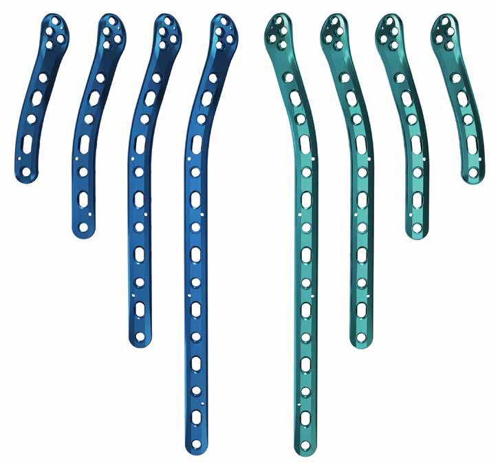
3 Plating System Olecranon Plates Coronoid Plates 3


4 Posterolateral Column Plates Lateral Column Plates Medial Column Plates 4


5 Plating Design Olecranon Plate Proximal screw cluster Prongs allow plate to sit on top of the triceps tendon Home Run screw Divergent locking screw trajectory Anatomic curvature including lateral tilt in the proximal region, distal lateral bow, and 6 proximal ulnar dorsal angulation (PUDA). Limited contact design Distal taper designed to reduce stress concentrations Posterolateral Distal Humerus Plate Proximal taper designed to reduce stress concentrations Limited contact design Converging and diverging distal screw cluster 5

 into the plate hole. A 2.")
 can be placed across the fracture, one on each side of the plate. 3 Nonlocking Distal Screw Placement With provisional reduction confirmed, drill with the 2.")
6 Olecranon Plate Surgical Technique Shawn W. O Driscoll, Ph.D., M.D. 1 Fracture Reduction and Plate Placement Attach the Proximal Targeting Guide ( ) to the plate with the Locking Bolt ( ). Flex the elbow 90, reduce the fracture and apply the plate. The prongs in the proximal end of the plate should penetrate the triceps tendon and provide provisional fixation. These prongs are not intended to compress the tendon and a gap between the plate and the bone should be visible on X-ray. 2 Provisional Wire Placement If a locking screw is to be utilized in the most proximal hole of the plate, thread the 2.3 mm Locking Drill Guide ( ) into the plate hole. A 2.0 mm Wire (WS-2009ST) is drilled through the locking drill guide and across the fracture site, penetrating the anterior metaphyseal cortex. Do not remove this wire until Step 6. Alternatively, two.062" Wires (WS-1607ST) can be placed across the fracture, one on each side of the plate. 3 Nonlocking Distal Screw Placement With provisional reduction confirmed, drill with the 2.8 mm Drill ( ), using a Depth Gauge ( ), measure depth and insert a 3.5 mm nonlocking screw through the slotted hole distal to the fracture site and into the ulnar shaft. Connect the T15 Hexalobe Driver ( ) to the Ratcheting Driver Handle ( ) and tighten the screw partially to allow for later compression. Bone taps are provided and recommended for patients with dense bone. Note: When implanting the Narrow Olecranon Plates ( or ), only the 2.7 mm Hexalobe Screws and associated instrumentation may be used 6
, be careful not to exit the bone. Drill depth may be read directly off of the laser line on the drill or with the 2.0 mm Depth Probe (80-0643).")

7 4 Proximal Locking Screw Placement Insert two 2.7 mm locking screws into the proximal holes on either side of the 2.0 mm wire, using the 2.0 mm Locking Drill Guide ( ). When drilling with the 2.0 mm Drill ( ), be careful not to exit the bone. Drill depth may be read directly off of the laser line on the drill or with the 2.0 mm Depth Probe ( ). The T8 Hexalobe Driver ( ) is used to insert the 2.7 mm screws. When using the T8 Driver, care should be taken to not overtighten the screw or apply more torque than necessary to seat the locking screw into the plate. Screws should be tightened by hand and not under power. Note: Plates designed for use on the left arm are blue in color. Plates designed for the right arm are green in color. Screw Diameter Drill Diameter 2.7 mm 2.0 mm 3.0 mm 2.3 mm 3.5 mm 2.8 mm 5 Fracture Site Compression If the plate length selected has two or more compression slots, the fracture site is compressed in the following manner. Insert a 3.5 mm nonlocking screw in dynamic compression mode into a distal slot along the ulnar shaft using the Offset Drill Guide (PL-2095). The proximal shaft screw must be slightly loosened to allow for compression. If a longer plate is used and further compression is required, partially insert another nonlocking screw into a distal slot in dynamic compression mode and then loosen the first two screws to allow for plate movement. 7
 in the path of the wire. Measure depth and insert the screw. If a 3.0 mm home run screw is desired, the 2.3 mm Locking Drill Guide (80-0622) and Drill (80-0627) are utilized.")


8 6 Final Screw Placement Remove the 2.0 mm wire from the most proximal plate hole and insert a locking 3.5 mm screw: attach the 2.8 mm Locking Drill Guide ( ) and use the 2.8 mm Drill ( ) in the path of the wire. Measure depth and insert the screw. If a 3.0 mm home run screw is desired, the 2.3 mm Locking Drill Guide ( ) and Drill ( ) are utilized. The proximal targeting guide may be removed at this time. The remaining locking screws are then inserted at the surgeon s discretion. 7 Postoperative Protocol Immediately after closure, the elbow is placed in a bulky non-compressive Jones dressing with an anterior plaster slab to maintain the elbow in extension. The initial rehabilitation is planned according to the extent of soft-tissue damage. When the fracture is associated with severe soft-tissue damage, the extremity is kept immobilized with the elbow in extension for three to seven days postoperatively. If the fracture is closed and there is no severe swelling or fracture blisters, the Jones dressing is removed after two days and an elastic non-constrictive sleeve is applied over an absorbent dressing placed on the wound. A physical therapy program including active and passive motion is then initiated. Techincal Objectives for Locking Olecranon Plates: Each screw should be as long as possible Locking screws should interlock to provide a stable fixed angle structure inside the bone fragment Plate should buttress against anterior pull of elbow flexors Plate should provide stable fixation of the ulnar shaft Plate should be applied with compression across the fracture Plate must be strong and stiff enough to resist bending before union occurs 8
 onto the proximal portion of the olecranon with the elbow flexed at 90.")
 may also be placed in the small K-wire hole between the cutting slots.")
 to drill the slot for future placement of a 3.5 mm screw. The 2.0 mm Drill (80-0318) is utilized to drill the two smaller, proximal holes for future placement of the 2.")
9 Olecranon Osteotomy Cutting Jig Surgical Technique Shawn W. O Driscoll, Ph.D., M.D. 1 Provisional Fixation Place the Olecranon Osteotomy Cutting Jig ( ) onto the proximal portion of the olecranon with the elbow flexed at 90. The jig is designed to sit on top of the triceps tendon. Secure the jig provisionally by placing a Plate Tack (PL-PTACK) into the plate tack holes in the jig. A.062" K-wire (WS-1607ST) may also be placed in the small K-wire hole between the cutting slots. 2 Pre-Drill Screw Holes The Olecranon Osteotomy Cutting Jig allows pre-drilling of the screw holes that will be used with future placement of the Olecranon Plate. Use a 2.8 mm Drill ( ) to drill the slot for future placement of a 3.5 mm screw. The 2.0 mm Drill ( ) is utilized to drill the two smaller, proximal holes for future placement of the 2.7 mm screws. 3 Create Osteotomy Select the cutting slot that provides the most optimal position for the chevron osteotomy. Using a thin-bladed oscillating saw (.025" in thickness), create an osteotomy about 1/3 of the way through the olecranon. Remove the Osteotomy Cutting Jig. Use the oscillating saw to join the two sides of the provisional cut. A thin-bladed osteotome is used to complete the osteotomy. 9
.")
.")


10 Distal Humerus Plates Surgical Technique Shawn W. O Driscoll, Ph.D., M.D. 1 Articular Fragment Reduction The articular fragments, which tend to be rotated toward each other in the axial plane, are reduced anatomically and provisionally held with.045" smooth K-wires (WS-1106ST). It is essential that these wires be placed close to the subchondral level to avoid interference with later screw placement, and away from where the plates will be placed on the lateral and medial columns (see Step 2). One or two strategically placed wires can then be used to provisionally hold the distal fragments in alignment with the humeral shaft. 2 Plate Placement and Provisional Fixation The selected medial and lateral plates are placed and held apposed to the distal humerus, while one smooth 2.0 mm K-wire (WS-2009ST) is inserted through hole #2 (numbered from distal to proximal) of each plate through the epicondyles and across the distal fragments to maintain provisional fixation. These 2.0 mm wires are left in place until Step 7 to aid in placing the locking screws in the distal fragments. Hole #1 Hole #2 Hole #3 Note: The medial and lateral distal humerus plates are designed to accept 3.0 mm and 3.5 mm hexalobe screws. The 2.7 mm hexalobe screws have a smaller head diameter and should NOT be used with the medial and lateral distal humerus plates. Lateral Column Plate Medial Column Plate 10


11 3 Initial Proximal Screw Placement With provisional reduction confirmed, drill with the 2.8 mm Drill ( ), measure depth ( ) and insert a 3.5 mm nonlocking screw into a slotted hole of each plate proximal to the fracture site. Connect the T15 Hexalobe Driver ( ) to the Ratcheting Driver Handle ( ) and tighten the screw partially, allowing some freedom for the plate to move proximally during compression later. (Because the undersurface of each plate is tubular in the metaphyseal and diaphyseal regions, the screw in the slotted hole only needs to be tightened slightly to provide provisional fixation of the entire distal humerus.) Bone taps are recommended for patients with dense bone. 4 Nonlocking Distal Screw Placement Drill and insert screws through hole #1 on both the medial and lateral side. The targeted drill guide cannot be used in hole #1 of the Medial Plate if the angle of the nonlocking screw exceeds 20. After drilling, measure depth and insert the appropriate size 3.5 mm nonlocking screw. The 3.0 mm screws may be used to enable more screws to be placed in the distal fragments to provide stability. 5 Compress Lateral Column Using a Large Tenaculum (MS-1280) to provide interfragmentary compression across the fracture at the supracondylar level, the lateral column is first fixed. A screw is inserted in the lateral plate in dynamic compression mode in a slotted hole proximal to the fracture site using the Offset Drill Guide (PL-2095). Tightening this screw further increases interfragmentary compression at the supracondylar level to the point of causing some distraction at the medial supracondylar ridge. The.045" wires used for provisional fixation may be removed at this point. 11
.")

12 6 Compress Medial Column The medial column is then compressed in a similar manner using the Large Tenaculum (MS-1280), and a 3.5 mm nonlocking screw is inserted in the medial plate in dynamic compression mode in a slotted hole proximal to the fracture site, using the Offset Drill Guide (PL-2095). If the plates are slightly under contoured, they can be compressed against the metaphysis with a large bone clamp, giving further supracondylar compression. Remove the 2.0 mm wires that were inserted in Step 2. 7 Tap Distal Plate Holes If using a 3.5 mm screw, use the 2.8 mm drill in the path of the wire. If using a 3.0 mm screw, the 2.3 mm drill is utilized. Measure drill depth ( ) to determine screw size. Connect the Plate Tap ( or ) to the T-Handle (MS-T1212) and tap the plate. The front end of the tap will act as a guide to aid the locking screw in following the correct trajectory. Turning the tap one-half turn at a time, tap the plate taking care not to insert the tap further than the start of the laser line on the tap threads (See Tapping Instructions on page 14). The T-Handle should only be used with the plate taps and not for locking or nonlocking screw insertion. The proximal slotted holes are NOT to be tapped. 8 Insert Distal Locking Screws Insert the appropriate size locking screws. Care should be taken to not overtighten the screw. The #3 holes on both the medial and lateral columns are optional. If these holes are used, be sure to use locking screws if locking screws have already been inserted in previous steps. 12
 into the locking hole in the plate.")


13 9 Insert Proximal Locking Screws The remaining locking shaft screws may be inserted at the surgeon s discretion. Note that the plate holes in the humeral shaft are pre-threaded, fixed angle screws. To insert the 3.5 mm or 3.0 mm locking screws, thread the appropriate size Locking Drill Guide ( or ) into the locking hole in the plate. Drill with the appropriate size Drill ( or ). Drill depth may be read directly off of the laser line on the drill or with the 2.3 mm Depth Probe ( ). Insert the appropriate size locking screws. 10 Postoperative Protocol Immediately after closure, the elbow is placed in a bulky non-compressive Jones dressing with an anterior plaster slab to maintain the elbow in extension. The initial rehabilitation is planned according to the extent of soft-tissue damage. When the fracture is associated with severe soft-tissue damage, the extremity is kept immobilized with the elbow in extension for three to seven days postoperatively. If the fracture is closed and there is no severe swelling or fracture blisters, the Jones dressing is removed after two days and an elastic non-constrictive sleeve is applied over an absorbent dressing placed on the wound. A physical therapy program including active and passive motion is then initiated. 13
14 Acumed Single Use Tapping Instrument Precautions: Tapping a plate using a plate tap will cause titanium debris to be generated, which should be removed. Failure to remove the plate debris can cause, among other complications, inflammation, cartilage damage and patient discomfort. The taps are single surgery use and should be discarded after each surgery or if the tap becomes dull or damaged. If the resistance increases while using a tap, discard the tap immediately. Breakage to the tap can occur due to excessive torque or levering and care should be taken to avoid such conditions. Should breakage occur, carefully remove all tap pieces. Tapping Instructions: Do not tap deeper than the start of the laser line Clean debris from tap after tapping each hole Irrigate hole prior to tapping Do not tap a slot Do not re-tap a hole (use a nonlocking screw) Tap by hand, not under power Angle of tapped hole must not exceed 20 Technical Objectives Checklist: Every screw should pass through a plate Each screw engages a fragment on the opposite side that is also attached to a plate Each screw should be as long as possible Each screw should engage as many fragments as possible The screws in the distal fragments should lock together by interdigitation, creating a fixed angle structure Plates should be applied such that compression is achieved at the supracondylar level for both columns Plates must be strong and stiff enough to resist breaking or bending before union occurs. Screw Diameter Drill Diameter 3.0 mm 2.3 mm 3.5 mm 2.8 mm 14


 and the 8\" Reduction Forceps (MS-1280) are provided in the system to aid in fracture reduction.")
.")
15 Posterolateral Plate Surgical Technique Shawn W. O Driscoll, Ph.D., M.D. 1 Articular Fragment Reduction Following exposure, the articular fragments are reduced anatomically and provisionally held using.045" K-wires (WS-1106ST). It is essential that these wires be placed close to the subchondral level to avoid interference with later screw placement, and away from where the plate will be placed on the posterolateral column. The Pointed Forceps (MS-45300) and the 8" Reduction Forceps (MS-1280) are provided in the system to aid in fracture reduction. Acumed Elbow Plating System Surgical Technique 2 Plate Placement and Provisional Fixation Apply the selected plate to the bone. K-wire holes are included on the plate for provisional fixation and accept.062" K-wires (WS-1607ST). Plate Tacks (PL-PTACK) may also be used through the plate holes to aid in provisional fixation. 3 Initial Proximal Screw Placement With provisional reduction confirmed, drill with the 2.8 mm Drill ( ), measure depth ( ) and insert a 3.5 mm nonlocking hexalobe screw through the slotted hole that is located proximally on the plate. Connect the T15 Hexalobe Driver ( ) to the Ratcheting Driver Handle ( ) and insert the screw. Bone taps are provided and recommended for patients with dense bone. 15
 into a plate hole. Select the 2.0 mm Drill (80-0318) and drill to the desired depth through the 2.0 mm locking drill guide.")
.")


16 4 Distal Screw Fixation and Supracondylar Compression The three most distal locking screws are inserted first by threading the 2.0 mm Locking Drill Guide ( ) into a plate hole. Select the 2.0 mm Drill ( ) and drill to the desired depth through the 2.0 mm locking drill guide. Drill depth may be read directly off of the laser band on the drill or with a 2.0 mm Depth Probe ( ). The most proximal of the four distal screws may be inserted for additional fixation of the distal fragments (shown in the illustration). Connect the T8 Hexalobe Driver ( ) to the Ratcheting Driver Handle ( ) and insert a 2.7 mm locking hexalobe screw until it is fully seated in the plate. Care should be taken to not overtighten the locking screws. Repeat this step for the remaining distal screws. To achieve supracondylar compression, the screw in the slotted hole should be loosened and the fracture compressed at the supracondylar level. 5 Insert Proximal Locking Screws The remaining locking shaft screws may be inserted at the surgeon s discretion. To insert the 3.5 mm or 3.0 mm locking screws, thread the appropriate size Locking Drill Guide ( or ) into the locking hole in the plate. Drill with the appropriate size Drill ( or ). Drill depth may be read directly off of the laser band on the drill or with the 2.3 mm Depth Probe ( ). Insert the appropriate size locking screws. 16
 to minimize the likelihood of compartment syndrome.")
17 6 Postoperative Protocol Immediately after closure, the elbow is placed in a bulky noncompressive Jones dressing with an anterior plaster slab to maintain the elbow in extension, and the upper extremity is elevated. The arm should be brought down from the elevated position frequently enough (perhaps once per hour) to minimize the likelihood of compartment syndrome. The initial rehabilitation is planned according to the extent of soft-tissue damage. When the fracture is associated with severe soft-tissue damage, the extremity is kept immobilized and elevated with the elbow in extension for three to seven days postoperatively. If the fracture is closed and there is no severe swelling or fracture blisters, the Jones dressing is removed after three days and an elastic non-constrictive sleeve is applied over an absorbent dressing placed on the wound. A physical therapy program including active and passive motion is then initiated. Screw Diameter Drill Diameter 2.7 mm 2.0 mm 3.0 mm 2.3 mm 3.5 mm 2.8 mm 17
.")
 the offset screw hole should sit over that fragment for proper screw")
18 Coronoid Plates Surgical Technique Shawn W. O Driscoll, Ph.D., M.D. 1 Fracture Fragment Fixation Expose the coronoid and ridge of the ulna through an anteromedial approach. Reduce and provisionally hold the fragments with smooth.045" K-wires (WS-1106ST). 2 PLATE PLACEMENT AND PROVISIONAL FIXATION Apply the Coronoid Plate so that the two prongs on the proximal section grasp and buttress the anteromedial facet of the coronoid. If the sublime tubercle, on which the anterior bundle of the MCL inserts, is also fractured (Anteromedial Subtype III fracture) the offset screw hole should sit over that fragment for proper screw position. The distal portion of the plate should extend along the ridge on the anteromedial side of the ulna. Several.045" K-wires may be used for provisional plate fixation through the K-wire holes in the plate. Note: Use caution when handling the plate as it has sharp prongs. Repeated and excessive bending may damage the plate causing it to not fit or function as intended. 18
 and T8 Hexalobe Driver (80-0759) are utilized for screw insertion.")

19 3 INSERT CENTRAL NONLOCKING SCREW The first screw inserted is a 2.7 mm nonlocking hexalobe screw into hole #1, which is the central plate hole. The 2.0 mm Drill ( ) and T8 Hexalobe Driver ( ) are utilized for screw insertion. When determining the screw lengths, make sure to compensate for any expected plate deformation if the bend does not fully seat the plate against the bone. As the screw is tightened, the plate will flex and contour to the bone. If the outer proximal hole begins to bend outward, tighten this first screw only partially, insert the most proximal screw, then go back to fully seat the central screw. Seating this screw may also cause the prongs on the proximal portion of the plate to buttress the coronoid and further compress the plate to the bone. Note: Tapping the bone prior to screw insertion with the Bone Tap ( ) may be needed for patients with dense bone. 4 CORONOID FIXATION Insert nonlocking screws into the proximal portion of the plate (holes #2 and #3). If K-wires were inserted for provisional fixation, they should be removed prior to drilling and inserting screws into the proximal portion of the plate. The offset screw hole, #4, is optional and can be filled with a nonlocking screw if the fracture extends to the sublime tubercle. Image intensification is strongly recommended to verify the trajectory of the nonlocking screws to ensure they avoid the articular surface. As these nonlocking screws are inserted, the plate will continue to contour to the bone. 4 (optional)
. Nonlocking screws can be used at the surgeon s discretion.")

20 5 INSERT REMAINING SCREWS To insert the 2.7 mm locking hexalobe screws, thread the 2.0 mm Locking Drill Guide ( ) into each distal plate hole and drill with the 2.0 mm Drill ( ). Insert the locking screws into holes #5 and #6 with the T8 Hexalobe Driver ( ). Nonlocking screws can be used at the surgeon s discretion. Care should be taken to not overtighten the screws or apply excess torque on the driver. 6 POSTOPERATIVE PROTOCOL Immediately after closure, the elbow is placed in a bulky non-compressive Jones dressing with an anterior plaster slab to maintain the elbow in a relatively extended position and the upper extremity is kept elevated for three days, bringing it down from the elevated position each hour for 5 10 minutes to avoid inadequate perfusion. The initial rehabilitation is planned according to the stability of the elbow, security of fracture fixation and the extent of soft tissue damage. Screw Diameter Drill Diameter 2.7 mm 2.0 mm 20
21 Ordering Information Olecranon Plates Olecranon Plate, Standard, 3-hole, Left (65 mm) Olecranon Plate, Standard, 3-hole, Right (65 mm) Olecranon Plate, Standard, 5-hole, Left (90 mm) Olecranon Plate, Standard, 5-hole, Right (90 mm) Olecranon Plate, Standard, 7-hole, Left (110 mm) Olecranon Plate, Standard, 7-hole, Right (110 mm) Olecranon Plate, Standard, 11-hole, Left (150 mm) Olecranon Plate, Standard, 11-hole, Right (150 mm) Olecranon Plate, Extended, 5-hole, Left (90 mm) Olecranon Plate, Extended, 5-hole, Right (90 mm) Olecranon Plate, Extended, 9-hole, Left (130 mm) Olecranon Plate, Extended, 9-hole, Right (130 mm) Optional Posterolateral Distal Humerus Plates Posterolateral Distal Humerus Plate, 15 Hole, Left (203 mm) Posterolateral Distal Humerus Plate, 15 Hole, Right (203 mm) Coronoid Plates Coronoid Plate, Standard, Left Coronoid Plate, Standard, Right Optional Coronoid Plates Coronoid Plate, Small, Left Coronoid Plate, Small, Right Optional Olecranon Plates Olecranon Plate, Standard, 15-hole, Left (190 mm) Olecranon Plate, Standard, 15-hole, Right (190 mm) Olecranon Plate, Narrow, 5-hole, Left (85 mm) Olecranon Plate, Narrow, 5-hole, Right (85 mm)
22 Ordering Information Distal Humerus Plates Locking Medial Plate, 7-Hole (84 mm) Locking Medial Plate, 8-Hole (88 mm) Locking Medial Plate, Long, 9-Hole (96 mm) Locking Medial Plate, Short, 9-Hole (95 mm) Locking Medial Plate, 12-Hole (130 mm) Locking Medial Plate, 16-Hole (175 mm) Locking Lateral Plate, 6-Hole, Left (58 mm) Locking Lateral Plate, 6-Hole, Right (58 mm) Locking Lateral Plate, 10-Hole, Left (100 mm) Locking Lateral Plate, 10-Hole, Right (100 mm) Locking Lateral Plate, 14-Hole, Left (142 mm) Locking Lateral Plate, 14-Hole, Right (142 mm) Locking Lateral Plate, 20-Hole, Left (206 mm) Locking Lateral Plate, 20-Hole, Right (206 mm) PL-LEM7 PL-LEM8 PL-LEM9L PL-LEM9S PL-LEM12 PL-LEM16 PL-LEL6L PL-LEL6R PL-LEL10L PL-LEL10R PL-LEL14L PL-LEL14R PL-LEL20L PL-LEL20R Posterolateral Distal Humerus Plate, 5 Hole, LT (78 mm) Posterolateral Distal Humerus Plate, 5 Hole, RT (78 mm) Posterolateral Distal Humerus Plate, 7 Hole, LT (103 mm) Posterolateral Distal Humerus Plate, 7 Hole, RT (103 mm) Posterolateral Distal Humerus Plate, 11 Hole, LT (152 mm) Posterolateral Distal Humerus Plate, 11 Hole, RT (152 mm)
23 3.5 mm Locking Hexalobe Screws 3.5 mm x 8 mm Locking Hexalobe Screw mm x 10 mm Locking Hexalobe Screw mm x 12 mm Locking Hexalobe Screw mm x 14 mm Locking Hexalobe Screw mm x 16 mm Locking Hexalobe Screw mm x 18 mm Locking Hexalobe Screw mm x 20 mm Locking Hexalobe Screw mm x 22 mm Locking Hexalobe Screw mm x 24 mm Locking Hexalobe Screw mm x 26 mm Locking Hexalobe Screw mm x 28 mm Locking Hexalobe Screw mm x 30 mm Locking Hexalobe Screw mm x 32 mm Locking Hexalobe Screw mm x 34 mm Locking Hexalobe Screw mm x 36 mm Locking Hexalobe Screw mm x 38 mm Locking Hexalobe Screw mm x 40 mm Locking Hexalobe Screw mm x 45 mm Locking Hexalobe Screw mm x 50 mm Locking Hexalobe Screw mm x 55 mm Locking Hexalobe Screw mm x 60 mm Locking Hexalobe Screw mm Nonlocking Hexalobe Screws 3.5 mm x 8 mm Nonlocking Hexalobe Screw mm x 10 mm Nonlocking Hexalobe Screw mm x 12 mm Nonlocking Hexalobe Screw mm x 14 mm Nonlocking Hexalobe Screw mm x 16 mm Nonlocking Hexalobe Screw mm x 18 mm Nonlocking Hexalobe Screw mm x 20 mm Nonlocking Hexalobe Screw mm x 22 mm Nonlocking Hexalobe Screw mm x 24 mm Nonlocking Hexalobe Screw mm x 26 mm Nonlocking Hexalobe Screw mm x 28 mm Nonlocking Hexalobe Screw mm x 30 mm Nonlocking Hexalobe Screw mm x 32 mm Nonlocking Hexalobe Screw mm x 34 mm Nonlocking Hexalobe Screw mm x 36 mm Nonlocking Hexalobe Screw mm x 38 mm Nonlocking Hexalobe Screw mm x 40 mm Nonlocking Hexalobe Screw mm x 45 mm Nonlocking Hexalobe Screw mm x 50 mm Nonlocking Hexalobe Screw mm x 55 mm Nonlocking Hexalobe Screw mm x 60 mm Nonlocking Hexalobe Screw mm x 65 mm Nonlocking Hexalobe Screw
24 Ordering Information 3.0 mm Locking Hexalobe Screws 3.0 mm x 8 mm Locking Hexalobe Screw mm x 10 mm Locking Hexalobe Screw mm x 12 mm Locking Hexalobe Screw mm x 14 mm Locking Hexalobe Screw mm x 16 mm Locking Hexalobe Screw mm x 18 mm Locking Hexalobe Screw mm x 20 mm Locking Hexalobe Screw mm x 22 mm Locking Hexalobe Screw mm x 24 mm Locking Hexalobe Screw mm x 26 mm Locking Hexalobe Screw mm x 28 mm Locking Hexalobe Screw mm x 30 mm Locking Hexalobe Screw mm x 32 mm Locking Hexalobe Screw mm x 34 mm Locking Hexalobe Screw mm x 36 mm Locking Hexalobe Screw mm x 38 mm Locking Hexalobe Screw mm x 40 mm Locking Hexalobe Screw mm x 45 mm Locking Hexalobe Screw mm x 50 mm Locking Hexalobe Screw mm x 55 mm Locking Hexalobe Screw mm x 60 mm Locking Hexalobe Screw mm Nonlocking Hexalobe Screws 3.0 mm x 8 mm Nonlocking Hexalobe Screw mm x 10 mm Nonlocking Hexalobe Screw mm x 12 mm Nonlocking Hexalobe Screw mm x 14 mm Nonlocking Hexalobe Screw mm x 16 mm Nonlocking Hexalobe Screw mm x 18 mm Nonlocking Hexalobe Screw mm x 20 mm Nonlocking Hexalobe Screw mm x 22 mm Nonlocking Hexalobe Screw mm x 24 mm Nonlocking Hexalobe Screw mm x 26 mm Nonlocking Hexalobe Screw mm x 28 mm Nonlocking Hexalobe Screw mm x 30 mm Nonlocking Hexalobe Screw mm x 32 mm Nonlocking Hexalobe Screw mm x 34 mm Nonlocking Hexalobe Screw mm x 36 mm Nonlocking Hexalobe Screw mm x 38 mm Nonlocking Hexalobe Screw mm x 40 mm Nonlocking Hexalobe Screw mm x 45 mm Nonlocking Hexalobe Screw mm x 50 mm Nonlocking Hexalobe Screw mm x 55 mm Nonlocking Hexalobe Screw mm x 60 mm Nonlocking Hexalobe Screw mm x 65 mm Nonlocking Hexalobe Screw
25 2.7 Locking Hexalobe Screws 2.7 mm x 8 mm Locking Hexalobe Screw mm x 10 mm Locking Hexalobe Screw mm x 12 mm Locking Hexalobe Screw mm x 14 mm Locking Hexalobe Screw mm x 16 mm Locking Hexalobe Screw mm x 18 mm Locking Hexalobe Screw mm x 20 mm Locking Hexalobe Screw mm x 22 mm Locking Hexalobe Screw mm x 24 mm Locking Hexalobe Screw mm x 26 mm Locking Hexalobe Screw mm x 28 mm Locking Hexalobe Screw mm x 30 mm Locking Hexalobe Screw mm x 32 mm Locking Hexalobe Screw Instrumentation T8 Stick-Fit Hexalobe Driver T15 Stick-Fit Hexalobe Driver mm Quick Release Drill mm Quick Release Drill mm Quick Release Drill mm Quick Release Drill MS-DC35 Bone Tap for 2.7 mm Hexalobe Screws Bone Tap for 3.0 mm Nonlocking Screws mm Long Tap Tip MS-LTT35 Plate Tap for 3.0 mm Screw Plate Tap for 3.5 mm Screw mm x 9" Guide Wire, Single Trocar WS-2009ST.045" x 6" SS Guide Wire WS-1106ST.062" x 6" SS Guide Wire WS-1607ST 2.7 Nonlocking Hexalobe Screws 2.7 mm x 8 mm Nonlocking Hexalobe Screw " x 6" Titanium Guide Wire (threaded) WT-1606STT.035" x 6" Titanium Guide Wire (threaded) WT-0906STT 2.7 mm x 10 mm Nonlocking Hexalobe Screw Plate Tack PL-PTACK 2.7 mm x 12 mm Nonlocking Hexalobe Screw mm x 14 mm Nonlocking Hexalobe Screw mm x 16 mm Nonlocking Hexalobe Screw These implants are available nonsterile or sterile-packed. Add -S to product number for sterile products. To order, contact your local Acumed Representative. 2.7 mm x 18 mm Nonlocking Hexalobe Screw mm x 20 mm Nonlocking Hexalobe Screw mm x 22 mm Nonlocking Hexalobe Screw mm x 24 mm Nonlocking Hexalobe Screw mm x 26 mm Nonlocking Hexalobe Screw mm x 28 mm Nonlocking Hexalobe Screw mm x 30 mm Nonlocking Hexalobe Screw mm x 32 mm Nonlocking Hexalobe Screw Tension Band Pins 70 mm Non-Sterile Tension Band Pin mm Non-Sterile Tension Band Pin
26 26 Notes:
27 Notes: 27
28 ELB00-05-H Effective: 01/ Acumed LLC Acumed 5885 NW Cornelius Pass Road Hillsboro, OR Office: Fax: acumed.net These materials contain information about products that may or may not be available in any particular country or may be available under different trademarks in different countries. The products may be approved or cleared by governmental regulatory organizations for sale or use with different indications or restrictions in different countries. Products may not be approved for use in all countries. Nothing contained on these materials should be construed as a promotion or solicitation for any product or for the use of any product in a particular way which is not authorized under the laws and regulations of the country where the reader is located. Specific questions physicians may have about the availability and use of the products described on these materials should be directed to their particular local sales representative. Specific questions patients may have about the use of the products described in these materials or the appropriateness for their own conditions should be directed to their own physician.
Elbow Plating System
 Elbow Plating System Elbow Plating System Acumed is a global leader of innovative orthopaedic and medical solutions. We are dedicated to developing products, service methods and approaches that improve
Elbow Plating System. Surgical Technique
 Elbow Plating System Surgical Technique Acumed is a global leader of innovative orthopaedic and medical solutions. We are dedicated to developing products, service methods, and approaches that improve
Mayo Clinic Congruent Elbow Plate System
 AcUMEDr Mayo Clinic Congruent Elbow Plate System Congruent Elbow Plate System Since 1988, Acumed has been designing solutions to the demanding situations facing orthopaedic surgeons, hospitals and their
Osteotomy System. Surgical Technique
 Osteotomy System Surgical Technique Acumed is a global leader of innovative orthopaedic and medical solutions. We are dedicated to developing products, service methods, and approaches that improve patient
Surgical Technique. Forearm Fracture Solutions
 Surgical Technique Forearm Fracture Solutions Acumed is a global leader of innovative orthopaedic and medical solutions. We are dedicated to developing products, service methods, and approaches that improve
Locking Radial Head Plates
 Locking Radial Head Plates Locking Radial Head Plates Since 1988, Acumed has been designing solutions to the demanding situations facing orthopaedic surgeons, hospitals and their patients. Our strategy
Acu-Loc Wrist Spanning Plate System. Surgical Technique
 Acu-Loc Wrist Spanning Plate System Surgical Technique Acumed is a global leader of innovative orthopaedic and medical solutions. We are dedicated to developing products, service methods, and approaches
Locking Ankle Plating System. Surgical Technique
 Locking Ankle Plating System Surgical Technique Acumed is a global leader of innovative orthopaedic and medical solutions. We are dedicated to developing products, service methods, and approaches that
Surgical Technique. Tension Band Pin System
 Surgical Technique Tension Band Pin System Acumed is a global leader of innovative orthopaedic and medical solutions. We are dedicated to developing products, service methods, and approaches that improve
Forearm Fracture Solutions. Surgical Technique
 Forearm Fracture Solutions Surgical Technique Acumed is a global leader of innovative orthopaedic and medical solutions. We are dedicated to developing products, service methods, and approaches that improve
Osteotomy System. The reference lines on the plate help facilitate the creation of the osteotomy, when a free hand cut is preferred.
 Osteotomy System Osteotomy System Since 1988, Acumed has been designing solutions for the demanding situations facing orthopaedic surgeons, hospitals and their patients. Our strategy has been to know the
Tension Band Pin System 2. Surgical Technique
 Tension Band Pin System 2 Surgical Technique Acumed is a global leader of innovative orthopaedic and medical solutions. We are dedicated to developing products, service methods, and approaches that improve
AcUMEDr. Olecranon Threaded Compression Rod
 AcUMEDr Olecranon Threaded Compression Rod Olecranon Threaded Compression Rod Since 1988, Acumed has been designing solutions to the demanding situations facing orthopaedic surgeons, hospitals and their
AcUMEDr. FoREARM ROD SYSTEM
 AcUMEDr FoREARM ROD SYSTEM FoREARM ROD SYSTEM Since 1988 Acumed has been designing solutions to the demanding situations facing orthopedic surgeons, hospitals and their patients. Our strategy has been
Hand Fracture System with Small Bone External Fixation System. Product Overview
 Hand Fracture System with Small Bone External Fixation System Product Overview Acumed Hand Fracture System The Acumed Hand Fracture System is designed to provide both standard and fracture-specific fixation
Tension Band Pin System
 Tension Band Pin System Tension Band Pin System Acumed is an industry leader in innovative solutions for extremities and trauma. We are dedicated to pioneering solutions that benefit the patient and drive
Forefoot/Midfoot Plating System
 Surgical Technique Forefoot/Midfoot Plating System Acumed is a global leader of innovative orthopaedic and medical solutions. We are dedicated to developing products, service methods, and approaches that
Forearm Fracture Solutions. Product Overview
 Forearm Fracture Solutions Product Overview Acumed Forearm Fracture Solutions Acumed Forearm Fracture Solutions includes plating and rodding systems with a range of diaphyseal radius and ulna fracture
Surgical Technique. Fibula Rod System
 Surgical Technique Fibula Rod System Acumed is a global leader of innovative orthopaedic and medical solutions. We are dedicated to developing products, service methods, and approaches that improve patient
Acu-Loc Wrist Plating System. Surgical Technique
 Acu-Loc Wrist Plating System Surgical Technique Acumed is a global leader of innovative orthopaedic and medical solutions. We are dedicated to developing products, service methods, and approaches that
Surgical Technique. Ankle Plating System
 Surgical Technique Ankle Plating System Acumed is a global leader of innovative orthopaedic and medical solutions. We are dedicated to developing products, service methods, and approaches that improve
AcUMEDr. Anatomic Midshaft Forearm Plates
 AcUMEDr Anatomic Midshaft Forearm Plates Anatomic Midshaft Forearm Plates Since 1988, Acumed has been designing solutions to the demanding situations facing orthopaedic surgeons, hospitals and their patients.
LCP Medial Distal Tibia Plate, without Tab. The Low Profile Anatomic Fixation System with Angular Stability and Optimal Screw Orientation.
 LCP Medial Distal Tibia Plate, without Tab. The Low Profile Anatomic Fixation System with Angular Stability and Optimal Screw Orientation. Technique Guide LCP Small Fragment System Table of Contents Introduction
Technique Guide. 3.5 mm LCP Olecranon Plates. Part of the Synthes locking compression plate (LCP) system.
 system.") Technique Guide 3.5 mm LCP Olecranon Plates. Part of the Synthes locking compression plate (LCP) system. Table of Contents Introduction 3.5 mm LCP Olecranon Plates 2 AO Principles 3 Indications 3 Clinical
Technique Guide 3.5 mm LCP Olecranon Plates. Part of the Synthes locking compression plate (LCP) system. Table of Contents Introduction 3.5 mm LCP Olecranon Plates 2 AO Principles 3 Indications 3 Clinical
3.5 mm LCP Extra-articular Distal Humerus Plate
 Part of the DePuy Synthes Locking Compression Plate (LCP ) System 3.5 mm LCP Extra-articular Distal Humerus Plate Surgical Technique Table of Contents Introduction 3.5 mm LCP Extra-articular Distal Humerus
Technique Guide. 3.5 mm LCP Low Bend Medial Distal Tibia Plates. Part of the Synthes locking compression plate (LCP) system.
 system.") Technique Guide 3.5 mm LCP Low Bend Medial Distal Tibia Plates. Part of the Synthes locking compression plate (LCP) system. Table of Contents Introduction 3.5 mm LCP Low Bend Medial Distal Tibia Plates
Technique Guide 3.5 mm LCP Low Bend Medial Distal Tibia Plates. Part of the Synthes locking compression plate (LCP) system. Table of Contents Introduction 3.5 mm LCP Low Bend Medial Distal Tibia Plates
Biotrak Resorbable Fixation System
 Surgical Technique Biotrak Resorbable Fixation System Acumed is a global leader of innovative orthopaedic and medical solutions. We are dedicated to developing products, service methods, and approaches
Technique Guide. 3.5 mm LCP Low Bend Medial Distal Tibia Plate Aiming Instruments. Part of the 3.5 mm LCP Percutaneous Instrument System.
 Technique Guide 3.5 mm LCP Low Bend Medial Distal Tibia Plate Aiming Instruments. Part of the 3.5 mm LCP Percutaneous Instrument System. Table of Contents Introduction 3.5 mm LCP Low Bend Medial Distal
Forefoot/Midfoot Plating System. Surgical Technique
 Forefoot/Midfoot Plating System Surgical Technique Acumed is a global leader of innovative orthopaedic and medical solutions. We are dedicated to developing products, service methods, and approaches that
Technique Guide. LCP Proximal Femoral Hook Plate 4.5/5.0. Part of the LCP Periarticular Plating System.
 Technique Guide LCP Proximal Femoral Hook Plate 4.5/5.0. Part of the LCP Periarticular Plating System. Table of Contents Introduction Features and Benefits 2 AO ASIF Principles 4 Indications 5 Surgical
3.5 mm LCP Hook Plate
 Part of the DePuy Synthes Locking Compression Plate (LCP ) System 3.5 mm LCP Hook Plate Surgical Technique Table of Contents Introduction 3.5 mm LCP Hook Plate 2 AO Principles 4 Indications 5 Clinical
Surgical Technique. Distal Humerus Locking Plate
 Surgical Technique Distal Humerus Locking Plate PERI-LOC Locked Plating System Distal Humerus Locking Plate Surgical Technique Table of Contents Introduction...2 Indications...3 Plate Features...3 Patient
Technique Guide. 3.5 mm LCP Periarticular Proximal Humerus Plate. Part of the Synthes locking compression plate (LCP) system.
 system.") Technique Guide 3.5 mm LCP Periarticular Proximal Humerus Plate. Part of the Synthes locking compression plate (LCP) system. Table of Contents Introduction 3.5 mm LCP Proximal Humerus Plate 2 AO Principles
Technique Guide 3.5 mm LCP Periarticular Proximal Humerus Plate. Part of the Synthes locking compression plate (LCP) system. Table of Contents Introduction 3.5 mm LCP Proximal Humerus Plate 2 AO Principles
Instrument and Implant for wrist fracture
 Instrument and Implant for wrist fracture Jansri Janpanya Product specialist The Bangkok Unitrade Co,.ltd. Objectives Type of LCP for distal radius Fx. The new LCP design for distal radius Fx. Have knowledge
Simply Complete. VA-LCP Elbow Plating System.
 Simply Complete. VA-LCP Elbow Plating System. Building on success Founded upon the established principles of Synthes LCP technology and the first generation elbow plates, the Variable Angle LCP Elbow Plating
3.5 mm LCP Olecranon Plates
 Part of the DePuy Synthes Locking Compression Plate (LCP ) System 3.5 mm LCP Olecranon Plates Surgical Technique Table of Contents Introduction 3.5 mm LCP Olecranon Plates 2 AO Principles 3 Indications
SMV Scientific Bone Plate and Screw System Surgical Technique
 SMV Scientific Bone Plate and Screw System Surgical Technique Description: The SMV Scientific Bone Plate and Screw System consists of non-locking plates and bone screw fasteners in a variety of lengths,
2.7 mm/3.5 mm Variable Angle LCP Elbow System DJ9257-B 1
 2.7 mm/3.5 mm Variable Angle LCP Elbow System DJ9257-B 1 System overview Simply complete: A comprehensive system, consisting of five (5) distal humerus plates and three (3) types of olecranon plates Implant
LCP Medial Proximal Tibial Plate 3.5. Part of the Synthes small fragment Locking Compression Plate (LCP) system.
 system.") LCP Medial Proximal Tibial Plate 3.5. Part of the Synthes small fragment Locking Compression Plate (LCP) system. Technique Guide This publication is not intended for distribution in the USA. Instruments
LCP Medial Proximal Tibial Plate 3.5. Part of the Synthes small fragment Locking Compression Plate (LCP) system. Technique Guide This publication is not intended for distribution in the USA. Instruments
Surgical Technique. Proximal Humerus Locking Plate
 Surgical Technique Proximal Humerus Locking Plate PERI-LOC Upper Extremity Locked Plating System 3.5mm & 4.5mm Proximal Humerus Locking PlatesCatalog Infor Table of Contents Introduction.........................................................2
Zimmer Periarticular Proximal Humeral Locking Plate
 Zimmer Periarticular Proximal Humeral Locking Plate Surgical Technique The Science of the Landscape Zimmer Periarticular Proximal Humeral Locking Plate 1 Surgical Technique Table of Contents Introduction
Ratcheting Compression Plating System. Surgical Technique
 Ratcheting Compression Plating System Surgical Technique Acumed is a global leader of innovative orthopaedic and medical solutions. We are dedicated to developing products, service methods, and approaches
AcUMEDr. Locking Proximal Humeral Plate. PoLARUSr PHPt
 AcUMEDr Locking Proximal Humeral Plate PoLARUSr PHPt PoLARUSr PHPt LOCKING PROXIMAL HUMERAL PLATE Since 1988 Acumed has been designing solutions to the demanding situations facing orthopedic surgeons,
Surgical Technique. Targeter Systems Overview
 Surgical Technique Targeter Systems Overview PERI-LOC Locked Plating System Targeter Systems Overview Table of contents Product overview... 2 Introduction... 2 Indications... 2 Design features and benefits...
Low Bend Distal Tibia Plates
 Part of the DePuy Synthes Locking Compression Plate (LCP ) System 3.5 mm LCP Low Bend Medial Distal Tibia Plates Surgical Technique Table of Contents Introduction 3.5 mm LCP Low Bend Medial Distal Tibia
Zimmer Small Fragment Universal Locking System. Surgical Technique
 Zimmer Small Fragment Universal Locking System Surgical Technique Zimmer Small Fragment Universal Locking System 1 Zimmer Small Fragment Universal Locking System Surgical Technique Table of Contents Introduction
Volar Distal Radius Plating System Surgical Technique
 Volar Distal Radius Plating System Surgical Technique Acu-Loc 2 Volar Distal Radius Plating System Acumed is a global leader of innovative orthopaedic and medical solutions. We are dedicated to developing
Surgical Technique. Olecranon Locking Plate
 Surgical Technique Olecranon Locking Plate PERI-LOC Locked Plating System Olecranon Locking Plate Surgical Techniquealog Infor Table of Contents Introduction...2 Indications...3 Plate Features...3 Patient
Conventus CAGE PH Surgical Techniques
 Conventus CAGE PH Surgical Techniques Conventus Orthopaedics The Conventus CAGE PH (PH Cage) is a permanent implant comprised of an expandable scaffold, made from nitinol and titanium, which is deployed
Surgical Technique. CONQUEST FN Femoral Neck Fracture System
 Surgical Technique CONQUEST FN Femoral Neck Fracture System Table of Contents Introduction... 3 Indications... 3 Product Overview... 4 Surgical Technique... 5 Patient Positioning... 5 Reduce the Fracture...
3.5 mm LCP Low Bend Medial Distal Tibia Plate Aiming Instruments
 Part of the 3.5 mm LCP 3.5 mm LCP Low Bend Medial Distal Tibia Plate Aiming Instruments Surgical Technique TABLE OF CONTENTS INTRODUCTION 3.5 mm LCP Low Bend Medial Distal Tibia Plate 2 Aiming Instruments
Olecranon Osteotomy Nail. For simple fractures and osteotomies of the olecranon.
 Olecranon Osteotomy Nail. For simple fractures and osteotomies of the olecranon. Technique Guide Discontinued June 2016; AVAILABLE FOR IMPLANT REMOVAL PURPOSES ONLY DSEM/TRM/0517/0843 Table of Contents
LCP Distal Humerus Plates
 The anatomic fixation system for the distal humerus with angular stability Surgical technique LCP Locking Compression Plate Contents Indications and contraindications 2 Implants 3 Instruments 5 Preparation
AcUMEDr. LoCKING CLAVICLE PLATE SYSTEM
 AcUMEDr LoCKING CLAVICLE PLATE SYSTEM LoCKING CLAVICLE PLATE SYSTEM Since 1988 Acumed has been designing solutions to the demanding situations facing orthopedic surgeons, hospitals and their patients.
Fibula Rod System. Lateral Malleolus Fracture Indications:
 Fibula Rod System Fibula Rod System Since 1988, Acumed has been designing solutions for the demanding situations facing orthopaedic surgeons, hospitals and their patients. Our strategy has been to know
Small Fragment Plating System. Securing optimal fixation through locked and compression plating technology
 Small Fragment Plating System Securing optimal fixation through locked and compression plating technology Contents Design Rationale Introduction Interfragmentary Fixation Insertion of a 3.5 mm Cortical
3.5 mm LCP Distal Humerus Plates
 Part of the DePuy Synthes Locking Compression Plate (LCP ) System 3.5 mm LCP Distal Humerus Plates Surgical Technique Table of Contents Introduction 3.5 mm LCP Distal Humerus Plates 2 AO Principles 4 Indications
Acutrak 2 Headless Compression Screw System Micro, Mini, and Standard Screws. Supplemental Use Guide Four Corner Fusion
 Acutrak 2 Headless Compression Screw System Micro, Mini, and Standard Screws Supplemental Use Guide Four Corner Fusion Acumed is a global leader of innovative orthopaedic and medical solutions. We are
Locking Forefoot/Midfoot Plating System
 Locking Forefoot/Midfoot Plating System Locking Forefoot/Midfoot Plating System Acumed is an industry leader in innovative solutions for extremities and trauma. We are dedicated to pioneering solutions
Zimmer MIS Periarticular 3.5mm Proximal Tibial Locking Plate
 Zimmer MIS Periarticular 3.5mm Proximal Tibial Locking Plate Surgical Technique The Science of the Landscape Zimmer MIS Periarticular 3.5mm Proximal Tibial Locking Plate Surgical Technique 1 Zimmer MIS
Complex Distal Humeral Fractures: Internal Fixation with a Principle-Based Parallel-Plate Technique. Surgical Technique
 This is an enhanced PDF from The Journal of Bone and Joint Surgery The PDF of the article you requested follows this cover page. Complex Distal Humeral Fractures: Internal Fixation with a Principle-Based
Acu-Loc 2 Wrist Plating System
 Surgical Technique Acu-Loc 2 Wrist Plating System Acumed is a global leader of innovative orthopaedic and medical solutions. We are dedicated to developing products, service methods, and approaches that
Supplemental Use Guide Standard Triple Arthrodesis
 Acutrak 2 Headless Compression Screw System 4.7 mm and 7.5 mm Screws Supplemental Use Guide Standard Triple Arthrodesis Acumed is a global leader of innovative orthopaedic and medical solutions. We are
Surgical Technique. Lower Extremity Plates and Straight Plates
 Surgical Technique Lower Extremity Plates and Straight Plates 2 Table of contents Overview...4 Indications... 4 Contraindications... 4 Screw Options... 5 Straight Plate Options... 6 Proximal Tibia Plate
Volar Distal Radius Plating System Surgical Technique
 Volar Distal Radius Plating System Surgical Technique Acu-Loc 2 Volar Distal Radius Plating System Acumed is a global leader of innovative orthopaedic and medical solutions. We are dedicated to developing
Surgical Technique. Wrist Plating System
 Surgical Technique Wrist Plating System Acumed is a global leader of innovative orthopaedic and medical solutions. We are dedicated to developing products, service methods, and approaches that improve
Acu-Loc 2 Wrist Plating System. Surgical Technique
 Acu-Loc 2 Wrist Plating System Surgical Technique Acumed is a global leader of innovative orthopaedic and medical solutions. We are dedicated to developing products, service methods, and approaches that
LCP Medial Proximal Tibial Plate 4.5/5.0. Part of the Synthes LCP periarticular plating system.
 LCP Medial Proximal Tibial Plate 4.5/5.0. Part of the Synthes LCP periarticular plating system. Technique Guide This publication is not intended for distribution in the USA. Instruments and implants approved
3. PATIENT POSITIONING & FRACTURE REDUCTION 3 8. DISTAL GUIDED LOCKING FOR PROXIMAL NAIL PROXIMAL LOCKING FOR LONG NAIL 13
 Contents IMPLANT FEATURES 2 1. INDICATIONS 3 2. PRE-OPERATIVE PLANNING 3 3. PATIENT POSITIONING & FRACTURE REDUCTION 3 4. INCISION 4 5. ENTRY POINT 4-6 6. PROXIMAL NAIL INSERTION 6-7 7. PROXIMAL LOCKING
Long Volar Plates for Diaphyseal-Metaphyseal Radius Fractures LCP. Dia-Meta Volar Distal Radius Plates. Surgical Technique
 Long Volar Plates for Diaphyseal-Metaphyseal Radius Fractures LCP Dia-Meta Volar Distal Radius Plates Surgical Technique Table of Contents Introduction LCP Dia-Meta Volar Distal Radius Plates 2 AO Principles
Small Fragment Plating System
 Small Fragment Plating System Securing optimal fixation through locked and compression plating technology SURGICAL TECHNIQUE RECOVERY FUNCTION SURVIVORSHIP DePuy believes in an approach to trauma surgery
TABLE OF CONTENTS. 2 (8144 Rev 2)
") 1 (8144 Rev 2) TABLE OF CONTENTS Introduction Conventus CAGE TM - Proximal Humerus...3 Indications and Contraindications...4 Surgical Summary...5 Patient Positioning & Approach...6 Surgical Technique Plate
1 (8144 Rev 2) TABLE OF CONTENTS Introduction Conventus CAGE TM - Proximal Humerus...3 Indications and Contraindications...4 Surgical Summary...5 Patient Positioning & Approach...6 Surgical Technique Plate
Olecranon Locking Plate II
 INDEX Indications Patient Position Fracture Reduction and Fixation Surgical Technique Step 1 Surgical Approach Step 2 Implantation Step 3 Proximal Locking Screw Insertion Step 4 Distal Screw Insertion
Hand Fracture System. Surgical Technique
 Hand Fracture System Surgical Technique Acumed is a global leader of innovative orthopaedic and medical solutions. We are dedicated to developing products, service methods, and approaches that improve
LCP Low Bend Medial Distal Tibia Plates 3.5 mm. Anatomic plates with low profile head for intra- and extraarticular fractures.
 LCP Low Bend Medial Distal Tibia Plates 3.5 mm. Anatomic plates with low profile head for intra- and extraarticular fractures. Surgical Technique This publication is not intended for distribution in the
OBSOLETED. LCP Medial Distal Tibia Plate, without Tab. The Low Profile Anatomic Fixation System with Angular Stability and Optimal Screw Orientation.
 LCP Medial Distal Tibia Plate, without Tab. The Low Profile Anatomic Fixation System with Angular Stability and Optimal Screw Orientation. Surgical Technique LCP Small Fragment System This publication
System. Humeral Nail. Surgical Technique
 System Humeral Nail Surgical Technique Contents IMPLANT FEATURES 2 1. INDICATIONS 3 2. PRE-OPERATIVE PLANNING 3 3. PATIENT POSITIONING & FRACTURE REDUCTION 3 4. INCISION 4 5. ENTRY POINT 4-6 6. PROXIMAL
DISTAL ELBOW SET. proximal ulna plate SURGICAL TECHNIQUE GUIDE
 SURGICAL TECHNIQUE GUIDE DISTAL ELBOW SET proximal ulna plate As described by: Jorge L. Orbay, M.D. Miami Hand & Upper Extremity Institute Miami, Florida DISTAL ELBOW SET proximal ulna plate Indications
Cannulated Angled Blade Plate 3.5 and 4.5, 90.
 Cannulated Angled Blade Plate 3.5 and 4.5, 90. Technique Guide This publication is not intended for distribution in the USA. Instruments and implants approved by the AO Foundation. Table of Contents Introduction
Distal Radius Plate Instrument and Implant Set. Discontinued December 2017 DSUS/TRM/0916/1063(1)
") Distal Radius Plate Instrument and Implant Set Surgical Technique Discontinued December 2017 DSUS/TRM/0916/1063(1) The Distal Radius Plates Indications For fixation of fractures and osteotomies, including
Distal Radius Plate Instrument and Implant Set Surgical Technique Discontinued December 2017 DSUS/TRM/0916/1063(1) The Distal Radius Plates Indications For fixation of fractures and osteotomies, including
Open reduction; plate fixation 1 Principles
 Executive Editor: Peter Trafton Authors: Martin Jaeger, Frankie Leung, Wilson Li Proximal humerus 11-A2 Open reduction, plate fixation Search search... Shortcuts All Preparations All Approaches All Reductions
The Flower Medial Column Fusion Plate
 The Flower Medial Column Fusion Plate PROCEDURE GUIDE www.flowerortho.com The Flower Foot & Ankle Application NC FUSION PLATE 2-HOLE COMPRESSION PLATE TMT FUSION PLATE LAPIDUS FUSION PLATE COMPRESSION
Surgical Technique. Locking Small Fragment Overview
 Surgical Technique Locking Small Fragment Overview PERI-LOC Locked Plating System Locking Small Fragment Overview Surgical Technique Table of contents Product overview... 2 Introduction... 2 Indications...
LCP Anterolateral Distal Tibia Plate 3.5. The low profile anatomic fixation system with optimal plate placement and angular stability.
 LCP Anterolateral Distal Tibia Plate 3.5. The low profile anatomic fixation system with optimal plate placement and angular stability. Technique Guide LCP Small Fragment System Table of Contents Introduction
VariAx 2 Elbow Locking Plate System. Operative technique
 VariAx 2 Elbow Locking Plate System Operative technique VariAx 2 Clavicle Locking Plate System Operative technique VariAx 2 Elbow Locking Plate System Contents 1. Introduction... 3 2. Indications, precautions
2.4 mm Variable Angle LCP Volar Extra-Articular Distal Radius System. For fragment-specific fracture fixation with variable angle locking technology.
 Technique Guide 2.4 mm Variable Angle LCP Volar Extra-Articular Distal Radius System. For fragment-specific fracture fixation with variable angle locking technology. Table of Contents Introduction 2.4
Technique Guide. 2.7 mm/3.5 mm LCP Distal Fibula Plates. Part of the Synthes locking compression plate (LCP) system.
 system.") Technique Guide 2.7 mm/3.5 mm LCP Distal Fibula Plates. Part of the Synthes locking compression plate (LCP) system. Table of Contents Introduction 2.7 mm/3.5 mm LCP Distal Fibula Plates 2 AO Principles
Technique Guide 2.7 mm/3.5 mm LCP Distal Fibula Plates. Part of the Synthes locking compression plate (LCP) system. Table of Contents Introduction 2.7 mm/3.5 mm LCP Distal Fibula Plates 2 AO Principles
Surgical Technique. Cannulated Angled Blade Plate 3.5 and 4.5, 90
 Surgical Technique Cannulated Angled Blade Plate 3.5 and 4.5, 90 Cannulated Angled Blade Plate 3.5 and 4.5, 90 Table of contents Indications/Contraindications 2 Implants 3 Surgical technique 5 Implant
Surgical Technique Guide
 Guide CAUTION: Federal Law (USA) restricts this device to sale by or on the order of a physician. INDICATIONS FOR USE The Align Anterior Ankle Fusion Plate is intended to facilitate arthrodesis of the
LCP Proximal Radius Plates 2.4. Plates for radial head rim and for radial head neck address individual fracture patterns of the proximal radius.
 Technique Guide LCP Proximal Radius Plates 2.4. Plates for radial head rim and for radial head neck address individual fracture patterns of the proximal radius. Table of Contents Introduction LCP Proximal
A locking plate system that expands a surgeon s options in trauma surgery. Zimmer NCB Plating System
 A locking plate system that expands a surgeon s options in trauma surgery Zimmer NCB Plating System The Power of Choice The power of having true intraoperative options is at your fingertips. Using standard
Technique Guide. 2.4 mm Variable Angle LCP Distal Radius System. For fragment-specific fracture fixation with variable angle locking technology.
 Technique Guide 2.4 mm Variable Angle LCP Distal Radius System. For fragment-specific fracture fixation with variable angle locking technology. Table of Contents Introduction 2.4 mm Variable Angle LCP
Technique Guide. LCP Distal Fibula Plates. Part of the Synthes locking compression plate (LCP) system.
 system.") Technique Guide LCP Distal Fibula Plates. Part of the Synthes locking compression plate (LCP) system. Table of Contents Introduction LCP Distal Fibula Plates 2 AO Principles 4 Indications 5 Surgical Technique
Technique Guide LCP Distal Fibula Plates. Part of the Synthes locking compression plate (LCP) system. Table of Contents Introduction LCP Distal Fibula Plates 2 AO Principles 4 Indications 5 Surgical Technique
LCP Anterolateral Distal Tibia Plate 3.5. The low profile anatomic fixation system with optimal plate placement and angular stability.
 LCP Anterolateral Distal Tibia Plate 3.5. The low profile anatomic fixation system with optimal plate placement and angular stability. Technique Guide LCP Small Fragment System Table of Contents Introduction
3.5 MM VA-LCP PROXIMAL TIBIA PLATE SYSTEM
 3.5 MM VA-LCP PROXIMAL TIBIA PLATE SYSTEM Part of the DePuy Synthes Variable Angle Periarticular Plating System SURGICAL TECHNIQUE TABLE OF CONTENTS INTRODUCTION 3.5 mm VA-LCP Proximal Tibial Plate 2 AO
Technique Guide. 4.5 mm LCP Proximal Tibia Plates. Part of the Synthes LCP Periarticular Plating System.
 Technique Guide 4.5 mm LCP Proximal Tibia Plates. Part of the Synthes LCP Periarticular Plating System. Table of Contents Introduction 4.5 mm LCP Proximal Tibia Plates 2 AO Principles 4 Indications 5 Surgical
Technique Guide. VA-LCP Distal Humerus Plates 2.7/3.5. The low-profile fixation system with variable angle locking technology.
 Technique Guide VA-LCP Distal Humerus Plates 2.7/3.5. The low-profile fixation system with variable angle locking technology. Image intensifier control Warning This description alone does not provide sufficient
LCP Medial Proximal Tibial Plate 3.5. Part of the Synthes small fragment Locking Compression Plate (LCP) system.
 system.") LCP Medial Proximal Tibial Plate 3.5. Part of the Synthes small fragment Locking Compression Plate (LCP) system. Surgical Technique This publication is not intended for distribution in the USA. Instruments
LCP Medial Proximal Tibial Plate 3.5. Part of the Synthes small fragment Locking Compression Plate (LCP) system. Surgical Technique This publication is not intended for distribution in the USA. Instruments