Table of Contents. Step One: Locate Medullary Canal 4 Step Two: Prepare Femur 4. Step Three: Size Femur 5. Step Four: Prepare Tibia 5
|
|
|
- Hugo Terry
- 5 years ago
- Views:
Transcription
1
2
3 Zimmer Natural-Knee Unicompartmental MIS Intramedullary Surgical Technique Zimmer MIS Surgical Technique for Unicompartmental Knee Arthroplasty Developed in conjunction with Aaron A. Hofmann, MD Professor of Orthopaedic Surgery University of Utah Medical Center Salt Lake City, Utah Table of Contents Introduction 2 Rationale 2 Preoperative Planning 3 Patient Preparation 3 Exposure 3 Surgical Technique 4 Step One: Locate Medullary Canal 4 Step Two: Prepare Femur 4 Step Three: Size Femur 5 Step Four: Prepare Tibia 5 Step Five: Perform Trial Reduction 6 Step Six: Implant Final Components 7 Closure 7
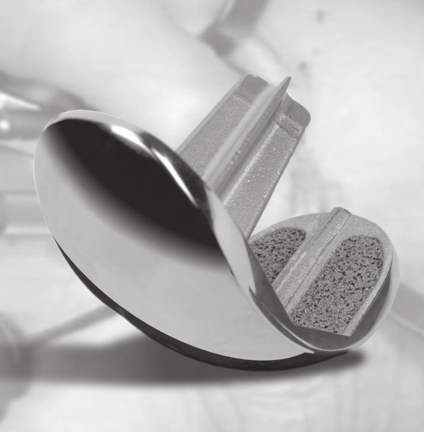
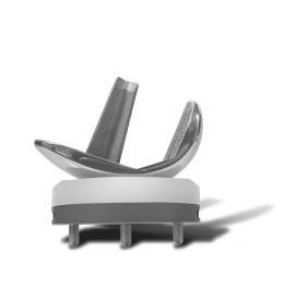
4 Zimmer Natural-Knee Unicompartmental MIS Intramedullary Surgical Technique Introduction Unicompartmental knee arthroplasty (UKA) has been shown to be a viable alternative to total knee arthroplasty for many patients with single-compartment disease. UKA has been successful even when mild to moderate (up to Grade II) chondromalacia is present in the patellofemoral joint as long as the contralateral compartment is preserved and both cruciate ligaments are functional. The MIS Instruments for the Natural- Knee Unicompartmental Knee System are designed to provide accurate, reproducible results using a minimally invasive technique. The goals of a minimally invasive surgical procedure are to: Facilitate the patient s recovery Provide less tissue trauma Provide less pain Provide earlier mobilization Provide shorter hospital stay Provide quicker rehabilitation Combined with surgeon judgment, proper patient selection, and appropriate use of the device, this step-by-step guide to the surgical technique discusses the procedure for component selection, bone preparation, trial reduction, and component implantation. It is strongly recommended that the surgeon read the complete procedure for details, notes, and technique tips. This technique is written for a medial compartment arthroplasty. The same technique is used for a lateral compartment arthroplasty with appropriate adjustments to reference the opposite compartment. For a lateral compartment arthroplasty, the proximity of the patella to the lateral condyle may require an extension of the incision so the patella can be adequately retracted medially. Rationale Instrumentation The basic goals of unicompartmental knee arthroplasty are to improve limb alignment and function, and to reduce pain. Routinely, an effort is made to minimize disruption of the surrounding soft tissue during the procedure. The development of instruments specifically designed to be used through a smaller exposure has had a significant impact on this effort, and has enhanced the surgeon s ability to make accurate bone cuts. Restoration of Normal Alignment Accurate limb alignment is described by the mechanical axis of the lower extremity, which is a straight line running from the center of the femoral head to the center of the ankle. When the center of the knee lies on or slightly lateral to this mechanical axis, the knee is said to be in neutral alignment. Unicompartmental knee disease typically reduces the joint space in the affected compartment, causing a malalignment of the joint. Full correction of the malalignment would return the knee to neutral alignment. When establishing alignment goals for unicompartmental arthroplasty, it is particularly important to avoid overcorrection of the limb as this may increase the stress in the contralateral compartment and heighten the potential for cartilaginous breakdown. Studies of unicompartmental procedures have shown that slight undercorrection of the limb alignment correlates to longterm survivorship. 1 It is important to recognize that the methods used to adjust alignment in total knee arthroplasty (TKA) are very different from those used in unicompartmental arthroplasty. In TKA, the angle of the femoral and tibial cuts determines the postoperative varus/ valgus alignment. In UKA, the angle of the cuts does not affect varus/valgus alignment. Instead, postoperative varus/valgus alignment is determined by the composite thickness of the prosthetic unicompartmental components. In the surgical technique for the Natural-Knee Unicompartmental Knee System, the distal femoral condyle is cut at a 6 angle to the anatomic axis of the femur. The proximal tibia is cut parallel to the femoral cut, which may be perpendicular to or at an angle of up to approximately 2-3 to the anatomic axis of the tibia, depending on patient anatomy and surgeon judgment. Enhanced Fixation Enhanced fixation helps stabilize the bone/implant interface and contributes to long-term arthroplasty success. Both porous and nonporous options are available on the femoral and tibial components. The porous components use Cancellous-Structured Titanium (CSTi ) Porous Coating while the nonporous components are grit-blasted. The tibial component is secured with smooth pegs to help prevent stress shielding and to enhance fixation. Cementless tibial fixation is further enhanced with a titanium cancellous bone screw, which augments the component s peripheral pegs. On the femoral component, the cruciate stem and a posterior runner contribute to component fixation as well as mediolateral and rotational stability.
5 Zimmer Natural-Knee Unicompartmental MIS Intramedullary Surgical Technique Preoperative Planning The primary objectives of preoperative planning are to choose the most appropriate implant size and to determine prosthetic placement while reestablishing joint kinematics and function. X-ray templates with 15% magnification are available for femoral and tibial component sizing to aid in meeting these objectives. Take standing weight-bearing A/P and lateral radiographs of the affected knee, and a long standing A/P radiograph showing the center of the femoral head, the knee, and as much of the tibia as possible (preferably including the ankle). Place a skin marker, such as an EKG electrode, over the center of the femoral head for later reference. Templating the Femur To achieve correct placement of the femoral template on the A/P view, select the size that best fits the posterior section of the condyle. Visualize the entire femur to rule out any structural abnormalities. Templating the Tibia Use both the A/P and lateral radiographs to template the tibia. Also, use the full-length tibial radiograph to check for varus or valgus alignment. The optimal size selected will have no overhang and will adequately cover the cortical rim of the tibia. No calculations for the tibia are necessary. The tibial assembly is aligned visually, relative to the mechanical axis of the tibia, so the tibial cut will be parallel to the femoral cut. Note: It is important to avoid overcorrection. An additional radiograph while stressing the limits of the tissues may be helpful in assessing the appropriate correction. Patient Preparation With the patient in the supine position, test the range of hip and knee flexion. If unable to achieve 120 of knee flexion, a larger incision may be necessary to create sufficient exposure. Wrap the ankle area with an elastic wrap. Do not place bulky drapes on the distal tibia, ankle, or foot. A bulky drape in this area will make it difficult to locate the center of the ankle, and may displace the MIS Ankle Alignment Guide, which may cause inaccurate cuts. Exposure Place the leg in a leg holder with the knee flexed 45. Begin the incision approximately one fingerbreadth from the medial edge of the patella, and extend it approximately 7cm-8cm to about 1cm below the joint line at the level of the tibial tubercle. Use a subvastus approach to expose the joint, making an inverted L-shaped incision over the affected femoral condyle. Incise the synovium following the same L-shaped incision. Use finger dissection to separate or undermine the soft tissue layers. Excise the fat pad as necessary to facilitate visualization, being careful not to cut the anterior horn of the lateral meniscus. Then incise the last fiber of the vastus medialis to free the muscle and allow lateral subluxation of the patella. Use a Z-retractor or bent Hohmann retractor to retract the patella laterally to expose the intercondylar notch. Reflect the soft tissue subperiosteally from the tibia along the joint line back toward, but not into, the collateral ligament. Use another Z-retractor medially to expose the proximal tibia. Then remove the anterior horn of the meniscus. Debride the joint and inspect it carefully. Remove all osteophytes. With medial compartment disease, osteophytes are commonly found on the lateral aspect of the medial tibial eminence and anterior to the origin of the ACL. By removing medial osteophytes, most varus deformities can be easily corrected passively. Also, remove peripheral osteophytes that interfere with the collateral ligaments and capsule. On the femur, remove intercondylar osteophytes to avoid impingement with the tibial spine or cruciate ligament. Final debridement will be performed before component implantation. Careful osteophyte removal may be important in achieving full extension.
6 Zimmer Natural-Knee Unicompartmental MIS Intramedullary Surgical Technique Surgical Technique Step One Locate Medullary Canal Center an 8mm (5/16-inch) drill distally on the patellar groove just anterior to both the posterior cruciate ligament and the true roof of the intercondylar notch. Holding the drill parallel to the shaft of the femur in both the A/P and lateral planes, drill the hole in the medullary canal. To help avoid contact with the medial cortex, aim the drill slightly laterally. When withdrawing the drill from the canal, slightly toggle the drill to allow venting that will help reduce pressurization of the medullary canal during the placement of subsequent guides. Suction the canal to remove intramedullary fat. Insert the Fluted T-handle by hand to open the medullary canal and ensure that the hole is aligned with the canal. If the T-handle is not easily inserted, reassess the orientation of the hole to ensure that it is in line with the femoral shaft. When the orientation of the hole has been confirmed, remove the Fluted T-handle. Step Two Prepare Femur Cut Distal Femoral Condyle The Distal Femoral Alignment/ Saw Guide is available in four configurations: right medial, left medial, right lateral, and left lateral. The guide will remove approximately 7mm of bone and cartilage from the distal femur, which is equal to the thickness of the femoral component. When properly positioned, the guide will result in a distal femoral cut with a 6 angle from the femoral anatomic axis in the frontal plane, and in 5 of flexion to minimize the risk of patellar impingement on the anterior flange of the femoral component. Insert the IM Rod into the canal until the stop on the rod contacts the bone. Then slide the appropriate Distal Femoral Alignment/Saw Guide over the rod until it contacts the bone. Stabilize the guide by turning the adjustable screw until it contacts the femoral condyle. Note: The 7mm resection includes the cartilage. If there is no cartilage or if eburnated bone is present, turn the adjustable screw clockwise to move the cutting slot distally to the preferred position. If, for example, the cutting slot is moved 2mm distally, the result will be a 5mm resection. The adjustable screw provides approximately 1mm of length adjustment for each complete rotation of the screw. Rotate the guide to allow optimal access with the saw blade, then predrill and insert a 5-inch by 1/8-inch pin through one of the two fixation holes on the side of the guide. For additional stability, insert a pin through one of the anterior holes near the cutting slot. Check the Distal Femoral Alignment/ Saw Guide to ensure that the saw blade will engage the distal femoral cortex at the meniscal abutment. If this is not clearly evident, mark the meniscal rest (junction of the tibiofemoral and patellofemoral joints) with methylene blue. Then insert the saw into the guide to determine if the blade will cut at the meniscal rest. If necessary, remove the pins and turn the adjustable screw to move the guide slightly proximally or distally to ensure a proper cut. Using a 1mm x 2 1/4-inch Synvasive Saw Blade ( ) or equivalent, cut the distal femur through the cutting slot on the guide. Remove the pins, and the Distal Femoral Alignment/Saw Guide. Leave the IM Rod in the canal to hold the patellar tendon out of the way for the remainder of the bone preparation. If desired, use a Z-retractor on the patellar tendon to achieve better exposure. Use a caliper to check the thickness of the resected bone fragment. Assume 1mm of bone loss due to the saw blade and 0.5mm for cartilage. If necessary, modify the cut surface of the distal condyle so that it is completely flat. This is extremely important for the placement of subsequent guides and for proper fit of the implant. Use a saw or rongeur to smooth any bony prominences that remain and contour the peripheral edge of the femur to restore anatomic shape.
7 Zimmer Natural-Knee Unicompartmental MIS Intramedullary Surgical Technique Drill Femoral Post Holes Apply the Distal Femoral Drill Guide with the flange resting against the posterior femoral condyle. Align the guide with the axis of the cut surface of the condyle with equal bone exposed on either side of the guide. Note: It is important to avoid the patellofemoral articulation centrally. Impact the set pins. Then use a 1/8- inch drill bit to drill the center hole to the second calibration mark on the drill. Insert a 3-inch x 1/8-inch pin. Use the 1/8-inch drill bit to drill the anterior and posterior post holes. Then remove the pin and use the Unicompartmental Slaphammer Extractor to remove the Distal Femoral Drill Guide. Cut Posterior Femoral Condyle Align the spikes on the Posterior Condyle Cutting Guide with the holes on the cut distal surface of the condyle and drive the spikes into the holes (Fig. 14). Attach the Unicompartmental Saw Capture to the cutting guide, and use an oscillating saw to resect 7mm of bone from the posterior condyle. Note: Be careful to protect the collateral ligament during the posterior resection. Ensure that the resected posterior surface is flat. Remove any prominences or uncut bone. To avoid bending the spikes, use the Unicompartmental Slaphammer Extractor to remove the Posterior Condyle Cutting Guide. Cut Posterior Chamfer Insert the Posterior Chamfer Cutting Guide into the two anterior drill holes. Attach the Unicompartmental Saw Capture to the cutting guide, and use a reciprocating saw to cut the posterior chamfer. Ensure that the resected surface is flat. Remove any prominences or uncut bone. Then use the Unicompartmental Slaphammer Extractor to remove the Posterior Chamfer Cutting Guide. Step Three Size Femur There are seven sizes of femoral implants and corresponding sizes of Femoral Sizing Templates. The outside contour of the templates matches the contour of the corresponding implant. Select the appropriate Femoral Sizing Template. The template should fit flush against the distal cut surface and lie below the meniscal abutment to avoid patellar impingement. If there is any anterior overhang, select a smaller size. Step Four Prepare Tibia Position MIS Tibial Assembly The MIS tibial assembly is a smaller version of the tibial assembly used with the Natural-Knee II Primary Prosthesis. Attach the MIS Ankle Alignment Guide to the appropriate MIS Proximal Tibial Alignment Guide (Left Medial/Right Lateral or Right Medial/Left Lateral). Then slide the corresponding MIS Tibial Saw Guide superiorly onto the locating pin on the proximal end of the alignment guide. The radiused cutout on the saw guide mates with the rod of the Proximal Tibial Alignment Guide. Depress the button on the proximal guide and raise the sleeve until the collar contacts the underside of the saw guide. Attach the ankle guide around the ankle. Place the knee in maximum flexion and position the superior portion of the proximal guide on the tibia, sliding the curved head around the patellar tendon. The guide should lie anterior to the ACL. The posterior slope of the tibia may range from The goal is to resect the tibia parallel to the joint surface in order to reproduce the posterior slope of the proximal tibia. Assess the angle of the tibial slope by placing a 5-inch by 1/8-inch pin on top or through the highest drill/pin hole of the Tibial Saw Guide. Then move the distal portion of the alignment guide anteroposteriorly until the pin rests parallel to the tibial plateau. To adjust rotational alignment, position the proximal alignment guide at the medial third of the tubercle. Slide the alignment guide medially or laterally at the ankle to adjust the varus/ valgus angle. Secure the position by
8 Zimmer Natural-Knee Unicompartmental MIS Intramedullary Surgical Technique tightening the knob. The distal end of the rod should point to the anterior tibial/fibular ligament for a 2 varus tibial cut. Thread the MIS Tibial Stylus into the superior hole on the saw guide. Then rotate the sleeve/box on the proximal alignment guide clockwise to lower the saw guide and stylus until the tip of the stylus is at the desired level for the appropriate resection. The cut will remove 7mm of bone below the tip of the stylus. One complete revolution of the nut will raise or lower the saw guide by approximately 1mm. Use a small rake retractor to pull the skin clear from the proximal tibia and the Tibial Saw Guide. To stabilize the saw guide, predrill and insert 1/8-inch (3.2mm) pins medially and laterally through the holes marked 7. This will set the guide for a 7mm resection. If the tibial defect is deeper, slide the saw guide off the pins and reinsert it through the holes marked 9, 11, or 13 for a corresponding 9mm, 11mm, or 13mm resection. Making a cut of at least 9mm will ensure that an adequate tibial articular surface thickness is used. Loosen the thumbscrew on the stylus to remove it from the MIS Proximal Tibial Alignment Guide. Then depress the button and lower the sleeve to allow the alignment guide assembly to be lifted off the Tibial Saw Guide. Make Sagittal Tibial Cut Use methylene blue to mark the location of the sagittal cut as close to the ACL as possible. If desired, use the Tibial Sizer/Drill Guide as a reference for marking the resection line. Using this reference line and the lines on the Tibial Saw Guide, make the vertical cut. This cut will serve as a reference point for the width of the horizontal proximal cut. Make Proximal Tibial Cut Place a saw blade or osteotome into the sagittal cut to help prevent undercutting the tibial spine when making the horizontal cut. Attach the Unicompartmental Saw Capture to the Tibial Saw Guide. Make the proximal tibial cut, being careful to avoid undercutting the medial tibial eminence. Then assure that a perfectly flat surface has been created by removing the saw capture and sighting along the superior surface of the saw guide, or by placing another saw guide or flat instrument on the surface of the bone cut. Slide the Tibial Saw Guide off the headless pins, and remove any posterior horn of the meniscus that remains. Size Tibia and Drill Tibial Screw Hole Use the Tibial Sizer/Drill Guides to select the size that best covers the tibial surface area without any overhang. This will determine the size of the Tibial Base Plate Provisional and the final tibial base plate component. The A/P dimension is the most critical; there is more room for adjustment in the M/L plane. Use a caliper to measure the thickness of the resected tibial fragment to ensure evenness of the cut. Then compare the fragment to the selected size Tibial Base Plate Provisional to ensure the best tibial coverage. Using the selected size Tibial Sizer/ Drill Guide, drill the central screw hole to the first calibration mark on the drill bit. If preferred, this hole can be drilled through the Tibial Base Plate Provisional. It is not necessary to drill peripheral holes for the tibial pegs. Step Five Perform Trial Reduction With all bone surfaces prepared, perform a trial reduction with the appropriate size Femoral Provisional, Tibial Base Plate Provisional, and Tibial Articular Surface Provisional. Position the Tibial Base Plate Provisional on the resected surface of the tibia, and impact it so the three short pegs engage the cancellous bone. The provisional should fit within the entire cortical rim without any overhand. If the all-polyethylene tibial component will be used, apply the Keel Broach over the Tibial Base Plate Provisional and impact it. Remove the IM Rod, and retract the patellar tendon as needed. Then slide the Tibial Articular Surface Provisional onto the Tibial Base Plate Provisional and lightly impact it. Tibial Articular Surface Provisionals are available in 9mm, 11mm, and 13mm thicknesses. There is also a 7mm provisional size available for the all-poly. Impact the correct size Femoral Provisional onto the prepared distal femur. The Femoral Provisional includes a cruciate cutter that will prepare the distal femoral surface
9 Zimmer Natural-Knee Unicompartmental MIS Intramedullary Surgical Technique to accept the cruciate stem of the femoral component. With the Femoral Provisional fully seated, use a 1/8-inch drill bit through the posterior drill hole in the provisional to create a groove for the antirotational posterior runner of the femoral component. Check the fit of the provisional components. If necessary, perform minor trimming of bone surfaces. Assess soft tissue balance using either the mechanical axis or the anatomic axis. If using the mechanical axis, use the preoperative x-ray with a skin marker centered over the femoral head. In unicompartmental arthroplasty, the mechanical axis should be centered over the tibial spine. Check joint stability in full extension and in flexion. In a varus knee, a valgus stress should allow for a 1mm to 2mm opening, and should allow for the insertion of the Laxity Checker, which is 2mm thick, in both full flexion and full extension. Overstuffing should be avoided, as this will transfer stress to the contralateral compartment. For example, in a varus knee, slightly undercorrect the alignment to avoid transferring excessive stress to the lateral compartment. After confirming that the tibial resection is correct, remove the headless pins from the anterior tibia. When stability and range of motion are satisfactory, use the Unicompartmental Slaphammer Extractor to remove the provisional components. Step Six Implant Final Components Obtain the final components and implant the tibial component first. Technique Tip: With the modest amount of bone removed, particularly from the tibia, there may be a sclerotic cut surface. If the resected surfaces of the tibia and/or femur are sclerotic, drill multiple holes with a small drill (2.0mm-3.2mm) to improve cement intrusion. Tibial Component If using a cemented component, apply cement. To facilitate insertion, flex the knee and externally rotate the tibia. If desired, place an opened and slightly moist sterile gauze sponge behind the tibia before implanting the components to help collect excess cement behind the tibia. Use the Modular Tibial Impactor to impact the tibial base plate, or the Allpolyethylene Tibial Impactor to impact the all-polyethylene tibial component. If using a cementless tibial base plate, insert a 40mm cancellous screw to secure it in place. Note: Do not use the Modular Tibial Impactor to impact an all-polyethylene tibial component. Remove the sterile gauze sponge slowly from behind the joint, and remove any excess cement. Tibial Articular Surface After the cement has cured, remove any remaining excess cement before the final placement of the tibial articular surface. Do not proceed with locking the final articular surface component until the cement has fully cured. Place the appropriate thickness tibial articular surface by engaging the component s posterior tabs, then applying compression on the anterior flat with the Articular Surface Impactor. Femoral Component If using the cemented component, apply cement. Engage the cruciate tip and the posterior runner of the femoral component, and impact the component carefully. Then check the final range of motion and joint stability. Closure Fashion an osteochondral plug from the resected tibial or posterior femoral fragments and insert it into the distal femoral hole to minimize bleeding. Thoroughly irrigate the knee and close in the standard manner over a suction drain. Apply a bulky, cotton roll compressive dressing. If desired, place the leg in a continuous passive motion (cpm) machine. A cold therapy machine may be used if desired. Then follow a standard knee rehabilitation protocol. 1. Cartier P, Seinouiller JL, Grelsamer RP. Unicompartmental knee arthroplasty 10-year minimum follow-up period. J Arthroplasty. 1996;11(7):
10 Please refer to package insert for complete product information, including contraindications, warnings, precautions, and adverse effects ML Printed in USA 2006 Zimmer, Inc. Contact your Zimmer representative or visit us at
Revolution. Unicompartmental Knee System
 Revolution Unicompartmental Knee System While Total Knee Arthroplasty (TKA) is one of the most predictable procedures in orthopedic surgery, many patients undergoing TKA are in fact excellent candidates
Zimmer Unicompartmental High Flex Knee. Intramedullary, Spacer Block Option and Extramedullary Minimally Invasive Surgical Techniques
 Zimmer Unicompartmental High Flex Knee Intramedullary, Spacer Block Option and Extramedullary Minimally Invasive Surgical Techniques ZIMMER UNICOMPARTMENTAL HIGH FLEX KNEE INTRAMEDULLARY, SPACER BLOCK
unicompartmental knee SURGICAL TECHNIQUE limacorporate.com
 unicompartmental knee SURGICAL TECHNIQUE limacorporate.com Index INTRAMEDULLARY (IM) SURGICAL PROCEDURE Introduction Page >> 04 Rationale Page >> 05 Preoperative Planning Page >> 07 Patient Preparation
TOTAL KNEE ARTHROPLASTY SYSTEM
 SURGICAL TECHNIQUE TOTAL KNEE ARTHROPLASTY SYSTEM 90-SRK-700000 B.0 0 Contents 1. Implant Sizing 2. Surgical Technique a. Incision and Exposure b. Distal Femoral Resection c. Tibial Resection d. Femoral
Zimmer Unicompartmental High Flex Knee. Spacer Block Surgical Technique
 Zimmer Unicompartmental High Flex Knee Spacer Block Surgical Technique INTRO Zimmer Unicompartmental High Flex Knee Spacer Block Surgical Technique Introduction Unicompartmental knee arthroplasty (UKA)
Zimmer NexGen MIS Tibial Component. Cemented Surgical Technique IMAGE TO COME
 Zimmer NexGen MIS Tibial Component Cemented Surgical Technique IMAGE TO COME Zimmer NexGen MIS Tibial Component Cemented Surgical Technique 1 Zimmer NexGen MIS Tibial Component Cemented Surgical Technique
Minimally Invasive Surgical Techniques for the Medial Compartment. Intramedullary, Spacer Block and Extramedullary techniques
 Minimally Invasive Surgical Techniques for the Medial Compartment Intramedullary, Spacer Block and Extramedullary techniques Unicompartmental High Flex Knee Intramedullary, Spacer Block and Extramedullary
Zimmer NexGen Tibial Stem Extension & Augmentation. Surgical Technique IMAGE TO COME. Stem Extensions and Augments
 Zimmer NexGen Tibial Stem Extension & Augmentation Surgical Technique IMAGE TO COME Stem Extensions and Augments Zimmer NexGen Tibial Stem Extension & Augmentation Surgical Technique 1 Zimmer NexGen Tibial
ANTERIOR REFERENCE NEXGEN COMPLETE KNEE SOLUTION. Multi-Reference 4-in-1 Femoral Instrumentation Anterior Reference Surgical Technique
 ANTERIOR REFERENCE NEXGEN COMPLETE KNEE SOLUTION Multi-Reference 4-in-1 Femoral Instrumentation Anterior Reference Surgical Technique For NexGen Cruciate Retaining & Legacy Posterior Stabilized Knees INTRODUCTION
Zimmer NexGen Trabecular Metal Tibial Tray
 Zimmer NexGen Trabecular Metal Tibial Tray Surgical Technique Zimmer NexGen Trabecular Metal Tibial Tray Surgical Technique Give Bone A Solid Hold Zimmer NexGen Trabecular Metal Tibial Tray Surgical Technique
NEXGEN COMPLETE KNEE SOLUTION S A. Tibial Stem Extension & Augmentation Surgical. ATechnique
 NEXGEN COMPLETE KNEE SOLUTION ATechnique Tibial Stem Extension & Augmentation Surgical INTRODUCTION The NexGen Complete Knee Solution Intramedullary Tibial Instruments have been designed to provide an
Distal Cut First Femoral Preparation
 Surgical Technique Distal Cut First Femoral Preparation Primary Total Knee Arthroplasty LEGION Total Knee System Femoral preparation Contents Introduction...3 DCF femoral highlights...4 Preoperative planning...6
TRK REVISION KNEE Surgical Technique
 1 TRK REVISION KNEE Surgical Technique 1. 2. 3. 4. 5. 6. 7. 8. 9. 10. INTERCONDYLAR RESECTION...... page FEMORAL STEM...... page NON CEMENTED FEMORAL STEM...... page TRIAL FEMORAL COMPONENTS...... page
POSTERIOR REFERENCE NEXGEN COMPLETE KNEE SOLUTION. Multi-Reference 4-in-1 Femoral Instrumentation Posterior Reference Surgical Technique
 POSTERIOR REFERENCE NEXGEN COMPLETE KNEE SOLUTION Multi-Reference 4-in-1 Femoral Instrumentation Posterior Reference Surgical Technique For NexGen Cruciate Retaining & Legacy Posterior Stabilized Knees
Total Knee Original System Primary Surgical Technique
 Surgical Procedure Total Knee Original System Primary Surgical Technique Where as a total hip replacement is primarily a bony operation, a total knee replacement is primarily a soft tissue operation. Excellent
MIS Cemented Tibial Component
 MIS Cemented Tibial Component NexGen Complete Knee Solution Surgical Technique Table of Contents Surgical Exposure... 2 Finish the Tibia... 2 Position Based on Anatomic Landmarks... 3 Lateral Posterior
Intramedullary Tibial Preparation
 Surgical Technique Intramedullary Tibial Preparation Primary Total Knee Arthroplasty LEGION Total Knee System Intramedullary tibial preparation Contents Introduction...2 IM tibial highlights...3 Preoperative
Triathlon Knee System
 Triathlon Knee System Express Instruments Surgical Protocol Posterior Stabilized & Cruciate Retaining TriathlonKneeSystem Express Instruments Surgical Protocol Acknowledgments..........................................................2
CONTRIBUTING SURGEON. Barry Waldman, MD Director, Center for Joint Preservation and Replacement Sinai Hospital of Baltimore Baltimore, MD
 CONTRIBUTING SURGEON Barry Waldman, MD Director, Center for Joint Preservation and Replacement Sinai Hospital of Baltimore Baltimore, MD System Overview The EPIK Uni is designed to ease the use of the
Minimally Invasive Surgical Techniques for the Lateral Compartment. Spacer Block, Intramedullary and Extramedullary techniques
 Minimally Invasive Surgical Techniques for the Lateral Compartment Spacer Block, Intramedullary and Extramedullary techniques Unicompartmental High Flex Knee Spacer Block, Intramedullary and Extramedullary
Extramedullary Tibial Preparation
 Surgical Technique Extramedullary Tibial Preparation Primary Total Knee Arthroplasty LEGION Total Knee System Extramedullary tibial preparation Contents Introduction...2 EM tibial highlights...3 Preoperative
NexGen Cruciate Retaining (CR) and Revision Instrumentation. Surgical Technique
 and Revision Instrumentation. Surgical Technique") NexGen Cruciate Retaining (CR) and Revision Instrumentation Surgical Technique Table of Contents Introduction... 4 Revision Arthroplasty... 4 Multi-Reference 4-in-1 Instrumentation System MICRO-MILL Instrumentation
NexGen Cruciate Retaining (CR) and Revision Instrumentation Surgical Technique Table of Contents Introduction... 4 Revision Arthroplasty... 4 Multi-Reference 4-in-1 Instrumentation System MICRO-MILL Instrumentation
Contact your Zimmer representative or visit us at
 Please refer to the package inserts for complete product information, including contraindications, warnings, precautions, and adverse effects. Contact your Zimmer representative or visit us at www.zimmer.com
Zimmer Trabecular Metal Ankle Interpositional Spacer and Trabecular Metal Ankle Fusion Spacer
 Zimmer Trabecular Metal Ankle Interpositional Spacer and Trabecular Metal Ankle Fusion Spacer Surgical Technique 2 Zimmer Trabecular Metal Ankle Interpositional Spacer and Trabecular Metal Ankle Fusion
Minimally Invasive. The M/G Unicompartmental Knee. <...after ten years of CLINICALSUCCESS. we ve drawn the line. >
 Minimally Invasive The M/G Unicompartmental Knee > < actual size of typical minimally invasive incision The MIS Minimally Invasive Solution
Surgical Technique. VISIONAIRE FastPak Instruments for the LEGION Total Knee System
 Surgical Technique VISIONAIRE FastPak Instruments for the LEGION Total Knee System VISIONAIRE FastPak for LEGION Instrument Technique* Nota Bene The technique description herein is made available to the
A novel cementless option. Zimmer NexGen Trabecular Metal Primary Patella Surgical Technique
 A novel cementless option Zimmer NexGen Trabecular Metal Primary Patella Surgical Technique Zimmer Trabecular Metal Primary Patella 1 Zimmer NexGen Trabecular Metal Primary Patella Surgical Technique
Unicondylar Surgical Technique
 Unicondylar Surgical Technique Contents Indications, Contra-indications and X-ray Templating 2 Approach and Exposure 3 Proximal Tibial Resection 4 Tibial Jig Alignment 6 Tibial Sizing 9 Balancing 10 Distal
Resurfacing Distal Femur. Orthopaedic Salvage System
 Resurfacing Distal Femur Orthopaedic Salvage System Primary Arthroplasty OSS 3cm Resurfacing Distal Femur Distal Femoral Resection Drill and ream the distal femur in the following sequence: (Figure 1)
Trabecular Metal Femoral Component. Surgical Technique
 Trabecular Metal Femoral Component Surgical Technique INTRO Trabecular Metal Femoral Component Surgical Technique Introduction The Persona Trabecular Metal Femoral Component is designed to unite stable
NATURAL MOTION TECHNOLOGY SURGICAL TECHNIQUE. EMPOWR 3D Knee. EMPOWR PS Knee
 NATURAL MOTION TECHNOLOGY EMPOWR 3D Knee EMPOWR PS Knee SURGICAL TECHNIQUE Contents System Features.... 3 Indications and Contraindications.... 4 Surgical Snap Shot.... Preoperative Planning.... Surgical
SIGMA High Performance Partial Knee. Unicondylar. Surgical Technique
 SIGMA High Performance Partial Knee Unicondylar Surgical Technique Table of Contents Surgical Technique X-ray Templating 3 Approach and Exposure 4 Proximal Tibial Resection 5 Tibial Jig Alignment 7 Tibial
Surgical Technique. VISIONAIRE Disposable Instruments for the LEGION Total Knee System
 Surgical Technique VISIONAIRE Disposable Instruments for the LEGION Total Knee System VISIONAIRE and LEGION Disposable instrument technique* Note: All disposable instruments are interchangeable with the
FOREWORD PRESERVATION UNICOMPARTMENTAL KNEE SYSTEM
 Surgical Technique FOREWORD PRESERVATION UNICOMPARTMENTAL KNEE SYSTEM Our surgeon design team first implanted the Preservation Unicompartmental Knee System in 2001. The system was developed with over
ANATOMIC SURGICAL TECHNIQUE. 5 in 1. Conventional instrumentation 07/11/2013
 ANATOMIC SURGICAL TECHNIQUE 5 in 1 Conventional instrumentation PRO.GB.933/1.0 Octobre 2013 2 Tibial step 3 Intramedullary technique - Based on the preoperative plan, drill the medullary canal with the
Uniglide. Unicompartmental Knee Replacement Mk III surgical technique
 Uniglide Unicompartmental Knee Replacement Mk III surgical technique Uniglide Contents Operative summary 4 Pre-operative assessment 6 Preparation 7 Incision 7 Approach 7 Medial procedure 8 Tibial preparation
FLK167 02/08. Biomet UK Ltd Waterton Industrial Estate Bridgend, South Wales CF31 3XA, United Kingdom. Tel Fax:
 FLK167 02/08 Biomet UK Ltd Waterton Industrial Estate Bridgend, South Wales CF31 3XA, United Kingdom Tel. 01656 655221 Fax: 01656 645454 Premier Instrumentation CR or PS Surgical Technique Vanguard Premier
TRUMATCH PERSONALIZED SOLUTIONS with the SIGMA High Performance Instruments
 TRUMATCH PERSONALIZED SOLUTIONS with the SIGMA High Performance Instruments Resection Guide System SURGICAL TECHNIQUE RESECTION GUIDE SURGICAL TECHNIQUE The following steps are an addendum to the SIGMA
JOINT RULER. Surgical Technique For Knee Joint JRReplacement
 JR JOINT RULER Surgical Technique For Knee Joint JRReplacement INTRODUCTION The Joint Ruler * is designed to help reduce the incidence of flexion, extension, and patellofemoral joint problems by allowing
U2 PSA. Revision Knee. Surgical Protocol
 U2 PSA TM Revision Knee Surgical Protocol Table of Contents 1 Component Removal... 1 2 Tibial Preparation... 1 2.1 Tibial Canal Preparation... 1 2.2 Proximal Tibial Resection... 2 2.3 Non Offset Tibial
Aesculap Orthopaedics Columbus MIOS
 Aesculap Orthopaedics Columbus MIOS Minimally Invasive Orthopaedic Solutions Manual TKA Surgical Technique MIOS 4-in-1 Cutting Block MIOS Distal Femoral Cutting Block MIOS Tibial Left and Right Cutting
NexGen Complete Knee Solution. Cruciate Retaining (CR) and Revision Instrumentation Surgical Technique for Cruciate Retaining Augmentable (CRA) Knees
 and Revision Instrumentation Surgical Technique for Cruciate Retaining Augmentable (CRA) Knees") NexGen Complete Knee Solution Cruciate Retaining (CR) and Revision Instrumentation Surgical Technique for Cruciate Retaining Augmentable (CRA) Knees INTRODUCTION This surgical technique document combines
NexGen Complete Knee Solution Cruciate Retaining (CR) and Revision Instrumentation Surgical Technique for Cruciate Retaining Augmentable (CRA) Knees INTRODUCTION This surgical technique document combines
Persona The Personalized Knee. Surgical Technique
 Persona The Personalized Knee Surgical Technique Table of Contents Introduction... 2 Constraint Options Preoperative Planning Surgical Approach Patient Preparation Magnet Usage Symbols Screw/Pin Information
Zimmer FuZion Instruments. Surgical Technique (Beta Version)
") Zimmer FuZion Surgical Technique (Beta Version) INTRO Surgical Technique Introduction Surgical goals during total knee arthroplasty (TKA) include establishment of normal leg alignment, secure implant fixation,
Zimmer FuZion Surgical Technique (Beta Version) INTRO Surgical Technique Introduction Surgical goals during total knee arthroplasty (TKA) include establishment of normal leg alignment, secure implant fixation,
ClassiQ. Scorpio. Anterior Referencing Surgical Protocol. Anterior Referencing. For use with Scorpio ClassiQ Instrument System
 Scorpio ClassiQ Anterior Referencing Surgical Protocol For use with Scorpio ClassiQ Instrument System For use with Scorpio ClassiQ Single Radius Total Knee System AR Anterior Referencing This document
Trabecular Metal Primary Patella
 Trabecular Metal Primary Patella NexGen Complete Knee Solution Surgical Technique Table of Contents Introduction... 2 Prepare the Patella... 2 Patella Reamer Technique... 3 Insetting Technique... 5 Universal
1. Pre-Operative Planning Skin Incision and Arthrotomy
 Stage of Operation 1. Pre-Operative Planning --------------------------1 2. Skin Incision and Arthrotomy ------------------1 3. Femoral valgus angle confirmation ------------2 4. Distal Femur Cutting -----------------------------3
Profix. Total Knee System. As described by
 Profix Total Knee System P R I M A R Y P R O C E D U R E As described by Leo A. Whiteside, M.D. Associate Research Professor Washington University School of Medicine Director, Biomechanical Research Laboratory
NexGen LPS Fixed Bearing Knee. Surgical Technique
 NexGen LPS Fixed Bearing Knee Surgical Technique Table of Contents Introduction...2 Preoperative Planning...2 Surgical Technique...3 Step One: Align the Tibia...3 MICRO-MILL Hand Piece Assembly and Usage
PRIMARY POROUS PATELLA WITH TRABECULAR METAL. Surgical Technique
 PRIMARY POROUS PATELLA WITH TRABECULAR METAL Surgical Technique 1 SURGICAL TECHNIQUE FOR NEXGEN PRIMARY POROUS PATELLA * WITH TRABECULAR METAL CONTENTS PREPARE THE PATELLA............... 2 Patella Reamer
Triathlon TS Knee System. Surgical Protocol
 Triathlon TS Knee System Surgical Protocol Triathlon TS Knee System Surgical Protocol Table of Contents Acknowledgments..........................................................2 Exposure...................................................................4
Constrained Posterior Stabilized (CPS) Surgical Technique
 Surgical Technique") Constrained Posterior Stabilized (CPS) Surgical Technique Constrained Posterior Stabilized (CPS) Surgical Technique INTRO Introduction The Constrained Posterior Stabilized (CPS) articular surfaces can
Constrained Posterior Stabilized (CPS) Surgical Technique Constrained Posterior Stabilized (CPS) Surgical Technique INTRO Introduction The Constrained Posterior Stabilized (CPS) articular surfaces can
Persona. The Personalized Knee. Trabecular Metal Tibia. Surgical Technique
 Persona The Personalized Knee Trabecular Metal Tibia Surgical Technique Table of Contents Resect the Tibia... 4 Size and Finish the Tibia... 4 Trial Fit... 6 Component Implantation... 7 Inserter/Implant
PIN GUIDE SYSTEM SURGICAL TECHNIQUE. with the SIGMA High Performance Instruments System. This publication is not intended for distribution in the USA.
 PIN GUIDE SYSTEM with the SIGMA High Performance Instruments System This publication is not intended for distribution in the USA. SURGICAL TECHNIQUE Pin Guide Surgical Technique The following steps are
Zimmer NexGen MIS Tibial Component. Cemented Surgical Technique. IMAGE TO COME Designed for minimally invasive procedures
 Zimmer NexGen MIS Tibial Component Cemented Surgical Technique IMAGE TO COME Designed for minimally invasive procedures Zimmer NexGen MIS Tibial Component Cemented Surgical Technique 1 Zimmer NexGen MIS
The ACL-PCL Substituting Knee. BioFoam Tibial Bases. Surgical Technique For Cementless Tibial Fixation
 The CL-PCL Substituting Knee BioFoam Surgical Technique For Cementless Tibial Fixation Surgical techniques and instrument recommendations were provided by: Michael nderson, MD Milwaukee, WI G. Lynn Rasmussen,
ZIMMER NEXGEN CRUCIATE RETAINING (CR) AND LEGACY KNEE POSTERIOR STABILIZED (LPS) TRABECULAR METAL MONOBLOCK TIBIAS. Surgical Technique Addendum
 AND LEGACY KNEE POSTERIOR STABILIZED (LPS) TRABECULAR METAL MONOBLOCK TIBIAS. Surgical Technique Addendum") ZIMMER NEXGEN CRUCIATE RETAINING (CR) AND LEGACY KNEE POSTERIOR STABILIZED (LPS) TRABECULAR METAL MONOBLOCK TIBIAS Surgical Technique Addendum SURGICAL TECHNIQUE FOR NEXGEN CRUCIATE RETAINING (CR) AND
ZIMMER NEXGEN CRUCIATE RETAINING (CR) AND LEGACY KNEE POSTERIOR STABILIZED (LPS) TRABECULAR METAL MONOBLOCK TIBIAS Surgical Technique Addendum SURGICAL TECHNIQUE FOR NEXGEN CRUCIATE RETAINING (CR) AND
OPERATIVE TECHNIQUE SKS Total Knee Replacement
 OPERATIVE TECHNIQUE SKS Total Knee Replacement Femoral preparation A40577 1 2 5 1: Alignment rod A30049 + A30124 2: Centromedullary rod A40224 4: T-handle A40232 6: Femoral measuring device A40411 + A40414
NexGen Trabecular Metal Tibial Tray. Surgical Technique
 NexGen Trabecular Metal Tibial Tray Surgical Technique Table of Contents Surgical Technique...2 Resect the Tibia...2 Finish the Tibia...2 Position Based Anatomic Landmarks...3 Position Based on Trial
Zimmer MOST Options System
 Zimmer MOST Options System Surgical Technique Modular Options for Severe Bone Loss and Trauma Zimmer MOST Options System Surgical Technique 1 MOST Options System Surgical Technique Developed in conjunction
TRUMATCH PERSONALIZED SOLUTIONS with the SIGMA High Performance Instruments
 TRUMATCH PERSONALIZED SOLUTIONS with the SIGMA High Performance Instruments Pin Guide System SURGICAL TECHNIQUE PIN GUIDE SURGICAL TECHNIQUE The following steps are an addendum to the SIGMA High Performance
NEXGEN COMPLETE KNEE SOLUTION. Revision Instrumentation Surgical Technique For Legacy Knee Constrained Condylar Knee
 NEXGEN COMPLETE KNEE SOLUTION Revision Instrumentation Surgical Technique For Legacy Knee Constrained Condylar Knee INTRODUCTION Revision total knee arthroplasty can be a very challenging task for any
Zimmer MIS Intramedullary Instrumentation
 Zimmer MIS Intramedullary Instrumentation Surgical Technique For NexGen Cruciate Retaining & NexGen Legacy Posterior Stabilized Knees Zimmer MIS Intramedullary Instrumentation Surgical Technique Surgical
AGC Total Knee System. Concise Surgical Technique Featuring EquiFlex Instrumentation
 AGC Total Knee System Concise Surgical Technique Featuring EquiFlex Instrumentation AGC TOTAL KNEE SYSTEM Concise Surgical Technique Instrumentation Disclaimer This brochure provides a description of the
Anterior Cut First Surgical Technique
 Anterior Cut First Surgical Technique GENESIS II Anterior Cut First Surgical Technique Table of Contents Introduction....................................... 2 Preop Planning....................................
Proven Gen-Flex Revision Knee System. Surgical Protocol
 Proven Gen-Flex Revision Knee System Surgical Protocol Proven Gen-Flex Revision Knee System Surgeon Design Team The Proven Gen-Flex Revision Knee System Instrumentation and Surgical Protocol were developed
14 mm x +30 mm Stem Extension
 14 mm x +30 mm Stem Extension Persona The Personalized Knee Surgical Technique Table of Contents Introduction...2 Preoperative Planning Surgical Approach Patient Preparation Magnet Usage Symbols Screw/Pin
S U R G I C A L T E C H N I Q U E David A. McQueen, MD Return to Menu
 S U R G I C A L T E C H N I Q U E David A. McQueen, MD TOTAL KNEE INSTRUMENTS Wichita Fusion Nail Introduction...1 Preoperative Planning...2 Surgical Technique...3-8 Wichita Fusion Nail Surgical Technique
Tibial & Femoral Opening Wedge Osteotomy System. Surgical Technique
 Tibial & Femoral Opening Wedge Osteotomy System Surgical Technique Opening Wedge Osteotomy Tibial & Femoral Opening Wedge Osteotomy 2 Prior to the osteotomy, a diagnostic arthroscopy is performed to verify
Patella Planing System
 REFERENCE GUIDE AND SURGICAL TECHNIQUE Patella Planing System SPECIALIST INSTRUMENTS AN ONLAY PATELLA PREPARATION SYSTEM TECHNIQUE FOR PRIMARY PATELLAR RESURFACING The Specialist 2 Patellar Planer System
Zimmer NexGen Rotating Hinge Knee Primary/ Revision
 Zimmer NexGen Rotating Hinge Knee Primary/ Revision Surgical Technique Designed for use in revision and difficult primary surgeries 1 INTRODUCTION The NexGen Rotating Hinge Knee Components are designed
Constrained Posterior Stabilized (CPS)
") Constrained Posterior Stabilized (CPS) Persona The Personalized Knee Surgical Technique Table of Contents Introduction... 2 Constraint Options Initial Knee Assessment... 3 Femoral Box Cut CPS Tibial Bearing
Constrained Posterior Stabilized (CPS) Persona The Personalized Knee Surgical Technique Table of Contents Introduction... 2 Constraint Options Initial Knee Assessment... 3 Femoral Box Cut CPS Tibial Bearing
Persona Partial Knee System. Surgical Technique
 Persona Partial Knee System Surgical Technique Table of Contents Introduction... 2 Cross-System Compatibility... 2 Surgical Technique... 3 Positioning the Limb... 3 Incision... 3 Osteophyte Excision...
Triathlon with Single-Use Instrumentation Optimize Your TKA Experience. Posterior Referencing Surgical Protocol
 Triathlon with Single-Use Instrumentation Optimize Your TKA Experience Posterior Referencing Surgical Protocol Introduction............................................................2 Assembly Instructions...................................................6
MRH Knee System Modular Peg Baseplate Surgical Protocol
 MRH Knee System Modular Peg Baseplate Surgical Protocol Using Monogram IM Revision Instruments 4 N 4 N Modular Rotating Hinge Knee System Using Monogram IM Revision Instruments Mr C R Howie FRCS Consultant
Passport. PR Surgical Protocol. Scorpio Total Knee System
 Passport PR Surgical Protocol Scorpio Total Knee System Table of Contents Scorpio Total Knee System Surgical Protocol for the Passport Instruments Exposure...2 FEMORAL PREPARATION...3 Femoral Intramedullary
Surgical Technique Final Trial Reduction and Component Implantation of
 Surgical Technique Final Trial Reduction and Component Implantation of TC *smith&nephew TC-PLUS PRIMARY Mobile Bearing TC-PLUS PRIMARY Mobile Bearing Final Trial Reduction and Component Implantation of
Inspiring people, Enriching lives
 TM Inspiring people, Enriching lives Encore Medical, L.P. 9800 Metric Blvd. Austin, Texas 78758 512-832-9500 www.encoremed.com Encore Medical, L.P. Cat. # 0152-017 2.5k/rev. A Encore and Foundation are
Mako Partial Knee Medial bicompartmental
 Mako Partial Knee Medial bicompartmental Surgical reference guide Mako Robotic-Arm Assisted Surgery Table of contents Implant compatibility.... 3 Pre-operative planning.... 4 Intra-operative planning....
Zimmer NexGen LPS-Flex Mobile and LPS-Mobile Bearing Knees. Surgical Technique United States Version IMAGE TO COME
 Zimmer NexGen LPS-Flex Mobile and LPS-Mobile Bearing Knees Surgical Technique United States Version IMAGE TO COME Zimmer NexGen LPS-Flex Mobile and LPS-Mobile Bearing Knees Surgical Technique 1 Zimmer
NexGen Intramedullary Instrumentation. Surgical Technique
 NexGen Intramedullary Instrumentation Surgical Technique Table of Contents Introduction... 2 Preoperative Planning... 2 Surgical Technique... 3 Step One: Size the Femur/Establish External Rotation...
LCS COMPLETE SURGICAL TECHNIQUE. MILESTONE Instruments with MBT Tray Preparation
 LCS COMPLETE MILESTONE Instruments with MBT Tray Preparation SURGICAL TECHNIQUE CONTENTS Preface 2 Surgical Concepts 3 Instruments 4 Surgical Technique Summary 6 Surgical Technique 8 Preparation 8 Incision
Technique Guide. *smith&nephew N8TIVE ACL Anatomic ACL Reconstruction System
 Technique Guide *smith&nephew N8TIVE ACL Anatomic ACL Reconstruction System N8TIVE ACL System The N8TIVE ACL Anatomic Reconstruction System provides a novel and simple approach to ACL repair. The N8TIVE
Triathlon Knee System. Anterior Referencing Surgical Protocol
 Triathlon Knee System Anterior Referencing Surgical Protocol Triathlon Knee System Anterior Referencing Surgical Protocol Table of Contents Acknowledgments..........................................................2
Orthopedic Bone Nail System - Distal Femoral Nail Surgical Technique Manual
 Orthopedic Bone Nail System - Distal Femoral Nail Surgical Technique Manual Note: The surgical procedures should be performed under the guidance of qualified skilled orthopedic surgeons, and this surgical
ELEOS Limb Salvage System
 ELEOS Limb Salvage System Surgical Technique Hinge Knee Replacement The ELEOS Limb Salvage System offers options for patients with significant bone loss due to cancer, trauma, or previous surgical procedures.
Triathlon Tritanium. Surgical Protocol. with Triathlon Cementless Beaded PA Femoral Component
 Triathlon Tritanium Surgical Protocol with Triathlon Cementless Beaded PA Femoral Component Triathlon Tritanium Surgical Protocol with Triathlon Cementless Beaded PA Femoral Component Description...............................
Triathlon Knee System. Universal Baseplate Surgical Protocol
 Triathlon Knee System Universal Baseplate Surgical Protocol Table of Contents Acknowledgments..........................................................2 Introduction...............................................................2
Knee. Surgical Protocol
 U2 TM PS CR Knee Surgical Protocol Table of Contents Pre-Operative Planning... Surgical Incision... 1 2 A. Femoral Preparation A.1. Pilot Hole... A.2. Femoral Valgus Angle Confirmation... A.3. Distal
BIOTECH FUTURE KNEE Minimal Invasive (and classic) Surgical Technique
 Surgical Technique") BIOTECH FUTURE KNEE Minimal Invasive (and classic) Surgical Technique Page No. Product description 2. Pre-operational planning 3. 1st step Femoral resection 4. P/S Femoral component 7. 2nd step Proximal
BIOTECH FUTURE KNEE Minimal Invasive (and classic) Surgical Technique Page No. Product description 2. Pre-operational planning 3. 1st step Femoral resection 4. P/S Femoral component 7. 2nd step Proximal
EXACTECH KNEE. Operative Technique ADDENDUM. Metaphyseal Cones. Surgeon focused. Patient driven. TM
 EXACTECH KNEE Operative Technique ADDENDUM Metaphyseal Cones Surgeon focused. Patient driven. TM TABLE OF CONTENTS INTRODUCTION...1 DESCRIPTION...1 DETAILED OPERATIVE TECHNIQUE Initial Preparation and
Microplasty Elite Total Knee Instrumentation
 Microplasty Elite Total Knee Instrumentation Vanguard Complete Knee System Surgical Technique Table of Contents Indications and Contraindications... 3 Introduction... 4 Patient Selection Preoperative
RESECTION GUIDE SYSTEM. TRUMATCH Personalized Solutions Surgical Technique with ATTUNE Knee INTUITION Instruments
 RESECTION GUIDE SYSTEM TRUMATCH Personalized Solutions Surgical Technique with ATTUNE Knee INTUITION Instruments RESECTION GUIDE SURGICAL TECHNIQUE The following steps are an addendum to the ATTUNE Knee
Passport AR. Surgical Technique. Total Knee Instrumentation. For use with Scorpio Single Axis Total Knee System
 Passport AR Surgical Technique Total Knee Instrumentation For use with Scorpio Single Axis Total Knee System Table of Contents Introduction...1 Summary...2 Exposure...3 FEMORAL PREPARATION...4 Femoral
Zimmer MIS Periarticular 3.5mm Proximal Tibial Locking Plate
 Zimmer MIS Periarticular 3.5mm Proximal Tibial Locking Plate Surgical Technique The Science of the Landscape Zimmer MIS Periarticular 3.5mm Proximal Tibial Locking Plate Surgical Technique 1 Zimmer MIS
The information contained in this document is intended for healthcare professionals only.
 The information contained in this document is intended for healthcare professionals only. Triathlon Knee System Posterior Stabilized & Cruciate Retaining Table of Contents Acknowledgments.......................................................1
Zimmer MIS Periarticular Distal Femoral Locking Plate
 For Clinical Evaluations Zimmer MIS Periarticular Distal Femoral Locking Plate Surgical Technique The Science of the Landscape Zimmer MIS Periarticular Distal Femoral Locking Plate Surgical Technique
Zimmer NexGen LPS-Flex Mobile and LPS-Mobile Bearing Knees. Surgical Technique IMAGE TO COME. Designed for rotation and safe high flexion
 Zimmer NexGen LPS-Flex Mobile and LPS-Mobile Bearing Knees Surgical Technique IMAGE TO COME Designed for rotation and safe high flexion Zimmer NexGen LPS-Flex Mobile and LPS-Mobile Bearing Knees Surgical
Operative Technique. Constrained Condylar. The Right Track It s not just a road we re on, it s a trail we re blazing.
 Operative Technique Constrained Condylar The Right Track It s not just a road we re on, it s a trail we re blazing. TABLE OF CONTENTS Introduction...1 Design rationale...1 operative technique Overview...2