OPERATIVE TECHNIQUE GALAXY UNYCO DIAPHYSEAL TIBIA
|
|
|
- Pearl Bryan
- 5 years ago
- Views:
Transcription


1 OPERATIVE TECHNIQUE GALAXY UNYCO DIAPHYSEAL TIBIA
2 CONTENTS INTRODUCTION 1 MAIN FEATURES 2 EQUIPMENT REQUIRED 5 UNI-CORTICAL SCREW INSERTION 6 DAMAGE CONTROL 14 Operative Technique Contributing Surgeon: S. Nayagam, MD T. Bégué, MD W. T. Gordon, MD
3 OPERATIVE TECHNIQUE 1 INTRODUCTION Rapid skeletal stabilisation with external fixation is used for some severe high energy tibial factures, especially in those with multiple injuries or from combat or natural disaster scenarios. This damage control surgery is part of a staged protocol where the temporary external fixation is an emergency procedure to be followed by definitive fracture fixation when conditions allow. In these scenarios, the external fixator has to be stable, versatile and quick to apply. Tibia fractures with severe soft tissue injuries present several problems due to contamination, loss of softtissue support, and disruption of the periosteal blood supply (Maurer, Yokoyama, Bhandari). These fractures are associated with high rates of complications including deep infection (Papakostidis, Chua, Bhandari). The management of open tibial fractures continue to challenge orthopaedic, plastic and vascular surgeons (Chua). The Galaxy UNYCO Diaphyseal Tibia Sterile Kit is an external fixation system conceived for temporary stabilization of tibial fractures, achieving excellent stability but without the screws perforating the medullary canal. The whole system offers the following unique benefits: For the patients: Designed to avoid contamination of the medullary canal Designed for a minimally invasive approach Designed to facilitate the conversion from temporary to definitive fixation In case of polytrauma and emergency situations, quick fracture stabilization procedures may positively influence lifesaving outcomes For the surgeons: Fewer steps in the operative technique Designed to facilitate the conversion from temporary to definitive fixation Designed to avoid contamination of the medullary canal Completely compatible with the Galaxy external fixator system, thereby enabling additional injuries of the lower limb to be stabilised and linked to the UNYCO assembly Simplicity in application enabling rapid familiarity and mastery of the system For the hospital: Designed to allow a minimally invasive approach which may optimize OR time and result in cost savings Prepacked sterile kits enabling efficient inventory management, better traceability and reduced logistic costs Bibliography Kulshrestha V. Incidence of infection after early intramedullary nailing of open tibial shaft fractures stabilized with pinless external fixators. Indian J Orthop Oct;42(4): Schütz M, Sudkamp N, Frigg R, Hoffman R, Stockle U, Haas N. Pinless external fixation: Indications and preliminary results in tibial shaft fractures. Clin Orthop Relat Res. 1998;347: Maurer DJ, Merkow RL, Gustilo RB. Infection after intramedullary nailing of severe open tibial fractures initially treated with external fixation. J Bone Joint Surg Am Jul;71(6): Yokoyama K, Uchino M, Nakamura K, Ohtsuka H, Suzuki T, Boku T, Itoman M. Risk factors for deep infection in secondary intramedullary nailing after external fixation for open tibial fractures. Injury Jun;37(6): Bhandari M, Guyatt GH, Swiontkowski MF, Schemitsch EH. Treatment of open fractures of the shaft of the tibia. J Bone Joint Surg Br Jan;83(1):62-8. Papakostidis C, Kanakaris NK, Pretel J, Faour O, Morell DJ, Giannoudis PV. Prevalence of complications of open tibial shaft fractures stratified as per the Gustilo-Anderson classification. Injury Dec;42(12): Chua W, Murphy D, Siow W, Kagda F, Thambiah J. Epidemiological analysis of outcomes in 323 open tibial diaphyseal fractures: a nine-year experience. Singapore Med J Jun;53(6): INTENDED USE The Galaxy UNYCO System is intended to be used for temporary bone stabilization in trauma and orthopedic procedures of the lower limb prior to definitive treatment. INDICATIONS Temporary stabilization of the tibia in conditions and procedures, such as: Comminuted open or closed tibial fractures extending from about 8cm below the knee to about 7 cm above the ankle joint Polytrauma patient Damage control orthopedics for fractures with severe soft tissue injuries Peri-prosthetic or peri-implant fractures Joint dislocations, intra- and extra-articular injuries where spanning fixation is needed Intra-operative fracture reduction Intermediate stabilization in staged surgery Infected non union pending second stage treatment, bone-loss or other reconstructive procedures The Galaxy UNYCO System is compatible with Galaxy Fixation System and bicortical screws. Galaxy Fixation System and bicortical screws must be used when Galaxy UNYCO is not indicated or available. The product is indicated for non-weight-bearing use.

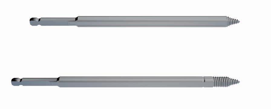
4 2 OPERATIVE TECHNIQUE MAIN FEATURES Part# Description UNYCO Cancellous Screw QC Shaft Ø 6mm UNYCO Cancellous Screw is designed both for diaphyseal and metaphyseal bone. The screw has a 6mm Ø shaft, is made of surgical grade stainless steel and is conical. The cortical thread is 5mm long, whereas the cancellous thread is 10mm long (so, the UNYCO Cancellous Screw thread is 15mm long). The special thread and tip design will only require screw insertion into the first cortex of the bone, avoiding perforation of the medullary canal. UNYCO Cancellous Screw Soft Tissue Reference Line Cancellous Thread Cortical Thread Ø 6mm Shank


.")


5 OPERATIVE TECHNIQUE 3 Large Multiscrew Clamp for UNYCO Screws M U The clamp is provided in M configuration, but it can be easily converted to U configuration by unlocking the arms with the universal Allen wrench and by re-positioning them (see below). This feature makes the system flexible and versatile. How to change configuration M 1. Unlock the locking screw of one arm using the 5mm Allen Wrench 2. Remove the locking screw 3. Re-position the arm in the new configuration and insert the locking screw 4. Tighten the locking screw using the 5mm Allen Wrench U Repeat the above mentioned steps 1-4 to position the second arm.


6 4 OPERATIVE TECHNIQUE The two arms subtend a 100 degree arc which facilitates screw insertion perpendicular to the bone surface. 100 The screw seats allow ± 10 variable angle screw positioning so that screws can be oriented independently.

 For manual screw insertion.")
7 OPERATIVE TECHNIQUE 5 EQUIPMENT REQUIRED Part# Description Qty Galaxy UNYCO Diaphyseal Tibia Box can accomodate: Galaxy UNYCO Mini Kit Tibia Sterile Rod D12mm L 350mm Sterile Galaxy UNYCO Mini Kit Instruments Sterile Galaxy UNYCO Mini Kit Tibia Sterile Limited Torque Wrench (out of Kit - available upon request) For manual screw insertion Galaxy UNYCO Mini Kit Instruments Sterile


 to it and check its correct")

8 6 OPERATIVE TECHNIQUE UNI-CORTICAL SCREW INSERTION Make a 5mm puncture in the skin. (Fig. 1) Fig. 1 Insert the first screw freehand, without the clamp, directly over the tibial crest (Fig. 2a) or medial or lateral (Fig. 2b) to it and check its correct position on the bone. Always attempt a perpendicular placement of the screw on the bone surface. Fig. 2 Fig. 2a Fig. 2b

 There will be instances when the cortex of cancellous bone is sufficiently hard that the torque limiter will activate and decouple the drilling and therefore stop further unnecessary advancement")
9 OPERATIVE TECHNIQUE 7 Screw Insertion Drill the screw perpendicular to the bone surface using a low speed power drill with the Power Drill Torque Limiter already mounted. The depth of insertion by the UNYCO Cancellous Screw in cancellous bone is controlled by the surgeon who stops advancing the drill when the reference line of the screw shank is flush with the skin surface. (Fig. 3a) There will be instances when the cortex of cancellous bone is sufficiently hard that the torque limiter will activate and decouple the drilling and therefore stop further unnecessary advancement of the screw. a Cancellous Bone The depth of penetration of the cortex of diaphyseal bone by the UNYCO Cancellous screw is controlled by the torque limiter. The torque limiter decouples the drill when the required torque has been reached. (Fig. 3b) b Cortical Bone Fig. 3 Apply the Large Multiscrew Clamp (93566) on the first screw and tighten the metal ring on the arm clockwise. (Fig. 4) NOTE: Once converging screws have been inserted, the clamps can no longer slide on the screw shafts. It is therefore important to determine the final distance (of 40mm or approximately 2 fingers breadth) of the clamp from the skin before inserting the second screw. Ideally, the clamp should be positioned at a distance of 40mm from the skin. (Fig. 4) 40mm Fig. 4
 as a template for screw insertion, insert the")


 Fig.")


10 8 OPERATIVE TECHNIQUE Using the Large Multiscrew Clamp (93566) as a template for screw insertion, insert the second screw in the contralateral arm, trying to be as perpendicular as possible to the bone surface. Check its correct position on the bone and if necessary partially tighten the metal ring on the arm clockwise so that the screw within its seat is free to move but without excessive play. (Fig. 5) Fig. 5 The clamp should not be pulled/pushed after the second screw is inserted. (Fig. 6) WARNING: On the tibia, the system stability is guaranteed only with 4 screws coupled with the Large Multiscrew Clamp in each bone segment. (Fig. 8) Fig. 6 Fig. 7 Fig. 8

 Fig.")


11 OPERATIVE TECHNIQUE 9 Once all screws in each arm have been inserted, tighten both metal rings fully with the 5mm Allen Wrench (30017). (Fig. 9) Fig. 9 Follow steps 2-5 to apply the second Large Multiscrew Clamp in the distal segment. (Fig. 10) Fig. 10 Join both Large Multiscrew Clamps with the rod leaving the clamps loosened to facilitate fracture reduction. (Fig. 11) Fig. 11

 Fig.")
 Fig.")

12 10 OPERATIVE TECHNIQUE Reduce the fracture, with X-ray guidance as necessary, holding the clamps to facilitate the reduction manoeuvre. (Fig. 12) Fig. 12 Lock the clamps first manually by turning the knurled metal ring clockwise. (Fig. 13) Fig. 13 If reduction is satisfactory, finally lock all the clamps firmly by tightening the cams with the 5mm Allen Wrench. (Fig. 14) Fig. 14
 attached to the same connecting")

 Fig.")
13 OPERATIVE TECHNIQUE 11 In case of a segmental fracture, the middle segment can be stabilised using a UNYCO Cancellous Screw in a Galaxy Large Clamp (93010) attached to the same connecting rod that links the two Large Multiscrew Clamps. Before drilling the UNYCO Cancellous Screw into the bone, partially tighten the metal ring on the clamp clockwise so that the screw within its seat is free to move but without excessive play. Once the screw has been inserted, tighten the clamp by hand. (Fig. 15) Fig. 15 Finally, lock the clamp with the Allen Wrench (30017). (Fig. 16) Fig. 16

 Fig. 17 Remove the uni-cortical screws. (Fig. 18) Fig.")
14 12 OPERATIVE TECHNIQUE CHANGING TO DEFINITIVE TREATMENT If the system is perceived as impediment for the correct definitive treatment application, remove the SYSTEM PARTS where needed. For instance, if there was a need to insert a plate on the medial side but maintain the overall reduction and alignment: Unlock the metal ring of the medial arm of the proximal Large Multiscrew Clamp. (Fig. 17) Fig. 17 Remove the uni-cortical screws. (Fig. 18) Fig. 18 Unlock the locking screw of the medial arm with the 5mm Allen Wrench. (Fig. 19) Fig. 19


15 OPERATIVE TECHNIQUE 13 Remove the medial arm. (Fig. 20) Fig. 20 If necessary repeat the procedure with the distal clamp. (Fig. 21) Fig. 21 In a similar fashion, if lateral submuscular plating was intended for the fracture, the lateral arms could be removed instead. In both scenarios described above, it is imperative there are 4 screws in each Large Multiscrew Clamp before any arm is disconnected. If intramedullary nailing of the fracture is envisaged as definitive treatment, it is usually not necessary to remove the fixator at all. However, appropriate sterile precautions would need to be taken to seal off the fixator from the remainder of the operative field.
 Ankle spanning configuration for peri-articular fractures or")
16 14 OPERATIVE TECHNIQUE DAMAGE CONTROL Knee spanning configuration for peri-articular fractures or ligamentous injuries of the knee Tibial application for peri-articular, diaphyseal or segmental fractures (as shown) Ankle spanning configuration for peri-articular fractures or ligamentous injuries


17 OPERATIVE TECHNIQUE 15 Knee spanning configuration for proximal tibial fracture associated with ligamentous instability of the knee Standard configuration for mid-shaft fracture of the tibia UNILATERAL FRAME DELTA FRAME Ankle spanning configuration for distal tibial fracture associated with ankle joint instability
18
19
20 Distributed by: Manufactured by: ORTHOFIX Srl Via Delle Nazioni 9, Bussolengo (Verona), Italy Telephone , Fax Instructions for Use: See actual package insert for Instructions for Use. Caution: Federal law (USA) restricts this device to sale by or on the order of a physician.proper surgical procedure is the responsibility of the medical professional. Operative techniques are furnished as an informative guideline. Each surgeon must evaluate the appropriateness of a technique based on his or her personal medical credentials and experience. Please refer to the Instructions for Use supplied with the product for specific information on indications for use, contraindications, warnings, precautions, adverse reactions and sterilization. MC-1512-OPT-E0 C 09/17
OPERATIVE TECHNIQUE. Galaxy UNYCO Diaphyseal Tibia Sterile Kit. Key contributors: S. Nayagam, MD T. Bégué, MD W.T. Gordon, MD
 OPERATIVE TECHNIQUE Galaxy UNYCO Diaphyseal Tibia Sterile Kit Key contributors: S. Nayagam, MD T. Bégué, MD W.T. Gordon, MD 1 INTRODUCTION 1 INDICATIONS 2 MAIN FEATURES 5 EQUIPMENT REQUIRED 6 UNYCO SCREW
Orthofix approach to Evidence Based Medicine
 VOICE OF DESIGN Orthofix approach to Evidence Based Medicine For years, clinical decision-making was based primarily on physician knowledge and expert opinion. Now, the medical community is searching for
OPERATIVE TECHNIQUE GALAXY UNYCO ANKLE BRIDGING DELTA FRAME UNILATERAL FRAME
 OPERATIVE TECHNIQUE GALAXY UNYCO ANKLE BRIDGING DELTA FRAME UNILATERAL FRAME CONTENTS INTRODUCTION 3 GALAXY UNYCO ANKLE DELTA APPROACH 4 GALAXY UNYCO ANKLE UNILATERAL APPROACH 12 Operative Technique Contributing
QUICK REFERENCE GUIDE. The PreFix Fixator (92000 Series) ALWAYS INNOVATING
 ALWAYS INNOVATING") 21 The PreFix Fixator (92000 Series) ALWAYS INNOVATING INTRODUCTION The PreFix fixator is designed to provide temporary external fixation. This may be needed when local facilities or the condition of the
21 The PreFix Fixator (92000 Series) ALWAYS INNOVATING INTRODUCTION The PreFix fixator is designed to provide temporary external fixation. This may be needed when local facilities or the condition of the
QUICK REFERENCE GUIDE. The XCaliber Articulated Ankle Fixator. By Dr. S. Berki, Dr. V. Caiaffa, Dr. F. Lavini and Dr. M. Manca
 QUICK REFERENCE GUIDE 18 The XCaliber Articulated Ankle Fixator By Dr. S. Berki, Dr. V. Caiaffa, Dr. F. Lavini and Dr. M. Manca GENERAL POINTS The XCaliber Articulated Ankle Fixator is made of radiolucent
QUICK REFERENCE GUIDE. The XCaliber Meta-Diaphyseal Fixator
 17 The XCaliber Meta-Diaphyseal Fixator GENERAL POINTS The XCaliber Fixator is made of radiolucent material for unobstructed X-ray visualization. The metallic bolts and the cam and bush of each ball-joint,
OPERATIVE TECHNIQUE GALAXY FIXATION SHOULDER
 OPERATIVE TECHNIQUE GALAXY FIXATION SHOULDER cop2 OPERATIVE TECHNIQUE INTRODUCTION 1 FEATURES OF SHOULDER COMPONENTS 2 EQUIPMENT REQUIRED 5 PREOPERATIVE PLANNING 6 SURGICAL PROCEDURE 8 POST OPERATIVE MANAGEMENT
QUICK REFERENCE GUIDE. Arthrodiatasis. Articulated Joint Distraction
 4 Arthrodiatasis Articulated Joint Distraction ARTHRODIATASIS OF THE HIP To prepare the assembly, remove the female component and replace it with the ProCallus articulated body for the hip. Remove cam
Knee spanning solutions
 Knee spanning solutions System features Indications Intended to be used on adults or pediatric patients as required for fracture fixation (open or closed); post-traumatic joint contracture which has resulted
pat hways Medtech innovation briefing Published: 14 December 2018 nice.org.uk/guidance/mib166
 pat hways Galaxy UNYCO for temporary stabilisation of lower limb fractures Medtech innovation briefing Published: 14 December 2018 nice.org.uk/guidance/mib166 Summary The technology described in this briefing
QUICK REFERENCE GUIDE. Arthrodesis. Joint Fusion. By Dr. S. Agostini and Dr. F. Lavini
 QUICK REFERENCE GUIDE 5 Arthrodesis Joint Fusion By Dr. S. Agostini and Dr. F. Lavini QUICK REFERENCE GUIDE ARTHRODESIS OF THE HIP Apply the ProCallus Fixator to the lateral aspect of the thigh. Insert
OPERATIVE TECHNIQUE. Knee Hinge (LRS Advanced System)
") OPERTIVE TECHNIQUE Knee Hinge (LRS dvanced System) 1 1 2 4 6 INTRODUCTION INDICTIONS FETURES ND BENEFITS EQUIPMENT REQUIRED KNEE HINGE SSEMBLY 8 17 TRUM KNEE DISLOCTION Orthofix wishes to thank the following
OPERTIVE TECHNIQUE Knee Hinge (LRS dvanced System) 1 1 2 4 6 INTRODUCTION INDICTIONS FETURES ND BENEFITS EQUIPMENT REQUIRED KNEE HINGE SSEMBLY 8 17 TRUM KNEE DISLOCTION Orthofix wishes to thank the following
N. Pennig Dynamic Wrist Fixator (37000 Series)
") N. Pennig Dynamic Wrist Fixator (000 Series) 0 Manufactured by: ORTHOFIX Srl Via Delle Nazioni, 0 Bussolengo (Verona), Italy Telephone + 0 000, Fax + 0 0 000 Pennig Dynamic Wrist Fixator Consisting of:
N. Pennig Dynamic Wrist Fixator (000 Series) 0 Manufactured by: ORTHOFIX Srl Via Delle Nazioni, 0 Bussolengo (Verona), Italy Telephone + 0 000, Fax + 0 0 000 Pennig Dynamic Wrist Fixator Consisting of:
Zimmer MIS Periarticular 3.5mm Proximal Tibial Locking Plate
 Zimmer MIS Periarticular 3.5mm Proximal Tibial Locking Plate Surgical Technique The Science of the Landscape Zimmer MIS Periarticular 3.5mm Proximal Tibial Locking Plate Surgical Technique 1 Zimmer MIS
LCP Distal Tibia Plate
 Surgical Technique LCP Locking Compression Plate Original Instruments and Implants of the Association for the Study of Internal Fixation AO/ASIF Table of contents Indications 3 Implants/Instruments 5 Surgical
OPERATIVE TECHNIQUE. The PreFix 2 Fixator
 OPERATIVE TECHNIQUE The PreFix 2 Fixator 1 INTRODUCTION 2 ASPECTS OF MRI COMPATIBILITY 4 FEATURES AND BENEFITS 5 EQUIPMENT REQUIRED 9 SURGICAL APPROACHES 12 LONG BONES 18 BLUE MULTI SCREW CLAMP 19 PELVIS
Technique Guide. 3.5 mm LCP Low Bend Medial Distal Tibia Plate Aiming Instruments. Part of the 3.5 mm LCP Percutaneous Instrument System.
 Technique Guide 3.5 mm LCP Low Bend Medial Distal Tibia Plate Aiming Instruments. Part of the 3.5 mm LCP Percutaneous Instrument System. Table of Contents Introduction 3.5 mm LCP Low Bend Medial Distal
Orthopedic Bone Nail System - Distal Femoral Nail Surgical Technique Manual
 Orthopedic Bone Nail System - Distal Femoral Nail Surgical Technique Manual Note: The surgical procedures should be performed under the guidance of qualified skilled orthopedic surgeons, and this surgical
PROXIMAL TIBIAL PLATE
 SURGICAL NÁSTROJE TECHNIQUE PRO ARTROSKOPII PROXIMAL INSTRUMENTS TIBIAL FOR PLATE ARTHROSCOPY Proximal Tibial Plate Description of medical device The Proximal Tibial Plate is used in epyphyseal and metaphyseal
LCP Medial Distal Tibia Plate, without Tab. The Low Profile Anatomic Fixation System with Angular Stability and Optimal Screw Orientation.
 LCP Medial Distal Tibia Plate, without Tab. The Low Profile Anatomic Fixation System with Angular Stability and Optimal Screw Orientation. Technique Guide LCP Small Fragment System Table of Contents Introduction
AcUMEDr. FoREARM ROD SYSTEM
 AcUMEDr FoREARM ROD SYSTEM FoREARM ROD SYSTEM Since 1988 Acumed has been designing solutions to the demanding situations facing orthopedic surgeons, hospitals and their patients. Our strategy has been
QUICK REFERENCE GUIDE. The Pennig Dynamic Wrist Fixator. Part A: Trans-articular application
 10 The Pennig Dynamic Wrist Fixator Part A: Trans-articular application B1 B2 B3 III IV TRANS-ARTICULAR APPLICATION The fractures that can be treated with this technique include AO type B and C fractures,
SMV Scientific Bone Plate and Screw System Surgical Technique
 SMV Scientific Bone Plate and Screw System Surgical Technique Description: The SMV Scientific Bone Plate and Screw System consists of non-locking plates and bone screw fasteners in a variety of lengths,
Conventus CAGE PH Surgical Techniques
 Conventus CAGE PH Surgical Techniques Conventus Orthopaedics The Conventus CAGE PH (PH Cage) is a permanent implant comprised of an expandable scaffold, made from nitinol and titanium, which is deployed
Correction System. Surgical Technique
 Re+Line Bunion Correction System Surgical Technique Bunion Correction System Easy insertion and medial placement accuracy using Landmark Guide technology 1 mm compression slot and fixed tines to encourage
QUICK REFERENCE GUIDE. The Orthofix Femoral Nailing System. By Prof. Dr. D. Pennig
 QUICK REFERENCE GUIDE The Orthofix Femoral Nailing System By Prof. Dr. D. Pennig Whenever possible, femoral fractures should be stabilized within the first 24 hours following injury, provided the patient
Large Distractor Femur
 Fracture Reduction and Provisional Stabilization Large Distractor Femur Surgical Technique Table of Contents Introduction Standard Femoral Distraction 2 Large Distractor System 4 Surgical Technique Prepare
3. Insert Tocar Sleeves Insert the NCB tissue protection sleeve assembly 1.6 to 10mm through a skin incision (Fig. 38).
.") NCB Proximal Humerus Plating System Surgical Technique 19 2. Temporary Plate Fixation The plate can be temporary fixed to the bone with 1.6mm K-wire through the proximal cannulated fixation screw of the
NCB Proximal Humerus Plating System Surgical Technique 19 2. Temporary Plate Fixation The plate can be temporary fixed to the bone with 1.6mm K-wire through the proximal cannulated fixation screw of the
Technique Guide. LCP Proximal Femoral Hook Plate 4.5/5.0. Part of the LCP Periarticular Plating System.
 Technique Guide LCP Proximal Femoral Hook Plate 4.5/5.0. Part of the LCP Periarticular Plating System. Table of Contents Introduction Features and Benefits 2 AO ASIF Principles 4 Indications 5 Surgical
Mini External Fixator.
 Mini External Fixator. Assembly and Surgical Technique This publication is not intended for distribution in the USA. Instruments and implants approved by the AO Foundation. Image intensifier control Warning
Galaxy Fixation System Lower Extremities Key contributor: S. Nayagam, MD Pr. A. C. Masquelet, MD
 OPERATIVE TECHNIQUE Galaxy Fixation System Lower Extremities Key contributor: S. Nayagam, MD Pr. A. C. Masquelet, MD 1 INTRODUCTION AND INDICATIONS 1 INDICATIONS IN TRAUMA 2 FEATURES AND BENEFITS 5 EQUIPMENT
Large External Fixator Delta Frame Ankle Bridge. For staged fixation of the distal tibia.
 Large External Fixator Delta Frame Ankle Bridge. For staged fixation of the distal tibia. Technique Guide Part of the Large External Fixation System Large External Fixator Delta Frame Ankle Bridge Technique
NCB Distal Femur System. Surgical Technique
 NCB Distal Femur System Surgical Technique NCB Distal Femur System Surgical Technique 3 Surgical Technique NCB Distal Femur System Table of Contents Introduction 4 Indications 8 Preoperative Planning
OPERATIVE TECHNIQUE GALAXY FIXATION SYSTEM LOWER EXTREMITIES
 OPERATIVE TECHNIQUE GALAXY FIXATION SYSTEM LOWER EXTREMITIES cop2 OPERATIVE TECHNIQUE INTRODUCTION 1 AND INDICATIONS INDICATIONS IN TRAUMA 1 FEATURES AND BENEFITS 2 CLAMP CLOSURE PROCEDURES 5 GALAXY FIXATION
Large External Fixator Delta Frame Ankle Bridge. Using pin clamps with outrigger posts.
 Large External Fixator Delta Frame Ankle Bridge. Using pin clamps with outrigger posts. Technique Guide Part of the Large External Fixation System Large External Fixator Delta Frame Ankle Bridge Technique
LCP Distal Humerus Plates
 The anatomic fixation system for the distal humerus with angular stability Surgical technique LCP Locking Compression Plate Contents Indications and contraindications 2 Implants 3 Instruments 5 Preparation
Knee Surgical Technique
 Knee Surgical Technique COMPASS Universal Hinge by Jimmy Tucker, M.D. Orthopaedic Surgeon Director, Arkansas Sports Medicine, P.A. Little Rock, Arkansas Table of contents Design features 3 Indications
External Skeletal Fixation (ESF)
") External Skeletal Fixation (ESF) Technique for fracture repair in animals Introduction External Skeletal Fixation is a versatile and effective technique for fracture repair in animals, rigidly stabilizing
External Skeletal Fixation (ESF) Technique for fracture repair in animals Introduction External Skeletal Fixation is a versatile and effective technique for fracture repair in animals, rigidly stabilizing
The Flower Straight Fibula Plate
 The Flower Straight Fibula Plate PROCEDURE GUIDE www.flowerortho.com The Flower Foot & Ankle Application STRAIGHT LOCKING FIBULA PLATE ANTERIOR LATERAL DISTAL TIBIAL PLATE MEDIAL DISTAL TIBIAL PLATE ANATOMIC
Patient Guide. Intramedullary Skeletal Kinetic Distractor For Tibial and Femoral Lengthening
 Patient Guide Intramedullary Skeletal Kinetic Distractor For Tibial and Femoral Lengthening Introduction You have decided to have a limb lengthening operation. The surgery you have chosen uses a device
OBSOLETED. LCP Medial Distal Tibia Plate, without Tab. The Low Profile Anatomic Fixation System with Angular Stability and Optimal Screw Orientation.
 LCP Medial Distal Tibia Plate, without Tab. The Low Profile Anatomic Fixation System with Angular Stability and Optimal Screw Orientation. Surgical Technique LCP Small Fragment System This publication
MetaFix Ludloff Plate
 Merete MetaFix Ludloff Plate Low Profile Locking Bone Plate System Surgical Technique and Ordering Information - Content - Content 1. Description.................................................. 3 2.
Zimmer MIS Periarticular Distal Femoral Locking Plate
 For Clinical Evaluations Zimmer MIS Periarticular Distal Femoral Locking Plate Surgical Technique The Science of the Landscape Zimmer MIS Periarticular Distal Femoral Locking Plate Surgical Technique
Zimmer Small Fragment Universal Locking System. Surgical Technique
 Zimmer Small Fragment Universal Locking System Surgical Technique Zimmer Small Fragment Universal Locking System 1 Zimmer Small Fragment Universal Locking System Surgical Technique Table of Contents Introduction
Sterile-Packaged Large External Fixator Kits. For treatment of long bone and pelvic fractures that require external fixation.
 Sterile-Packaged Large External Fixator Kits. For treatment of long bone and pelvic fractures that require external fixation. Large External Fixator Ankle Frame Kit Large External Fixator Trauma Kit Large
TransFx External Fixation System Large and Intermediate Surgical Technique
 TransFx External Fixation System Large and Intermediate Surgical Technique Choice, Simplicity, Transition TransFx External Fixation System Large and Intermediate Surgical Technique 1 Surgical Technique
Hoffmann II Micro External Fixation System
 Trauma Hoffmann II Micro External Fixation System Indications for the Hand Components 9 3 4 5 1 2 7 8 6 1 Hoffmann II Micro Multi-Pin Clamp 2 Hoffmann II Micro 90 Multi-Pin Clamp 3 Hoffmann II Micro Rod
OPERATIVE TECHNIQUE ANKLE ARTHRODESIS USING THE TRUELOK RING FIXATION SYSTEM
 OPERATIVE TECHNIQUE ANKLE ARTHRODESIS USING THE TRUELOK RING FIXATION SYSTEM 1 INTRODUCTION 2 COMPONENTS REQUIRED 2 CLEANING AND STERILIZATION 2 INDICATIONS 3 ASSEMBLY 5 PATIENT POSITIONING 5 APPLICATION
EVOS MINI with IM Nailing
 Case Series Dr. John A. Scolaro EVOS MINI with IM Nailing A series of studies Introduction Intramedullary nailing has become the standard for many long bone fractures. Fracture reduction prior to nail
LCP Low Bend Medial Distal Tibia Plates 3.5 mm. Anatomic plates with low profile head for intra- and extraarticular fractures.
 LCP Low Bend Medial Distal Tibia Plates 3.5 mm. Anatomic plates with low profile head for intra- and extraarticular fractures. Surgical Technique This publication is not intended for distribution in the
DFS STANDARD FIXATOR DFS ANKLE CLAMP DFS T-CLAMP
 DFS STANDAD FIXATO DFS ANKLE CLAMP DFS T-CLAMP SUGICAL TECHNIQUE Dr. James V. Nepola Professor of Orthopaedics University of Iowa Hospitals and Clinics Iowa City, Iowa Patent No. 5,662,650 C ontents DynaFix
LCP Medial Proximal Tibial Plate 3.5. Part of the Synthes small fragment Locking Compression Plate (LCP) system.
 system.") LCP Medial Proximal Tibial Plate 3.5. Part of the Synthes small fragment Locking Compression Plate (LCP) system. Technique Guide This publication is not intended for distribution in the USA. Instruments
LCP Medial Proximal Tibial Plate 3.5. Part of the Synthes small fragment Locking Compression Plate (LCP) system. Technique Guide This publication is not intended for distribution in the USA. Instruments
Zimmer ITST Intertrochanteric/ Subtrochanteric Fixation System. Abbreviated Surgical Technique
 Zimmer ITST Intertrochanteric/ Subtrochanteric Fixation System Abbreviated Surgical Technique ITST System Abbreviated Surgical Technique Indications The ITST Intramedullary Nail is indicated for use in
MiniRail System. Part B: Foot Applications. By Dr. B. Magnan, Dr. E. Rodriguez and Dr. G. Vito
 Q U I C K R E F E R E N C E G U I D E 14 MiniRail System Part B: Foot Applications By Dr. B. Magnan, Dr. E. Rodriguez and Dr. G. Vito ORDERING INFORMATION MiniRail System Kit, M190C Contents: M 101 Standard
TransFx External Fixation System Large and Intermediate Surgical Technique
 TransFx External Fixation System Large and Intermediate Surgical Technique TransFx External Fixation System Large and Intermediate Surgical Technique 1 Surgical Technique For TransFx External Fixation
3.5 mm LCP Low Bend Medial Distal Tibia Plate Aiming Instruments
 Part of the 3.5 mm LCP 3.5 mm LCP Low Bend Medial Distal Tibia Plate Aiming Instruments Surgical Technique TABLE OF CONTENTS INTRODUCTION 3.5 mm LCP Low Bend Medial Distal Tibia Plate 2 Aiming Instruments
Distal Ulnar Locking Plate
 INDEX Indications Patient Position Surgical Technique - Step 1 Approach - Step 2 Plate Contouring - Step 3 Fracture Reduction - Step 4 Distal Plate Fixation - Step 5 Confirm Proper Reconstruction - Step
3.5 mm Locking Attachment Plate
 For Treatment of Periprosthetic Fractures 3.5 mm Locking Attachment Plate Surgical Technique Table of Contents Introduction 3.5 mm Locking Attachment Plate 2 Indications 4 Surgical Technique Preparation
LCP Anterolateral Distal Tibia Plate 3.5. The low profile anatomic fixation system with optimal plate placement and angular stability.
 LCP Anterolateral Distal Tibia Plate 3.5. The low profile anatomic fixation system with optimal plate placement and angular stability. Technique Guide LCP Small Fragment System Table of Contents Introduction
3.5 mm LCP Extra-articular Distal Humerus Plate
 Part of the DePuy Synthes Locking Compression Plate (LCP ) System 3.5 mm LCP Extra-articular Distal Humerus Plate Surgical Technique Table of Contents Introduction 3.5 mm LCP Extra-articular Distal Humerus
PediLoc 3.5mm and 4.5mm Contour Femur Plate Surgical Technique
 PediLoc 3.5mm and 4.5mm Contour Femur Plate Surgical Technique Surgical Technique Contour Femur Plate The technique description herein is made available to the healthcare professional to illustrate the
A locking plate system that expands a surgeon s options in trauma surgery. Zimmer NCB Plating System
 A locking plate system that expands a surgeon s options in trauma surgery Zimmer NCB Plating System The Power of Choice The power of having true intraoperative options is at your fingertips. Using standard
quad-plate eight-plate Easy application, solid growth for kids Not an actual patient
 quad-plate eight-plate Easy application, solid growth for kids Not an actual patient Up to 60 screw angulation Increases the maximum angular correction and the possibility to cover also severe pathology
Technique Guide. 3.5 mm LCP Low Bend Medial Distal Tibia Plates. Part of the Synthes locking compression plate (LCP) system.
 system.") Technique Guide 3.5 mm LCP Low Bend Medial Distal Tibia Plates. Part of the Synthes locking compression plate (LCP) system. Table of Contents Introduction 3.5 mm LCP Low Bend Medial Distal Tibia Plates
Technique Guide 3.5 mm LCP Low Bend Medial Distal Tibia Plates. Part of the Synthes locking compression plate (LCP) system. Table of Contents Introduction 3.5 mm LCP Low Bend Medial Distal Tibia Plates
Hallux Valgus Deformity OPERATIVE TECHNIQUE
 Hallux Valgus Deformity OPERATIVE TECHNIQUE Table of Contents 1 ORDERING INFORMATION 3 OPERATIVE TECHNIQUE 9 INDICATIONS FOR USE The surgical technique shown is for illustrative purposes only. The technique(s)
LCP Anterolateral Distal Tibia Plate 3.5. The low profile anatomic fixation system with optimal plate placement and angular stability.
 LCP Anterolateral Distal Tibia Plate 3.5. The low profile anatomic fixation system with optimal plate placement and angular stability. Technique Guide LCP Small Fragment System Table of Contents Introduction
Technique Guide. TomoFix Osteotomy System. A comprehensive plating system for stable fixation of osteotomies around the knee.
 Technique Guide TomoFix Osteotomy System. A comprehensive plating system for stable fixation of osteotomies around the knee. Table of Contents Introduction TomoFix Osteotomy System 2 AO Principles 4 Indications
Surgical Technique. Cannulated Angled Blade Plate 3.5 and 4.5, 90
 Surgical Technique Cannulated Angled Blade Plate 3.5 and 4.5, 90 Cannulated Angled Blade Plate 3.5 and 4.5, 90 Table of contents Indications/Contraindications 2 Implants 3 Surgical technique 5 Implant
3. PATIENT POSITIONING & FRACTURE REDUCTION 3 8. DISTAL GUIDED LOCKING FOR PROXIMAL NAIL PROXIMAL LOCKING FOR LONG NAIL 13
 Contents IMPLANT FEATURES 2 1. INDICATIONS 3 2. PRE-OPERATIVE PLANNING 3 3. PATIENT POSITIONING & FRACTURE REDUCTION 3 4. INCISION 4 5. ENTRY POINT 4-6 6. PROXIMAL NAIL INSERTION 6-7 7. PROXIMAL LOCKING
TIPMED EXTERNAL FIXATION SYSTEMS
 TIPMED EXTERNAL FIXATION SYSTEMS ANATOMICAL LOCATIONS FOR EXTERNAL FIXATION SYSTEMS Humeral Dynamic Axial Fixator Elbow Fixator Pelvic Dynamic Axial Fixator Pennig Wrist Fixator Hand Fixator Finger Fixator
Monolateral External Fixation System for Trauma and Orthopaedics
 MEFiSTO Monolateral External Fixation System for Trauma and Orthopaedics Surgical Technique Original Instruments and Implants of the Association for the Study of Internal Fixation AO/ASIF MEFiSTO Table
EXTERNAL FIXATION SYSTEM
 EXTERNAL FIXATION SYSTEM The Apex Pin Fixation System is a one step procedure reducing insertion time and reducing insertion temperature. There are three types of fixation pins: Self-drilling / Self-tapping
U2 PSA. Revision Knee. Surgical Protocol
 U2 PSA TM Revision Knee Surgical Protocol Table of Contents 1 Component Removal... 1 2 Tibial Preparation... 1 2.1 Tibial Canal Preparation... 1 2.2 Proximal Tibial Resection... 2 2.3 Non Offset Tibial
Gentle Guided Growth to Correct Knock Knees and Bowed Legs in Children
 PATIENT INFORMATION Gentle Guided Growth to Correct Knock Knees and Bowed Legs in Children The Guided Growth System eight-plate quad-plate INTRODUCTION Children need gentle guidance and correction in many
Surgical Technique. Anterolateral and Medial Distal Tibia Locking Plates
 Surgical Technique Anterolateral and Medial Distal Tibia Locking Plates PERI-LOC Periarticular Locked Plating System Anterolateral and Medial Distal Tibia Locking Plates Surgical Technique Contents Product
The Vilex FUZETM. Dual Thread Screw & Intramedullary Nail in One Implant. The Ultimate TTC Arthrodesis Internal Fixator
 The Vilex FUZETM Dual Thread Screw & Intramedullary Nail in One Implant The Ultimate TTC Arthrodesis Internal Fixator Introduction The Vilex FUZE TM TTC Arthrodesis Compression Nail combines the attributes
Surgical Technique. Intramedullary locked Nailing With Screws for Humerus Fractures Solid/Cannulated. Humeral Interlocking Nail.
 Screws for Humerus Fractures Surgical Technique Humeral Interlocking Nail Approved by Humerus Nail Kit Code 08050001 Contents Introduction Implant design Indications Pre-operative planning Patient positioning
The Flower Four Corner Fusion Plate
 The Flower Four Corner Fusion Plate PROCEDURE GUIDE www.flowerortho.com The Flower Upper Extremity Application PROXIMAL HUMERUS PLATE SMALL BONE PLATES FOUR CORNER FUSION PLATE ANATOMIC DISTAL RADIUS PLATE
Technique Guide. 2.7 mm/3.5 mm LCP Distal Fibula Plates. Part of the Synthes locking compression plate (LCP) system.
 system.") Technique Guide 2.7 mm/3.5 mm LCP Distal Fibula Plates. Part of the Synthes locking compression plate (LCP) system. Table of Contents Introduction 2.7 mm/3.5 mm LCP Distal Fibula Plates 2 AO Principles
Technique Guide 2.7 mm/3.5 mm LCP Distal Fibula Plates. Part of the Synthes locking compression plate (LCP) system. Table of Contents Introduction 2.7 mm/3.5 mm LCP Distal Fibula Plates 2 AO Principles
LCP Extra-articular Distal Humerus Plate.
 Technique Guide LCP Extra-articular Distal Humerus Plate. The anatomically shaped and angular stable fixation system for extraarticular fractures of the distal humerus. Table of Contents Introduction
Hoffmann II External Fixation System
 Hoffmann II External Fixation System Modular System for Long Bones Pelvis Introduction In 1938, Raoul Hoffmann, a surgeon from Geneva, Switzerland, designed a revolutionary External Fixation System. The
Technique Guide Hammertoe Correction System
 Technique Guide Hammertoe Correction System TM The ToeMATE Hammertoe Correction System is an easy to implant bone screw system intended for the correction of hammertoe deformity. It is provided in a complete
LCP Medial Proximal Tibial Plate 4.5/5.0. Part of the Synthes LCP periarticular plating system.
 LCP Medial Proximal Tibial Plate 4.5/5.0. Part of the Synthes LCP periarticular plating system. Technique Guide This publication is not intended for distribution in the USA. Instruments and implants approved
Correction System. Surgical Technique
 Nextra Hammertoe Correction System Surgical Technique Maximized Bone Purchase* Stable and Secure Phalanx Optimized Screw Design Adjustable Bone-to-Bone Apposition Progressive Ratchet Tightening Mechanism
Clinical. Solutions. Synthes Solutions. Foot and Ankle.
 Clinical Solutions Foot and Ankle. Foot and Ankle. Fractures of the tibial shaft Fractures of the distal fibula Fractures of the distal tibia Fractures and osteotomies of the calcaneus Arthrodesis Fractures,
Technique Guide. DHS Blade. For osteoporotic bone.
 Technique Guide DHS Blade. For osteoporotic bone. Table of Contents Introduction Features and Benefits 2 Indications and Contraindications 4 Clinical Cases 5 Surgical Technique Implantation 6 Implant
MEFiSTO. Monolateral External Fixation System for Trauma and Orthopaedics.
 MEFiSTO. Monolateral External Fixation System for Trauma and Orthopaedics. Surgical Technique This publication is not intended for distribution in the USA. Instruments and implants approved by the AO Foundation.
THE NATURAL FIT. Surgical Technique. Hip Knee Spine Navigation
 THE NATURAL FIT Surgical Technique Hip Knee Spine Navigation MiniMAX Surgical Technique Hip Knee Spine Navigation INTRODUCTION The MiniMAX TM is a cementless anatomic stem available in 9 right sizes and
Mandible External Fixator II. Provides treatment for fractures of the maxillofacial area.
 Mandible External Fixator II. Provides treatment for fractures of the maxillofacial area. Technique Guide This publication is not intended for distribution in the USA. Instruments and implants approved
Surgical Technique. Targeter Systems Overview
 Surgical Technique Targeter Systems Overview PERI-LOC Locked Plating System Targeter Systems Overview Table of contents Product overview... 2 Introduction... 2 Indications... 2 Design features and benefits...
AxSOS Locking Plate System
 AxSOS Locking Plate System Operative Technique Small Fragment Basic Fragment 1 2 Contents Page 1. Introduction 4 2. Features & Benefits 5 4 and 5mm Compression Plates 5 Reconstruction and 1/3 Tubular Locking
TRUMATCH PERSONALIZED SOLUTIONS with the SIGMA High Performance Instruments
 TRUMATCH PERSONALIZED SOLUTIONS with the SIGMA High Performance Instruments Resection Guide System SURGICAL TECHNIQUE RESECTION GUIDE SURGICAL TECHNIQUE The following steps are an addendum to the SIGMA
Technique Guide. 2.4 mm Variable Angle LCP Distal Radius System. For fragment-specific fracture fixation with variable angle locking technology.
 Technique Guide 2.4 mm Variable Angle LCP Distal Radius System. For fragment-specific fracture fixation with variable angle locking technology. Table of Contents Introduction 2.4 mm Variable Angle LCP
Large External Fixator Modular Knee Bridge. Using multi-pin clamps.
 Large External Fixator Modular Knee Bridge. Using multi-pin clamps. Technique Guide Part of the Large External Fixation System Large External Fixator Modular Knee Bridge Technique Overview Insert Schanz
Surgical Technique. Distal Humerus Locking Plate
 Surgical Technique Distal Humerus Locking Plate PERI-LOC Locked Plating System Distal Humerus Locking Plate Surgical Technique Table of Contents Introduction...2 Indications...3 Plate Features...3 Patient
System. Humeral Nail. Surgical Technique
 System Humeral Nail Surgical Technique Contents IMPLANT FEATURES 2 1. INDICATIONS 3 2. PRE-OPERATIVE PLANNING 3 3. PATIENT POSITIONING & FRACTURE REDUCTION 3 4. INCISION 4 5. ENTRY POINT 4-6 6. PROXIMAL
Surgical Technique. Fibula Rod System
 Surgical Technique Fibula Rod System Acumed is a global leader of innovative orthopaedic and medical solutions. We are dedicated to developing products, service methods, and approaches that improve patient
Femur Condylar Plate System Procedural Steps.
 Femur Condylar Plate System Procedural Steps www.carbo-fix.com 1 Table of Contents Introduction..3 Instrumentation Set... 8 Procedural Steps:...... 12 Ordering Information 19 2 Introduction The CarboFix