Excretory urography (EU) or IVP US CT & radionuclide imaging
|
|
|
- Judith Griffith
- 5 years ago
- Views:
Transcription
1
2
3 Excretory urography (EU) or IVP US CT & radionuclide imaging
4 MRI arteriography studies requiring catherization or direct puncture of collecting system
5 EU & to a lesser extent CT provide both functional & anatomical informations. US & MRI provide anatomical informations. Radionuclide scanning provides functional informations only.
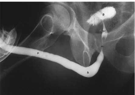
6 The first line No ionising radiatrion no contrast
7 IV injection of iodinated contrast media Iodine concentration is 300 mg I /Kg BW has been largely replaced by US& CT
8 Fasting for at least 6 hours Laxative taken 30 hr & 24 hr prior to the time of examination. No fluid restriction in cases of impaired renal function.

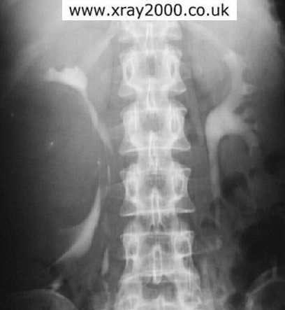
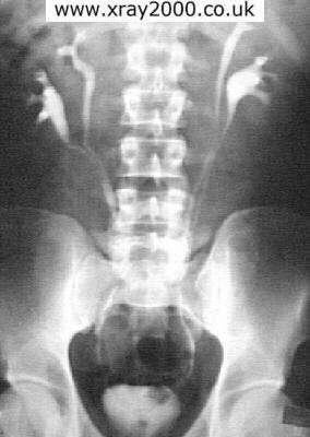
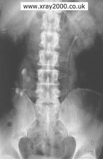
9 Plain film: full length, frontal view?calcification
10 Urinary calculi (stones are missed if no plain film is taken). Diffuse nephrocalcinosis. Localized nephrocalcinosis (TB, tumors). Prostatic calcifications.

11 Assess the renal size Assess the renal outlines

12 Assess the calices


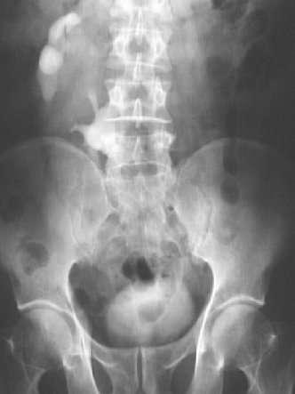
13 Only portion of the ureter is seen Course: along the transverse processes of the lumbar vertebrae. Dilatation of the renal pelvis & ureter
14 Dilatation of the renal pelvis & ureter obstruction congenital variant vesicouretric reflux

15 Full bladder film Outline indentations

16

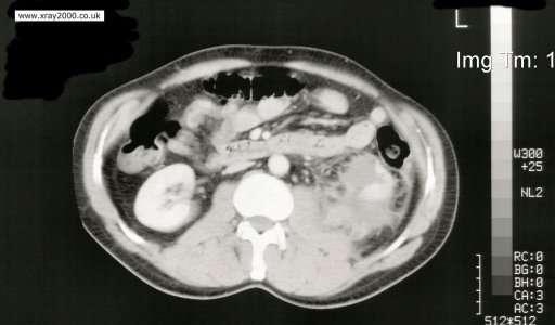
17 CT urography More sensitive for detecting stones Allows charactarisation of renal lesions Assess surrounding structures No superimposition of structures
18 o Retrograde & antegrade pyelography o Voiding cystourethrography: o Urethrography:

19

20
21 Most of the UT stones are radiopaque Pure uric acid & xanthine stones Radiopaque stones can be identified on plain film CT & US can identify all types of stones

22 Staghorn stone large stone filling the whole of the PCS & taking its shape PLAIN FILM

23 Urinary tract obstruction The principal feature is dilatation of PC system & ureters
24 stones Tumors blood clot strictures Congenital :PUJ obstruction, posterior urethral valve enlarged prostate compression from adjacent retroperitoneal structures or masses
25 splaying of the renal sinus fat due to pooling of urine within the dilated PCS The proximal ureter can be easily identified but the distal ureter is usually obscured by overlying bowel.

26 NORMAL US HYDRONEPHROSIS
27 Plain films may demonstrate stones Delayed nephrogram In acute obstruction, dense nephrogram In intermittent obstruction, IVU may be normal between attacks In prolonged obstruction there will be atrophy of the kidney

28

29
30 simple renal cysts malignant tumors Multiple masses : multiple simple renal cysts, polycystic disease, lymphoma Abscess benign tumors hydatid metastasis.
31 -a rounded lucency in the nephrogram. -bulging of renal outlines -displacement of calyces -enlargement of the kidney -calcification in a small proportion of renal carcinomas or wall of a cyst
32


33 determine if the mass is cystic or solid


34 further assess masses and stage renal cancer
35
36 predisposing factors: Stones Reflux Obstructive lesios Diabetes mellitus
37 Most patients with Acute infections of the urinary tract do not require urgent imaging investigations. US: may show diffuse or focal swelling of kidneys, evidence of the predisposing lesion (stones, obstruction, reflux) complications (abscess, scarring)

38 Scar formation Dilatation of calyces in scarred areas. Overall reduction in renal size. Dilatation of affected collecting system due to reflux. Reflux may be demonstrated at micturating cystourethrography.
39 IVP & CT are the major imaging modalities Aim of imaging: 1-assess the renal perfusion 2-ensure that opposite kidney is normal 3-show the extent of renal parenchymal damage 4- Demonstrate injury to other abdominal organs
40
41
42 unilateral or bilateral The upper ureter may be ectopic The dilated lower ureter may prolapse into bladder (ureterocele)
43

44 usually in the lower abdomen & rotated some cases, both lie on same side of pelvis & fused. more prone to complications
45

46 failure of separation of the lower poles. May be an incidental finding or associated with PUJ obstruction & stone formation

47 autosomal dominant polycystic kidney disease (adult type) autosomal recessive polycystic kidney disease (infantile type)
48 other kidney show compensatory hypertrophy. Radionuclide scanning used for diagnosis as well US & CT.
49 Absent kidney : nephrectomy, renal agenesis Non functioning kidney : Ureteric obstruction Renal artery occlusion : Acute renal vein thrombosis :

50 Nephrocalcinosis wide spread calcification in the cortex or medulla of the kidney PLAIN FILM
51 Absent kidney : nephrectomy, renal agenesis Non functioning kidney : Ureteric obstruction Renal artery occlusion : Acute renal vein thrombosis
52
53 Most of tumors are TCC May obstruct the ureters US: solid mass. CT & MRI determine the extent of tumor spread outside the bladder walls
54 Usually secondary to chronic bladder outlet obstruction May be congenital Predispose to infection, stone formation & occasionally tumors may arise within them. Filled with contrast in IVP, cystourethrography & in post voiding film Readily diagnosed with US, CT & MRI.
R adio logical investigations of urinary system
 R adio logical investigations of urinary system There are 4 main radiological Ix: 1 IVU: Intravenous urography. 2- U/S 3-CT scan 4-Radioisotope scan. Others (not frequently used): MRI, arteriography, antegrade
PROFESSIONAL SKILLS 1 3RD YEAR SEMESTER 6 RADIOGRAPHY. THE URINARY SYSTEM Uz. Fatema shmus aldeen Tel
 PROFESSIONAL SKILLS 1 3RD YEAR SEMESTER 6 RADIOGRAPHY THE URINARY SYSTEM Uz. Fatema shmus aldeen Tel. 0925111552 Professional skills-2 THE URINARY SYSTEM The urinary system (review anatomy and physiology)
Proceedings of the 34th World Small Animal Veterinary Congress WSAVA 2009
 www.ivis.org Proceedings of the 34th World Small Animal Veterinary Congress WSAVA 2009 São Paulo, Brazil - 2009 Next WSAVA Congress : Reprinted in IVIS with the permission of the Congress Organizers IMAGING
Chapter 6: Genitourinary and Gastrointestinal Systems 93
 Chapter 6: Genitourinary and Gastrointestinal Systems 93 Chapter 6 Genitourinary and Gastrointestinal Systems Embryology Three sets of excretory organs or kidneys develop in human embryos: Pronephros:
URINARY SYSTEM I. Kidneys II. Nephron Unit and Urine Formation
 URINARY SYSTEM I. Kidneys A. Location and Structure 1. Retroperitoneal 2. Between T12 and L3 3. Rt. kidney slightly lower 4. Two bean shaped organs 5. Adrenal gland 6. Internal construction a. Renal cortex
Hydronephrosis. What is hydronephrosis?
 What is hydronephrosis? Hydronephrosis Hydronephrosis describes the situation where the urine collecting system of the kidney is dilated. This may be a normal variant or it may be due to an underlying
Pediatric Ure-Radiology*
 Pediatric Ure-Radiology* HERMAN GROSSMAN, M.D. Professor of Radiology and Pediatrics, Duke University Medical Center, Durham, North Carolina "Routine" radiologic studies do not, often enough, concentrate
Obstetrics Content Outline Obstetrics - Fetal Abnormalities
 Obstetrics Content Outline Obstetrics - Fetal Abnormalities Effective February 2007 10 16% renal agenesis complete absence of the kidneys occurs when ureteric buds fail to develop Or degenerate before
CYSTIC DISEASES of THE KIDNEY. Dr. Nisreen Abu Shahin
 CYSTIC DISEASES of THE KIDNEY Dr. Nisreen Abu Shahin 1 Types of cysts 1-Simple Cysts 2-Dialysis-associated acquired cysts 3-Autosomal Dominant (Adult) Polycystic Kidney Disease 4-Autosomal Recessive (Childhood)
Obstructive Uropathy. PATHOPHYSIOLOGIC CHANGES UUO vs BUO. Arry Rodjani Urology Department Ciptomangunkusumo Hospital Jakarta
 Obstructive Uropathy PATHOPHYSIOLOGIC CHANGES UUO vs BUO Arry Rodjani Urology Department Ciptomangunkusumo Hospital Jakarta INTRODUCTION Obstructive uropathy refers to the functional or anatomic obstruction
RENAL SCINTIGRAPHY IN THE 21 st CENTURY
 RENAL SCINTIGRAPHY IN THE 21 st CENTURY 99m Tc- MAG 3 with zero time injection of Furosemide (MAG 3 -F 0 ) : A Fast and Easy Protocol, One for All Indications Clinical Experience Congenital Disorders PROTOCOL
Acute flank pain in children: Imaging considerations
 Acute flank pain in children: Imaging considerations Carlos J. Sivit MD Rainbow Babies and Children s Hospital Case Western Reserve School of Medicine Flank pain Results from distention of ureter or renal
ISUOG Basic Training. Distinguishing between Normal & Abnormal Appearances of the Urinary Tract. Seshadri Suresh, India
 ISUOG Basic Training Distinguishing between Normal & Abnormal Appearances of the Urinary Tract Seshadri Suresh, India Learning objectives 13 & 14 At the end of the lecture you will be able to: describe
Fetal Renal Malformations: The Role of Ultrasound in Diagnosis & Management
 Fetal Renal Malformations: The Role of Ultrasound in Diagnosis & Management 12 weeks Alfred Abuhamad, M.D. Eastern Virginia Medical School 13 weeks 2nd trimester Medullary pyramids Renal Sinus Cortex 2nd
Contents. Review anatomy of the urinary tract Imaging modalities
 Contents Review anatomy of the urinary tract Imaging modalities The Urinary Tract Kidneys ตาแหน งไต (position) อย ใน retroperitoneum ระด บ T12-L3 โดยไต ขวาจะม ระด บตากว าไตซ ายเล กน อย ร ปร าง (shape)
IMAGING OF THE UROGENITAL TRACT
 IMAGING OF THE UROGENITAL TRACT 1 A) URINARY TRACT There are many methods of imaging the urinary tract but plain abdominal X-ray and ultrasound scan are usually done first in most cases, especially in
Outline. Introduction to imaging modalities of the urinary system. Case base learning of common diseases in urinary tract
 Outline Introduction to imaging modalities of the urinary system Case base learning of common diseases in urinary tract Outline Introduction to imaging modalities of the urinary system Case base learning
Outline. Introduction to imaging modalities of the urinary system. Case base learning of common diseases in urinary tract
 Outline Introduction to imaging modalities of the urinary system Case base learning of common diseases in urinary tract Diagnostic Investigations in Urinary System PLAIN KUB EXCRETORY UROGRAPHY RETROGRADE
Abdominal Ultrasound : Aorta, Kidneys, Bladder
 Abdominal Ultrasound : Aorta, Kidneys, Bladder Nilam J. Soni, MD, MSc Associate Professor of Medicine Divisions of Hospital Medicine and Pulmonary/Critical Care Medicine Department of Medicine University
US in non-traumatic acute abdomen. Lalita, M.D. Radiologist Department of radiology Faculty of Medicine ChiangMai university
 US in non-traumatic acute abdomen Lalita, M.D. Radiologist Department of radiology Faculty of Medicine ChiangMai university Sagittal Orientation Transverse (Axial) Orientation Coronal Orientation Intercostal
Acute Pyelonephritis
 Acute Pyelonephritis Variant 1: Acute pyelonephritis. Uncomplicated patient (eg, no history of diabetes or immune compromise or history of stones or obstruction or prior renal surgery or lack of response
Urinary system Ultrasound (Renal & Urinary bladder)
") Urinary system Ultrasound (Renal & Urinary bladder) Edited & Presented by ; Hussien A.B ALI DINAR. Msc.Phd ISRRT Associate Member Lecturer (National university) Reporting Sonographer (PHC) Objective By
Urinary system Ultrasound (Renal & Urinary bladder) Edited & Presented by ; Hussien A.B ALI DINAR. Msc.Phd ISRRT Associate Member Lecturer (National university) Reporting Sonographer (PHC) Objective By
Urinary tract obstruction
 Urinary tract obstruction Common causes : stone, blood clot Radiographic findings depend on I. Level of obstruction II. Severity of obstruction : partial or complete III. Timing of obstruction Pathophysiology
Imaging Ejaculatory Disorders and Hematospermia
 ATHENS 4-6 October 2018 European Society of Urogenital Radiology Imaging Ejaculatory Disorders and Hematospermia Parvati Ramchandani, MD Professor, Radiology and Surgery University of Pennsylvania Medical
Uroradiology For Medical Students
 Uroradiology For Medical Students Lesson 4: Cystography & Urethrography - Part 2 American Urological Association Review Cystography is useful in evaluating the bladder, the urethra and the competence of
Radiological Assessment of the Kidney in Patients with Hematuria
 March 2005 Radiological Assessment of the Kidney in Patients with Hematuria Jeremy L. McKay, Harvard Medical School Year III Hematuria Signs and Symptoms Microscopic or gross hematuria Abdominal pain Fever
Sex: 女 Age: 51 Occupation: 無 Admission date:92/07/22
 Sex: 女 Age: 51 Occupation: 無 Admission date:92/07/22 Chief complaint Unknown fever for one month Hand tremor and left huge renal tumor was noted Present illness Suffered from fever for one month, hand
ASSESSING THE PLAIN ABDOMINAL RADIOGRAPH M A A M E F O S U A A M P O F O
 ASSESSING THE PLAIN ABDOMINAL RADIOGRAPH M A A M E F O S U A A M P O F O Introduction The abdomen (less formally called the belly, stomach, is that part of the body between the thorax (chest) and pelvis,
Kristina M. Nowitzki, M.D., Ph.D. and Hao S. Lo, M.D. University of Massachusetts Medical School, Worcester, MA
 Kristina M. Nowitzki, M.D., Ph.D. and Hao S. Lo, M.D. University of Massachusetts Medical School, Worcester, MA Outline I. Introduction highlighting normal renal enhancement physiology including normal
Developmental Abnormalities of the Kidneys and GU System
 A5 Developmental Abnormalities of the Kidneys and GU System Erin Parilla, MD Neonatologist Pediatrix Medical Group, Tampa, FL The speaker has signed a disclosure form and indicated she has no significant
Radiographic Procedures III (RAD 228)
") Radiographic Procedures III (RAD 228) Urinary System RADIOGRAPHIC EXAMINATIONS Urinary System Antegrade Exam IVU Functional test Hypertensive evaluation as per protocol Retrograde Exams Retrograde Urography
Radiographic Procedures III (RAD 228) Urinary System RADIOGRAPHIC EXAMINATIONS Urinary System Antegrade Exam IVU Functional test Hypertensive evaluation as per protocol Retrograde Exams Retrograde Urography
URINARY TRACT IMAGING - BASIC PRINCIPLES
 URINARY TRACT IMAGING - BASIC PRINCIPLES Clinical Radiology Every physician needs a basic understanding of diagnostic imaging to understand how to order the appropriate studies and to understand the resulting
Role of imaging in evaluation of genitourinary i trauma Spectrum of GU injuries Relevance of imaging findings in determining management Focus on MDCT
 Genitourinary Tract Injuries 6 th Nordic Course Scott D. Steenburg, MD Assistant Professor University of Maryland Department of Radiology Division of Trauma and Emergency Radiology R Adams Cowley Shock
8/14/2017. Kidney location & visualization. Brief Review with tips & Case Based Illustrations. Size = x L2. Size =
 Dr. Russell Tucker, DACVR Brief Review with tips & Case Based Illustrations Kidney location & visualization K9 Kidneys: Rt @ T13-L1 Lt @ L2-L4 Kidney visualization K9 Kidneys: Rt @ T13-L1 Lt @ L2-L4 Size
Chapter IV. Angionephrography in Simple Renal Cysts
 Acta Radiologica ISSN: 0001-6926 (Print) (Online) Journal homepage: http://www.tandfonline.com/loi/iaro20 Chapter IV. Angionephrography in Simple Renal Cysts To cite this article: (1957) Chapter IV. Angionephrography
Perineal Sonography in Diagnosis of an Ectopic Ureteric Opening Into the Urethra
 Case Series Perineal Sonography in Diagnosis of an Ectopic Ureteric Opening Into the Urethra S. Boopathy Vijayaraghavan, MD, DMRD Objective. To study the role of perineal sonography in the diagnosis of
The Kidneys. (L., ren; Gk, nephros; hence the adjectives renal and nephric) & Suprarenal (Adrenal) Glands. Dr Maan Al-Abbasi PhD, MBChB
 & Suprarenal (Adrenal) Glands. Dr Maan Al-Abbasi PhD, MBChB") The Kidneys (L., ren; Gk, nephros; hence the adjectives renal and nephric) & Suprarenal (Adrenal) Glands Dr Maan Al-Abbasi PhD, MBChB Functions of Urinary System Regulate electrolytes (K+, Na+, etc) Regulate
The Kidneys (L., ren; Gk, nephros; hence the adjectives renal and nephric) & Suprarenal (Adrenal) Glands Dr Maan Al-Abbasi PhD, MBChB Functions of Urinary System Regulate electrolytes (K+, Na+, etc) Regulate
Request Card Task ANSWERS
 Request Card Task ANSWERS Medical Student Workbook Author: Dr Sam Leach, SpR Case 1 What differential diagnoses are most likely? Which investigation is most appropriate? Case 1 The most likely diagnosis
1. Congenital Anomalies of Kidney and Ureter 1
 CONTENTS 1. Congenital Anomalies of Kidney and Ureter 1 1.1 Antenatal Pelviureteric Junction Obstruction 1 1.2 Bilateral Pelviureteric Junction Obstruction 3 1.3 Circumcaval Ureter 6 1.4 Crossed Renal
Find Medical Solutions to Your Problems HYDRONEPHROSIS. (Distension of Renal Calyces & Pelvis)
") HYDRONEPHROSIS (Distension of Renal Calyces & Pelvis) Hydronephrosis is the distension of the renal calyces and pelvis due to accumulation of the urine as a result of the obstruction to the outflow of
HYDRONEPHROSIS (Distension of Renal Calyces & Pelvis) Hydronephrosis is the distension of the renal calyces and pelvis due to accumulation of the urine as a result of the obstruction to the outflow of
Abdominal ultrasound:
 Abdominal ultrasound: Non-traumatic acute abdomen Wittanee Na-ChiangMai, MD Department of Radiology ChiangMai University 26/04/2017 Contents Technique of examination Normal anatomy Emergency conditions
Lec-8 جراحة بولية د.نعمان
 4th stage Lec-8 جراحة بولية د.نعمان 11/10/2015 بسم هللا الرحمن الرحيم Ureteric, Vesical, & urethral stones Ureteric Calculus Epidemiology like renal stones Etiology like renal stones Risk factors like
Urinary Tract Abnormalities
 Urinary Tract Abnormalities Dr Hennie Lombaard Senior Specialist Maternal and Fetal Medcine Department of Obstetrics and Gynecology Level 7 Pretoria Academic Hospital Pictures from The 18 to 23 weeks scan
My Patient Has Abdominal Pain PoCUS of the Biliary Tract and the Urinary Tract
 My Patient Has Abdominal Pain PoCUS of the Biliary Tract and the Urinary Tract Objectives PoCUS for Biliary Disease PoCUS for Renal Colic PoCUS for Urinary Retention Biliary Disease A patient presents
Uroradiology Tutorial For Medical Students
 Uroradiology Tutorial For Medical Students Lesson 3: Cystography & Urethrography Part 1 American Urological Association Introduction Conventional radiography of the urinary tract includes several diagnostic
The functional anatomy of the urinary system. Human Anatomy Department Dr. Anastasia Bendelic
 The functional anatomy of the urinary system Human Anatomy Department Dr. Anastasia Bendelic Plan Development of the kidneys and their abnormalities Development of the urinary ways and their abnormalities
Five Views of Transitional Cell Carcinoma: One Man s Journey
 September 2006 Five Views of Transitional Cell Carcinoma: One Man s Journey Amsalu Dabela, Harvard Medical School III Outline Overview: Renal Anatomy Our Patient s Story Diagnostic Imaging Studies Appearance
Kidney & Urinary Tract Ultrasound. Fatina Fadel Hafez Bazaraa
 Kidney & Urinary Tract Ultrasound Fatina Fadel Hafez Bazaraa Ultrasonography Ultrasound Available Rapid Inexpensive Painless & no sedation needed No adverse effects/ complications Can be repeated Useful
Genitourinary Radiology In-Training Test Questions for Diagnostic Radiology Residents
 Genitourinary Radiology In-Training Test Questions for Diagnostic Radiology Residents March, 2013 Sponsored by: Commission on Education Committee on Residency Training in Diagnostic Radiology 2013 by American
Cystitis cystica is a rare chronic reactive inflammatory
 JOURNAL OF ENDOUROLOGY CASE REPORTS Volume 3.1, 2017 Mary Ann Liebert, Inc. Pp. 34 38 DOI: 10.1089/cren.2017.0010 Case Report Cystitis Cystica as a Large Solitary Bladder Cyst Stephanie Potts, MBChB and
Lecture 56 Kidney and Urinary System
 Lecture 56 Kidney and Urinary System The adrenal glands are located on the superomedial aspect of the kidney The right diagram shows a picture of the kidney with the abdominal walls and organs removed
Audit of split-bolus CT urography for the investigation of haematuria over a 12 month period at two district general hospitals
 Audit of split-bolus CT urography for the investigation of haematuria over a 12 month period at two district general hospitals Poster No.: C-1349 Congress: ECR 2010 Type: Educational Exhibit Topic: Genitourinary
Kidney Case 1 SURGICAL PATHOLOGY REPORT
 Kidney Case 1 Surgical Pathology Report February 9, 2007 Clinical History: This 45 year old woman was found to have a left renal mass. CT urography with reconstruction revealed a 2 cm medial mass which
Plain abdomen The standard films are supine & erect AP views (alternative to erect, lateral decubitus film is used in ill patients).
.") Plain abdomen The standard films are supine & erect AP views (alternative to erect, lateral decubitus film is used in ill patients). The stomach can be readily identified by its location, gastric rugae
Plain abdomen The standard films are supine & erect AP views (alternative to erect, lateral decubitus film is used in ill patients). The stomach can be readily identified by its location, gastric rugae
Caveat sonologist Mistakes to avoid in Kidney Ultrasound
 Caveat sonologist Mistakes to avoid in Kidney Ultrasound Simon Freeman Derriford Hospital, Plymouth simonfreeman@nhs.net Bear trap 1 Report: There is a 4cm solid mass arising from the left kidney likely
MICTURATING CYSTOURETHROGRAPHY- A PICTORIAL ESSAY
 PICTORIAL REVIEW MICTURATING CYSTOURETHROGRAPHY- A PICTORIAL ESSAY Palle Lalitha, 1 M. Ch. Balaji Reddy, 1 K. Jagannath Reddy, 1 Vijaya Kumari 2 1 2 Department of Radiology, Focus Diagnostic Center, Punjagutta,
Imaging the Urinary Tract
 Imaging the Urinary Tract Laura Armbrust, DVM, DACVR Gregory F. Grauer, DVM, MS, DACVIM Kansas State University Radiographic and ultrasound imaging in addition to history, physical examination, and clinicopathologic
Separating and Distorted Nephroliths Signs of Renal Squamous Cell Carcinoma
 Chin J Radiol 2003; 28: 203-208 203 Separating and Distorted Nephroliths Signs of Renal Squamous Cell Carcinoma TZE-YU LEE SHEUNG-FAT KO CHUNG-CHENG HUANG YU-FENG CHENG Department of Radiology, Chang Gung
Intrarenal reflux and the scarred kidney
 Archives of Disease in Childhood, 1974, 49, 531. Intrarenal reflux and the scarred kidney G. L. ROLLESTON, T. M. J. MALING, and C. J. HODSON* From the Department of Radiology, Christchurch Hospital and
The Urinary System. Medical Assisting Third Edition. Booth, Whicker, Wyman, Pugh, Thompson The McGraw-Hill Companies, Inc. All rights reserved
 The Urinary System PowerPoint presentation to accompany: Medical Assisting Third Edition Booth, Whicker, Wyman, Pugh, Thompson 30-2 Learning Outcomes 30.1 Describe the structure, location, and functions
Autosomal Dominant Polycystic Kidney Disease
 Case Studies [1] July 01, 2014 By Amar Udare, MBBS [2] Case History: 45-year-old female with vague pain in the abdomen. Case History: A 45-year-old female presented with vague pain in the abdomen. A USG
Kidneys and Urinary Tract Content Outline. Anatomy Coverings. Location. (Effective February 2007) (16%-24%)
 (16%-24%)") Kidneys and Urinary Tract Content Outline (Effective February 2007) (16%-24%) Anatomy Coverings true capsule perirenal fat surrounds capsule Gerota s fascia separates perirenal from extraperitoneal fat
Kidneys and Urinary Tract Content Outline (Effective February 2007) (16%-24%) Anatomy Coverings true capsule perirenal fat surrounds capsule Gerota s fascia separates perirenal from extraperitoneal fat
Index. mri.theclinics.com. Note: Page numbers of article titles are in boldface type.
 Index Note: Page numbers of article titles are in boldface type. A Angiogenesis, and cancer of prostate, 689 690 Angiography, MR. See MR angiography. Apoptosis, MR imaging of, 637 Apparent diffusion coefficient,
1. Hypogonadism is usually encountered in the following conditions, except
 1. Hypogonadism is usually encountered in the following conditions, except A. Congenital adrenal hyperplasia B. Noonan Syndrome C. Prader-Willi Syndrome D. Bardet-Biedl Syndrome 2. A 6 year old girl with
Radiological changes of renal papillary necrosis
 Kidney International, Vol. 13 (1978), pp. 93-1 06 Radiological changes of renal papillary necrosis NILs LINDVALL Department of Diagnostic Radiology, Karolinska Sjukhuset, Stockholm, Sweden Necrosis of
Acute renal colic Radiological investigation in patients with renal colic
 Acute renal colic Radiological investigation in patients with renal colic Mikael Hellström Professor Department of Radiology Sahlgrenska University Hospital Göteborg University 0.9-1.8/1.000 inhabitants
IVU ((INTRAVENOUSUROGRAM))
)") IVU ((INTRAVENOUSUROGRAM)) Anatomy The urinary system consists of : 2 kidneys, 2 ureters,1 bladder, 1 urethra Renal pelvis Minor calyx Major calyx Renal parenchyma Proximal ureter Pelvi-uretric junction
IVU ((INTRAVENOUSUROGRAM)) Anatomy The urinary system consists of : 2 kidneys, 2 ureters,1 bladder, 1 urethra Renal pelvis Minor calyx Major calyx Renal parenchyma Proximal ureter Pelvi-uretric junction
Obstructive Nephropathy
 Obstructive Nephropathy Liza A. Lucero RN, FNP-C, MSN Renal Medicine Associates Conflicts No conflict of interests Obstructive Nephropathy Objectives Definition of Obstructive Nephropathy Causes Clinical
GU Ultrasound in First Trimester
 Fetal Renal Malformations: The Role of Ultrasound in Diagnosis & Management Outline 1. Renal Anomalies Urinary Tract Dilation Aberrant Early Development Defects Terminal Maturation Alfred Abuhamad, M.D.
Development of the urinary system
 Development of the urinary system WSO School of Biomedical Sciences, University of Hong Kong. 3 sets of kidneys developing in succession (temporally and spatially) : Pronephros ] Mesonephros ]- Intermediate
IVU ((INTRAVENOUSUROGRAM))
)") IVU ((INTRAVENOUSUROGRAM)) Anatomy The urinary system consists of the following : 2 kidneys, 2 ureters,1 bladder, 1 urethra Renal pelvis Minor calyx Major calyx Proximal ureter Pelvi-uretric junction
IVU ((INTRAVENOUSUROGRAM)) Anatomy The urinary system consists of the following : 2 kidneys, 2 ureters,1 bladder, 1 urethra Renal pelvis Minor calyx Major calyx Proximal ureter Pelvi-uretric junction
Index. Note: Page numbers of article titles are in boldface type.
 Magn Reson Imaging Clin N Am 12 (2004) 587 591 Index Note: Page numbers of article titles are in boldface type. A Adenoma(s), adrenal, gadolinium-enhanced MR imaging in, 533 534 hyperfunctioning versus
CLINICAL PRESENTATION AND RADIOLOGY QUIZ QUESTION
 Donald L. Renfrew, MD Radiology Associates of the Fox Valley, 333 N. Commercial Street, Suite 100, Neenah, WI 54956 1/22/2011 Radiology Quiz of the Week # 4 Page 1 CLINICAL PRESENTATION AND RADIOLOGY QUIZ
Abdomen and Retroperitoneum Ultrasound Protocols
 Abdomen and Retroperitoneum Ultrasound Protocols Reviewed By: Anna Ellermeier, MD Last Reviewed: March 2018 Contact: (866) 761-4200, Option 1 **NOTE for all examinations: 1. If documenting possible flow
XANTHOGRANULOMATOUS PYELONEPHRITIS: radiologic review.
 XANTHOGRANULOMATOUS PYELONEPHRITIS: radiologic review. Poster No.: C-0557 Congress: ECR 2014 Type: Educational Exhibit Authors: M. Barral, J. M. Sánchez Crespo, J. C. Pérez Herrera, J. L. 1 2 3 1 1 1 Ortega
Information for Patients
 Information for Patients Congenital Malformation in the Urinary Tract: Ureteral Duplication, Ureterocele, and Ectopic Ureter English Table of contents Ureteral Duplication... 3 Symptoms and Diagnosis...
Imaging of liver and pancreas
 Imaging of liver and pancreas.. Disease of the liver Focal liver disease Diffusion liver disease Focal liver disease Benign Cyst Abscess Hemangioma FNH Hepatic adenoma HCC Malignant Fibrolamellar carcinoma
Urinary System. Chapter 17 7/19/11. Introduction
 7/19/11 Chapter 17 Urinary System Introduction A. The urinary system consists of two kidneys that filter the blood, two ureters, a urinary bladder, and a urethra to convey waste substances to the outside.
Imaging the Urogenital System
 maging the Urogenital System Tony Pease, DVM, MS, DACVR Assistant Professor of Radiology North Carolina State University Reading Thrall Chapters 42-46 Prostate Gland Not visible radiographically in normal
Congenital Pediatric Anomalies: A Collection of Abdominal Scintigraphy Findings: An Imaging Atlas
 ISPUB.COM The Internet Journal of Nuclear Medicine Volume 5 Number 1 Congenital Pediatric Anomalies: A Collection of Abdominal Scintigraphy Findings: An Imaging Atlas V Vijayakumar, T Nishino Citation
Forms: Etiology ureter-occlusion! Ureter-occlusion
 Surgical anatomy Surgery of the Kidney Dr. T. Németh, DVM, Ph.D Surgical Diseases of the Kidney Hydronephrosis Renal injuries Surgical Diseases of the Kidney Hydronephrosis Renal injuries Hydronephrosis
Appendix 5. EFSUMB Newsletter. Gastroenterological Ultrasound
 EFSUMB Newsletter 87 Examinations should encompass the full range of pathological conditions listed below A log book listing the types of examinations undertaken should be kept Training should usually
CASE REPORT RENAL TUBERCULOSIS CAUSE OF RENAL REPLACEMENT LIPOMATOSIS : A RARE ASSOCIATION
 CASE REPORT RENAL TUBERCULOSIS CAUSE OF RENAL REPLACEMENT LIPOMATOSIS : A RARE ASSOCIATION DR ANAND AARTI 1, DR CHANDAK PRIYA 2,DR SURESH PARVATHY 3 1. PROF AND HOD, DEPARTMENT OF RADIODIAGNOSIS, GOVERNMENT
Hydronephrosis. Nephrosis. Refers to the kidney
 What is hydronephrosis? Hydro Nephrosis Refers to water or fluid Refers to the kidney A build-up of fluid (urine) in the kidney is the medical term for a build-up of urine in the kidney. As the urine builds
Nephrographic and Pyelographic Analysis of CT Urography: Principles, Patterns, and Pathophysiology
 Genitourinary Imaging Review Wolin et al. CT Urography Principles, Patterns, and Genitourinary Imaging Review FOCUS ON: Ely A. Wolin 1 David S. Hartman J. Ryan Olson Wolin EA, Hartman DS, Olson JR Keywords:
A Case of Calcified Ureteritis Cystica: An Indiscernible Condition from Ureterolithiasis
 Prague Medical Report / Vol. 110 (2009) No. 3, p. 245 249 245) A Case of Calcified Ureteritis Cystica: An Indiscernible Condition from Ureterolithiasis Alicioglu B. 1, Kaplan M. 2, Aktoz T. 3, Atakan I.
Guidelines, Policies and Statements D5 Statement on Abdominal Scanning
 Guidelines, Policies and Statements D5 Statement on Abdominal Scanning Disclaimer and Copyright The ASUM Standards of Practice Board have made every effort to ensure that this Guideline/Policy/Statement
Urologic investigations
 Urologic investigations د. Laboratory studies EXAMINATION OF URINE Urinalysis: Urinalysis is one of the most important and useful urologic tests available. Reasons for inadequate urinalyses include: (1)
IMAGING PROFILE OF CHILDREN BIRTH TO 12 YEARS PRESENTING WITH FIRST URINARY TRACT INFECTION (UTI) AT A TERTIARY CARE HOSPITAL
 AT A TERTIARY CARE HOSPITAL") IMAGING PROFILE OF CHILDREN BIRTH TO 12 YEARS PRESENTING WITH FIRST URINARY TRACT INFECTION (UTI) AT A TERTIARY CARE HOSPITAL Yengkhom Rameshwor Singh 1, Okram Pusparani Devi 2, Tonjam Hemchand Singh 3
IMAGING PROFILE OF CHILDREN BIRTH TO 12 YEARS PRESENTING WITH FIRST URINARY TRACT INFECTION (UTI) AT A TERTIARY CARE HOSPITAL Yengkhom Rameshwor Singh 1, Okram Pusparani Devi 2, Tonjam Hemchand Singh 3
Renal tumors of adults
 Renal tumors of adults Urinary Tract Tumors 2%-3% of all cancers in adults. The most common malignant tumor of the kidney is renal cell carcinoma. Tumors of the lower urinary tract are twice as common
Imaging spectrum of genitourinary tuberculosis: Our experience at a tertiary care centre of a third world country
 Imaging spectrum of genitourinary tuberculosis: Our experience at a tertiary care centre of a third world country Poster No.: C-361 Congress: ECR 2009 Type: Educational Exhibit Topic: Genitourinary Authors:
National Defense Medical Center, Taipei, Taiwan.
 CONGENITAL SEMINAL VESICLE CYST ASSOCIATED WITH IPSILATERAL RENAL AGENESIS MIMICKING BLADDER OUTLET OBSTRUCTION: A CASE REPORT AND REVIEW OF THE LITERATURE Chien-Chang Kao, 1 Ching-Jiunn Wu, 2 Guang-Huan
1/25/13 Right partial nephrectomy followed by completion right radical nephrectomy.
 History and Physical Case Scenario 1 45 year old white male presents with complaints of nausea, weight loss, and back pain. A CT of the chest, abdomen and pelvis was done on 12/8/12 that revealed a 12
By GEORGE E. NELIGAN, M.C., M.A., B.M,, B.Ch. (Oxon.), F.R.C.S. (Swrgeon with charge of Out-patients and Surgeon in charge of the Genito-Urinary
, F.R.C.S. (Swrgeon with charge of Out-patients and Surgeon in charge of the Genito-Urinary") 426 POST-GRADUATE MEDICAL JOURNAL November, 1935 RENAL TUMOURS. By GEORGE E. NELIGAN, M.C., M.A., B.M,, B.Ch. (Oxon.), F.R.C.S. (Swrgeon with charge of Out-patients and Surgeon in charge of the Genito-Urinary
426 POST-GRADUATE MEDICAL JOURNAL November, 1935 RENAL TUMOURS. By GEORGE E. NELIGAN, M.C., M.A., B.M,, B.Ch. (Oxon.), F.R.C.S. (Swrgeon with charge of Out-patients and Surgeon in charge of the Genito-Urinary
IMAGING OF UPPER UT TCC
 IMAGING OF UPPER UT TCC IS THERE AN EVIDENCE BASED STRATEGY? S A MOUSSA FRCS Ed, FRCR WESTERN GENERAL HOSPITAL EDINBURGH UPPER TRACT TCC 0.7-4% of patients with primary bladder cancer develops UT-TCC.
URINARY SYSTEM. Lecturer Dr.Firdous M.Jaafar Department of anatomy/histology section Lecture 3
 URINARY SYSTEM Lecturer Dr.Firdous M.Jaafar Department of anatomy/histology section Lecture 3 Objectives 1- Describe the structure of the urinary bladder, 2- Describe the structure of the ureters, bladder,
Excretory Cystograms After Voiding
 July, 1947 17 Excretory Cystograms After Voiding JAMES R. DILLON,* M.D., Sani Franicisco Y ANY pathological conditions of the upper urinary tract and bladder can be diagnosed and differentiated by excretory
RADIOLOGY OF THE URINARY TRACT CHAPTER 9 239
 RADIOLOGY OF THE URINARY TRACT CHAPTER 9 239 in length. They lie cephalad to the kidneys, with the right just posterior to the inferior vena cava (IVC) and the left anteromedial to the upper pole of the
CLINICS IN DIAGNOSTIC IMAGING (18)
") I R DÏOLOGICAL, CASE j CLINICS IN DIAGNOSTIC IMAGING (18) K L Chan, K W Chan, W C G Peh SINGAPORE MED J 1996; Vol 37: 536-540 CASE REPORT A one-year three-month old boy was incidentally noted to have a
I R DÏOLOGICAL, CASE j CLINICS IN DIAGNOSTIC IMAGING (18) K L Chan, K W Chan, W C G Peh SINGAPORE MED J 1996; Vol 37: 536-540 CASE REPORT A one-year three-month old boy was incidentally noted to have a
URINARY SYSTEM. These organs lie posterior or inferior to the. (membrane).
.") URINARY SYSTEM I. INTRODUCTION Each kidney is made up of about a million tiny tubules called nephrons. Each nephron individually filters the blood and makes urine and it does the job completely, from start
URINARY SYSTEM I. INTRODUCTION Each kidney is made up of about a million tiny tubules called nephrons. Each nephron individually filters the blood and makes urine and it does the job completely, from start