Sex: 女 Age: 51 Occupation: 無 Admission date:92/07/22
|
|
|
- Clarence Chapman
- 5 years ago
- Views:
Transcription
1 Sex: 女 Age: 51 Occupation: 無 Admission date:92/07/22
2 Chief complaint Unknown fever for one month Hand tremor and left huge renal tumor was noted
3 Present illness Suffered from fever for one month, hand tremor and anemia with Hb 6.0 was noted in 仁康 Hospital. Referred to our Hematologist OPD because of anemia.
4 Present illness CT and sono was arranged, a large mass occupying the left kidney was found. Transferred to Urologist for further evaluation and treatment.
5 Physical examination Ill looking Conjunctiva: pale Left abdominal tenderness
6 Lab data 92/07/22 CBC RBC:4.19 ; HGB:11.0 HCT: 31.9 ; MCV: 76.3 ; PLT:539 electrolyte: Ca: 10.9 U/A Occult blood: 2+
7 Sono (2003/07/18) a huge mixed echoic tumor(13x11cm) at L't kidney Impression: L't Renal tumor
")
8 CT (2003/07/18) pre- enhanced

9 CT pre-enhanced enhanced The possibility of renal lymphoma or metastasis or left adrenal malignant mass or retroperitoneal mass is less likely.

10 CT post-enhanced the interfaces between the posterior gastric walls, pancreatic tail, splenic hilum and the huge left renal mass are partially blurred. Left renal tumor invading to stomach, pancreatic tail and splenic hilum
 with central necrosis occupying the upper and middle poles of left kidney")
11 CT post-enhanced a large hetrogeneous enhanced mass(8.6 cm x 9.0 cm x 9.1 cm in size) with central necrosis occupying the upper and middle poles of left kidney with perirenal invasion.

12 CT post-enhanced some non-enhanced decreased attenuated materials at the left perirenal space.

13 CT post-enhanced no definite abnormal enlarged para- aortic lymph nodes.

14 CXR (2003/07/22) mild obliteration of L t costophrenic angle, pleuritis or small amount of pleural effusion can not be R/O.

15 KUB (2003/07/22) Relatively clear R t renal shadow, but the L t renal shadow is not well demonstrated in this film.
")
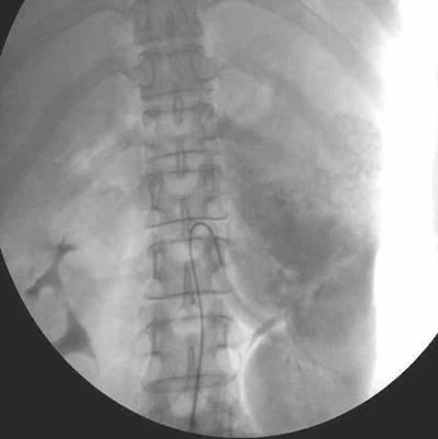
16 I.V.P (2003/07/24) 1MIN, 5MIN

17 I.V.P 10MIN There is shadow of huge tumor mass, mainly occupy at L t upper pole region Impression: Poor functioning of Lt kidney,with huge tumor at Lt pole kidney.
18 L t Renal angiography (2003/07/24) Huge tumor arise from upper pole of Lt kidney.
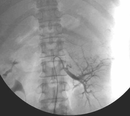
19 L t Renal angiography
20 Summary of image finding A large hetrogeneous enhanced mass with central necrosis occupying the upper and middle poles of left kidney with perirenal invasion. There is found involvement of Lt renal artery, splenic artery and some collateral supplies from the adjacent arteries. Left renal cell carcinoma (may be stage IV A) with stomach, pancreatic tail and splenic hilum invasion is more favored.
21 D/D Renal cell carcinoma I.V.P: distortion of the renal contour, enlargement of a portion of the kidney, and calcifications. CT:solid and highly enhancing mass. Transitional cell carcinoma I.V.P: Filling defects in the upper urinary tract CT:obstruction and dilatation of the ureter and pelvis proximal to the lesion; ureteral wall thickening.
22 D/D Angiomyolipoma 1.a benign renal neoplasm composed of fat, vascular, and smooth muscle elements. 2.main presenting symptoms are related to intratumoral or retroperitoneal hemorrhage. 3. displaces the renal parenchyma and distorts the collecting system, and sometimes causes renal destruction. 4.CT:shows a mixed-attenuation mass interspersed with areas of low attenuation (fat) and areas of high attenuation (blood).
23 D/D Oncocytoma 1. occur within a well-defined fibrous capsule, with tumor tissue rarely penetrating the renal capsule, pelvis, collecting system, or perinephric fat. 2. The diagnosis of oncocytoma is predominantly pathologic.
24 Impression Renal cell carcinoma
25 Operation method Left radical nephrectomy Ligated renal artery, vein and ureter, then cut them. Dissected Gerota fascia with surrounding structure. Remove kidney; repair diaphragm and remove the bleeding spleen.
26 Pathological finding The tumor is solid, golden-yellow with marked hemorrhage and necrosis. The tumor involves the capsule and perirenal adipose tissue but not beyond the Gerota fascia.
27 Pathological finding Renal artery, renal vein and ureter in the hilar area are free of the tumor. The adrenal gland is not involved by the tumor. The tumor also extended to the peripelvic adipose tissue.
28 Pathological finding The diaphragm reveals focal hemorrhage but no tumor involvement. Zero out of 4 lymph nodes dissected out from the perirenal and perisplenic shows metastatic carcinoma (perirenal( perirenal: 0/3, perisplenic: : 0/1).
29 Renal Cell Carcinoma 3% of adult malignancies and 90-95% 95% of neoplasms arising from the kidney. RCC is more common in men than in women (ratio, 2:1) aged years One fourth to one third of patients have metastatic disease at the time of presentation.
30 Risk factors increased age, male sex, smoking, excessive weight, chronic dialysis use, several genetic syndromes (familial RCC, von Hippel-Lindau syndrome, and tuberous sclerosis).
31 Pathophysiology Spread by means of direct local invasion of adjacent structures, such as the adrenal glands, liver, spleen, colon or pancreas, can occur. RCCs have a propensity to extend into the renal vein and, subsequently, into the inferior vena cava. The lungs are the most common sites of distant metastases.
32 Pathophysiology RCCs can be staged by using the Robson classification Stage 1: RCCs are confined to the kidney Stage 2: RCCs extend to the adrenal gland or perinephric tissues but not beyond the Gerota fascia Stage 3a: tumors extend into the renal vein or vena cava Stage 3b: tumors involve the regional nodes Stage 3c: tumors involve both regional nodes and the renal vein or vena cava. Stage 4a: tumors extend beyond the Gerota fascia. Stage 4b: tumors have distant metastases
33 Mortality/Morbidity The prognosis is worst for patients with metastatic disease at presentation and best for patients with small masses confined to the kidney. Unresectable RCCs have a 5-year 5 year survival rate of less than 2%.
34 Clinical Details Most common presentations Hematuria (40%) Flank pain (40%) Palpable mass in the flank or abdomen (25%) Incidental detection has increased on ultrasonographic (US) images.
35 Other signs and symptoms Weight loss (33%) Fever (20%) Hypertension (20%) Hypercalcemia (5%) Night sweats Malaise Varicocele,, usually left sided, due to obstruction of the testicular vein (2% of males)
36 Imaging Studies Excretory urography CT scan Ultrasonography Arteriography Venography MRI
37 Sono RCC can be isoechoic, hypoechoic, or hyperechoic relative to the remainder of the renal parenchyma. US is used primarily to differentiate solid masses from simple cysts.
38 CT On nonenhanced CT, RCCs may appear isoattenuating, hypoattenuating,, or hyperattenuating relative to the remainder of the kidney. Calcifications may be present. On contrast-enhanced CT, RCC is usually solid, and evidence of necrosis is often present. RCC may also appear as a completely solid and highly enhancing mass.
39 I.V.P mass effect on the collecting system, distortion of the renal contour, enlargement of a portion of the kidney, and calcifications.
40 Histologic Findings Clear cell carcinoma the most common histologic type; other phenotypes: granular carcinoma, mixed histology, and sarcomatoid-spindle spindle cell
41 Operation method Radical nephrectomy: complete removal of the Gerota fascia and its contents, including a resection of kidney, perirenal fat, and ipsilateral adrenal gland, with or without ipsilateral lymph node dissection.
42 THANK YOU!!!
Kidney Case 1 SURGICAL PATHOLOGY REPORT
 Kidney Case 1 Surgical Pathology Report February 9, 2007 Clinical History: This 45 year old woman was found to have a left renal mass. CT urography with reconstruction revealed a 2 cm medial mass which
the urinary system pathology Dr. Fairoz A Eltorgman
 the urinary system pathology Dr. Fairoz A Eltorgman Tumors of the renal pelvis & kidney Benign tumors of the renal pelvis: Hemangioma Leiomyoma Malignant tumors: Transitional cell carcinoma Squamous cell
ID data. Sex: female Age: 46y/o Birthday: 1955/10/13
 ID data Sex: female Age: 46y/o Birthday: 1955/10/13 Chief Complain Right upper quadrate abdominal tenderness for one month. Present illness (1) This 46 years old female patient was in a healthy condition
Renal Parenchymal Neoplasms
 Renal Parenchymal Neoplasms د. BENIGN TUMORS : Benign renal tumors include adenoma, oncocytoma, angiomyolipoma, leiomyoma, lipoma, hemangioma, and juxtaglomerular tumors. Renal Adenomas : The adenoma is
Role of imaging in RCC. Ultrasonography. Solid lesion. Cystic RCC. Solid RCC 31/08/60. From Diagnosis to Treatment: the Radiologist Perspective
 Role of imaging in RCC From Diagnosis to Treatment: the Radiologist Perspective Diagnosis Staging Follow up Imaging modalities Limitations and pitfalls Duangkamon Prapruttam, MD Department of Therapeutic
1/25/13 Right partial nephrectomy followed by completion right radical nephrectomy.
 History and Physical Case Scenario 1 45 year old white male presents with complaints of nausea, weight loss, and back pain. A CT of the chest, abdomen and pelvis was done on 12/8/12 that revealed a 12
Renal tumors of adults
 Renal tumors of adults Urinary Tract Tumors 2%-3% of all cancers in adults. The most common malignant tumor of the kidney is renal cell carcinoma. Tumors of the lower urinary tract are twice as common
Male genital tract tumors. SiCA. Division of Urology, Department of Surgery, Faculty of Medicine Siriraj Hospital.
 Male genital tract tumors Division of Urology, Department of Surgery, Faculty of Medicine Siriraj Hospital. adenocarcinoma Prostate Cancer most common male cancer in western countries more detected in
Autosomal Dominant Polycystic Kidney Disease
 Case Studies [1] July 01, 2014 By Amar Udare, MBBS [2] Case History: 45-year-old female with vague pain in the abdomen. Case History: A 45-year-old female presented with vague pain in the abdomen. A USG
Excretory urography (EU) or IVP US CT & radionuclide imaging
 or IVP US CT & radionuclide imaging") Excretory urography (EU) or IVP US CT & radionuclide imaging MRI arteriography studies requiring catherization or direct puncture of collecting system EU & to a lesser extent CT provide both functional
Excretory urography (EU) or IVP US CT & radionuclide imaging MRI arteriography studies requiring catherization or direct puncture of collecting system EU & to a lesser extent CT provide both functional
RENAL CELL CARCINOMA 2 to 3% of All New Visceral Cancers Peak Incidence is 6th Decade M:F = 2:1 Grossly is a Bright Yellow, Necrotic Mass with a Pseud
 GENITOURINARY PATHOLOGY Kathleen M. O Toole Toole, M.D. RENAL CELL CARCINOMA 2 to 3% of All New Visceral Cancers Peak Incidence is 6th Decade M:F = 2:1 Grossly is a Bright Yellow, Necrotic Mass with a
Renal masses - the role of diagnostic imaging
 Renal masses - the role of diagnostic imaging Poster No.: C-2471 Congress: ECR 2015 Type: Educational Exhibit Authors: V. Rai#; Bjelovar/HR Keywords: Cysts, Cancer, Structured reporting, Ultrasound, MR,
Five Views of Transitional Cell Carcinoma: One Man s Journey
 September 2006 Five Views of Transitional Cell Carcinoma: One Man s Journey Amsalu Dabela, Harvard Medical School III Outline Overview: Renal Anatomy Our Patient s Story Diagnostic Imaging Studies Appearance
GUIDELINES ON RENAL CELL CANCER
 20 G. Mickisch (chairman), J. Carballido, S. Hellsten, H. Schulze, H. Mensink Eur Urol 2001;40(3):252-255 Introduction is characterised by a constant rise in incidence over the last 50 years, with a predominance
Urological Tumours 1 Kidney tumours 2 Bladder tumours
 Urological Tumours 1 Kidney tumours 2 Bladder tumours Tim Bracey SpR Histopathology Derriford Hospital Kidney tumours What are we going to talk about?! Anatomy of urinary tract! Types of kidney tumours!
NAACCR Webinar Series 1
 NAACCR 2009 2010 Webinar Series Collecting Cancer Data: Kidney 1 Questions Please use the Q&A panel to submit your questions Send questions to All Panelist 2 Fabulous Prizes 3 NAACCR 2009 2010 Webinar
Pediatric Retroperitoneal Masses Radiologic-Pathologic Correlation
 Acta Radiológica Portuguesa, Vol.XVIII, nº 70, pág. 61-70, Abr.-Jun., 2006 Pediatric Retroperitoneal Masses Radiologic-Pathologic Correlation Marilyn J. Siegel Mallinckrodt Institute of Radiology, Washington
Outline. Introduction to imaging modalities of the urinary system. Case base learning of common diseases in urinary tract
 Outline Introduction to imaging modalities of the urinary system Case base learning of common diseases in urinary tract Outline Introduction to imaging modalities of the urinary system Case base learning
Outline. Introduction to imaging modalities of the urinary system. Case base learning of common diseases in urinary tract
 Outline Introduction to imaging modalities of the urinary system Case base learning of common diseases in urinary tract Diagnostic Investigations in Urinary System PLAIN KUB EXCRETORY UROGRAPHY RETROGRADE
Imaging in gastric cancer
 Imaging in gastric cancer Gastric cancer remains a deadly disease because of late diagnosis. Adenocarcinoma represents 90% of malignant tumors. Diagnosis is based on endoscopic examination with biopsies.
Guidelines, Policies and Statements D5 Statement on Abdominal Scanning
 Guidelines, Policies and Statements D5 Statement on Abdominal Scanning Disclaimer and Copyright The ASUM Standards of Practice Board have made every effort to ensure that this Guideline/Policy/Statement
Chief Complaint. Retroperitoneal cystic mass incidentally found at health examination center.
 Personal Information Age: 34 y/o Sex: female Past history: major systemic medical history(-) surgical history(-), family history(-) Denied food or drug allergy Chief Complaint Retroperitoneal cystic mass
Personal data. Age : 63 Gender : male
 Personal data Age : 63 Gender : male Chief complain No specific symptom or discomfort A hepatic mass, found by abdominal sonography of routine health exam on 88-12-08 Past history 1984-3-3 Old CVA with
Bilateral Renal Angiomyolipomas with Invasion of the Renal Vein: A Case Report
 Case Study TheScientificWorldJOURNAL (2008) 8, 145 148 TSW Urology ISSN 1537-744X; DOI 10.1100/tsw.2008.29 Bilateral Renal Angiomyolipomas with Invasion of the Renal Vein: A Case Report C. Blick, N. Ravindranath,
Normal Sonographic Anatomy
 hapter 2:The Liver DUNSTAN ABRAHAM Normal Sonographic Anatomy Homogeneous, echogenic texture (Figure 2-1) Measures approximately 15 cm in length and 10 12.5 cm anterior to posterior; measurement taken
Renal Tumors in Adult Saudi Patients: A Review of 43 Cases
 Riyadh F. Talic, FRCS(Ed); Salah R. El Faqih, FRCS From the Division of Urology, Department of Surgery, King Khalid University Hospital, King Saud University, Riyadh. Address reprint requests and correspondence
PDF created with pdffactory Pro trial version
 Neuroblastoma Tumor derived from neural crest cell that form the sympathetic ganglia&adrenal medulla. Causes *unknown. *familial neuroblastoma has been reported but is rare. * The incidence is 1:100,000
2 to 3% of All New Visceral Cancers Peak Incidence is 6th Decade M:F = 2:1 Grossly is a Bright Yellow, Necrotic Mass with a Pseudocapsule
 GENITOURINARY PATHOLOGY Kathleen M. O Toole, M.D. Renal Cell Carcinoma 2 to 3% of All New Visceral Cancers Peak Incidence is 6th Decade M:F = 2:1 Grossly is a Bright Yellow Necrotic Mass Grossly is a Bright
Uroradiology For Medical Students
 Uroradiology For Medical Students Lesson 8 Computerized Tomography 2 American Urological Association Objectives In this lesson you will: Gain more experience reading CT images Learn how computer generated
Daniela Faivovich K., MS VII Universidad de Chile Gillian Lieberman, MD Harvard Medical School
 Daniela Faivovich K., MS VII Universidad de Chile Gillian Lieberman, MD Harvard Medical School May 21st, 2010 56 year old male patient History of hypertension, hyperlipidemia and insulin-resistance 2009:
Solitary Contralateral Adrenal Metastases after Nephrectomy for Renal Cell Carcinoma
 Original Report ISSN 1537-744X; DOI 10.1100/tsw.2004.39 Solitary Contralateral Adrenal after Nephrectomy for Renal Cell Carcinoma Nikolaos Antoniou, M.D. and Demetrios Karanastasis, M.D. General Hospital
Manchester Cancer. Guidelines for the management of renal cancer
 Guidelines for the management of renal cancer Approved by the urology pathway board September 2014 To be reviewed September 2016 Renal Cancer Guidelines 1. Introduction 1.1 Kidney cancer accounts for 3%
Role of MDCT in Radiological evaluation of Renal Masses and its beneficial effects on patient management.
 International Journal of advances in health sciences (IJHS) ISSN 2349-7033 Vol2, Issue1, 2015, pp56-63 http://www.ijhsonline.com Research Article Role of MDCT in Radiological evaluation of Renal Masses
THE ABDOMEN SUPRARENAL GLANDS KIDNEY URETERS URINARY BLADDER
 THE ABDOMEN SUPRARENAL GLANDS KIDNEY URETERS URINARY BLADDER THE SUPRARENAL GLANDS The suprarenal (adrenal) glands lie immediately superior and slightly anterior to the upper pole of either kidney. Golden
Imaging of Kidney Cancer
 119 Imaging of Kidney Cancer RADIOLOGIC CLINICS OF NORTH AMERICA Radiol Clin N Am 45 (2007) 119 147 Jingbo Zhang, MD*, Robert A. Lefkowitz, MD, Ariadne Bach, MD - Detection and diagnosis - CT scan Solid
Partial Nephrectomy Techniques for Renal Preservation: Historical and Modern Approaches
 Partial Nephrectomy Techniques for Renal Preservation: Historical and Modern Approaches Cary N Robertson MD FACS Associate Professor Division of Urology Associate Director Urologic Oncology Duke Cancer
GUIDELINES ON RENAL CELL CARCINOMA
 GUIDELINES ON RENAL CELL CARCINOMA B. Ljungberg (chairman), D.C. Hanbury, M.A. Kuczyk, A.S. Merseburger, P.F.A. Mulders, J-J. Patard, I.C. Sinescu Introduction This EAU guideline was prepared to help urologists
Renal cell carcinoma (RCC)
") Renal cell carcinoma (RCC) Introduction The most common solid renal tumor. Accounts for 2 3% of all adult malignancies. It is the 3 rd most common urological tumor in men and the 2 nd in women. It is th
Renal cell carcinoma (RCC) Introduction The most common solid renal tumor. Accounts for 2 3% of all adult malignancies. It is the 3 rd most common urological tumor in men and the 2 nd in women. It is th
UICC TNM 8 th Edition Errata
 UICC TNM 8 th Edition Errata ions are in italics Page 28 Oropharynx p16 positive Pathological Stage II,T2 N2 M0 T3 N0,N1 M0 Stage II,T2 N2 M0 T3,T4 N0,N1 M0 Page 61 Oesophagus Adenocarcinoma Pathological
Tumors of kidney and urinary bladder
 Tumors of kidney and urinary bladder Overview of kidney tumors Benign and malignant Of the benign: papillary adenoma -cortical -small (0.5cm) -in 40% of population -clinically insignificant The most common
International Journal of Medical and Health Sciences
 International Journal of Medical and Health Sciences Journal Home Page: http://www.ijmhs.net ISSN:2277-4505 Case Report Squamous Cell Carcinoma Of Renal Pelvis- A Rare Case Report S. S. Inamdar* 1, Sainath
CASE REPORT RENAL TUBERCULOSIS CAUSE OF RENAL REPLACEMENT LIPOMATOSIS : A RARE ASSOCIATION
 CASE REPORT RENAL TUBERCULOSIS CAUSE OF RENAL REPLACEMENT LIPOMATOSIS : A RARE ASSOCIATION DR ANAND AARTI 1, DR CHANDAK PRIYA 2,DR SURESH PARVATHY 3 1. PROF AND HOD, DEPARTMENT OF RADIODIAGNOSIS, GOVERNMENT
MDCT signs differentiating retroperitoneal and intraperitoneal lesions- diagnostic pearls
 MDCT signs differentiating retroperitoneal and intraperitoneal lesions- diagnostic pearls Poster No.: C-0987 Congress: ECR 2015 Type: Educational Exhibit Authors: D. V. Bhargavi, R. Avantsa, P. Kala; Bangalore/IN
Case Scenario 1: Thyroid
 Case Scenario 1: Thyroid History and Physical Patient is an otherwise healthy 80 year old female with the complaint of a neck mass first noticed two weeks ago. The mass has increased in size and is palpable.
How To Approach Renal Masses? - Differential Diagnosis On Image
 How To Approach Renal Masses? - Differential Diagnosis On Image Poster No.: C-1646 Congress: ECR 2015 Type: Educational Exhibit Authors: A. E. A. G. Costa, A. Gomes, A. Duarte, I. Távora; Lisbon/PT Keywords:
The new TNM staging for renal cell carcinoma: what and why the urologists want to know.
 The new TNM staging for renal cell carcinoma: what and why the urologists want to know. Poster No.: C-1132 Congress: ECR 2011 Type: Educational Exhibit Authors: Y. Y. Lim, A. Hattab, A. Bradley ; Manchester/UK,
Bladder Case 1 SURGICAL PATHOLOGY REPORT. Procedure: Cystoscopy, transurethral resection of bladder tumor (TURBT)
") Bladder Case 1 February 17, 2007 Specimen (s) received: Bladder Tumor Pre-operative Diagnosis: Bladder Cancer Post operative Diagnosis: Bladder Cancer Procedure: Cystoscopy, transurethral resection of
Bladder Case 1 February 17, 2007 Specimen (s) received: Bladder Tumor Pre-operative Diagnosis: Bladder Cancer Post operative Diagnosis: Bladder Cancer Procedure: Cystoscopy, transurethral resection of
Guidelines on Renal Cell
 Guidelines on Renal Cell Carcinoma (Text update March 2009) B. Ljungberg (Chairman), D.C. Hanbury, M.A. Kuczyk, A.S. Merseburger, P.F.A. Mulders, J-J. Patard, I.C. Sinescu Introduction Renal cell carcinoma
Recommendations for cross-sectional imaging in cancer management, Second edition
 www.rcr.ac.uk Recommendations for cross-sectional imaging in cancer management, Second edition Renal and adrenal tumours Faculty of Clinical Radiology www.rcr.ac.uk Contents Renal cell carcinoma 3 Clinical
Index. Note: Page numbers of article titles are in boldface type.
 Magn Reson Imaging Clin N Am 12 (2004) 587 591 Index Note: Page numbers of article titles are in boldface type. A Adenoma(s), adrenal, gadolinium-enhanced MR imaging in, 533 534 hyperfunctioning versus
CT abdomen and pelvis
 CT abdomen and pelvis General indications: Assessment of vague abdominal symptoms (pain, colics,distenstion,...) Varifecation of a lesion discovered by other diagnostic modalities as US, barium,ivp, Staging
Dr.Dafalla Ahmed Babiker Jazan University
 Dr.Dafalla Ahmed Babiker Jazan University Brain tumors are the second commonest malignancy in children Infratentorial tumors are more common As a general rule they do not metastasize out of the CNS, but
Sarcomatoid renal cell carcinoma: A case report and literature review
 Sarcomatoid renal cell carcinoma: A case report and literature review Michael Reiter 1*, Ryan Schwope 1, Arthur Clarkson 2 1. Department of Radiology, Brooke Army Medical Center, San Antonio USA 2. Department
What s Your Diagnosis??? Renée Fahrenholz, Class of 2012
 Renée Fahrenholz, Class of 2012 What s Your Diagnosis??? Signalment Emma, a 9 year old, Female, Spayed, Domestic Short Haired Feline Presenting Complaint Weight loss, vomited the morning of her visit,
Genitourinary Neoplasms Updated for 2012 Requirements and CSv02.04
 Presentation Outline Genitourinary Neoplasms Updated for 2012 Requirements and CSv02.04 X:\FCDS_PUB\wwwroot\downloads\Teleconfere nces\2013 FCDS Educational Webcast Series February 28, 2013 General Information
Genitourinary Neoplasms Updated for 2012 Requirements and CSv02.04
 Genitourinary Neoplasms Updated for 2012 Requirements and CSv02.04 X:\FCDS_PUB\wwwroot\downloads\Teleconfere nces\2013 FCDS Educational Webcast Series February 28, 2013 1 Steven Peace, BS, CTR Susan Smith
Role of imaging in evaluation of genitourinary i trauma Spectrum of GU injuries Relevance of imaging findings in determining management Focus on MDCT
 Genitourinary Tract Injuries 6 th Nordic Course Scott D. Steenburg, MD Assistant Professor University of Maryland Department of Radiology Division of Trauma and Emergency Radiology R Adams Cowley Shock
Adrenal masses in infancy and childhood: A clinical and radiological overview M. Mearadji
 Adrenal masses in infancy and childhood: A clinical and radiological overview M. Mearadji International Foundation for Pediatric Imaging Aid Introduction Neoplastic adrenal masses usually originate from
UICC TNM 8 th Edition Errata
 UICC TNM 8 th Edition Errata ions are in italics Head and Neck Tumours Pages 20, p27, p34, p38, p41, and p49 ly pn2a Metastasis in a single ipsilateral lymph node, less than 3cm in greatest dimension with
Birthday: 1952/07/31 Date of admission:1999/12/30 Age:48 y/o Past medication:esrd under regular HD for 5+ years; denied DM and HTN
 Birthday: 1952/07/31 Date of admission:1999/12/30 Age:48 y/o Past medication:esrd under regular HD for 5+ years; denied DM and HTN Chief Complaint : 1)intermittent LLQ cramping pain for 2 months 2) LGI
The Spleen. Dr Fahad Ullah
 The Spleen BY Dr Fahad Ullah Spleen The spleen is an largest lymphoid organ shaped like a shoe that lies relative to the 9th and 11th ribs and is located in the left hypochondrium. Thus, the spleen is
Basic of Ultrasound Physics E FAST & Renal Examination. Dr Muhammad Umer Ihsan MBBS,MD, DCH CCPU,DDU1,FACEM
 Basic of Ultrasound Physics E FAST & Renal Examination Dr Muhammad Umer Ihsan MBBS,MD, DCH CCPU,DDU1,FACEM What is Sound? Sound is Mechanical pressure waves What is Ultrasound? Ultrasounds are sound waves
Imaging characterization of renal clear cell carcinoma
 Imaging characterization of renal clear cell carcinoma Poster No.: C-0327 Congress: ECR 2011 Type: Educational Exhibit Authors: S. Ballester 1, A. Gaser 2, M. Dotta 1, M. F. CAPPA 1, F. Hammar 1 ; 1 2
Case Discussion Splenic Abscess
 Case Discussion Splenic Abscess Personal Data Gender: male Birth Date: 1928/Mar/06th Allergy: Mefenamic Smoking: 0.5 PPD for 55 years Alcohol: negative (?) 4 Months Ago Abdominal pain: epigastric area
25 TH ICRO DEHRADUN STAGING OF GENITOURINARY MALIGNANCIES
 25 TH ICRO DEHRADUN STAGING OF GENITOURINARY MALIGNANCIES SPEAKER DR DEEPAK ABROL CLINICAL ONCOLOGIST JAND K HEALTH SERVICES CONSULTANT ONCOLOGIST MAHARISHI DAYANAND HOSPITAL AND MEDICAL RESEARCH CENTER
Boot Camp Case Scenarios
 Boot Camp Case Scenarios Case Scenario 1 Patient is a 69-year-old white female. She presents with dyspnea on exertion, cough, and right rib pain. Patient is a smoker. 9/21/12 CT Chest FINDINGS: There is
Solitary Fibrous Tumor of the Kidney with Massive Retroperitoneal Recurrence. A Case Presentation
 246) Prague Medical Report / Vol. 113 (2012) No. 3, p. 246 250 Solitary Fibrous Tumor of the Kidney with Massive Retroperitoneal Recurrence. A Case Presentation Sfoungaristos S., Papatheodorou M., Kavouras
Lecture 56 Kidney and Urinary System
 Lecture 56 Kidney and Urinary System The adrenal glands are located on the superomedial aspect of the kidney The right diagram shows a picture of the kidney with the abdominal walls and organs removed
Complex case Presentations
 Complex case Presentations Case Presentations April 2016 Lisa M Pickering Case presentations: chromophobe renal carcinoma 60 year old man. ECOG PS 0 No significant comorbodities August 2009: L radical
Anatomy of the renal system. Professor Nawfal K. Al-Hadithi
 Anatomy of the renal system Professor Nawfal K. Al-Hadithi Objectives To describe the posterior abdominal wall To identify the main anatomical landmarks of the kidneys & ureters To describe the suprarenal
Radio-Pathologic Workup of a Retroperitoneal Abdominal Mass
 Radio-Pathologic Workup of a Retroperitoneal Abdominal Mass Joe Carlson Advanced Radiology Clerkship Harvard Medical School Year IV September 12, 2002 84 year old Male Presented to PCP With Abdominal Pain
My Patient Has Abdominal Pain PoCUS of the Biliary Tract and the Urinary Tract
 My Patient Has Abdominal Pain PoCUS of the Biliary Tract and the Urinary Tract Objectives PoCUS for Biliary Disease PoCUS for Renal Colic PoCUS for Urinary Retention Biliary Disease A patient presents
Radiology- Pathology Conference 4/29/2012. Lymph Nodes. John McGrath
 Radiology- Pathology Conference 4/29/2012 Lymph Nodes John McGrath 1 Presentation material is for education purposes only. All rights reserved. 2012 URMC Radiology Page 1 of 24 Case 1: 51 year-old male
Wilms Tumor and Neuroblastoma
 Wilms Tumor and Neuroblastoma Wilm s Tumor AKA: Nephroblastoma the most common intra-abdominal cancer in children. peak incidence is 2 to 3 years of age Biology somatic mutations restricted to tumor tissue
!! 2 to 3% of All New Visceral Cancers.!! Peak Incidence is 6th Decade!! M:F = 2:1
 !! Kathleen M. O Toole, M.D.!! 2 to 3% of All New Visceral Cancers!! Peak Incidence is 6th Decade!! M:F = 2:1!! Grossly is a Bright Yellow, Necrotic Mass with a Pseudocapsule 1 !!Conventional RCC! Clear
Brief History. Identification : Past History : HTN without regular treatment.
 Brief History Identification : Name : 陳 x - Admission : 94/10/06 Gender : male Age : 75 y/o Chief Complaint : Urinary difficulty for months. Past History : HTN without regular treatment. Brief History
ASSESSING THE PLAIN ABDOMINAL RADIOGRAPH M A A M E F O S U A A M P O F O
 ASSESSING THE PLAIN ABDOMINAL RADIOGRAPH M A A M E F O S U A A M P O F O Introduction The abdomen (less formally called the belly, stomach, is that part of the body between the thorax (chest) and pelvis,
Surgically Discovered Xanthogranulomatous Pyelonephritis Invading Inferior Vena Cava with Coexisting Renal Cell Carcinoma
 Case Study TheScientificWorldJOURNAL (2009) 9, 5 9 TSW Urology ISSN 1537-744X; DOI 10.1100/tsw.2009.6 Surgically Discovered Xanthogranulomatous Pyelonephritis Invading Inferior Vena Cava with Coexisting
Partial Nephrectomy Planning: Everybody s s doing it, you can to
 Partial Nephrectomy Planning: Everybody s s doing it, you can to Brian R. Herts, MD Associate Professor of Radiology Head, Abdominal Imaging, Imaging Institute & Staff, The Glickman Urological and Kidney
GENERAL ABDOMINAL IMAGING PERITONEAL SPACE, PANCREAS, & SPLEEN. VMB 960 March 25, 2013
 GENERAL ABDOMINAL IMAGING PERITONEAL SPACE, PANCREAS, & SPLEEN VMB 960 March 25, 2013 REFERENCE Chapters 35-36 Pages 650-678 Chapter 37 Pages 694-701 Chapter 3 Pages 38-49 OBJECTIVES Radiography and Ultrasound
 Title Metastatic renal tumor from involvement: a case report the lun Ishihara, Satoshi; Kobayashi, Sator Author(s) Takeuchi, Toshimi; Kuriyama, Manabu Kawada, Yukimichi; Takahashi, Yoshi Isogai, Kazutoshi
Atypical kidney tumors and pseudotumors: Imaging features in 44 patients.
 Atypical kidney tumors and pseudotumors: Imaging features in 44 patients. Poster No.: C-1472 Congress: ECR 2011 Type: Scientific Exhibit Authors: M. Kasbi, Y. kallel, M. basly, Z. fitouri, K. Nouira, Y.
KIDNEY HEALTH. Kidney Masses and Localized Kidney Tumors: A Patient Guide
 KIDNEY HEALTH Kidney Masses and Localized Kidney Tumors: A Patient Guide Table of Contents Kidney & Adrenal Health Committee Renal Mass Committee.... 2 Patient Story.... 3 Introduction: I have a kidney
CT 101 :Pancreas and Spleen
 CT 101 :Pancreas and Spleen Shikha Khullar,, MD, MPH Division of Radiology University of South Alabama The Pancreas Normal Pancreas 3 Phase Pancreatic CT Non contrast Arterial phase : 30-35 35 second
Separating and Distorted Nephroliths Signs of Renal Squamous Cell Carcinoma
 Chin J Radiol 2003; 28: 203-208 203 Separating and Distorted Nephroliths Signs of Renal Squamous Cell Carcinoma TZE-YU LEE SHEUNG-FAT KO CHUNG-CHENG HUANG YU-FENG CHENG Department of Radiology, Chang Gung
Alison Douglass Gillian Lieberman, MD. November. Colon Cancer. Alison Douglass, Harvard Medical School Year III Gillian Lieberman, MD
 November Colon Cancer Alison Douglass, Harvard Medical School Year III Our Patient Mr. K. is a 67 year old man with no prior medical problems other than hemorrhoids which have caused occasional rectal
The Incidental Renal Mass in the Primary Care Setting
 The Incidental Renal Mass in the Primary Care Setting Adele M. Caruso, MSN, CRNP Adult Nurse Practitioner The Perelman School of Medicine at the University of Pennsylvania Abstract There are approximately
3. Guidelines for Reporting Bladder Cancer, Prostate Cancer and Renal Tumours
 60 3. Guidelines for Reporting Bladder Cancer, Prostate Cancer and Renal Tumours Compilation and editing and of this volume: Prof. Chandu de Silva (Consultant Histopathologist) List of contributors Consultant
Protocol applies to specimens from patients with Wilms tumor (nephroblastoma) or other renal tumors of childhood.
 or other renal tumors of childhood.") Wilms Tumor Protocol applies to specimens from patients with Wilms tumor (nephroblastoma) or other renal tumors of childhood. Procedures Cytology (No Accompanying Checklist) Incisional Biopsy (Needle or
Wilms Tumor Protocol applies to specimens from patients with Wilms tumor (nephroblastoma) or other renal tumors of childhood. Procedures Cytology (No Accompanying Checklist) Incisional Biopsy (Needle or
6/5/2010. Renal vein invasion & Capsule Penetration (T3a) Adrenal Gland involvement (T4 vs. M1) Beyond Gerota s Fascia? (?T4).
 Adrenal Gland involvement (T4 vs. M1) Beyond Gerota s Fascia? (?T4).") GU Cancer Staging: Updates and Challenging Areas 13 th Current Issues in Surgical Pathology San Francisco, CA June 5, 2010 Jeffry P. Simko, PhD, MD Associate Professor Departments of Urology and Anatomic
GU Cancer Staging: Updates and Challenging Areas 13 th Current Issues in Surgical Pathology San Francisco, CA June 5, 2010 Jeffry P. Simko, PhD, MD Associate Professor Departments of Urology and Anatomic
Multiple Primary Quiz
 Multiple Primary Quiz Case 1 A 72 year old man was found to have a 12 mm solid lesion in the pancreatic tail by computed tomography carried out during a routine follow up study of this patient with adult
Pancreas Quizzes c. Both A and B a. Directly into the blood stream (not using ducts)
") Pancreas Quizzes Quiz 1 1. The pancreas produces hormones. Which type of hormone producing organ is the pancreas? a. Endocrine b. Exocrine c. Both A and B d. Neither A or B 2. Endocrine indicates hormones
Pancreas Quizzes Quiz 1 1. The pancreas produces hormones. Which type of hormone producing organ is the pancreas? a. Endocrine b. Exocrine c. Both A and B d. Neither A or B 2. Endocrine indicates hormones
Abdomen and Retroperitoneum Ultrasound Protocols
 Abdomen and Retroperitoneum Ultrasound Protocols Reviewed By: Anna Ellermeier, MD Last Reviewed: March 2018 Contact: (866) 761-4200, Option 1 **NOTE for all examinations: 1. If documenting possible flow
Q129. Which of the following is NOT true about lymph node?
 Q129. Which of the following is NOT true about lymph node? (1). Normal lymph node is not seen in the ultrasound image (2). It is general that high frequency probe is used due to normal lymph node is located
Policies, Standards, and Guidelines. Guidelines for Abdominal Ultrasound Examination
 Policies, Standards, and Guidelines Guidelines for Abdominal Ultrasound Examination Approved by Council Feb 2018 Disclaimer and Copyright The ASUM Standards of Practice Board have made every effort to
Radiological Investigations of Abdominal Trauma
 76 77 Investigations of Abdominal Trauma Introduction: Trauma to abdominal organs is a common cause of patient morbidity and mortality among trauma patients. Causes of abdominal trauma include blunt injuries,
Interesting case. Vikas Kundra, M.D., Ph.D. October Vikas Kundra, M.D., Ph.D.
 Interesting case October 2012 Disclosure Information Vikas Kundra, M.D, Ph.D. I have no financial relationships to disclose. I WILL NOT include discussion of investigational or off-label use of a product
Imaging Guided Biopsy. Edited & Presented by ; Hussien A.B ALI DINAR. Msc Lecturer,Reporting Sonographer
 Imaging Guided Biopsy Edited & Presented by ; Hussien A.B ALI DINAR. Msc Lecturer,Reporting Sonographer Objective By the End of this lessons you should : Define what biopsy Justify Aim to perform biopsy
RECURRENT ADRENAL DISEASE. Megan Applewhite Endorama 2/19/2015 SR , SC
 RECURRENT ADRENAL DISEASE Megan Applewhite Endorama 2/19/2015 SR 2412318, SC 3421561 Category: Adrenal Attendings: Angelos & Grogan PATIENT #1 36yo woman with a hx of Cushing s Syndrome and right adrenalectomy
ROLE OF COMPUTED TOMOGRAPHY IN EVALUATION OF ASCITES Ramesh Chander 1, Kamlesh Gupta 2, Arvinder Singh 3, V. K. Rampal 4, Mohit Khandelwal 5
 ROLE OF COMPUTED TOMOGRAPHY IN EVALUATION OF ASCITES Ramesh Chander 1, Kamlesh Gupta 2, Arvinder Singh 3, V. K. Rampal 4, Mohit Khandelwal 5 HOW TO CITE THIS ARTICLE: Ramesh Chander, Kamlesh Gupta, Arvinder