Retinal Nerve Fiber Layer Measurements in Myopia Using Optical Coherence Tomography
|
|
|
- Ariel Day
- 6 years ago
- Views:
Transcription
1 Original Article Philippine Journal of OPHTHALMOLOGY Retinal Nerve Fiber Layer Measurements in Myopia Using Optical Coherence Tomography Dennis L. del Rosario, MD and Mario M. Yatco, MD University of Santo Tomas Hospital, España, Manila Correspondence: Dennis L. del Rosario, MD University of Santo Tomas Hospital, España, Manila, Philippines Tel. no.: Disclosure: The authors have no proprietary or financial interest in any product used or cited in this study. ABSTRACT Objective: To evaluate the relationship between retinal nerve fiber layer (RNFL) thickness measured by optical coherence tomography (OCT) and the degree of myopia. Methods: A total of 28 eyes of 14 healthy subjects with myopia, 9 eyes with low myopia (spherical equivalent [SE] between D and D) and 19 eyes with moderate to high myopia (SE >-3.0 D), were included. Total average and mean quadrant RNFL thicknesses were measured by Stratus OCT. Associations between RNFL measurements and spherical equivalent refraction were evaluated by linear regression analysis. Results: Twenty two out of 28 eyes were classified as below normal limits with reference to the normative database of the RNFL thickness. There was a significant correlation between refraction and average RNFL thickness (p=0.04), indicating that for every decrease of 1.00 D in refraction (more myopic), there was a 2.86 µm decrease in the average RNFL thickness. The most frequently abnormal sector was at the nasal quadrant, where 78% of myopic eyes were below normal limits. A significant correlation was seen between refraction and nasal RNFL thickness (p=0.02), showing a decrease of 3.37 µm in the nasal RNFL thickness for every 1.00 D decrease. There was also a significant correlation between refraction and inferior RNFL thickness (p=0.007). For every decrease of 1.00 D in refraction, there was a decrease of 6.27 µm in the inferior RNFL thickness. There were no significant correlations between the refraction and the superior and temporal RNFL thickness (p = 0.12 and 0.64, respectively). Conclusion: There was a decrease in the average RNFL thickness as the refractive error became more myopic, suggesting the need for the Stratus OCT RNFL normative database to have corrective factors for refractive error among myopic patients. Although both the nasal and inferior quadrants positively correlated with myopia, majority of the nasal quadrant showed below normal thickness based on the Stratus OCT normative database. A careful interpretation of RNFL measurements, especially the nasal quadrant, should be done among myopic subjects to avoid mislabeling them as glaucoma suspects. Keywords: Myopia, Retinal nerve fiber layer, Optical coherence tomography, Stratus OCT, Refractive error Philipp J Ophthalmol 2014;39:39-44 January - June
2 Myopia is a common ocular abnormality worldwide with a prevalence in adults of 22.7% and 26.2%, as reported in the Baltimore Eye Survey and the Beaver Dam Study, respectively. 1 2 Among adult Chinese in Singapore, it was estimated that 38.7% of adult Chinese were myopic and 9.1% were high myopes. 3 From the clinical and socioeconomic standpoints, these are major concerns especially among East Asian countries where there are projections for increasing prevalence rate and severity of the condition. 4 One of the potentially blinding ocular diseases found to have a strong relationship with myopia is glaucoma 5 which is characterized by progressive degeneration of retinal ganglion cells, one of the common ocular diseases seen among Filipinos. An important approach in the detection of early structural change in glaucoma is based on assessment of the retinal nerve fiber layer (RNFL) using the optical coherence tomography (OCT). Numerous studies have confirmed that RNFL measurement is sensitive in detecting glaucoma, and the extent of RNFL damage correlates with the severity of functional deficits in the visual field. 6 8 Some patients can lose up to 40% of their RNFL before any visual field defect is detected. The Stratus OCT utilizes a normal population database for RNFL measurements, developed by the manufacturer and packaged within the software. However, it did not include individuals with moderate or high degrees of myopia and there were only 11 Asians out of the 328 subjects included in the establishment of the normative reference values for RNFL thickness measurement. 9 RNFL thinning is indicative of glaucomatous damage; however, it remains uncertain whether RNFL thickness would vary with the degree of myopia where eyes have longer axial lengths and stretched-out retina. Conflicting data existed regarding the influence of myopia on peripapillary retinal nerve fiber layer (RNFL) thickness. Some studies showed no association whereas others found significant correlations. 9 This study investigated if there is a significant relationship between the degree of myopia and RNFL thickness as measured by optical coherence tomography. METHODOLOGY Adult Filipinos, between years of age, with myopia of 0.75 D and no ocular or systemic conditions that can affect the refraction, were recruited from July 2012 to August 2012 at the outpatient department of the Department of Ophthalmology, University of Santo Thomas Hospital. Subjects less than 18 years old, spherical equivalent <-0.75 D, diagnosed glaucoma or glaucoma suspect with CD ratio 0.5, those with frank presbyopia, and those with ocular diseases such as cataract and any form of retinopathy were excluded. All subjects underwent complete ophthalmologic examination consisting of visual acuity determination, refraction, applanation tonometry, slit lamp evaluation, stereoscopic biomicroscopy of the optic nerve head at the slit lamp using a 90 D lens, and fundoscopy. OCT was performed with the Stratus OCT (Carl Zeiss, Meditec Inc.). RNFL thickness was measured with the fast RNFL scanning protocol (256 A-scans). Average measurements of three sequential circular scans with a diameter of 3.4 mm centered on the optic disc were recorded. All the scans had signal strength of at least 7. Parameters including average RNFL thickness and mean RNFL thickness in each quadrant were generated automatically in the analysis report of the Stratus OCT. Correlations between refraction (spherical equivalent) and RNFL thicknesses were determined by linear regression analysis and expressed as the Pearson coefficient of correlation (r), with p<0.05 considered statistically significant. RESULTS Twenty-eight eyes of 14 myopic subjects were included in the study. The mean age and spherical equivalent were 25 years (range, 22 35) and D (range, to -6.00), respectively. The average RNFL thickness in myopic eyes with D to D was µm (range, to μm) and in myopic eyes with >-3.00 D µm (range,77.67 to μm). A significant proportion of myopic eyes (78%) were classified as below normal limits, with reference to the normative database of the retinal nerve fiber layer measurement of the Stratus OCT (Table 1). 40 Philippine Academy of Ophthalmology
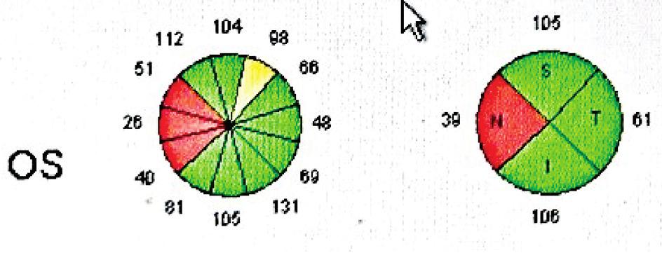
3 Philippine Journal of OPHTHALMOLOGY TABLE 1. Average RNFL thickness and RNFL quadrant thickness in RNFL (µm) REFRACT- AVE ION THICK- SUPER- NASAL INFE- TEMPO- (SE) NESS IOR RIOR RAL TABLE 2. Correlation of RNFL thickness (μm) and refraction (D) in RNFL Coefficient ρ F thickness (μm) value value Average Superior Inferior Nasal Temporal There was, however, no significant correlation between the refraction and the superior and temporal RNFL thickness (p = 0.13 and 0.64, respectively) (Figures 2 and 3; Table 2). There was a significant correlation between refraction and average RNFL thickness (p=0.036) (Figure 1). For every 1.00 D decrease in refraction (more myopic), there was a µm decrease in the average RNFL thickness (Table 2). Figure 2. Refraction (SE) and superior RNFL thickness (µm) in Figure 1. Refraction (SE) and average RNFL thickness (μm) in Figure 3. Refraction (SE) and temporal RNFL thickness (µm) in January - June


4 -0.75 sph sph sph sph sph Figure 6. Representative retinal nerve fiber layer scans from normal subjects with different degrees of myopia. 42 Philippine Academy of Ophthalmology
5 Philippine Journal of OPHTHALMOLOGY Figure 4. Refraction (SE) and nasal RNFL thickness (µm) in 28 myopic eyes. Figure 5. Refraction (SE) and inferior RNFL thickness (µm) in The most frequently abnormal sector was at the nasal quadrant, where 78% of the myopic eyes were below normal limits. A significant correlation was seen between refraction and nasal RNFL thickness (p=0.02). There was a corresponding decrease of 3.37 µm in the nasal RNFL thickness for every 1.00 D decrease in refraction (more myopic) (Figure 4 and Table 2). There was also a significant correlation between refraction and inferior RNFL thickness (p=0.007). For every decrease of 1.00 D in refraction (more myopic), there was a decrease of µm in the inferior RNFL thickness (Figure 5 and Table 2). Representative RNFL scans of the different degrees of myopia are shown in Figure 6. DISCUSSION Our findings indicated that the average retinal nerve fiber layer thickness decreases as the refraction becomes more myopic. These data supported the findings of Schweitzer 10 and Leung 11 that there is a positive correlation between refraction and RNFL thickness. This is in contrast to the results found by Hoh 12 where the mean RNFL thickness did not vary with the degree of myopia when investigated by OCT. In our study, we found that there was a decrease in the RNFL thickness at the nasal quadrant, 78% of which were below normal limits based on the normative database of the Stratus OCT. A positive correlation between the degree of myopia and the nasal RNFL thickness was observed. There was a decrease of 3.37 µm for every 1.00 D decrease in refraction. This data supported the findings of Leung 11 that Chinese myopic subjects have thin nasal RNFL, a finding not found in the largely Caucasian population where Rauscher noted that thin RNFL in myopic subjects occurred preferentially at the superior and inferior quadrants. 9 We also found that there was a positive correlation between the degree of myopia and RNFL thickness at the inferior quadrant but the majority were within normal range and only 3% were below 95%. The Stratus OCT used in this study provided superior image quality and higher resolution scanning of the RNFL compared to earlier models. Although newer generation OCTs are now available, the Stratus OCT remains to be widely used in the Philippines. The Stratus OCT provides a database with agematched controls for comparative analysis aiding the detection of ocular diseases involving the RNFL but the validity of applying this database to healthy myopes is yet to be verified. 11 The study of Rauscher 9 pointed out that extremes of refractive error were excluded and the normative database lack the exclusionary criteria based on refractive error. Moreover, there were only 11 Asian eyes out of 328 included in the study establishing the database where majority were whites (205 eyes). Statistical analyses were done between RNFL thickness and age, right vs left eye, and gender, but not the ethnicity. Therefore, it would be of interest to examine whether there is a significant difference in RNFL thickness measurements between Asians and non-asians. Several explanations for RNFL thinning in January - June
6 myopia have been reported. 10 Myopic globe elongation may seem to stretch and thin the RNFL because of mechanical forces. 11 Myopic retinal degeneration may account for decrease RNFL thickness and a larger disc with normal ganglion cells may provide a greater surface area for axon distribution. 13 On the other hand, the decreased RNFL thickness among myopes could represent a predisposing factor for future development of glaucoma that could partly explain the increase incidence among myopes in some studies like the Blue Mountain Eye Study where data showed strong relationship between open-angle glaucoma and myopia. 5 In conclusion, RNFL measurements may vary with refraction. There is a decrease in the average RNFL thickness as the refractive error becomes more myopic which may suggest the need for the Stratus OCT RNFL normative database to have corrective factors for refractive error among myopic patients and to verify these data with the use of the new generation Cirrus OCT. 8. Medeiros FA, Zangwill LM, Bowd C, et al. Comparison of the GDx VCC scanning laser polarimeter, HRT II confocal scanning laser ophthalmoscope, and Stratus OCT optical coherence tomography for the detection of glaucoma. Arch Ophthalmol 2004;122: Rauscher FM, Sekhon N, Feuer WJ, et al. Myopia affects retinal nerve fiber layer measurements as determined by optical coherence tomography. J Glaucoma 2009:18: Schweitzer KD, Ehmann D, Garcia R. Nerve fibre layer changes in highly myopic eyes by optical coherence tomography. Can J Ophthalmol 2009; 44: Leung CK, Mohamed S, Leung KS, et al. Retinal nerve fiber layer measurements in myopia: an optical coherence tomography study. Invest Ophthalmol Vis Sci 2006;47: Hoh ST, Lim MC, Seah SK, et al. Peripapillary retinal nerve fiber layer thickness variations with myopia. Ophthalmology 2006;113: Kremmer S, Zadow T, Steuhl KP, et al. Scanning laser polarimetry in myopic and hyperopic subjects. Graefes Arch Clin Exp Ophthalmol 2004;242: Although both nasal and inferior quadrants positively correlate with myopia, majority of the nasal quadrant showed below normal thickness based on the Stratus OCT normative database. A careful interpretation of RNFL measurements, especially the nasal quadrant, should be done among myopic subjects to avoid mislabeling them as glaucoma suspects. REFERENCES 1. Katz J, Tielsch JM, Sommer A. Prevalence and risk factors for refractive errors in an adult inner city population. Invest Ophthalmol Vis Sci 1997;38: Wang Q, Klein BE, Klein R, Moss SE. Refractive status in the Beaver Dam Eye Study. Invest Ophthalmol Vis Sci 1994;35: Wong TY, Foster PJ, Hee J, et al. Prevalence and risk factors for refractive errors in adult Chinese in Singapore. Invest Ophthalmol Vis Sci 2000;41: Seet B, Wong TY, Tan DT, et al. Myopia in Singapore: taking a public health approach. Br J Ophthalmol 2001;85: Mitchell P, Hourihan F, Sandbach J, et al. The relationship between glaucoma and myopia: the Blue Mountains Eye Study. Ophthalmology 1999;106: Leung CK, Chan WM, Yung WH, et al. Comparison of macular and peripapillary measurements for the detection of glaucoma: an optical coherence tomography study. Ophthalmology 2005;112: Hoffmann EM, Medeiros FA, Sample PA, et al. Relationship between patterns of visual field loss and retinal nerve fiber layer thickness measurements. Am J Ophthalmol 2006;141: Philippine Academy of Ophthalmology
Evaluation of retinal nerve fiber layer thickness parameters in myopic population using scanning laser polarimetry (GDxVCC)
") Dada T et al Original article Evaluation of retinal nerve fiber layer thickness parameters in myopic population using scanning laser polarimetry (GDxVCC) Dada T1, Aggarwal A1, Bali SJ2, Sharma A1, Shah
Dada T et al Original article Evaluation of retinal nerve fiber layer thickness parameters in myopic population using scanning laser polarimetry (GDxVCC) Dada T1, Aggarwal A1, Bali SJ2, Sharma A1, Shah
STRUCTURE & FUNCTION An Integrated Approach for the Detection and Follow-up of Glaucoma. Module 3a GDx
 STRUCTURE & FUNCTION An Integrated Approach for the Detection and Follow-up of Glaucoma Module 3a GDx Educational Slide Deck Carl Zeiss Meditec, Inc. November 2005 1 Structure & Function Modules Module
Assessment of Retinal Nerve Fiber Layer Changes by Cirrus High-definition Optical Coherence Tomography in Myopia
 Divya Singh et al Original REASEARCH 10.5005/jp-journals-10028-1223 Assessment of Retinal Nerve Fiber Layer Changes by Cirrus High-definition Optical Coherence Tomography in Myopia 1 Divya Singh, 2 Sanjay
Retinal nerve fiber layer thickness in Indian eyes with optical coherence tomography
 Original articles in Indian eyes with optical coherence tomography Malik A, Singh M, Arya SK, Sood S, Ichhpujani P Department of Ophthalmology Government Medical College and Hospital, Sector 32, Chandigarh,
Noel de Jesus Atienza, MD, MSc and Joseph Anthony Tumbocon, MD
 Original Article Philippine Journal of OPHTHALMOLOGY Diagnostic Accuracy of the Optical Coherence Tomography in Assessing Glaucoma Among Filipinos. Part 1: Categorical Outcomes Based on a Normative Database
Comparative evaluation of time domain and spectral domain optical coherence tomography in retinal nerve fiber layer thickness measurements
 Original article Comparative evaluation of time domain and spectral domain optical coherence tomography in retinal nerve fiber layer thickness measurements Dewang Angmo, 1 Shibal Bhartiya, 1 Sanjay K Mishra,
Relationship between the GDx VCC and Stratus OCT in Primary Open Angle Glaucoma
 Relationship between the GDx VCC and Stratus OCT in Primary Open Angle Glaucoma Reza Zarei, MD 1 Mohammad Soleimani, MD 2 Sasan Moghimi, MD 3 Mohammad Yaser Kiarudi, MD 2 Mahmoud Jabbarvand, MD 1 Yadollah
S Morishita, T Tanabe, S Yu, M Hangai, T Ojima, H Aikawa, N Yoshimura. Clinical science
 Department of Ophthalmology and Visual Sciences, Kyoto University Graduate School of Medicine, Kyoto, Japan Correspondence to: Dr T Tanabe, Department of Ophthalmology, The Tazuke Kofukai Medical Institute,
OtticaFisiopatologica
 Anno quindicesimo dicembre 2010 How to assess the retinal nerve fiber layer thickness Antonio Ferreras Miguel Servet University Hospital, Zaragoza. Aragón Health Sciences Institute University of Zaragoza
Relationship Between Structure
 Original Article Relationship Between Structure and Function of the Optic Nerve Head-Glaucoma versus Normal Dr Savita Bhat, Dr Anna Elias, Dr Siddharth Pawar, Dr S.J. Saikumar, Dr Alpesh Rajput, superior,
Seiji T. Takagi, Yoshiyuki Kita, Asuka Takeyama, and Goji Tomita. 1. Introduction. 2. Subjects and Methods
 Ophthalmology Volume 2011, Article ID 914250, 5 pages doi:10.1155/2011/914250 Clinical Study Macular Retinal Ganglion Cell Complex Thickness and Its Relationship to the Optic Nerve Head Topography in Glaucomatous
Rates of Abnormal Retinal Nerve Fiber Layer and Ganglion Cell Layer OCT Scans in Healthy Myopic Eyes: Cirrus Versus RTVue
 CLINICAL SCIENCE Rates of Abnormal Retinal Nerve Fiber Layer and Ganglion Cell Layer OCT Scans in Healthy Myopic Eyes: Cirrus Versus RTVue Jean-Claude Mwanza, MD, MPH, PhD; Fouad E. Sayyad, MD; Ahmad A.
International Journal of Health Sciences and Research ISSN:
 International Journal of Health Sciences and Research www.ijhsr.org ISSN: 2249-9571 Original Research Article Conversion of Ocular Hypertensives into Glaucoma: A Retrospective Study Aditi Singh 1, Shibi
CLINICAL SCIENCES. Felipe A. Medeiros, MD; Linda M. Zangwill, PhD; Christopher Bowd, PhD; Robert N. Weinreb, MD
 CLINICAL SCIENCES Comparison of the GDx VCC Scanning Laser Polarimeter, HRT II Confocal Scanning Laser Ophthalmoscope, and Stratus OCT Optical Coherence Tomograph for the Detection of Glaucoma Felipe A.
Position of retinal blood vessels correlates with retinal nerve fibre layer thickness profiles as measured with GDx VCC and ECC
 Department of Ophthalmology, Medical University of Vienna, Austria Correspondence to Clemens Vass, Department of Ophthalmology and Optometry, Medical University of Vienna, General Hospital, Währinger Gürtel
CLINICAL SCIENCES. Comparison of Glaucoma Diagnostic Capabilities of Cirrus HD and Stratus Optical Coherence Tomography
 CLINICAL SCIENCES Comparison of Glaucoma Diagnostic Capabilities of Cirrus HD and Stratus Optical Coherence Tomography Seong Bae Park, MD; Kyung Rim Sung, MD, PhD; Sung Yong Kang, MD; Kyung Ri Kim, BS;
Evaluation of myopia on retinal nerve fiber layer thickness measured by Spectralis optical coherence tomography
 2716 Evaluation of myopia on retinal nerve fiber layer thickness measured by Spectralis optical coherence tomography YI ZHA, JINFEI ZHUANG, DA LIN, WANGQIANG FENG, HAIHUA ZHENG and JIANQIU CAI Ophthalmology
Optical Coherence Tomography-Measured Nerve Fiber Layer and Macular Thickness in Emmetropic, High-Myopic and High-Hyperopic Eyes
 Optical Coherence Tomography-Measured Nerve Fiber Layer and Macular Thickness in Emmetropic, High-Myopic and High-Hyperopic Eyes Mohammad-Mehdi Parvaresh, MD 1 Marjan Imani, MD 2 Mohsen Bahmani-Kashkouli,
International Journal of Scientific & Engineering Research, Volume 4, Issue 12, December ISSN
 International Journal of Scientific & Engineering Research, Volume 4, Issue 12, December-2013 108 Name of Chief and Corresponding Author : Dr Chandrima Paul TITLE : Comparison of glaucoma diagnostic ability
Advances in OCT Murray Fingeret, OD
 Disclosures Advances in OCT Murray Fingeret, OD Consultant Alcon, Allergan, Bausch & Lomb, Carl Zeiss Meditec, Diopsys, Heidelberg Engineering, Reichert, Topcon Currently Approved OCT Devices OCT Devices
MEDICAL POLICY. Proprietary Information of Excellus Health Plan, Inc. A nonprofit independent licensee of the BlueCross BlueShield Association
 MEDICAL POLICY SUBJECT: OPHTHALMOLOGIC TECHNIQUES PAGE: 1 OF: 7 If the member's subscriber contract excludes coverage for a specific service it is not covered under that contract. In such cases, medical
Scanning Laser Polarimetry and Optical Coherence Tomography for Detection of Retinal Nerve Fiber Layer Defects
 접수번호 : 2008-105 Korean Journal of Ophthalmology 2009;23:169-175 ISSN : 1011-8942 DOI : 10.3341/kjo.2009.23.3.169 Scanning Laser Polarimetry and Optical Coherence Tomography for Detection of Retinal Nerve
To assess the glaucoma diagnostic ability of Fourier Domain Optical Coherence Tomography
 American Journal of Engineering Research (AJER) e-issn : 2320-0847 p-issn : 2320-0936 Volume-02, Issue-11, pp-104-110 www.ajer.org Research Paper Open Access To assess the glaucoma diagnostic ability of
The Effect of Pupil Dilation on Scanning Laser Polarimetry With Variable Corneal Compensation
 C L I N I C A L S C I E N C E The Effect of Pupil Dilation on Scanning Laser Polarimetry With Variable Corneal Compensation Amjad Horani, MD; Shahar Frenkel, MD, PhD; Eytan Z. Blumenthal, MD BACKGROUND
EXPERIMENTAL AND THERAPEUTIC MEDICINE 6: , 2013
 268 Comparison of optic nerve morphology in eyes with glaucoma and eyes with non-arteritic anterior ischemic optic neuropathy by Fourier domain optical coherence tomography YUXIN YANG 1, HAITAO ZHANG 1,
Investigation of the relationship between central corneal thickness and retinal nerve fiber layer thickness in ocular hypertension
 Acta Medica Anatolia Volume 2 Issue 1 2014 Investigation of the relationship between central corneal thickness and retinal nerve fiber layer thickness in ocular hypertension Remzi Mısır 1, Sinan Sarıcaoğlu
Available online at Pelagia Research Library. Advances in Applied Science Research, 2013, 4(6):
:") Available online at www.pelagiaresearchlibrary.com Advances in Applied Science Research, 2013, 4(6):201-206 ISSN: 0976-8610 CODEN (USA): AASRFC Comparison of glaucoma diagnostic ability of retinal nerve
Available online at www.pelagiaresearchlibrary.com Advances in Applied Science Research, 2013, 4(6):201-206 ISSN: 0976-8610 CODEN (USA): AASRFC Comparison of glaucoma diagnostic ability of retinal nerve
MATERIALS AND METHODS
 Glaucoma Analysis of Peripapillary Retinal Nerve Fiber Distribution in Normal Young Adults Seung Woo Hong, 1,2 Myung Douk Ahn, 2 Shin Hee Kang, 1,3 and Seong Kyu Im 1,4 PURPOSE. To determine the anatomic
Comparison of Retinal Nerve Fiber Layer Thickness between Stratus and Spectralis OCT
 pissn: 1011-8942 eissn: 2092-9382 Korean J Ophthalmol 2011;25(3):166-173 DOI: 10.3341/kjo.2011.25.3.166 Original Article Comparison of Retinal Nerve Fiber Layer Thickness between Stratus and Spectralis
Diagnostic Accuracy of the Optical Coherence Tomography in Assessing Glaucoma Among Filipinos. Part 2: Optic Nerve Head and Retinal
 Original Article Philippine Journal of OPHTHALMOLOGY Diagnostic Accuracy of the Optical Coherence Tomography in Assessing Glaucoma Among Filipinos. Part 2: Optic Nerve Head and Retinal Nerve Fiber Layer
Optical coherence tomography (OCT) is a noninvasive,
 is a noninvasive,") Ability of Stratus OCT to Detect Progressive Retinal Nerve Fiber Layer Atrophy in Glaucoma Eun Ji Lee, 1,2 Tae-Woo Kim, 1,2 Ki Ho Park, 2 Mincheol Seong, 3 Hyunjoong Kim, 4 and Dong Myung Kim 2 PURPOSE.
Ability of Stratus OCT to Detect Progressive Retinal Nerve Fiber Layer Atrophy in Glaucoma Eun Ji Lee, 1,2 Tae-Woo Kim, 1,2 Ki Ho Park, 2 Mincheol Seong, 3 Hyunjoong Kim, 4 and Dong Myung Kim 2 PURPOSE.
Retinal Nerve Fiber Layer Measurement Variability with Spectral Domain Optical Coherence Tomography
 pissn: 1011-8942 eissn: 2092-9382 Korean J Ophthalmol 2012;26(1):32-38 http://dx.doi.org/10.3341/kjo.2012.26.1.32 Retinal Nerve Fiber Layer Measurement Variability with Spectral Domain Optical Coherence
Diagnostic Accuracy of Scanning Laser Polarimetry with Enhanced versus Variable Corneal Compensation
 Diagnostic Accuracy of Scanning Laser olarimetry with Enhanced versus Variable Corneal T. A. Mai, MD, N. J. Reus, MD, hd, H. G. Lemij, MD, hd urpose: To compare the diagnostic accuracy of scanning laser
Structural examina.on: Imaging
 ManaMa: Glaucoma Structural examina.on: Imaging Luís Abegão Pinto, MD, PhD Department of Ophthalmology CHLC Lisbon Faculty of Medicine, Lisbon University 1 11-10- 2013 Structural changes Qualitative changes
Il contributo dell'angio-oct: valutazione integrata della componente nervosa e vascolare della malattia glaucomatosa
 SIMPOSIO G.O.A.L. - LE NUOVE FRONTIERE DIAGNOSTICHE E LE LINEE DI INDIRIZZO AMBULATORIALI DEL GLAUCOMA Coordinatore e moderatore: D. Mazzacane Presidente: L. Rossetti Il contributo dell'angio-oct: valutazione
CLINICAL SCIENCES. optic neuropathy characterized
 CLINICAL SCIENCES Spectral-Domain Optical Coherence Tomography for Detection of Localized Retinal Nerve Fiber Layer Defects in Patients With Open-Angle Glaucoma Na Rae Kim, MD; Eun Suk Lee, MD, PhD; Gong
Relationship between GDx VCC and Stratus OCT in juvenile glaucoma
 (2009) 23, 2182 2186 & 2009 Macmillan Publishers Limited All rights reserved 09-222X/09 $32.00 www.nature.com/eye CLINICAL STUDY Relationship between GDx VCC and Stratus OCT in juvenile glaucoma R Zareii,
Scanning laser polarimetry (SLP) provides real-time, objective
 provides real-time, objective") The Effect of Atypical Birefringence Patterns on Glaucoma Detection Using Scanning Laser Polarimetry with Variable Corneal Compensation Christopher Bowd, Felipe A. Medeiros, Robert N. Weinreb, and Linda
The Effect of Atypical Birefringence Patterns on Glaucoma Detection Using Scanning Laser Polarimetry with Variable Corneal Compensation Christopher Bowd, Felipe A. Medeiros, Robert N. Weinreb, and Linda
Effect of refractive errors/axial length on peripapillary retinal nerve fibre layer thickness (RNFL) measured by Topcon SD-OCT
 measured by Topcon SD-OCT") 1054 RESEARCH ARTICLE Effect of refractive errors/axial length on peripapillary retinal nerve fibre layer thickness (RNFL) measured by Topcon SD-OCT Ayisha Kausar, 1 Nadia Akhtar, 2 Farooq Afzal, 3 Khalid
1054 RESEARCH ARTICLE Effect of refractive errors/axial length on peripapillary retinal nerve fibre layer thickness (RNFL) measured by Topcon SD-OCT Ayisha Kausar, 1 Nadia Akhtar, 2 Farooq Afzal, 3 Khalid
MEDICAL POLICY. Proprietary Information of YourCare Healthcare
 MEDICAL POLICY PAGE: 1 OF: 7 If the member's subscriber contract excludes coverage for a specific service it is not covered under that contract. In such cases, medical policy criteria are not applied.
Detection of Progressive Retinal Nerve Fiber Layer Loss in Glaucoma Using Scanning Laser Polarimetry with Variable Corneal Compensation
 Detection of Progressive Retinal Nerve Fiber Layer Loss in Glaucoma Using Scanning Laser Polarimetry with Variable Corneal Compensation Felipe A. Medeiros, Luciana M. Alencar, Linda M. Zangwill, Christopher
Ganglion cell complex scan in the early prediction of glaucoma
 Original article in the early prediction of glaucoma Ganekal S Nayana Super Specialty Eye Hospital and Research Center, Davangere, Karnataka, India Abstract Objective: To compare the macular ganglion cell
Comparison of Retinal Nerve Fiber Layer Profile in Subjects with Myopia and Emmetropia Using Scanning Laser Polarimetry
 Comparison of Retinal Nerve Fiber Layer Profile in Subjects with Myopia and Emmetropia Using Scanning Laser Polarimetry Reza Zarei, MD 1 Ramin Daneshvar, MD 2 Mohammad Soleimani, MD 3 Mohammad Razzaghi,
Translating data and measurements from stratus to cirrus OCT in glaucoma patients and healthy subjects
 Romanian Journal of Ophthalmology, Volume 60, Issue 3, July-September 2016. pp:158-164 GENERAL ARTICLE Translating data and measurements from stratus to cirrus OCT in glaucoma patients and healthy subjects
RETINAL NERVE FIBER LAYER
 CLINICAL SCIENCES The Effect of Scan Diameter on Retinal Nerve Fiber Layer Thickness Measurement Using Stratus Optic Coherence Tomography Giacomo Savini, MD; Piero Barboni, MD; Michele Carbonelli, MD;
* Şükrü Bayraktar, MD, Zerrin Bayraktar, MD, and Ömer Faruk Yilmaz, MD
 Journal of Glaucoma 10:163 169 2001 Lippincott Williams & Wilkins, Inc. Influence of Scan Radius Correction for Ocular Magnification and Relationship Between Scan Radius With Retinal Nerve Fiber Layer
Scanning Laser Tomography to Evaluate Optic Discs of Normal Eyes
 Scanning Laser Tomography to Evaluate Optic Discs of Normal Eyes Hiroshi Nakamura,* Toshine Maeda,* Yasuyuki Suzuki and Yoichi Inoue* *Eye Division of Olympia Medical Clinic, Tokyo, Japan; Department of
Diagnostic Accuracy of OCT with a Normative Database to Detect Diffuse Retinal Nerve Fiber Layer Atrophy: Diffuse Atrophy Imaging Study METHODS
 Glaucoma Diagnostic Accuracy of OCT with a Normative Database to Detect Diffuse Retinal Nerve Fiber Layer Atrophy: Diffuse Atrophy Imaging Study Jin Wook Jeoung, 1,2 Seok Hwan Kim, 1,3 Ki Ho Park, 1,2
Macular Ganglion Cell Complex Measurement Using Spectral Domain Optical Coherence Tomography in Glaucoma
 Med. J. Cairo Univ., Vol. 83, No. 2, September: 67-72, 2015 www.medicaljournalofcairouniversity.net Macular Ganglion Cell Complex Measurement Using Spectral Domain Optical Coherence Tomography in Glaucoma
THE BASIC PATHOLOGIC CHANGE IN GLAUCOMA IS
 Quantitative Assessment of Atypical Birefringence Images Using Scanning Laser Polarimetry With Variable Corneal Compensation HARMOHINA BAGGA, MD, DAVID S. GREENFIELD, MD, AND WILLIAM J. FEUER, MS PURPOSE:
OPTOMETRY RESEARCH PAPER
 C L I N I C A L A N D E X P E R I M E N T A L OPTOMETRY RESEARCH PAPER Interocular symmetry of retinal nerve fibre layer thickness in healthy eyes: a spectral-domain optical coherence tomographic study
Yau, GSK; Lee, JWY; Woo, TTY; Wong, RLM; Wong, YHI. Citation BioMed Research International, 2015, v. 2015, article no
 Title Central Macular Thickness in Children with Myopia, Emmetropia, and Hyperopia: An Optical Coherence Tomography Study Author(s) Yau, GSK; Lee, JWY; Woo, TTY; Wong, RLM; Wong, YHI Citation BioMed Research
Although measurements of the optic disc and retinal nerve
 Longitudinal Variability of Optic Disc and Retinal Nerve Fiber Layer Measurements Christopher Kai-shun Leung, 1,2 Carol Yim-lui Cheung, 1 Dusheng Lin, 1,3 Chi Pui Pang, 1 Dennis S. C. Lam, 1 and Robert
The Role of the RNFL in the Diagnosis of Glaucoma
 Chapter 1. The Role of the RNFL in the Diagnosis of Glaucoma Introduction Glaucoma is an optic neuropathy characterized by a loss of of retinal ganglion cells and their axons, the Retinal Nerve Fiber Layer
Thickness Changes in the Fovea and Peripapillary Retinal Nerve Fiber Layer Depend on the Degree of Myopia
 Thickness Changes in the Fovea and Peripapillary Retinal Nerve Fiber Layer Depend on the Degree of Myopia Sung-Won Choi, MD, Seok-Joon Lee, MD Department of Ophthalmology, Wonju Christian Hospital. Yonsei
A Formula to Predict Spectral Domain Optical Coherence Tomography (OCT) Retinal Nerve Fiber Layer Measurements Based on Time Domain OCT Measurements
 Retinal Nerve Fiber Layer Measurements Based on Time Domain OCT Measurements") pissn: 1011-8942 eissn: 2092-9382 Korean J Ophthalmol 2012;26(5):369-377 http://dx.doi.org/10.3341/kjo.2012.26.5.369 Original Article A Formula to Predict Spectral Domain Optical Coherence Tomography (OCT)
pissn: 1011-8942 eissn: 2092-9382 Korean J Ophthalmol 2012;26(5):369-377 http://dx.doi.org/10.3341/kjo.2012.26.5.369 Original Article A Formula to Predict Spectral Domain Optical Coherence Tomography (OCT)
The evaluation of retinal nerve fiber layer in pigment dispersion syndrome and pigmentary glaucoma using scanning laser polarimetry
 European Journal of Ophthalmology / Vol. 13 no. 4, 2003 / pp. 377-382 The evaluation of retinal nerve fiber layer in pigment dispersion syndrome and pigmentary glaucoma using scanning laser polarimetry
OCT in the Diagnosis and Follow-up of Glaucoma
 OCT in the Diagnosis and Follow-up of Glaucoma Karim A Raafat MD. Professor Of Ophthalmology Cairo University Hmmmm! Do I have Glaucoma or not?! 1 Visual Function 100% - N Gl Structure : - 5000 axon /
Performance of time-domain and spectral-domain Optical Coherence Tomography for glaucoma screening
 Performance of time-domain and spectral-domain Optical Coherence Tomography for glaucoma screening Boel Bengtsson, Sabina Andersson and Anders Heijl Department of Clinical Sciences, Ophthalmology in Malmo,
Reproducibility of Retinal Nerve Fiber Layer Thickness Measurements Using Spectral Domain Optical Coherence Tomography
 ORIGINAL STUDY Reproducibility of Retinal Nerve Fiber Layer Thickness Measurements Using Spectral Domain Optical Coherence Tomography Huijuan Wu, MD, PhD,*w Johannes F. de Boer, PhD,z and Teresa C. Chen,
CLINICAL SCIENCES. Effect of Partial Posterior Vitreous Detachment on Retinal Nerve Fiber Layer Thickness as Measured by Optical Coherence Tomography
 CLNCAL CENCE Effect of Partial Posterior Vitreous Detachment on Retinal Nerve Fiber Layer Thickness as Measured by Optical Coherence Tomography Priti Batta, MD; Harry M. Engel, MD; Anurag hrivastava, MD;
Study of Retinal Nerve Fiber Layer Thickness Within Normal Hemivisual Field in Primary Open-Angle Glaucoma and Normal-Tension Glaucoma
 Study of Retinal Nerve Fiber Layer Thickness Within Normal Hemivisual Field in Primary Open-Angle Glaucoma and Normal-Tension Glaucoma Chiharu Matsumoto, Shiroaki Shirato, Mai Haneda, Hiroko Yamashiro
Clinical Use of OCT in Assessing Glaucoma Progression
 r e v i e w Clinical Use of OCT in Assessing Glaucoma Progression Jacek Kotowski, MD; Gadi Wollstein, MD; Lindsey S. Folio, BS; Hiroshi Ishikawa, MD; Joel S. Schuman, MD ABSTRACT Detection of disease progression
Research Article Repeatability of Perimacular Ganglion Cell Complex Analysis with Spectral-Domain Optical Coherence Tomography
 Ophthalmology Volume 2015, Article ID 605940, 5 pages http://dx.doi.org/10.1155/2015/605940 Research Article Repeatability of Perimacular Ganglion Cell Complex Analysis with Spectral-Domain Optical Coherence
Joon Mo Kim 1, Ki Ho Park 2*, Seon Jeong Kim 2, Hyo Ju Jang 1, Eun Noh 3, Mi Jeung Kim 2, Tae Woo Kim 4, Dong Myung Kim 2 and Joseph Caprioli 5
 Kim et al. BMC Ophthalmology 2013, 13:67 RESEARCH ARTICLE Open Access Comparison of localized retinal nerve fiber layer defects in highly myopic, myopic, and non-myopic patients with normal-tension glaucoma:
Detection of Glaucoma Using Scanning Laser Polarimetry with Enhanced Corneal Compensation
 Detection of Glaucoma Using Scanning Laser Polarimetry with Enhanced Corneal Compensation Felipe A. Medeiros, Christopher Bowd, Linda M. Zangwill, Chirag Patel, and Robert N. Weinreb From the Hamilton
C a t a r a c t G l a u c o m a R e t i n a R e f r a c t i v e. The GDxVCC Early answers and ongoing assessment for glaucoma
 C a t a r a c t G l a u c o m a R e t i n a R e f r a c t i v e The GDxVCC Early answers and ongoing assessment for glaucoma The quantifiable approach to quality care Only Humphrey GPA software Early insight
Eye Movements, Strabismus, Amblyopia, and Neuro-Ophthalmology
 Eye Movements, Strabismus, Amblyopia, and Neuro-Ophthaology Scanning Laser Polarimetry, but Not Optical Coherence Tomography Predicts Permanent Visual Field Loss in Acute Nonarteritic Anterior Ischemic
Ability of Scanning Laser Polarimetry (GDx) to Discriminate among Early Glaucomatous, Ocular Hypertensive and Normal Eyes in the Korean Population
 to Discriminate among Early Glaucomatous, Ocular Hypertensive and Normal Eyes in the Korean Population") Korean J Ophthalmol Vol. 18:1-8, 2004 Ability of Scanning Laser Polarimetry (GDx) to Discriminate among Early Glaucomatous, Ocular Hypertensive and Normal Eyes in the Korean Population Sun Young Lee, MD,
Korean J Ophthalmol Vol. 18:1-8, 2004 Ability of Scanning Laser Polarimetry (GDx) to Discriminate among Early Glaucomatous, Ocular Hypertensive and Normal Eyes in the Korean Population Sun Young Lee, MD,
Reliability analyses of the GDx nerve-fiber analyzer
 VOL. 29 NO. 2 PHILIPPINE JOURNAL OF Ophthalmology APRIL ORIGINAL ARTICLE - JUNE 2004 1, 2, 3 Patricia M. Khu, MD, MS Edgardo U. Dorotheo, MD 1 Lawrence Tinio, MD 2 Cynthia P. Cordero, MS 4 1, 2, 3 Manuel
Retinal nerve fiber layer measured by Heidelberg retina tomograph and nerve fiber analyzer
 European Journal of Ophthalmology / Vol. 15 no. 2, 2005 / pp. 246-254 Retinal nerve fiber layer measured by Heidelberg retina tomograph and nerve fiber analyzer M. IESTER 1,2, A. MERMOUD 1 1 Hopital Ophtalmique
Jong Chul Han, Da Ye Choi, and Changwon Kee. 1. Introduction
 Ophthalmology Volume 2015, Article ID 641204, 7 pages http://dx.doi.org/10.1155/2015/641204 Clinical Study The Different Characteristics of Cirrus Optical Coherence Tomography between Superior Segmental
Characterization of Peripapillary Atrophy Using Spectral Domain Optical Coherence Tomography
 pissn: 1011-8942 eissn: 2092-9382 Korean J Ophthalmol 2010;24(6):353-359 DOI: 10.3341/kjo.2010.24.6.353 Characterization of Peripapillary trophy Using Spectral Domain Optical Coherence Tomography Original
Optical coherence tomography (OCT) is a new noninvasive
 is a new noninvasive") Use of Optical Coherence Tomography to Assess Variations in Macular Retinal Thickness in Myopia Marcus C. C. Lim, 1 Sek-Tien Hoh, 1 Paul J. Foster, 1,2 Tock-Han Lim, 3,4 Sek-Jin Chew, 1 Steve K. L. Seah,
Use of Optical Coherence Tomography to Assess Variations in Macular Retinal Thickness in Myopia Marcus C. C. Lim, 1 Sek-Tien Hoh, 1 Paul J. Foster, 1,2 Tock-Han Lim, 3,4 Sek-Jin Chew, 1 Steve K. L. Seah,
A comparison of HRT II and GDx imaging for glaucoma detection in a primary care eye clinic setting
 (2007) 21, 1050 1055 & 2007 Nature Publishing Group All rights reserved 0950-222X/07 $30.00 www.nature.com/eye CLINICAL STUDY A comparison of HRT II and GDx imaging for glaucoma detection in a primary
Sensitivity and specificity of new GDx parameters Colen TP, Tang NEML, Mulder PGH and Lemij HG Submitted for publication CHAPTER 7
 Sensitivity and specificity of new GDx parameters Colen TP, Tang NEML, Mulder PGH and Lemij HG Submitted for publication CHAPTER 7 61 Abstract Purpose The GDx is a scanning laser polarimeter that assesses
Spectral- and time-domain optical coherence tomography measurements of macular thickness in young myopic eyes
 Liu et al. Diagnostic Pathology 2014, 9:38 RESEARCH Open Access Spectral- and time-domain optical coherence tomography measurements of macular thickness in young myopic eyes Lin Liu, Jun Zou *, Lili Jia,
Peripapillary Retinal Thickness. Maps in the Evaluation of Glaucoma Patients: A Novel Concept.
 Peripapillary Retinal Thickness Maps in the Evaluation of Glaucoma Patients: A Novel Concept The Harvard community has made this article openly available. Please share how this access benefits you. Your
Correspondence should be addressed to Verena Prokosch;
 Hindawi Ophthalmology Volume 2017, Article ID 8014294, 6 pages https://doi.org/10.1155/2017/8014294 Research Article Comparison between the Correlations of Retinal Nerve Fiber Layer Thickness Measured
Scanning Laser Polarimetry in Patients with Acute Attack of Primary Angle Closure
 Scanning Laser Polarimetry in Patients with Acute Attack of Primary Angle Closure Jimmy S. M. Lai*, Clement C. Y. Tham, Jonathan C. H. Chan*, Nelson K. F. Yip, Wilson W. T. Tang, Patrick S. H. Li*, Jane
Reproducibility of Nerve Fiber Layer Thickness Measurements by Use of Optical Coherence Tomography
 Reproducibility of Nerve Fiber Layer Thickness Measurements by Use of Optical Coherence Tomography Eytan Z. Blumenthal, MD, 1 Julia M. Williams, BS, 1 Robert N. Weinreb, MD, 1 Christopher A. Girkin, MD,
Glaucoma: Diagnostic Modalities
 Glaucoma: Diagnostic Modalities - Dr. Barun Kumar Nayak, Dr. Sarika Ramugade Glaucoma is a leading cause of blindness in the world, especially in older people. Early detection and treatment by ophthalmologist
Scanning laser polarimetry (SLP) incorporates a confocal
 incorporates a confocal") Scanning Laser Polarimetry with Enhanced Corneal Compensation and Optical Coherence Tomography in Normal and Glaucomatous Eyes Mitra Sehi, 1 Stephen Ume, 1 David S. Greenfield, 1 and Advanced Imaging in
Scanning Laser Polarimetry with Enhanced Corneal Compensation and Optical Coherence Tomography in Normal and Glaucomatous Eyes Mitra Sehi, 1 Stephen Ume, 1 David S. Greenfield, 1 and Advanced Imaging in
Ophthalmology Department, Lozano Blesa University Hospital, c/ San Juan Bosco 15, Zaragoza, Spain 2
 Ophthalmology Volume 2012, Article ID 107053, 6 pages doi:10.1155/2012/107053 Clinical Study Comparison of Retinal Nerve Fiber Layer Thickness Measurements in Healthy Subjects Using Fourier and Time Domain
LARGE DISCS WITH LARGE CUPS A DIAGNOSTIC CHALLENGE IN AFRICAN PATIENTS. Darshana Soma
 LARGE DISCS WITH LARGE CUPS A DIAGNOSTIC CHALLENGE IN AFRICAN PATIENTS Darshana Soma A research report submitted to the Faculty of Health Sciences, University of the Witwatersrand, Johannesburg, in partial
Introduction. Hemma Resch, Gabor Deak, Ivania Pereira and Clemens Vass. e225. Acta Ophthalmologica 2012
 Comparison of optic disc parameters using spectral domain cirrus high-definition optical coherence tomography and confocal scanning laser ophthalmoscopy in normal eyes Hemma Resch, Gabor Deak, Ivania Pereira
Linking structure and function in glaucoma
 CET CONTINUING Sponsored by 1 CET POINT Linking structure and function in glaucoma 50 Dr Samantha McGinnigle PhD, BSc (Hons), MCOptom, AHEA This article will give an overview of the latest imaging technology
Differences between Non-arteritic Anterior Ischemic Optic Neuropathy and Open Angle Glaucoma with Altitudinal Visual Field Defect
 pissn: 1011-8942 eissn: 2092-9382 Korean J Ophthalmol 2015;29(6):418-423 http://dx.doi.org/10.3341/kjo.2015.29.6.418 Original Article Differences between Non-arteritic Anterior Ischemic Optic Neuropathy
Repeatability and Reproducibility of Macular Thickness Measurements Using Fourier Domain Optical Coherence Tomography
 10 The Open Ophthalmology Journal, 009, 3, 10-14 Open Access Repeatability and Reproducibility of Macular Thickness Measurements Using Fourier Domain Optical Coherence Tomography Alison Bruce 1, Ian E.
Research Article The Pattern of Retinal Nerve Fiber Layer and Macular Ganglion Cell-Inner Plexiform Layer Thickness Changes in Glaucoma
 Hindawi Ophthalmology Volume 2017, Article ID 78365, 8 pages https://doi.org/10.1155/2017/78365 Research Article The Pattern of Retinal Nerve Fiber Layer and Macular Ganglion Cell-Inner Plexiform Layer
Normative spectral domain optical coherence tomography data on macular and retinal nerve fiber layer thickness in Indians
 AOP*** 1 Original Article Normative spectral domain optical coherence tomography data on macular and retinal nerve fiber layer thickness in Indians Bindu Appukuttan, Anantharaman Giridhar, Mahesh Gopalakrishnan,
Changes in Peripapillary Retinal Nerve Fiber Layer Thickness after Pattern Scanning Laser Photocoagulation in Patients with Diabetic Retinopathy
 pissn: 1011-8942 eissn: 2092-9382 Korean J Ophthalmol 2014;28(3):220-225 http://dx.doi.org/10.3341/kjo.2014.28.3.220 Original Article Changes in Peripapillary Retinal Nerve Fiber Layer Thickness after
Correlating Structure With Function in End-Stage Glaucoma
 C L I N I C A L S C I E N C E Correlating Structure With Function in End-Stage Glaucoma Eytan Z. Blumenthal, MD; Amjad Horani, MD; Rajesh Sasikumar, MD; Chandrasekhar Garudadri, MD; Addepalli Udaykumar,
Progressive glaucomatous optic disc atrophy is characterized
 Glaucoma Longitudinal Analysis of Progression in Glaucoma Using Spectral-Domain Optical Coherence Tomography Julia M. Wessel, 1 Folkert K. Horn, 1 Ralf P. Tornow, 1 Matthias Schmid, 2 Christian Y. Mardin,
Key words: Glaucoma, Imaging, Ophthalmoscopy, Optic neuropathy, Topography
 194 Review Article Evaluating the Optic Nerve and Retinal Nerve Fibre Layer: The Roles of Heidelberg Retina Tomography, Scanning Laser Polarimetry and Optical Coherence Tomography Sek-Tien Hoh, 1 MBBS,
The prevalence of myopia has been increasing over
 Clinical Research Comparison of peripapillary retinal nerve fiber layer thickness between myopia severity groups and controls Evelyn Li Min Tai 1,2, Jiunn Loong Ling 1,2, Eng Hui Gan 1,2, Hussein Adil
Clinical Study Evaluation of Peripapillary Nerve Fiber Layer after Dexamethasone Implantation (Ozurdex) in Branch Retinal Vein Occlusions
 in Branch Retinal Vein Occlusions") Ophthalmology Volume 2016, Article ID 2050796, 4 pages http://dx.doi.org/10.1155/2016/2050796 Clinical Study Evaluation of Peripapillary Nerve Fiber Layer after Dexamethasone Implantation (Ozurdex) in
Ophthalmology Volume 2016, Article ID 2050796, 4 pages http://dx.doi.org/10.1155/2016/2050796 Clinical Study Evaluation of Peripapillary Nerve Fiber Layer after Dexamethasone Implantation (Ozurdex) in
Measurement of Choroidal Thickness in Normal Eyes Using 3D OCT-1000 Spectral Domain Optical Coherence Tomography
 pissn: 111-8942 eissn: 292-9382 Korean J Ophthalmol 212;26(4):255-259 http://dx.doi.org/1.3341/kjo.212.26.4.255 Original Article Measurement of Choroidal Thickness in Normal Eyes Using 3D OCT-1 Spectral
Assessing the Relationship between Central Corneal Thickness and Retinal Nerve Fiber Layer Thickness in Healthy Subjects
 1 1 1 1 1 1 Assessing the Relationship between Central Corneal Thickness and Retinal Nerve Fiber Layer Thickness in Healthy Subjects Authors: Tarkan Mumcuoglu, 1* Kelly A Townsend, 1* Gadi Wollstein, 1
Reports. Macular Thickness as a Potential Biomarker of Mild Alzheimer s Disease
 Reports Macular Thickness as a Potential Biomarker of Mild Alzheimer s Disease Although several postmortem findings in the retina of patients with Alzheimer s disease (AD) are available, 1 new biomarkers