Why the dog? Analogy of the anatomy
|
|
|
- Annis McCormick
- 5 years ago
- Views:
Transcription


 o Meniscal release")


1 Why the dog? Analogy of the anatomy Surgically Induced canine OA models: Anterior (cranial) cruciate ligament transection model Pond MJ, Nuki G. Ann Rheum Dis 1973 (and > 100 others) Meniscal disruption models o Menisectomy model Lindhorst E, et al. J Orthop Res 2000 (and others) o Meniscal release model Luther JK, et al. Vet Surg 2009 Groove model Marijnissen AC, et al. OA Cartilage 2002 (and others) Arthrotomy vs. Arthroscopy Outcome Measures Clinical Relevance Clinical Canine Patients
2 Histological/histochemical Grading System Mankin HJ, et al J Bone Joint Surg Am Category Subcategory Structure Normal Surface irregularities Pannus and surface irregularities Clefts to transitional zone Clefts to radial zone Clefts to calcified zone Complete disorganization Cells Normal Diffuse hypercellularity Cloning Hypocellularity Proteoglycan staining (Safranin O) Normal Slight reduction Moderate reduction Severe reduction No dye noted Tidemark integrity Intact Crossed by blood vessels Scor e 0 1 Total Inadequate differentiation between mild OA and moderate OA Ostergaard et al. Ann Rheum Dis 1999 Inadequate reproducibility and validity Ostergaard et al. Arthritis Rheum 1997 Inadequate representation/weight of the relative importance Lack of a standardized sampling method Lack of global assessment of articular cartilage Lack of assessment of the joint as a whole

3 Grade (key feature) 0 (surface intact, cartilage morphology intact) 1 (surface intact) 2 (surface discontinuity) 3 (vertical fissures/clefts) 4 (erosion) 5 (denudation) 6 (deformation) Associated criteria Matrix: Normal architecture Cells: Intact, appropriate orientation Matrix: Superficial zone intact, edema, and/or superficial fibrillation, focal superficial matrix condensation Cells: Death, proliferation (clusters), hypertrophy, superficial zone As above + matrix discontinuity at superficial zone (deep fibrillation) +/- cationic stain matrix depletion upper 1/3 of cartilage +/- focal perichondral increased stain (mid zone) +/- disorientation of chondrone columns Cells: Death, proliferation (clusters), hypertrophy As above + vertical fissures into mid zone, branched fissures +/- cationic stain depletion into lower 2/3 of cartilage (deep zone) +/- new collagen formation Cells: Death, regeneration (clusters), hypertrophy, cartilage domains adjacent to fissures Cartilage matrix loss: delamination of superficial layer, mid layer cyst formation Excavation: matrix loss superficial layer and mid zone Surface: sclerotic bone or reparative tissue including fibrocartilage within denuded surface. Microfracture with repair limited to bone surface. Bone remodelling (more than osteophyte formation only) including microfracture with fibrocartilaginous and osseous repair extending above the previous surface OA score = grade x *stage *% area involvement in a tissue section 0: no OA, 1: < 10%, 2: 10-25%, 3: 25-50%, 4: >50%
 2.26 0.")
4 Articular cartilage: Human vs. Dog Human femoral condyle Canine femoral condyle S M D C Human Canine Thickness (mm) Cell density (10 4 /mm 3 ) Stockwell RA. J Anat 1971

 and modified from Masterbergen et al")
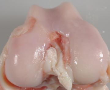
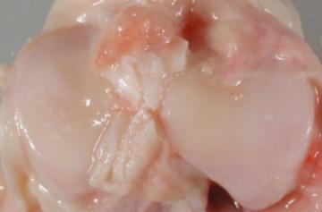
5 Macroscopic assessment of cartilage E A Smooth surface 0 B Slightly fibrillated/roughened surface 1 C Fibrillated surface with focal partial thickness lesions 2 D Deep lesions with surrounding damage 3 E Large areas of severe damage 4 Macroscopic cartilage scoring for each weight bearing compartment, based on Outerbridgeclassification (J Bone Joint SurgBr 1961) and modified from Masterbergen et al (Rheumatology 2006)


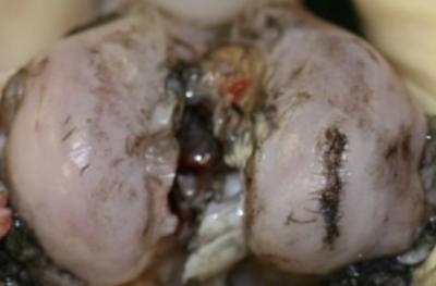
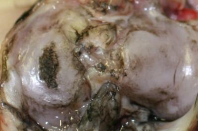
6 Macroscopic assessment of cartilage Indian ink staining % area of cartilage damage
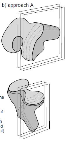
7 Sample collection for histopathology FEMUR TIBIA

8 Scoring System Cartilage & Osteochondral tissues Tissue Articular cartilage Osteochondral tissue Categories (use one or more) Cartilage structure Chondrocytes Proteoglycan staining intensity Collagen integrity Tide mark integrity Subchondral bone changes C D 1/3 D 1/3 of section scored D + 1/3 of section scored D + 1/3 of section scored C = = 8 Pathological Changes in Each Category < 1/3 (Focal) EXTENT OF SECTION AFFECTED < 2/3 (Multifocal, Focally extensive) >2/3 (Multifocal, Diffuse ) A Normal B Less severe pathology C D E Most severe pathology
9 Cartilage Pathology: Structural change SEVERITY OF CARTILAGE PATHOLOGY Characteristics EXTENT OF SECTION AFFECTED < 1/3 < 2/3 >2/3 A Normal volume, smooth surface with intact superficial zone B Slight surface irregularities including fibrillations/fissures in superficial zone C Clefts/fissures to mid zone and/or erosion of superficial zone D Cleft/fissures that extend to deep zone and/or erosion through mid zone E Full thickness loss/deformation of cartilage
 of fibrillation in superficial zone = 1 b b.")
 erosion through mid zone = 9 SEVERITY OF CARTILAGE PATHOLOGY Characteristics EXTENT OF SECTION")

10 Cartilage Pathology: Structural Change examples a a. Small focal area (<1/3) of fibrillation in superficial zone = 1 b b. Focally extensive area of erosion of superficial zone (<2/3) = 4 c c. Diffuse (>2/3) erosion through mid zone = 9 SEVERITY OF CARTILAGE PATHOLOGY Characteristics EXTENT OF SECTION AFFECTED < 1/3 < 2/3 >2/3 A Normal volume, smooth surface with intact superficial zone B Slight surface irregularities including fibrillations/fissures in superficial zone C Clefts/fissures to mid zone and/or erosion of superficial zone D Cleft/fissures that extend to deep zone and/or erosion through mid zone E Full thickness loss/derangement of cartilage


 =")
11 Cartilage Pathology: Chondrocyte change SEVERITY OF CHONDROCYTE PATHOLOGY Characteristics EXTENT OF SECTION AFFECTED < 1/3 < 2/3 >2/3 A Normal B Relative hypocellularity at the articular surface or hypercellularity with occasional superficial clones C Frequent clones, small cell clones predominate D Frequent clones, large cell clones predominate E Cell loss predominates A C D Normal = 0 Frequent small clones (duos and trios) = 2 Frequent large cell clones = 3


12 Cartilage Pathology: Proteoglycan staining Toluidine blue CATIONIC STAINING (PROTEOGLYCAN) Characteristics Safranin O EXTENT OF SECTION AFFECTED < 1/3 < 2/3 >2/3 A Normal B Reduction of staining in the superficial zone C Reduction of staining into the mid zone D Reduction of staining into the deep zone E Full depth reduction of staining


13 Cartilage Pathology: Collagen integrity A B C Collagen type II Collagen type I Picrosirius red staining with polarized light COLLAGEN DERRANGEMENT Characteristics EXTENT OF SECTION AFFECTED < 1/3 < 2/3 >2/3 A Normal B Loss of integrity of superficial zone C Loss of integrity of surface and mid zones D Loss of integrity of surface, mid and deep zones 3 6 9


14 Osteochondral pathology: Tidemark integrity TIDEMARK INTEGRITY EXTENT OF SECTION AFFECTED < 1/3 < 2/3 >2/3 A Intact and distinct B Not consistent or distinct (loss and/or duplication) C Loss of tidemark which is crossed by blood vessels A. Apparently normal tidemark = 0 B. Indistinct tidemark = 1 B.Duplication of tidemark = 1 C.Loss of tidemark with vascular penetration = 2


15 Osteochondral Pathology: Subchondral bone change EXTENT OF SECTION SUBCHONDRAL BON CHANGES AFFECTED < 1/3 < 2/3 >2/3 A Apparently normal thickness B Mild to moderate increase in thickness C Marked increase in thickness and/or subchondral pseudocysts A B C Increased in thickness Subchondral pseudocysts

16 Synovial changes
 Lining cells characteristics (Synoviocyte changes) Lateral (1/3) Medial (1/3) Axial (1/3) A Normal (1 to 2 cell layers of thin synoviocytes) 0 0 0 B")
17 Scoring system: Synovium Tissue Categories (use one or more) Synovium Synoviocytes Tissue morphology Cellular infiltrates Lateral Medial Axial 3 sections of synovium (medial, axial, and SEVERITY OF PATHOLOGY lateral compartments if possible) Lining cells characteristics (Synoviocyte changes) Lateral (1/3) Medial (1/3) Axial (1/3) A Normal (1 to 2 cell layers of thin synoviocytes) B Hypertrophy and/or mild to moderate hyperplasia C Marked hyperplasia (> 6 cell layers) Lining characteristics (Tissue morphologic changes) A Normal B Short villi formation C Finger-like projections Cell infiltration characteristics A No inflammatory cell infiltration B Mild to moderate inflammatory cell infiltration C Marked inflammatory cell infiltration, lymphoid proliferation 2 4 6

 0 0 0 B Hypertrophy and/or mild to moderate hyperplasia 1 2 3 C Marked")
18 Synovial Pathology: Lining synoviocytes changes A B C Apparently normal synoviocytes= 0 Mild hyperplasia = 1 Marked hyperplasia = 2 SEVERITY OF PATHOLOGY Lining cells characteristics SECTION AFFECTED 1/3 1/3 1/3 A Normal (1 to 2 cell layers of thin synoviocytes) B Hypertrophy and/or mild to moderate hyperplasia C Marked hyperplasia (>6 cell layers) 2 4 6


19 Synovial Pathology: Synovial tissue morphologic changes A B C Apparently normal synovium= 0 Short villi formation = 1 Fronds-like projections = 2 SEVERITY OF PATHOLOGY Lining characteristics (Tissue morphologic changes) SECTION AFFECTED 1/3 1/3 1/3 A Normal B Short villi formation C Finger-like projections 2 4 6


20 Synovial Pathology: Cellular infiltration A B C No cellular infiltrates = 0 Mild lymphocytic infiltrates = 1 Lymphoid proliferation = 2 SEVERITY OF PAHTOLOGY Cellular infiltration characteristics SECTION AFFECTED 1/3 1/3 1/3 A No inflammatory cell infiltration B Mild to moderate inflammatory cell infiltration C Marked inflammatory cell infiltration, lymphoid proliferation 2 4 6


 Meniscus structure")

21 Meniscal Pathology: Sampling & Categories Cross section of meniscus Tissue Meniscus Tissue sampling for histology Lateral 1 Medial Categories (use one or more) Meniscus structure Matrix content Cellular proliferation 3. Posterior 2. Middle 1. Anterior
 Tissue architecture Anterior 1/3 Middle Posterior 1/3 A Normal 0 0 0 B Mild disruption 1 1 1 C Moderate")

22 Meniscal Pathology c 3 sections of med. & lat. meniscus SEVERITY OF PATHOLOGY (anterior, middle, posterior if possible) Tissue architecture Anterior 1/3 Middle Posterior 1/3 A Normal B Mild disruption C Moderate disruption with loss of tissue D More than 50% loss of tissue architecture Matrix Content A Normal B Mild alterations in matrix content C Moderate alterations in matrix content D Severe loss of matrix content Proliferative Response A None B Mild proliferation of cells at synovial-meniscal junction C Proliferation of cells at synovial junction and extending into tissue or along surface D Marked proliferation of cells involving majority of remaining tissue 3 3 3
International Cartilage Repair Society
 OsteoArthritis and Cartilage (2006) 14, 13e29 ª 2005 OsteoArthritis Research Society International. Published by Elsevier Ltd. All rights reserved. doi:10.1016/j.joca.2005.07.014 Osteoarthritis cartilage
Disclosures: C.B. Raub: None. B.C. Hansen: None. T. Yamaguchi: None. M.M. Temple-Wong: None. K. Masuda: None. R.L. Sah: None.
 En Face Microscopy of Rabbit Knee Articular Cartilage Following Anterior Cruciate Ligament Transection Reveals Early Matrix Damage, Chondrocyte Loss and Cloning Christopher B. Raub, PhD, Bradley C. Hansen,
The OARSI histopathology initiative e recommendations for histological assessments of osteoarthritis in the guinea pig 1
 Osteoarthritis and Cartilage 18 (2010) S35eS52 The OARSI histopathology initiative e recommendations for histological assessments of osteoarthritis in the guinea pig 1 V.B. Kraus y *, J.L. Huebner y, J.
TITLE: Local Blockade of CCL21 and CXCL13 Signaling as a New Strategy to Prevent and Treat Osteoarthritis
 AWARD NUMBER: W1XWH-15-1-31 TITLE: Local Blockade of CCL1 and CXCL13 Signaling as a New Strategy to Prevent and Treat Osteoarthritis PRINCIPAL INVESTIGATOR: Bouchra Edderkaoui., Ph.D. CONTRACTING ORGANIZATION:
AIMS We will all come across osteo- and rheumatoid arthritis whatever our clinical practice Overview of pathology of osteoarthritis, its assessment an
 Osteoarthritis and Rheumatoid Arthritis Mr. Guy Barham FY1 & FY2 Orthopaedic Curriculum June 2007 AIMS We will all come across osteo- and rheumatoid arthritis whatever our clinical practice Overview of
ABNORMAL SOFTENING IN ARTICULAR CARTILAGE
 1209 ABNORMAL SOFTENING IN ARTICULAR CARTILAGE Its Relationship to the Collagen Framework NEIL D. BROOM Abnormal softening in articular cartilage is related to the presence of collagen fibers strongly
CASE REPORT GIANT OSTEOCHONDRAL LOOSE BODY OF THE KNEE JOINT
 Journal of Musculoskeletal Research, Vol. 4, No. 2 (2000) 145 149 World Scientific Publishing Company ORIGINAL CASE REPORT ARTICLES GIANT OSTEOCHONDRAL LOOSE BODY OF THE KNEE JOINT Mustafa Yel *,, Mustafa
Osteoarthritis. Dr Anthony Feher. With special thanks to Dr. Tim Williams and Dr. Bhatia for allowing me to use some of their slides
 Osteoarthritis Dr Anthony Feher With special thanks to Dr. Tim Williams and Dr. Bhatia for allowing me to use some of their slides No Financial Disclosures Number one chronic disability in the United States
Nanomechanical Symptoms in Cartilage Precede Histological Osteoarthritis Signs after the Destabilization of Medial Meniscus in Mice
 Nanomechanical Symptoms in Cartilage Precede Histological Osteoarthritis Signs after the Destabilization of Medial Meniscus in Mice Basak Doyran 1, Wei Tong 2, Qing Li 1, Haoruo Jia 2, Xianrong Zhang 3,
Imaging of Articular Cartilage
 Clinical Imaging of Articular Cartilage Imaging of Articular Cartilage Prof. Dr. K. Verstraete Ghent University Introduction : Articular Cartilage Histology and biochemical composition Review of Imaging
TREATMENT OF CARTILAGE LESIONS
 TREATMENT OF CARTILAGE LESIONS Angelo J. Colosimo, MD -Head Orthopaedic Surgeon University of Cincinnati Athletics -Director of Sports Medicine University of Cincinnati Medical Center -Associate Professor
SSSR. 1. Nov Ankle. Postoperative Imaging of Cartilage Repair. and Lateral Ligament Reconstruction
 Ankle Postoperative Imaging of Cartilage Repair and Lateral Ligament Reconstruction Andrea B. Rosskopf, MD University Hospital Balgrist Imaging of Cartilage Repair Why? To assess the technical success
Osteoarthritis. RA Hughes
 Osteoarthritis RA Hughes Osteoarthritis (OA) OA is the most common form of arthritis and the most common joint disease Most of the people who have OA are older than age 45, and women are more commonly
Basics of Cartilage Restoration Introduction of TruFit
 Basics of Cartilage Restoration Introduction of TruFit Philip A. Davidson, MD Heiden Orthopaedics Park City, Utah USA Smith & Nephew Seminar London, UK October 2008 Cartilage Restoration A wide realm between..
OSTEOCHONDRAL ALLOGRAFT RECONSTRUCTION FOR MASSIVE BONE DEFECT
 OSTEOCHONDRAL ALLOGRAFT RECONSTRUCTION FOR MASSIVE BONE DEFECT Angelo J. Colosimo, MD -Head Orthopaedic Surgeon University of Cincinnati Athletics -Director of Sports Medicine University of Cincinnati
MRI KNEE WHAT TO SEE. Dr. SHEKHAR SRIVASTAV. Sr.Consultant KNEE & SHOULDER ARTHROSCOPY
 MRI KNEE WHAT TO SEE Dr. SHEKHAR SRIVASTAV Sr.Consultant KNEE & SHOULDER ARTHROSCOPY MRI KNEE - WHAT TO SEE MRI is the most accurate and frequently used diagnostic tool for evaluation of internal derangement
Discovery of a Small Molecule Inhibitor of the Wnt Pathway (SM04690) as a Potential Disease Modifying Treatment for Knee Osteoarthritis
 as a Potential Disease Modifying Treatment for Knee Osteoarthritis") Discovery of a Small Molecule Inhibitor of the Wnt Pathway (SM469) as a Potential Disease Modifying Treatment for Knee Osteoarthritis Vishal Deshmukh, Ph.D., Charlene Barroga, Ph.D., Yong Hu, Ph.D., John
Discovery of a Small Molecule Inhibitor of the Wnt Pathway (SM469) as a Potential Disease Modifying Treatment for Knee Osteoarthritis Vishal Deshmukh, Ph.D., Charlene Barroga, Ph.D., Yong Hu, Ph.D., John
Degenerative Lesions Of The Patellofemoral Joint: An Autopsy Study
 Degenerative Lesions Of The Patellofemoral Joint: An Autopsy Study Laura Cutignola-Kelley Norman A. Johanson Abstract: 30 knee joints were collected from individuals who were between 55 and 85 at the time
Discovery of a Small Molecule Inhibitor of the Wnt Pathway as a Potential Disease Modifying Treatment for Knee Osteoarthritis
 Discovery of a Small Molecule Inhibitor of the Wnt Pathway as a Potential Disease Modifying Treatment for Knee Osteoarthritis Charlene Barroga, Ph.D., Yong Hu, Ph.D., Vishal Deshmukh, Ph.D., and John Hood,
Department of Plastic Surgery, Royal Melbourne Hospital, Australia
 ARTICULAR CARTILAGE LOSS IN LONG-STANDING IMMOBILISATION OF INTERPHALANGEAL JOINTS By P. L. FIELD, F.R.C.S., and J. T. HUESTON,/Vi.S., F.R.C.S., F.R.A.C.S. Department of Plastic Surgery, Royal Melbourne
OSTEOARTHRITIS and CARTILAGE
 Osteoarthritis and Cartilage (1996) 4, 9-22 1996 Osteoarthritis Research Society 1063-4584/96/010009 + 14 $12.00/Q OSTEOARTHRITIS and CARTILAGE Experimental osteoarthritis in dogs: a comparison of the
Arthrographic study of the rheumatoid knee.
 Annals of the Rheumatic Diseases, 1981, 40, 344-349 Arthrographic study of the rheumatoid knee. Part 2. Articular cartilage and menisci KYOSUKE FUJIKAWA, YOSHINORI TANAKA, TSUNEYO MATSUBAYASHI, AND FUJIO
Cartilage. Dr. Heba Kalbouneh Associate Professor of Anatomy and Histology
 Cartilage Dr. Heba Kalbouneh Associate Professor of Anatomy and Histology 1 Cartilage is a specialized type of connective tissue designed to give support, bear weight and withstand tension, torsion and
Spontaneous Repair of Full- Thickness Defects of Articular Cartilage in a Goat Model A PRELIMINARY STUDY
 53 Spontaneous Repair of Full- Thickness Defects of Articular Cartilage in a Goat Model A PRELIMINARY STUDY BY DOUGLAS W. JACKSON, MD, PEGGY A. LALOR, PHD, HAROLD M. ABERMAN, DVM, AND TIMOTHY M. SIMON,
Rakesh Patel, MD 4/9/09
 Rakesh Patel, MD 4/9/09 Chondral Injuries Very common Present in 63-66% patients undergoing arthroscopy 11-19% full-thickness lesions Up to 79% patients with ACL deficient knee have some form of chondral
MENISCAL INJURY. Meniscus. Anterior Roots. Medial Meniscus. Lateral Meniscus. Posterior Roots. MRI and Arthroscopic Findings
 Meniscus Anterior Roots MENISCAL INJURY MRI and Arthroscopic Findings Medial Meniscus AH PH PH AH Lateral Meniscus Rawiwan Pattaweerakul Naresuan University Hospital Posterior Roots Meniscus Normal Meniscus
Classification of Acetabular Cartilage Lesions. Claudio Mella, MD
 Classification of Acetabular Cartilage Lesions Claudio Mella, MD Acetabular cartilage lesions are frequently found during hip arthroscopy. The arthroscopic view offers an exceptional perspective to assess
Non-Invasive Characterization of Cartilage Properties Using MR Imaging
 Non-Invasive Characterization of Cartilage Properties Using MR Imaging by Sophia Natalie Ziemian Department of Biomedical Engineering Duke University Date: Approved: Farshid Guilak, Supervisor Lori A.
RECENT ADVANCES IN CLINICAL MR OF ARTICULAR CARTILAGE
 In Practice RECENT ADVANCES IN CLINICAL MR OF ARTICULAR CARTILAGE By Atsuya Watanabe, MD, PhD, Director, Advanced Diagnostic Imaging Center and Associate Professor, Department of Orthopedic Surgery, Teikyo
Medical Practice for Sports Injuries and Disorders of the Knee
 Sports-Related Injuries and Disorders Medical Practice for Sports Injuries and Disorders of the Knee JMAJ 48(1): 20 24, 2005 Hirotsugu MURATSU*, Masahiro KUROSAKA**, Tetsuji YAMAMOTO***, and Shinichi YOSHIDA****
Imaging the Knee 17/10/2017. Friction syndrome Common in runners or cyclists Fluid between ITB and Lateral femoral condyle
 17/10/2017 Imaging the Knee Alicia M. Yochum RN, DC, DACBR, RMSK Iliotibial Band Syndrome Ligamentous Tears (ACL, PCL, MCL, LCL) Meniscal Tears Cartilage Degeneration Quadriceps/Patellar tendinosis Osteochondral
Unicompartmental Knee Resurfacing
 Disclaimer This movie is an educational resource only and should not be used to manage knee pain. All decisions about the management of knee pain must be made in conjunction with your Physician or a licensed
The Meniscus. History. Anatomy. Anatomy. Blood Supply. Attachments
 History The Meniscus W. Randall Schultz, MD, MS Austin, TX January 23, 2016 Meniscus originally thought to represent vestigial tissue 1883 first reported meniscal repair (Annandale) Total menisectomy treatment
Medial Knee Osteoarthritis Precedes Medial Meniscal Posterior Root Tear with an Event of Painful Popping
 Medial Knee Osteoarthritis Precedes Medial Meniscal Posterior Root Tear with an Event of Painful Popping Dhong Won Lee, M.D, Ji Nam Kim, M.D., Jin Goo Kim, M.D., Ph.D. KonKuk University Medical Center
MY PATIENT HAS KNEE PAIN. David Levi, MD Chief, Division of Musculoskeletal l limaging Atlantic Medical Imaging
 MY PATIENT HAS KNEE PAIN David Levi, MD Chief, Division of Musculoskeletal l limaging Atlantic Medical Imaging Causes of knee pain Non traumatic Trauma Osteoarthritis Patellofemoral pain Menisci or ligaments
MRI of Cartilage. D. BENDAHAN (PhD)
") MRI of Cartilage D. BENDAHAN (PhD) Centre de Résonance Magnétique Biologique et Médicale UMR CNRS 7339 Faculté de Médecine de la Timone 27, Bd J. Moulin 13005 Marseille France david.bendahan@univ-amu.fr
MRI of Cartilage D. BENDAHAN (PhD) Centre de Résonance Magnétique Biologique et Médicale UMR CNRS 7339 Faculté de Médecine de la Timone 27, Bd J. Moulin 13005 Marseille France david.bendahan@univ-amu.fr
Calcium Pyrophosphate Deposition in Nonhuman Primates
 Vet. Pathol. : 59-596 (984) Calcium Pyrophosphate Deposition in Nonhuman Primates E. D. ROBERTS, G. B. BASKIN, E. WATSON, W. G. HENK, T. C. SHELTON, and M. S. BOWEN Departments of Veterinary Pathology,
The Effect of Varus Stress on the Moving Rabbit Knee Joint
 The Effect of Varus Stress on the Moving Rabbit Knee Joint KOSUKE OGATA, M.D., LEO A. WHITESIDE, M.D., PEGGY A. LESKER, B.S. AND DAVID J. SIMMONS, PH.D. Many attempts have been made to induce osteoarthritis
Types of osteoarthritis
 ARTHRITIS Osteoarthritis is a degenerative joint disease is the most common joint disorder. It is a frequent part of aging and is an important cause of physical disability in persons older than 65 years
"BONE BRUISES" OF THE KNEE: A REVIEW
 "BONE BRUISES" OF THE KNEE: A REVIEW Chad E. Mathis, M.D. Ken Noonan, M.D. Kosmas Kayes, M.D. ABSTRACT Magnetic resonance (MR) imaging is often used - to assess the location and degree of ligamentous wm.
The cellfree matrix for autoregeneration of articular cartilage defects
 The cellfree matrix for autoregeneration of articular cartilage defects What is Amedrix GmbH? Amedrix GmbH, located in Esslingen near Stuttgart, is a medical biotech company which develops highly innovative
BASELINE QUESTIONNAIRE (SURGEON)
") SECTION A: STUDY INFORMATION Subject ID: - - Study Visit: Baseline Site Number: Date: / / Surgeon ID: SECTION B: INITIAL SURGEON HISTORY B1. Previous Knee Surgery: Yes No Not recorded B2. Number of Previous
SECTION A: STUDY INFORMATION Subject ID: - - Study Visit: Baseline Site Number: Date: / / Surgeon ID: SECTION B: INITIAL SURGEON HISTORY B1. Previous Knee Surgery: Yes No Not recorded B2. Number of Previous
LIGAMENTS AND TENDONS
 Harvard-MIT Division of Health Sciences and Technology HST.523J: Cell-Matrix Mechanics Prof. Myron Spector Massachusetts Institute of Technology Harvard Medical School Brigham and Women s s Hospital VA
triquetrum in rheumatoid arthritis
 Ann. rheum. Dis. (1976), 35, 46 Early abnormalities of pisiform and triquetrum in rheumatoid arthritis DONALD RESNICK From the Department of Radiology, Veterans Administration Hospital, San Diego, and
Histologic change of cartilage layer of osteochondritis dissecans before and after fixation in the knee
 1 Histologic change of cartilage layer of osteochondritis dissecans before and after fixation in the knee Mitsuo Ochi, M.D. PhD Professor and chairman Department of Orthopaedic Surgery Graduate School
HOW DO WE DIAGNOSE LAMENESS IN YOUR HORSE?
 HOW DO WE DIAGNOSE LAMENESS IN YOUR HORSE? To help horse owners better understand the tools we routinely use at VetweRx to evaluate their horse s soundness, the following section of this website reviews
= BONE & JOINT = ANATOMY & NORMAL US FINDINGS
 Dongguk Univeristy 1 = BONE & JOINT = ANATOMY & NORMAL US FINDINGS 2012.4.14. 동국대일산병원재활의학과이호준 Dongguk Univeristy 2 = BONE = ANATOMY (& HISTOLOGY) Dongguk Univeristy 3 Bone : Histology Epiphysis Filled
Osteochondral regeneration. Getting to the core of the problem.
 Osteochondral regeneration. Getting to the core of the problem. TM TM Bio-mimetic, biointegratable and resorbable Flexible and easy to shape Straightforward one-step procedure Promotes a guided osteo-chondral
3 Sternoclavicular Joints
 3 Sternoclavicular Joints Anne Grethe Jurik and Flemming Brandt Soerensen 29 Contents 3.1 Introduction.......................................................... 29 3.2 Macroscopic Anatomy.................................................
What is the most effective MRI specific findings for lateral meniscus posterior root tear in ACL injuries
 What is the most effective MRI specific findings for lateral meniscus posterior root tear in ACL injuries Kazuki Asai 1), Junsuke Nakase 1), Kengo Shimozaki 1), Kazu Toyooka 1), Hiroyuki Tsuchiya 1) 1)
Mid-Term Clinical Outcomes of Atelocollagenassociated Autologous Chondrocyte Implantation for the Repair of Chondral Defects of the Knee
 International Society of Arthroscopy, Knee Surgery and Orthopaedic Sports Medicine Cancun, Mexico MAY 12 16, 2019 Mid-Term Clinical Outcomes of Atelocollagenassociated Autologous Chondrocyte Implantation
Lasers in Surgery and Medicine 38: (2006)
") Lasers in Surgery and Medicine 38:852 865 (2006) Determination of Characteristics of Degenerative Joint Disease Using Optical Coherence Tomography and Polarization Sensitive Optical Coherence Tomography
Lasers in Surgery and Medicine 38:852 865 (2006) Determination of Characteristics of Degenerative Joint Disease Using Optical Coherence Tomography and Polarization Sensitive Optical Coherence Tomography
Chapter 6: Skeletal System: Bones and Bone Tissue
 Chapter 6: Skeletal System: Bones and Bone Tissue I. Functions A. List and describe the five major functions of the skeletal system: 1. 2. 3.. 4. 5.. II. Cartilage A. What do chondroblasts do? B. When
Joint and Epiphyseal Progenitor Cells Revitalize Tendon Graft and Form Mineralized Insertion Sites in Murine ACL Reconstruction Model
 Joint and Epiphyseal Progenitor Cells Revitalize Tendon Graft and Form Mineralized Insertion Sites in Murine ACL Reconstruction Model Yusuke Hagiwara 1,2, Nathaniel A. Dyment 3, Douglas J. Adams 3, Shinro
Which compound is reponsible for the viscous character of the ground substance?
 1 2 Which type of collagen forms the coarse collagen fibres in dense regular and irregular connective tissues? Which compound is reponsible for the viscous character of the ground substance? 3 Which class
OSTEOPHYTOSIS OF THE FEMORAL HEAD AND NECK
 908 RDIOLOGIC VIGNETTE OSTEOPHYTOSIS OF THE FEMORL HED ND NECK DONLD RESNICK Osteophytes are frequently considered the most characteristic abnormality of degenerative joint disease. In patients with osteoarthritis,
ADVANCED IMAGING OF THE KNEE
 MENISCAL ANATOMY ADVANCED IMAGING OF THE KNEE MENISCAL ABNORMALITIES MENISCAL FUNCTION MENISCAL FUNCTION load transmission shock absorption stability The menisci DO NOT function as primary stabilizers
OSTEOCHONDRAL ALLOGRAFTS AND AUTOGRAFTS IN THE TREATMENT OF FOCAL ARTICULAR CARTILAGE LESIONS
 Status Active Medical and Behavioral Health Policy Section: Surgery Policy Number: IV-115 Effective Date: 10/22/2014 Blue Cross and Blue Shield of Minnesota medical policies do not imply that members should
Contrasting alteration patterns of different cartilage plates in knee articular cartilage after spinal cord injury in rats
 (2009) 47, 218 224 & 2009 International Society All rights reserved 1362-4393/09 $32.00 www.nature.com/sc ORIGINAL ARTICLE Contrasting alteration patterns of different cartilage plates in knee articular
Cruciate Ligament. Summary of the Doctoral Thesis
 Study of the Effect of Excessive Tibial Plateau Angle on Degenerative Changes of Canine Cranial Cruciate Ligament Summary of the Doctoral Thesis Tom Ichinohe Graduate School of Veterinary Medicine and
MR imaging of the knee in marathon runners before and after competition
 Skeletal Radiol (2001) 30:72 76 International Skeletal Society 2001 ARTICLE W. Krampla R. Mayrhofer J. Malcher K.H. Kristen M. Urban W. Hruby MR imaging of the knee in marathon runners before and after
MRI of the Knee: Part 2 - menisci. Mark Anderson, M.D. University of Virginia Health System
 MRI of the Knee: Part 2 - menisci Mark Anderson, M.D. University of Virginia Health System Learning Objectives At the end of the presentation, each participant should be able to: describe the normal anatomy
Small animal osteoarthritis
 Vet Times The website for the veterinary profession https://www.vettimes.co.uk Small animal osteoarthritis Author : Kelly Bowlt Categories : Vets Date : April 13, 2009 Normal joints A healthy synovial
AN OVERVIEW : Cartilage Treatment. Eric Thiel, MD. WVAM Conference 01/25/2019
 AN OVERVIEW : Cartilage Treatment Eric Thiel, MD WVAM Conference 01/25/2019 No Disclosure The Science of Hyaline Cartilage I. HYALINE CARTILAGE BASIC SCIENCE Normal Anatomy Cartilage is hypocellular, avascular,
Chapter 7. Anatomy of the Triangular Fibrocartilage Complex: Current Concepts. Introduction. Anatomy. Histology
 Chapter 7 Anatomy of the Triangular Fibrocartilage Complex: Current Concepts Introduction The triangular fibrocartilage complex (TFCC) is one of the intrinsic ligaments of the wrist. It is often injured
Validity of histopathological grading of articular cartilage from osteoarthritic knee joints
 208 Osteoarthritis Research Unit, Institute for Inflammation Research, National University Hospital/Rigshospitalet, Copenhagen, Denmark K Ostergaard J Petersen K Bendtzen Department of Pathology, National
9/18/18. Welcome- MSK Ultrasound Workshop. Introduction to Musculoskeletal Ultrasound. Acknowledgement of Country. The Workshop.
 Acknowledgement of Country Welcome- MSK Ultrasound Workshop I would like to acknowledge that this meeting is being held on the traditional lands of the Wurundjeri and Boonwurrung people and pay my respect
When (How) MRI Became the Gold Standard Hollis G. Potter, MD
 MRI Became the Gold Standard Hollis G. Potter, MD") When (How) MRI Became the Gold Standard Hollis G. Potter, MD potterh@hss.edu Target audience: Radiologists and imaging scientists interested in assessing MRI of cartilage Outcome/Objectives: 1. To become
When (How) MRI Became the Gold Standard Hollis G. Potter, MD potterh@hss.edu Target audience: Radiologists and imaging scientists interested in assessing MRI of cartilage Outcome/Objectives: 1. To become
AUTOLOGOUS CHONDROCYTE IMPLANTATION FOR CHONDRAL KNEE DAMAGE B.A. Jalba 1, C.S. Jalba 2, F. Gherghina 3, M. Cruce 3
 AUTOLOGOUS CHONDROCYTE IMPLANTATION FOR CHONDRAL KNEE DAMAGE B.A. Jalba 1, C.S. Jalba 2, F. Gherghina 3, M. Cruce 3 1-EMERGENCY CLINICAL HOSPITAL FLOREASCA BUCHAREST 2-EMERGENCY CLINICAL HOSPITAL SFANTUL
Compact bone; Many parallel Haversian canals contain: small blood vessels. very small nerve. Interconnected by Volkmann s canals.
 Special characteristics of COMPACT BONE (dense bone) Thick; well vascularized Osteocytes and lamellae Concentric rings around blood vessels Most bones: outer compact bone inner spongy bone Marrow cavity
International Cartilage Repair Society
 Osteoarthritis and Cartilage (2003) 11, 78 84 2003 OsteoArthritis Research Society International. Published by Elsevier Science Ltd. All rights reserved. 1063 4584/03/$30.00/0 doi:10.1053/joca.2002.0870
Why do they fail?? TOM MINAS MD MS. The Management of Failed Cartilage Repair Procedures PALM BEACH FL
 The Management of Failed Cartilage Repair Procedures Why do they fail?? TOM MINAS MD MS DIRECTOR, CARTILAGE REPAIR CENTER, PALEY ORTHOPEDIC INSTITUTE, PALM BEACH FL PROFESSOR EMERITUS, HARVARD MEDICAL
Knee: Cruciate Ligaments
 72 Knee: Cruciate Ligaments R. Kent Sanders Sagittal oblique 2.5-mm sequences along the plane of the anterior cruciate ligament (ACL) typically yield three to four images of the ACL, with the first medial
Effects of Immobilization on Structure of Cell Layers In Tibial Articular Cartilage
 Effects of Immobilization on Structure of Cell Layers In Tibial Articular Cartilage Effects of Immobilization on Structure of Cell Layers In Tibial Articular Cartilage OGIWARA Yuh* FUJIKAWA Kaoru** OHSAKO
ACL Athletic Career. ACL Rupture - Warning Features Intensive pain Immediate swelling Locking Feel a Pop Dead leg Cannot continue to play
 FIMS Ambassador Tour to Eastern Europe, 2004 Belgrade, Serbia Montenegro Acute Knee Injuries - Controversies and Challenges Professor KM Chan OBE, JP President of FIMS Belgrade ACL Athletic Career ACL
4 2 Osteoarthritis 1
 Osteoarthritis 1 Osteoarthritis ( OA) Osteoarthritis is a chronic disease and the most common of all rheumatological disorders. It particularly affects individuals over the age of 65 years. The prevalence
Summary. Introduction. Materials and methods
 Osteoarthritis and Cartilage (2000) 8, 303 308 2000 OsteoArthritis Research Society International 1063 4584/00/040303+06 $35.00/0 doi:10.1053/joca.1999.0305, available online at http://www.idealibrary.com
Gout. Crystal deposition disease: Imaging perspectives. Crystal associated arthropathies. Clinical Stages of Gout 07/06/60
 Crystal associated arthropathies Crystal deposition disease: Imaging perspectives Warapat Virayavanich, MD Ramathibodi hospital, Mahidol University Commonly seen arthropathy MSU (gout) CPPD HADD Uncommon
High-resolution measurements of the multilayer ultra-structure of articular cartilage and their translational potential He et al.
 High-resolution measurements of the multilayer ultra-structure of articular cartilage and their translational potential He et al. He et al. Arthritis Research & Therapy He et al. Arthritis Research & Therapy
MRI of the Knee: Part 4 - normal variants that may simulate disease. Mark Anderson, M.D. University of Virginia
 MRI of the Knee: Part 4 - normal variants that may simulate disease Mark Anderson, M.D. University of Virginia discuss the most common normal variants in the pediatric knee that may simulate pathology
Knee: Meniscus Back to Basics
 Knee: Meniscus Back to Basics Kyung Jin Suh kyungjin.suh@gmail.com Doctor Radiology, Daegu, KOREA Medial Lateral 7.7 10.2 11.6 9.6 10.6 mm Posterior > Anterior horn 10.6 mm Posterior = Anterior horn Medial
Knee Joint Anatomy 101
 Knee Joint Anatomy 101 Bone Basics There are three bones at the knee joint femur, tibia and patella commonly referred to as the thighbone, shinbone and kneecap. The fibula is not typically associated with
Corporate Medical Policy
 Corporate Medical Policy Autologous Chondrocyte Implantation File Name: Origination: Last CAP Review: Next CAP Review: Last Review: autologous_chondrocyte_implantation 4/1996 6/2017 6/2018 6/2017 Description
International Cartilage Repair Society
 OsteoArthritis and Cartilage (2004) 12, 201 216 2003 OsteoArthritis Research Society International. Published by Elsevier Ltd. All rights reserved. doi:10.1016/j.joca.2003.11.001 The use of photooxidized,
New Directions in Osteoarthritis Research
 New Directions in Osteoarthritis Research Kananaskis October 22, 2015 Nick Mohtadi MD MSc FRCSC No conflicts of interest related to this presentation 1 Osteoarthritis: Disease? Fact of Life? Strong family
Articular cartilage repair using collagen type I hydrogels Clincal results
 Articular cartilage repair using collagen type I hydrogels Clincal results Ulrich Nöth, MD Department of Orthopaedic Surgery, König-Ludwig-Haus University of Würzburg, Germany Orthopädisches Zentrum für
OSTEOARTHRITIS AS AN ORGAN DISEASE
 OSTEOARTHRITIS AS AN ORGAN DISEASE Peter Muir BVSc, MVetClinStud, PhD, DACVS, DECVS University of Wisconsin-Madison, School of Veterinary Medicine, Madison, WI, USA Osteoarthritis (OA) essentially represents
The biomechanical and histological effects of posterior cruciate ligament rupture on the medial tibial plateau
 Deng et al. Journal of Orthopaedic Surgery and Research (2017) 12:48 DOI 10.1186/s13018-017-0551-x RESEARCH ARTICLE Open Access The biomechanical and histological effects of posterior cruciate ligament
Knee Contusions and Stress Injuries. Laura W. Bancroft, M.D.
 Knee Contusions and Stress Injuries Laura W. Bancroft, M.D. Objectives Review 5 types of contusion patterns Pivot shift Dashboard Hyperextension Clip Lateral patellar dislocation Demonstrate various stress
CONSULTATION DURING SURGERY / NOT A FINAL DIAGNOSIS. FROZEN SECTION DIAGNOSIS: - A. High grade sarcoma. Wait for paraffin sections results.
 Pathology Report Date: 3/5/02 A, B. Biopsy right distal femur- high grade spindle cell sarcoma Immunohistochemistry studies are pending to further classify the nature of the tumor. CONSULTATION DURING
evicore MSK joint surgery procedures requiring prior authorization
 evicore MSK joint surgery procedures requiring prior authorization Moda Health Commercial Group and Individual Members* Updated 1/30/2018 *Check EBT to verify member enrollment in evicore program Radiology
Keywords Osteoarthritis; Knee; Pathology; Biopsy
 Carlos Antônio Garrido 1, Tania Clarete Fonseca Vieira Sales Sampaio 2, Frederico de Souza Ferreira 3 Objectives: To compare the modified Ahlbäck radiological classification with macroscopic analysis of
Effect of aspirin treatment on chondromalacia patellae
 Annals of the Rheumatic Diseases, 1981, 40, 37-41 Effect of aspirin treatment on chondromalacia patellae GEORGE BENTLEY, IAN J. LESLIE, AND DAVID FISCHER From the University Department of Orthopaedic and
Chapter 9 Articulations Articulations joints where two bones interconnect. Two classification methods are used to categorize joints:
 Chapter 9 Articulations Articulations joints where two bones interconnect Two classification methods are used to categorize joints: Functional classification Structural classification Functional classification
Arthroscopy / MRI Correlation Conference. Department of Radiology, Section of MSK Imaging Department of Orthopedic Surgery 7/19/16
 Arthroscopy / MRI Correlation Conference Department of Radiology, Section of MSK Imaging Department of Orthopedic Surgery 7/19/16 Case 1: 29 YOM with recurrent shoulder dislocations Glenoid Axial T1FS
To classify the joints relative to structure & shape
 To classify the joints relative to structure & shape To describe the anatomy of the hip joint To describe the ankle joint To memorize their blood & nerve supply JOINTS: Joints are sites where skeletal
Treatment of meniscal lesions and isolated lesions of the anterior cruciate ligament of the knee in adults
 QUICK REFERENCE GUIDE Treatment of meniscal s and isolated s of the anterior cruciate ligament of the knee in adults June 2008 AIM OF THE GUIDELINES To encourage good practices in the areas of meniscal
Advanced Hip Arthroscopy
 Advanced Hip Arthroscopy S C OT T D. M A R T I N, M D D I R E C T O R, J O I N T P R E S E R V A T I O N D I R E C T O R, M G H S P O R T S M E D I C I N E F E L L O W S H I P A S S O C I A T E P R O F
TRANSLATIONAL AND CLINICAL RESEARCH
 TRANSLATIONAL AND CLINICAL RESEARCH Intra-Articular Injection of Mesenchymal Stem Cells for the Treatment of Osteoarthritis of the Knee: A Proof-of-Concept Clinical Trial a Department of Orthopedic Surgery,