Rakesh Patel, MD 4/9/09
|
|
|
- Gwendolyn Rose
- 5 years ago
- Views:
Transcription

1 Rakesh Patel, MD 4/9/09

2 Chondral Injuries Very common Present in 63-66% patients undergoing arthroscopy 11-19% full-thickness lesions Up to 79% patients with ACL deficient knee have some form of chondral injury Shindle MK. J Bone Joint Surg Am. 2006;88:27-46.

3 Cartilage Trauma and Degeneration Does not heal spontaneously relative avascularity of articular cartilage If injury extends to subchondral bone, usually more substantial natural healing process Repair tissue composition of hyaline cartilage and fibrocartilage Shindle MK. J Bone Joint Surg Am. 2006;88:27-46.
4 Surgical procedures Restoration Replacement Allograft Relief- Osteotomy Resection Joint Arthroplasty
5 Chondral Restoration/Replacement Debridement/Abrasion Arthroplasty Microfracture Autograft (OAT and Mosaicplasty) Allograft Autologous Chondrocyte Implantation Biodegradable Pins


6 Ho, Y. Y. et al. Radiographics 2007;27: Post-operative MR Followup MR imaging and arthroscopy complementary MR is less invasive than arthroscopy MR more comprehensive evaluation of repaired tissue

7 Post-operative Followup Standard MR imaging techniques may be used postoperatively International Cartilage Repair Society recommendations (Table 1) Postoperative appearance of the joints after repair varies according to the surgical technique and stage of healing Important to be familiar with surgical techniques and characteristic MR imaging features at various postoperative intervals Choi, Y. S. et al. Radiographics 2008;28:
8 Future Techniques Newer techniques, which include delayed gadolinium-enhanced imaging and mapping of T1ρ and T2 values, may provide useful supplemental information about the histologic and biochemical contents of reparative tissue
 Choi, Y. S. et al. Radiographics 2008;28:1043-1059")
9 MR Imaging - Basics Degree of defect filling (should be same thickness as normal cartilage) Extent of integration with adjacent tissues (should be continuous) Graft appearance Underlying bone appearance Presence of proud subchondral bone formation (should be smooth) Choi, Y. S. et al. Radiographics 2008;28:
10 Restoration Two methods Enhance intrinsic healing capacity of tissue Stimulate growth of fibrocartilage from bone marrow stem cells Mainly Type I collagen rather than Type II collagen in hyaline cartilage Structural properties are inferior to normal tissue Regenerate new cartilage
11 Restoration Mechanical Repair - Debridement Abrasion Arthroplasty Microfracture

12 Mechanical Repair Unstable cartilage at defect is removed with arthroscopic debridement and lavage Usually palliative, short term relief Cleanup

13 Abrasion Arthroplasty Exposed surface bone is excised by burr or shaver to a depth of 1-3 mm beneath cartilage defect Results in formation of fibrin clot at the defect site

14 Microfracture

15 Microfracture Unstable cartilage debrided Stable edge of viable cartilage formed Helps hold the marrow clot Calcified cartilage layer removed by curette Angled awl used to create 2-4 mm deep pits perpendicular in subchondral bone Spaced 3-4 mm apart Start at periphery and move inwards Non-power tools preferred due to decreased risk of thermal necrosis

16 Microfracture Exposed bone is debrided of all remaining unstable cartilage Stable perpendicular edge of viable cartilage is formed around the defect Helps hold the marrow clot Calcified cartilage layer removed by curette Angled awl used to create 2-4 mm deep pits perpendicular in subchondral bone beneath the cartilage defect Spaced 3-4 mm apart Start at periphery and move inwards Non-power tools preferred due to decreased risk of thermal necrosis Microfracture holes are continued into the central portion of the defect. Awl is penetrating subchondral bone approximately 2-4mm in depth. Steadman JR. et al. Clin Ortho Rel Resrch 2001;391S:S

17 Microfracture Exposed bone is debrided of all remaining unstable cartilage Stable perpendicular edge of viable cartilage is formed around the defect Helps hold the marrow clot Calcified cartilage layer removed by curette Angled awl used to create 2-4 mm deep pits perpendicular in subchondral bone beneath the cartilage defect Spaced 3-4 mm apart Start at periphery and move inwards Non-power tools preferred due to decreased risk of thermal necrosis Marrow elements including blood and fat droplents can be seen coming from microfracture holes. Steadman JR. et al. Clin Ortho Rel Resrch 2001;391S:S
18 Microfracture Most commonly performed cartilage repair procedure Likely no improvement for at least 3-6 months Improvement slowly and steadily for 2 years Best short-term results observed with good fill grade, low body-mass index, and short duration of preoperative symptoms Will help decrease pain and return to function short term Several high profile athletes never returned to full function after surgery Some returned too early in the recovery period and reinjured same/contralateral knee Recommended for focal grade III/IV lesion surrounded by normal cartilage in young patient

19 Microfracture MR features Appearance of lesion evolves over time Early postoperative period Thin and indistinct 1-2 yrs after surgery Filled defect Smooth and well-defined Bone overgrowth in 25-49% patients Does not have negative effect on clinical outcomes Signal of reparative fibrocartilage Hyperintense due to less organized matrix and increased water mobility Signal decreases as tissue matures Subchondral bone marrow edema decreases Treatment failure Incomplete filling of defect with thin and irregular tissue Persistent bone marrow edema 12 months overgrowth of subchondral bone (arrow), thin overlying reparative fibrocartilage, and hyperintense signal in native cartilage Choi, Y. S. et al. Radiographics 2008;28:


20 Choi, Y. S. et al. Radiographics 2008;28: Microfracture 6 months after surgery Sagittal inversion-recovery fast SE and sagittal intermediate-weighted fast SE images acquired 6 months after a microfracture procedure in a 29-year-old man show hyperintense signal in the repair cartilage with good fill over the lateral femoral condyle (arrow)
 Choi, Y. S. et al.")
21 Microfracture 6 months after surgery 6 months after surgery Sagittal and coronal intermediate-weighted fast SE images acquired 6 months after a microfracture procedure in a 50-yearold man show hyperintense signal and superficial irregularity in the reparative cartilage over the medial femoral condyle (arrow) Choi, Y. S. et al. Radiographics 2008;28:

22 Microfracture 6 months after surgery 4 years after surgery Four year follow-up exam (right) shows superficial irregularity of the reparative fibrocartilage, but no exposed subchondral bone Choi, Y. S. et al. Radiographics 2008;28:
23 Restoration Transplantation of chondral/osteochondral plugs Autologous Allograft Transplantation of chondrocytes


24 OATS-mini.pdf Osteochondral Autograft Transplantation OAT Transfer of osteochondral plug from non-weight bearing region of knee to site of chondral damage Most common harvest site Lateral femoral condyle nonweight bearing surface Intercondylar region Peripheral portion of LFC Most frequently performed in knee and ankle

25 Osteochondral Autograft Transplantation OAT Transfer of osteochondral plug from non-weight bearing region of knee to site of chondral damage Most common harvest site Lateral femoral condyle nonweight bearing surface Intercondylar region Peripheral portion of LFC Most frequently performed in knee and ankle
26 Osteochondral Autograft Transplantation Key to success is viability of chondrocytes Best suited for 1-4 cm 2 lesions, osteochondritis dissecans, and osteonecrosis Preferred treatment at some institutions for patella defects Large grafts may produce incongruent surface Articular surface should be flush Proud plug is subjected to increased shear forces
27 Osteochondral Autograft Transplantation Advantages Graft availability Absence of disease transmission compared to allograft Less likely to evoke immune response Single-stage operation Overall success rate 80-90%, suitable for small or medium sized defects Disadvantages Donor site morbidity limited graft volume availability and age of cartilage Difficulty recreating contour of articular surface

28 Mosaicplasty Several smaller osteochondral grafts are transferred to defect rather than single large graft Diameter mm Depth mm Well suited for smaller osteochondral defects 1-4 cm 2 in younger patients Donor site integrity is generally maintained Chondral defect treated with osteochondral autograft transplantation in a 25-year-old man Recht, M. P. et al. Radiographics 2002;22:

29 Autografts MR assessment Degree of defect filling by osteochondral plug Morphologic characteristics of autologous bone Cartilage surface contour Peripheral integration of reparative cartilage and bone Donor site assessment Choi, Y. S. et al. Radiographics 2008;28: years after surgery Coronal intermediate-weighted fast SE image acquired 2 years after mosaicplasty in a 36-year-old woman shows good integration of osteochondral plugs into the medial femoral condyle, with a slight prominence of cartilage over the most medial plug (arrow)

30 Autografts MR assessment Peripheral integration of reparative cartilage and bone Gaps between cartilage plug and native cartilage can be seen Fluid signal at interface between graft and native bone suggest incomplete incorporation and potential instability Restoration of normal curvature is important Axial intermediate-weighted image after mosaicplasty in 17 year old boy. Subchondral plate is flush, small fissure at medial margin of articular surface. Plugs have incorporated. Choi, Y. S. et al. Radiographics 2008;28:
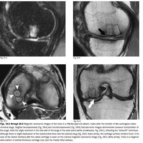
 images acquired 5 years after autologous osteochondral transplantation in a 23-year-old man show that the")

31 Autograft Restoration of curvature 5 years after surgery Sagittal intermediate-weighted fast SE and 3D fat-suppressed T1-weighted GRE) images acquired 5 years after autologous osteochondral transplantation in a 23-year-old man show that the osseous portions of the plugs (arrows) are proud relative to the subchondral bone (ie, extend into the repair tissue), whereas the reparative cartilage lies relatively flush with the native cartilage Choi, Y. S. et al. Radiographics 2008;28:


32 Incongruent Autograft Graft incongruence in a 27-year-old man after osteochondral autograft transplantation Recht, M. P. et al. Radiographics 2002;22:

33 Autograft 0 months 4 months 16 months Nho SJ, et al. Am J Sports Med 2008; 36:
34


35 Autograft 3 month after surgery Sagittal inversion-recovery fast SE and coronal intermediate-weighted fast SE images acquired 3 months after mosaicplasty in a 40-year-old man. Good incorporation of plugs, however there is focal collapse of subchondral bone, central cystic change, and subchondral depression. Donor site is filled with fibrocartilage. Choi, Y. S. et al. Radiographics 2008;28:


36 Autograft Failure 5 months after surgery Poor integration of graft with native bone Subchondral cysts with fluid signal Graft osteonecrosis Depressed subchondral bone Severe fibrillation of adjacent cartilage Persistent synovitis Normal effusion and synovitis may persist up to 2 years Persistent bone marrow edema in/around graft Normal edema in 50% patients during first 12 months with gradual reduction afterwards Sagittal and coronal intermediate-weighted fast SE images acquired 5 months after transplantation of an osteochondral autograft over the lateral femoral condyle in a 14-year-old boy show a failure of repair Choi, Y. S. et al. Radiographics 2008;28:

37 Osteochondral Allograft Cadaver harvest is transplanted Useful for large defects No donor-site morbidity Risks of immune rejection and disease transmission Cell Viability 10-30%

38 Osteochondral Allograft Jamali AA, et al. Clin Ortho Rel Research, 2005; 437:


39 Osteochondral Allograft 20 months Early postoperative period (0-3 months) has bone marrow edema Late postoperative period (3-6 months) decreased bone marrow edema Rejection or incomplete incorporation Edema more than 12 months Fluid signal at graft-host interface Surface collapse Patient on right has intense edema and proud graft anteriorly indicating poor integration Sagittal fast SE images acquired with and without fat suppression 20 months after a fresh osteochondral allograft in a 32-year-old woman show intense bone marrow edema in the graft Choi, Y. S. et al. Radiographics 2008;28:

40 Allograft Revision with Autograft plug 20 months post initial surgery 18 months after second surgery Choi, Y. S. et al. Radiographics 2008;28:

41 Autologous chondrocyte implantation Two-stage procedure introduced in 1994 First biopsy of normal hyaline cartilage Typically trochlea or intercondylar notch In vitro culture of chondrocytes for 6 wks Second reimplant condrocytes into cartilage defect that is covered by watertight autologous periosteal flap or commercially available membrane Periosteal flap harvested from proximal medial tibia flap secured by fibrin glue or sutures


42 Autologous chondrocyte implantation Two-stage procedure First biopsy of normal hyaline cartilage Typically trochlea or intercondylar notch In vitro culture of chondrocytes for 6 wks Second reimplant condrocytes into cartilage defect that is covered by watertight autologous periosteal flap or commercially available membrane Periosteal flap harvested from proximal medial tibia flap secured by fibrin glue or sutures Ho, Y. Y. et al. Radiographics 2007;27:
43 Autologous Chondrocyte Implantation Three-Stage Healing Process Stage I proliferative stage 0-7 wk, soft jellylike tissue formed Stage II transition stage 7-12 wk, type II collagen framework and proteoglycans Stage III remodeling and maturation stage 13wk-3yrs, tissue similar to native hyaline cartilage forms Best suited for well contained or shallow defects 2-10 cm 2 Also used for hip, elbow, ankle and glenohumeral joint
44 MR Observations of Cartilage Repair Tissue (MOCART) May help compare different cartilage repair outcomes Good interobserver reproducibility for scoring Choi, Y. S. et al. Radiographics 2008;28:

45 Autologous Chondrocyte Implantation MR imaging effective to evaluate underfilling Fill less than 50% depth of adjacent native cartilage 2% of cases underfilling severe enough to need further surgery Graft hypertrophy can occur 3-7 months after surgery in 10-63% patients Axial MR image obtained in a 21-year-old man 17 months after autologous chondrocyte implantation shows near-perfect graft incorporation at the right patella (arrow) Ho, Y. Y. et al. Radiographics 2007;27:

46 Ho, Y. Y. et al. Radiographics 2007;27: Autologous Chondrocyte Implantation MR imaging effective to evaluate underfilling Fill less than 50% depth of adjacent native cartilage 2% of cases underfilling severe enough to need further surgery Graft hypertrophy can occur 3-7 months after surgery in 10-63% patients May cause catching in 25% Coronal spoiled GRE MRA in 18 yr old 44 months after ACI shows heterogenous hypertrophic tissue repair (arrow)


47 Autologous Chondrocyte Implantation 6 months Signal Intensity-PD Initial hyperintense Late postoperative period signal decreases steadily until approaching that of native cartilage 20 months Sagittal intermediate-weighted fast SE images acquired after implantation of an autologous chondrocyte graft over the medial femoral condyle in a 31-year-old man show gradual maturation of the graft Choi, Y. S. et al. Radiographics 2008;28:
 Ho, Y.")
48 Autologous Chondrocyte Implantation Integration between repair tissue and native tissue is complete when no fissure is present between the two Coronal spoiled GRE MR arthrograms obtained in a 51-year-old man 8 months after autologous chondrocyte implantation show near-perfect incorporation of cartilage at the right medial femoral condyle, with only a tiny cartilage fissure (arrow) Ho, Y. Y. et al. Radiographics 2007;27:

49 Autologous Chondrocyte Implantation Edema in subchondral bone typically decreases with time Persistence beyond 12 months requires close follow-up Fluid signal between repair tissue and subchondral bone indicates delamination Most common first 6 months Subchondral cyst beneath interface also suggests failure of intergration Less common complications Intraarticular adhesions Hypertrophic synovitis Coronal STIR image obtained after autologous chondrocyte implantation in a 39-year-old man shows signal intensity characteristic of edema in the marrow adjacent to the site of cartilage repair Ho, Y. Y. et al. Radiographics 2007;27:

50 Autologous Chondrocyte Implantation Edema in subchondral bone typically decreases with time Persistence beyond 12 months requires close follow-up Fluid signal between repair tissue and subchondral bone indicates delamination Most common first 6 months Occurs in 5% patients With/without displacement, loose body Subchondral cyst beneath interface also suggests failure of intergration Less common complications Intraarticular adhesions Hypertrophic synovitis Ho, Y. Y. et al. Radiographics 2007;27: Sagittal spoiled GRE MR arthrogram, obtained in a 30-year-old man 10 months after a right patellar autologous chondrocyte implantation, shows displaced delamination with folding of a retropatellar cartilage flap (arrow)

")
51 Autologous Chondrocyte Implantation Edema in subchondral bone typically decreases with time Persistence beyond 12 months requires close follow-up Fluid signal between repair tissue and subchondral bone indicates delamination Most common first 6 months Subchondral cyst beneath interface also suggests failure of intergration Less common complications Intraarticular adhesions Hypertrophic synovitis Ho, Y. Y. et al. Radiographics 2007;27: Coronal and sagittal spoiled GRE MR arthrograms obtained in a 59-year-old woman 1 month after autologous chondrocyte implantation show a small cyst (arrow) and adjacent partial delamination (arrowhead) in the interface between native and transplanted cartilage at the left lateral femoral condyle

52 Autologous Chondrocyte Implantation Edema in subchondral bone typically decreases with time Persistence beyond 12 months requires close follow-up Fluid signal between repair tissue and subchondral bone indicates delamination Most common first 6 months Subchondral cyst beneath interface also suggests failure of intergration Less common complications Intraarticular adhesions 5-10% patients Hypertrophic synovitis Ho, Y. Y. et al. Radiographics 2007;27: \) 58-year-old man 18 months after autologous chondrocyte implantation and surgical elevation of the tibial tubercle, shows low-signal-intensity material (arrow) suggestive of intraarticular adhesions in the infrapatellar fat pad)


53 Fixation with Biodegradable Pins Biodegradable pins made of polydioxanone or other Osteochondral fractures Chondral flaps Allografts Strength lost 18-36wks Generally resorb within 6-24 months Resultant synthetic debris cleared predominately by macrophages Sirlin, C. B. et al. Am. J. Roentgenol. 2001;176:83-90


54 Biodegradable Pins Biodegradable Pins Linear low T1 signal intensity during first 6 months By end of 1 st year, pin sites have high linear T2 signal due to hydrolyzed debris or fluid After 2 years, 80% pins are not visible at MR 6 mo 12 mo Sirlin, C. B. et al. Am. J. Roentgenol. 2001;176:83-90


55 Biodegradable Pins Biodegradable Pins Linear low T1 signal intensity during first 6 months By end of 1 st year, pin sites have high linear T2 signal due to hydrolyzed debris or fluid After 2 years, 80% pins are not visible at MR 3 mo 6 mo Sirlin, C. B. et al. Am. J. Roentgenol. 2001;176:83-90


56 Biodegradable Pins Unstable Osteochondritis Dissecans Coronal and sagittal intermediate-weighted fast SE images acquired in a 16-year-old boy show features of unstable osteochondritis dissecans, with a focal fissure (arrow) that extends into the interface between the donor site and devitalized subchondral bone Choi, Y. S. et al. Radiographics 2008;28:
 intermediate-weighted fast SE images acquired in a")

57 Osteochondritis Dissecans status post pinning Coronal (a) and sagittal (b) intermediate-weighted fast SE images acquired in a 16-year-old boy show features of unstable osteochondritis dissecans status post pinning Choi, Y. S. et al. Radiographics 2008;28:
58 Conclusion Chondral Surgery Microfracture Autograft Allograft Autologous Condrocyte Implantation Biodegradable Pins MR Follow-up evaluation 3-6 months, 1 yr images Extent of defect filling Degree of peripheral tissue integration Signal and structure of repair tissue Native bone/cartilage integrity
59 References Choi YS, et al. MR Imaging of Cartilage Repair in the Knee and Ankle. Radiographics. 2008; 28: Ho YY, et al. Postoperative Evaluation of the Knee after Autologous Chondrocyte Implantation: What Radiologists Need to Know. Radiographics. 2007; 27: Resnick D, et al. Internal Derangements of Joints, 2 nd Ed Recht MP, Kramer J. MR Imaging of the Postoperative Knee: A Pictorial Essay. Radiographics. 2002;22: Shindle MK, et al. Magnetic Resonance Imaging of Cartilage in the Athlete: Current Techniques and Spectrum of Disease. Journal Bone and Joint Surgery Am. 2006;88: Sirlin CB, et al. Polydioxanone Biodegradable Pins in the Knee: MR Imaging. AJR. 2001; 176: Smith GD, et al. A clinical review of cartilage repair techniques. Journal Bone and Joint Surgery Br. 2005;87: Steadman JR, et al. Microfracture: Surgical Technique and Rehabilitation to Treat Chondral Defects. Clinical Orthopaedics and Related Research. 2001; 391S: S362-S369. Nho SJ, et al. Magnetic Resonance Imaging and Clinical Evaluation of Patellar Resurfacing With Press-Fit Osteochondral Autograft Plugs. Am J Sports Med 2008; 36: Jamali AA, et al. Fresh Osteochondral Allografts Results in the Patellofemoral Joint. Clinical Orthopaedics and Related Research. 2005; 437: Shindle, MK. Magnetic Resonance Imaging of Cartilage in the Athlete: Current Techniques and Spectrum of Disease. Journal Bone and Joint Surgery, 2006; 88:
Cartilage Repair Options
 Imaging of Cartilage Repair Carl S. Winalski, MD Imaging Institute Department of Biomedical Engineering Cleveland Clinic Cartilage Repair Options Direct repair Marrow stimulation Autologous transplantation
TREATMENT OF CARTILAGE LESIONS
 TREATMENT OF CARTILAGE LESIONS Angelo J. Colosimo, MD -Head Orthopaedic Surgeon University of Cincinnati Athletics -Director of Sports Medicine University of Cincinnati Medical Center -Associate Professor
OSTEOCHONDRAL ALLOGRAFT RECONSTRUCTION FOR MASSIVE BONE DEFECT
 OSTEOCHONDRAL ALLOGRAFT RECONSTRUCTION FOR MASSIVE BONE DEFECT Angelo J. Colosimo, MD -Head Orthopaedic Surgeon University of Cincinnati Athletics -Director of Sports Medicine University of Cincinnati
OSTEOCHONDRAL ALLOGRAFTS AND AUTOGRAFTS IN THE TREATMENT OF FOCAL ARTICULAR CARTILAGE LESIONS
 Status Active Medical and Behavioral Health Policy Section: Surgery Policy Number: IV-115 Effective Date: 10/22/2014 Blue Cross and Blue Shield of Minnesota medical policies do not imply that members should
Knee Preservation and Articular Cartilage Restoration
 Knee Preservation and Articular Cartilage Restoration With Special Thanks to Aaron Krych, MD and Riley Willims, MD Zak Knutson, MD Articular Cartilage Layer of tissue covering the bone which are part of
Basics of Cartilage Restoration Introduction of TruFit
 Basics of Cartilage Restoration Introduction of TruFit Philip A. Davidson, MD Heiden Orthopaedics Park City, Utah USA Smith & Nephew Seminar London, UK October 2008 Cartilage Restoration A wide realm between..
Rehabilitation Protocol:
 Rehabilitation Protocol: Patellofemoral resurfacing: Osteochondral Autograft Transplantation (OATS), Autologous Chondrocyte Implantation (ACI) and Microfracture Department of Orthopaedic Surgery Lahey
Osteochondritis Dissecans of the Knee. M Lucas Murnaghan MD, MEd, FRCSC
 Osteochondritis Dissecans of the Knee M Lucas Murnaghan MD, MEd, FRCSC Outline 1. Clinical Presentation 2. Investigations 3. Classification 4. Non-operative Treatment 5. Operative Treatment 6. Treatment
Horizon Scanning Centre November Spheroids of human autologous matrix-associated chondrocytes (Chondrosphere) for articular cartilage defects
 for articular cartilage defects") Horizon Scanning Centre November 2014 Spheroids of human autologous matrix-associated chondrocytes (Chondrosphere) for articular cartilage defects SUMMARY NIHR HSC ID: 8515 This briefing is based on information
Horizon Scanning Centre November 2014 Spheroids of human autologous matrix-associated chondrocytes (Chondrosphere) for articular cartilage defects SUMMARY NIHR HSC ID: 8515 This briefing is based on information
Histologic change of cartilage layer of osteochondritis dissecans before and after fixation in the knee
 1 Histologic change of cartilage layer of osteochondritis dissecans before and after fixation in the knee Mitsuo Ochi, M.D. PhD Professor and chairman Department of Orthopaedic Surgery Graduate School
CARTILAGE REPAIR PROCEDURES IN LARGE CARTILAGE DEFECTS
 CARTILAGE REPAIR TECHNIQUES CARTILAGE REPAIR PROCEDURES IN LARGE CARTILAGE DEFECTS Written by Steffano Zaffagnini, Francesco Perdisa and Giuseppe Filardo, Italy Knee articular cartilage defects greater
AUTOLOGOUS CHONDROCYTE IMPLANTATION FOR FOCAL ARTICULAR CARTILAGE LESIONS
 CARTILAGE LESIONS Non-Discrimination Statement and Multi-Language Interpreter Services information are located at the end of this document. Coverage for services, procedures, medical devices and drugs
BASELINE QUESTIONNAIRE (SURGEON)
") SECTION A: STUDY INFORMATION Subject ID: - - Study Visit: Baseline Site Number: Date: / / Surgeon ID: SECTION B: INITIAL SURGEON HISTORY B1. Previous Knee Surgery: Yes No Not recorded B2. Number of Previous
SECTION A: STUDY INFORMATION Subject ID: - - Study Visit: Baseline Site Number: Date: / / Surgeon ID: SECTION B: INITIAL SURGEON HISTORY B1. Previous Knee Surgery: Yes No Not recorded B2. Number of Previous
ARTICULAR CARTILAGE RESTORATION: A REVIEW OF CURRENTLY AVAILABLE METHODS FOR REPAIR OF ARTICULAR CARTILAGE DEFECTS
 ARTICULAR CARTILAGE RESTORATION: A REVIEW OF CURRENTLY AVAILABLE METHODS FOR REPAIR OF ARTICULAR CARTILAGE DEFECTS AMERICAN ACADEMY OF ORTHOPAEDIC SURGEONS 76TH ANNUAL MEETING FEBRUARY 25-28, 2009 LAS
Autografts and Allografts in the Treatment of Focal Articular Cartilage Lesions
 Autografts and Allografts in the Treatment of Focal Articular Cartilage Lesions Policy Number: 7.01.78 Last Review: 2/2018 Origination: 8/2002 Next Review: 2/2019 Policy Blue Cross and Blue Shield of Kansas
Chondral Injuries in the Athlete
 Chondral Injuries in the Athlete Michael J. Stuart MD Professor of Orthopedic Surgery Chair, Division of Sports Medicine Mayo Clinic 2013 MFMER slide-1 Michael J. Stuart MD February 5, 2014 Financial Relationships
Case Report. Byung Ill Lee, MD and Byoung Min Kim, MD Department of Orthopedic Surgery, Soonchunhyang University Hospital, Seoul, Korea
 Case Report Knee Surg Relat Res 2015;27(4):263-268 http://dx.doi.org/10.5792/ksrr.2015.27.4.263 pissn 2234-0726 eissn 2234-2451 Knee Surgery & Related Research Concomitant Osteochondral utograft Transplantation
Osteochondral Knee Injuries. Marc N Ialenti, MD PGY-4 8/19/14
 Osteochondral Knee Injuries Marc N Ialenti, MD PGY-4 8/19/14 Articular Cartilage Complex, layered structure Viscoelastic material Resist shear forces Variable loadbearing Range of motion Position Essential
Mid-Term Clinical Outcomes of Atelocollagenassociated Autologous Chondrocyte Implantation for the Repair of Chondral Defects of the Knee
 International Society of Arthroscopy, Knee Surgery and Orthopaedic Sports Medicine Cancun, Mexico MAY 12 16, 2019 Mid-Term Clinical Outcomes of Atelocollagenassociated Autologous Chondrocyte Implantation
Why the dog? Analogy of the anatomy
 Why the dog? Analogy of the anatomy Surgically Induced canine OA models: Anterior (cranial) cruciate ligament transection model Pond MJ, Nuki G. Ann Rheum Dis 1973 (and > 100 others) Meniscal disruption
CARTILAGE LESIONS IN THE PATELLOFEMORAL JOINT
 GENERAL OVERVIEW CARTILAGE LESIONS IN THE PATELLOFEMORAL JOINT Written by Mats Brittberg, Sweden The general low level of understanding of problems in the patellofemoral joint is reflected in the large
Osteochondral Allograft Transplantation and Autograft Transfer System (OATS/mosaicplasty) in the Treatment of Articular
 in the Treatment of Articular") Osteochondral Allograft Transplantation and Autograft Transfer System (OATS/mosaicplasty) in the Treatment of Articular Cartilage Lesions Corporate Medical Policy File name: Osteochondral Allograft Transplantation
Osteochondral Allograft Transplantation and Autograft Transfer System (OATS/mosaicplasty) in the Treatment of Articular Cartilage Lesions Corporate Medical Policy File name: Osteochondral Allograft Transplantation
FAI syndrome with or without labral tear.
 Case This 16-year-old female, soccer athlete was treated for pain in the right groin previously. Now has acute onset of pain in the left hip. The pain was in the groin that was worse with activities. Diagnosis
POSITION STATEMENT The Use of Osteochondral Transplantation for the Treatment of Osteochondral Lesions of the Talus
 Position Statement POSITION STATEMENT The Use of Osteochondral Transplantation for the Treatment of Osteochondral Lesions of the Talus The American Orthopaedic Foot & Ankle Society (AOFAS) endorses the
Corporate Medical Policy
 Corporate Medical Policy Autologous Chondrocyte Implantation File Name: Origination: Last CAP Review: Next CAP Review: Last Review: autologous_chondrocyte_implantation 4/1996 6/2017 6/2018 6/2017 Description
Imaging of Articular Cartilage
 Clinical Imaging of Articular Cartilage Imaging of Articular Cartilage Prof. Dr. K. Verstraete Ghent University Introduction : Articular Cartilage Histology and biochemical composition Review of Imaging
Case Report Arthroscopic Microfracture Technique for Cartilage Damage to the Lateral Condyle of the Tibia
 Case Reports in Orthopedics Volume 2015, Article ID 795759, 5 pages http://dx.doi.org/10.1155/2015/795759 Case Report Arthroscopic Microfracture Technique for Cartilage Damage to the Lateral Condyle of
Stability of Post Traumatic Osteochondritis Dissecans of the Knee: MR Imaging Findings
 Chin J Radiol 2005; 30: 199-204 199 Stability of Post Traumatic Osteochondritis Dissecans of the Knee: MR Imaging Findings YU-CHUNG HUNG 1 JON-KWAY HUANG 1,2 Department of Radiology 1, Mackay Memorial
Disclosures. How to approach cartilage repair. Articular Cartilage Problems: Surface Options
 Disclosures I have the following potential conflicts of interest: Consulting payments/royalties and research support directly related to products discussed: Vericel (ACI) [consultant] SLACK publishing
Articular Cartilage Surgical Restoration Options
 Articular Cartilage Surgical Restoration Options Randy Schwartzberg, M.D. Assistant Professor - UCF College of Medicine Rationale Our bodies do not make articular/hyaline cartilage. gics injections to
Autologous Osteochondral Transplantation for Osteochondral Lesions of the Talus: Functional and T2 MRI Outcomes at Mid to Long-term follow-up
 Autologous Osteochondral Transplantation for Osteochondral Lesions of the Talus: Functional and T2 MRI Outcomes at Mid to Long-term follow-up September 2014 Seán Flynn, Keir Ross, Charles P. Hannon, Hunter
PATIENT GUIDE TO CARTILAGE INJURIES
 Lucas Wymore, MD Sports Medicine 23000 Moakley Street Suite 102 Leonardtown MD 20650 Office Phone: 301-475-5555 Office Fax: 301-475- 5914 Email: lwymore@somdortho.com PATIENT GUIDE TO CARTILAGE INJURIES
Arthroscopy / MRI Correlation Conference. Department of Radiology, Section of MSK Imaging Department of Orthopedic Surgery 7/19/16
 Arthroscopy / MRI Correlation Conference Department of Radiology, Section of MSK Imaging Department of Orthopedic Surgery 7/19/16 Case 1: 29 YOM with recurrent shoulder dislocations Glenoid Axial T1FS
SSSR. 1. Nov Ankle. Postoperative Imaging of Cartilage Repair. and Lateral Ligament Reconstruction
 Ankle Postoperative Imaging of Cartilage Repair and Lateral Ligament Reconstruction Andrea B. Rosskopf, MD University Hospital Balgrist Imaging of Cartilage Repair Why? To assess the technical success
CARTILAGE REPAIR INTRODUCTION. M. BERRUTO and G.M. PERETTI 1,2. Received January 6, Accepted January 8, 2013
 CARTILAGE REPAIR INTRODUCTION M. BERRUTO and G.M. PERETTI 1,2 Gaetano Pini Orthopedic Institute, Milan; 1 Department of Biomedical Sciences for Health, University of Milan, 2 IRCCS Galeazzi Orthopedic
Theoretical Background Cartilage
 Theoretical Background Cartilage NEW TECHNIQUES FOR CARTILAGE REPAIR AND REPLACEMENT Kevin R. Stone, M.D. Michael J. Mullin, ATC, PTA Injury to the cartilage of the knee joint is one of the most common
Cartilage Restoration, Part 2
 Basic Science Update Cartilage Restoration, Part 2 Techniques, Outcomes, and Future Directions J. Winslow Alford,* MD, and Brian J. Cole, MD, MBA From *Shoulder and Sports Medicine, West Bay Orthopedics,
Origins of PF Pain. Genesis of Iatrogenic Patellofemoral Pain
 Origins of PF Pain Genesis of Iatrogenic Patellofemoral Pain ISAKOS: DonJoy Consensus Meeting: Understanding Patellofemoral Pain Saturday, May 26, 2007 8:00-12:30 Talk: 7 minutes Improper Techniques Iatrogenic:
Knee Cartilage Transplants
 Knee Cartilage Transplants Date of Origin: 3/2005 Last Review Date: 8/23/2017 Effective Date: 8/23/2017 Dates Reviewed: Developed By: Medical Necessity Criteria Committee I. Description Allograft transplants
Ideal Candidate for Cartilage Restoration. Large or Complex Lesions
 Complex Biological Knee Reconstruction: Bipolar, Multifocal Lesions and Osteoarthritis William Bugbee, MD Attending Physician, Scripps Clinic 18 th International Sports Medicine Fellow s Conference Ideal
Osteochondral Problems of the Knee
 Arthroscopy: The Journal of Arthroscopic & Related Surgery Online October 1999, Supplement 1 Volume 15 Number 7
Cartilage Restoration, Part 2
 Cartilage Restoration, Part 2 Basic Science Update Techniques, Outcomes, and Future Directions J. Winslow Alford,* MD, and Brian J. Cole, MD, MBA From *Shoulder and Sports Medicine Division, West Bay Orthopedics,
Autologous Chondrocyte Implantation. Gerard Hardisty FRACS
 Autologous Chondrocyte Implantation Gerard Hardisty FRACS Disclosure Orthopaedic Surgeons Strong as an OX and half as bright Orthopaedic Innovation Arthroscopy Joint replacement Trauma management MIS Early
Medical Policy An independent licensee of the Blue Cross Blue Shield Association
 Autografts and Allografts in the Treatment of Focal Page 1 of 30 Medical Policy An independent licensee of the Blue Cross Blue Shield Association Title: Autografts and Allografts in the Treatment of Focal
MEDICAL POLICY SUBJECT: OSTEOCHONDRAL GRAFTING
 MEDICAL POLICY PAGE: 1 OF: 7 If the member's subscriber contract excludes coverage for a specific service it is not covered under that contract. In such cases, medical policy criteria are not applied.
New Directions in Osteoarthritis Research
 New Directions in Osteoarthritis Research Kananaskis October 22, 2015 Nick Mohtadi MD MSc FRCSC No conflicts of interest related to this presentation 1 Osteoarthritis: Disease? Fact of Life? Strong family
Introduction Knee Anatomy and Function Making the Diagnosis
 Introduction Knee injuries are a very common problem among active individuals. It is important for us to understand how your knee was injured. Most knee injuries are associated with non-contact mechanisms.
Greetings From SCOI. Richard D. Ferkel, M.D
 Greetings From SCOI Richard D. Ferkel, M.D OLT In the Athlete Operative Treatment and Return to Play The Following relationships exist: Royalties and stock options Smith and Nephew Consulting income
Rehabilitation Protocol: Distal Femoral/Proximal Tibial Microfracture and Osteochondral Autograft Transplantation (OATS)
") Rehabilitation Protocol: Distal Femoral/Proximal Tibial Microfracture and Osteochondral Autograft Transplantation (OATS) Department of Orthopaedic Surgery Lahey Hospital & Medical Center, Burlington 781-744-8650
Rehabilitation Protocol: Distal Femoral/Proximal Tibial Microfracture and Osteochondral Autograft Transplantation (OATS) Department of Orthopaedic Surgery Lahey Hospital & Medical Center, Burlington 781-744-8650
Osteochondral allografting for all other joints is not covered as the evidence is insufficient to determine the
 Medical Coverage Policy Osteochondral Autologous Chrondrocyte Implantation for Focal Articular Cartilage Lesions EFFECTIVE DATE: 05 20 2008 POLICY LAST UPDATED: 10 16 2018 OVERVIEW A variety of procedures
Knee Articular Cartilage Restoration: From cells to the patient. Professor Lars Engebretsen, University of Oslo, Norway
 Knee Articular Cartilage Restoration: From cells to the patient Professor Lars Engebretsen, University of Oslo, Norway Much of this started in 1994: I 1989 Grande et al -cartilage cell transplantation
evicore MSK joint surgery procedures requiring prior authorization
 evicore MSK joint surgery procedures requiring prior authorization Moda Health Commercial Group and Individual Members* Updated 1/30/2018 *Check EBT to verify member enrollment in evicore program Radiology
OCD: Beyond Microfracture. Disclosures. OCD Talus: My Approach 2/23/2018
 OCD: Beyond Microfracture Gregory C Berlet MD, FRCS(C), FAOA Orthopedic Foot and Ankle Center Columbus Ohio Disclosures Consultant/Speaker Bureau/Royalties/ Stock: Wright Medical, Stryker, ZimmerBiomet,
Autologous Chondrocyte Implantation and Other Cell-based Treatments of Focal Articular Cartilage Lesions. Original Policy Date
 MP 7.01.36 Autologous Chondrocyte Implantation and Other Cell-based Treatments of Focal Articular Cartilage Lesions Medical Policy Section Surgery Issue 12:2013 Original Policy Date 12:2013 Last Review
Osteo-chondral Transplantation (OATS) The Unhappy ACL. Dr Ivan Popoff Knee, Elbow & Shoulder Surgery
 The Unhappy ACL. Dr Ivan Popoff Knee, Elbow & Shoulder Surgery") Osteo-chondral Transplantation (OATS) The Unhappy ACL The Unhappy ACL ACL reconstruction highly successful surgery for restoring stability and return to pre-injury levels of sporting activity? Return to
Osteo-chondral Transplantation (OATS) The Unhappy ACL The Unhappy ACL ACL reconstruction highly successful surgery for restoring stability and return to pre-injury levels of sporting activity? Return to
Knee MRI Update Case Review 2009 Russell C. Fritz, M.D. National Orthopedic Imaging Associates San Francisco, CA
 Knee MRI Update Case Review 2009 Russell C. Fritz, M.D. National Orthopedic Imaging Associates San Francisco, CA Meniscal Tears -linear increased signal extending to an articular surface is the hallmark
MSK Covered Services. Musculoskeletal: Joint Metal-on-metal total hip resurfacing, including acetabular and femoral components
 CPT CODE S2118 MSK Covered Services Musculoskeletal: Joint Metal-on-metal total hip resurfacing, including acetabular and femoral components 23000 Removal of subdeltoid calcareous deposits, open 23020
Osteochondral Grafting
 Last Review Date: January 19, 2017 Number: MG.MM.SU.68 Medical Guideline Disclaimer Property of EmblemHealth. All rights reserved. The treating physician or primary care provider must submit to EmblemHealth
Evaluation & Treatment of the Injured Athlete Autograft OATS versus Osteochondral Allograft Technique: Indications, Problems, Outcomes
 Evaluation & Treatment of the Injured Athlete Autograft OATS versus Osteochondral Allograft Technique: Indications, Problems, Outcomes C H R I S T I A N L AT T E R M A N N C H I E F O F S P O R T S M E
Marrow (MSC) Stimulation Techniques: Microfracture/Microfracture Plus/Cartiform Kai Mithoefer, MD
 Stimulation Techniques: Microfracture/Microfracture Plus/Cartiform Kai Mithoefer, MD") Marrow (MSC) Stimulation Techniques: Microfracture/Microfracture Plus/Cartiform Kai Mithoefer, MD Harvard Vanguard Medical Associates New England Baptist Hospital Boston, USA Cartilage Repair Marrow Stimulation
Marrow (MSC) Stimulation Techniques: Microfracture/Microfracture Plus/Cartiform Kai Mithoefer, MD Harvard Vanguard Medical Associates New England Baptist Hospital Boston, USA Cartilage Repair Marrow Stimulation
Medical Practice for Sports Injuries and Disorders of the Knee
 Sports-Related Injuries and Disorders Medical Practice for Sports Injuries and Disorders of the Knee JMAJ 48(1): 20 24, 2005 Hirotsugu MURATSU*, Masahiro KUROSAKA**, Tetsuji YAMAMOTO***, and Shinichi YOSHIDA****
Osteochondral Autografts and Allografts in the Treatment of Focal Articular Cartilage Lesions. Original Policy Date
 MP 7.01.61 Osteochondral Autografts and Allografts in the Treatment of Focal Articular Cartilage Lesions Medical Policy Section Surgery Issue 12/2013 Original Policy Date 12/2013 Last Review Status/Date
Osteochondral Lesions of the Talus A Unique Surgical Approach. Mark J. Mendeszoon, DPM, FACFAS, FACFAOM
 Osteochondral Lesions of the Talus A Unique Surgical Approach Mark J. Mendeszoon, DPM, FACFAS, FACFAOM Introduction Osteochondral lesions of the talar dome can cause significant functional impairment and
Osteochondritis Dissecans
 Osteochondritis Dissecans Introduction Osteochondritis dissecans (OCD) is a problem that affects the knee, mostly at the end of the big bone of the thigh (the femur). A joint surface damaged by OCD doesn't
Tibial Cartilage Defects
 Tibial Cartilage Defects Kevin C. Wang 1 Rachel M. Frank 2 Brian J. Cole 3 Email brian.cole@rushortho.com 1 2 3 Department of Orthopedics, Icahn School of Medicine at Mount Sinai, New York, NY, USA Department
8/10/2016. Treatment of Articular Cartilage Injuries: Osteochondral autograft Osteochondral allograft DISCLOSURES CONSIDERATIONS:
 Treatment of Articular Cartilage Injuries: Center for Cartilage Repair and Restoration University of Kentucky Osteochondral autograft Osteochondral allograft Christian Lattermann, MD Professor for Orthopaedics
AUTOLOGOUS CHONDROCYTE IMPLANTATION FOR CHONDRAL KNEE DAMAGE B.A. Jalba 1, C.S. Jalba 2, F. Gherghina 3, M. Cruce 3
 AUTOLOGOUS CHONDROCYTE IMPLANTATION FOR CHONDRAL KNEE DAMAGE B.A. Jalba 1, C.S. Jalba 2, F. Gherghina 3, M. Cruce 3 1-EMERGENCY CLINICAL HOSPITAL FLOREASCA BUCHAREST 2-EMERGENCY CLINICAL HOSPITAL SFANTUL
ACI < > < > +/- MSC MSC MASS
 Articular Cartilage Injury Natural History Management of Small Articular Cartilage Lesions Kai Mithoefer, MD Harvard Vanguard Medical Associates Harvard Medical School New England Baptist Hospital Boston,
The cellfree matrix for autoregeneration of articular cartilage defects
 The cellfree matrix for autoregeneration of articular cartilage defects What is Amedrix GmbH? Amedrix GmbH, located in Esslingen near Stuttgart, is a medical biotech company which develops highly innovative
Treatment Options for Articular Cartilage Defects of the Knee
 Innovations Treatment Options for Articular Cartilage Defects of the Knee Alvin J. Detterline Steven Goldberg ernard R. ach, Jr. rian J. Cole The treatment of symptomatic articular cartilage defects of
Page: 1 of 21. Autologous Chondrocyte Implantation for Focal Articular Cartilage Lesions
 Page: 1 of 21 Last Review Status/Date: September 2015 Focal Articular Cartilage Lesions Description A variety of procedures are being developed to resurface articular cartilage defects. Autologous chondrocyte
UvA-DARE (Digital Academic Repository) Treatment of osteochondral defects of the talus van Bergen, C.J.A. Link to publication
 Treatment of osteochondral defects of the talus van Bergen, C.J.A. Link to publication") UvA-DARE (Digital Academic Repository) Treatment of osteochondral defects of the talus van Bergen, C.J.A. Link to publication Citation for published version (APA): van Bergen, C. J. A. (2014). Treatment
UvA-DARE (Digital Academic Repository) Treatment of osteochondral defects of the talus van Bergen, C.J.A. Link to publication Citation for published version (APA): van Bergen, C. J. A. (2014). Treatment
Why do they fail?? TOM MINAS MD MS. The Management of Failed Cartilage Repair Procedures PALM BEACH FL
 The Management of Failed Cartilage Repair Procedures Why do they fail?? TOM MINAS MD MS DIRECTOR, CARTILAGE REPAIR CENTER, PALEY ORTHOPEDIC INSTITUTE, PALM BEACH FL PROFESSOR EMERITUS, HARVARD MEDICAL
The management of articular cartilage injuries in the
 1 2 3 4 5 6 7 8 Q1 Osteochondral Allograft Transplantation and Osteochondral Autograft Transfer D12XXAustin V. Stone, D13XXMD, PhD, D14XXDavid R. Christian, D15XXBS, D16XXMichael L. Redondo, D17XXMA, BS,
Articular cartilage repair using collagen type I hydrogels Clincal results
 Articular cartilage repair using collagen type I hydrogels Clincal results Ulrich Nöth, MD Department of Orthopaedic Surgery, König-Ludwig-Haus University of Würzburg, Germany Orthopädisches Zentrum für
Increased Failure Rate of Autologous Chondrocyte Implantation After Previous Treatment With Marrow Stimulation Techniques
 Increased Failure Rate of Autologous Chondrocyte Implantation After Previous Treatment With Marrow Stimulation Techniques Tom Minas,* MD, MS, Andreas H. Gomoll, MD, Ralf Rosenberger, MD, Ronald O. Royce,
Failed Cartilage Repair
 Chapter 27 Failed Cartilage Repair Robert C. Grumet, Sarvottam Bajaj, and Brian J. Cole The management of traumatic and degenerative cartilage lesions is a known challenge given the limited vascularity
AN OVERVIEW : Cartilage Treatment. Eric Thiel, MD. WVAM Conference 01/25/2019
 AN OVERVIEW : Cartilage Treatment Eric Thiel, MD WVAM Conference 01/25/2019 No Disclosure The Science of Hyaline Cartilage I. HYALINE CARTILAGE BASIC SCIENCE Normal Anatomy Cartilage is hypocellular, avascular,
Retrospective study of cell-free collagen matrix for cartilage repair
 SCIENCE / RESEARCH Survey / Review 515 Andreas Breil-Wirth 1, Lars von Engelhardt 1,2, Stefan Lobner 1, Jörg Jerosch 1 Retrospective study of cell-free collagen matrix for cartilage repair n Study objective:
POLICY PRODUCT VARIATIONS DESCRIPTION/BACKGROUND RATIONALE DEFINITIONS BENEFIT VARIATIONS DISCLAIMER CODING INFORMATION REFERENCES POLICY HISTORY
 Original Issue Date (Created): July 1, 2002 Most Recent Review Date (Revised): January 28, 2014 Effective Date: April 1, 2014 POLICY PRODUCT VARIATIONS DESCRIPTION/BACKGROUND RATIONALE DEFINITIONS BENEFIT
Hip, Knee and Shoulder Surgery
 Hip, Knee and Shoulder Surgery Policy Number: MM.06.030 Lines of Business: HMO; PPO; QUEST Integration; Medicare Advantage Section: Surgery Place(s) of Service: Outpatient; Inpatient Original Effective
CASE REPORT GIANT OSTEOCHONDRAL LOOSE BODY OF THE KNEE JOINT
 Journal of Musculoskeletal Research, Vol. 4, No. 2 (2000) 145 149 World Scientific Publishing Company ORIGINAL CASE REPORT ARTICLES GIANT OSTEOCHONDRAL LOOSE BODY OF THE KNEE JOINT Mustafa Yel *,, Mustafa
Comparison of Outcomes of Osteochondral Transplantation with Autografts and Allografts for Osteochondral Lesions of the Talus
 Comparison of Outcomes of Osteochondral Transplantation with Autografts and Allografts for Osteochondral Lesions of the Talus Yoshiharu Shimozono, MD, Eoghan T Hurley, MB, BCh, Timothy W Deyer, MD, John
A Patient s Guide to Osteochondritis Dissecans of the Knee
 A Patient s Guide to Osteochondritis Dissecans of the Knee 2350 Royal Boulevard Suite 200 Elgin, IL 60123 Phone: 847.931.5300 Fax: 847.931.9072 DISCLAIMER: The information in this booklet is compiled from
Rehabilitation Guidelines Following Microfracture Procedures to the Knee
 UW HEALTH SPORTS REHABILITATION Rehabilitation Guidelines Following Microfracture Procedures to the Knee There are two types of cartilage in the knee: meniscus and articular. One type of cartilage is the
Clinical Policy Title: Autologous chondrocyte implantation
 Clinical Policy Title: Autologous chondrocyte implantation Clinical Policy Number: 14.03.07 Effective Date: March 1, 2017 Initial Review Date: February 15, 2017 Most Recent Review Date: February 6, 2018
A Patient s Guide to Articular Cartilage Problems of the Knee. William T. Grant, MD
 A Patient s Guide to Articular Cartilage Problems of the Knee Dr. Grant is a talented orthopedic surgeon with more than 30 years of experience helping people return to their quality of life. He and GM
Lower Extremity-Articular. Cartilage Injuries Epidemiology. S. Bajaj, M.O. Petrera and B.J. Cole
 001_000_C:Orthopaedic 29-07-2010 11:00 Pagina 1 Lower Extremity-Articular Cartilage Injuries S. Bajaj, M.O. Petrera and B.J. Cole aaa Abstract Articular cartilage provides for a smooth low-friction articulation,
Novel Techniques in Articular Cartilage Restoration
 21 Novel Techniques in Articular Cartilage Restoration Adam B. Yanke, MD and Brian J. Cole, MD, MBA PART ONE: DENOVO NT Introduction DeNovo NT (Zimmer) is minced (1-mm cubes) juvenile allograft cartilage
This presentation is the intellectual property of the author. Contact them for permission to reprint and/or distribute.
 MRI of the Knee Jennifer Swart, M.D. Musculoskeletal Radiology South Texas Radiology Group Outline Coils, Patient Positioning Acquisition Parameters, Planes and Pulse Sequences Knee Arthrography Normal
Osteochondritis Dissecans
 P R O C E D U R E 1 4 Ammar Anbari, Adam B. Yanke, and Brian J. Cole ch014-x4397.indd 221 4/11/2008 10:50:21 AM 222 Treatment Options Conservative management Fixation in situ Elevate the OCD lesion, débride
Specialists in Joint Replacement, Spinal Surgery, Orthopaedics and Sport Injuries. Cartilage Surgery. The Knee.
 Specialists in Joint Replacement, Spinal Surgery, Orthopaedics and Sport Injuries Cartilage Surgery The Knee CARTILAGE INJURY Treatment of cartilage injury remains one of the most significant challenges
This presentation is the intellectual property of the author. Contact them at for permission to reprint and/or distribute.
 MRI of the Knee Jennifer Swart, M.D. Musculoskeletal Radiology South Texas Radiology Group Financial Disclosure Dr. Jennifer Swart has no relevant financial relationships with commercial interests to disclose.
Mr Aslam Mohammed FRCS, FRCS (Orth) Consultant Orthopaedic Surgeon Specialising in Lower Limb Arthroplasty and Sports Injury
 Consultant Orthopaedic Surgeon Specialising in Lower Limb Arthroplasty and Sports Injury") Mr Aslam Mohammed FRCS, FRCS (Orth) Consultant Orthopaedic Surgeon Specialising in Lower Limb Arthroplasty and Sports Injury I qualified from the Welsh National School of Medicine in Cardiff in 1984. I
Mr Aslam Mohammed FRCS, FRCS (Orth) Consultant Orthopaedic Surgeon Specialising in Lower Limb Arthroplasty and Sports Injury I qualified from the Welsh National School of Medicine in Cardiff in 1984. I
First Metatarsal Head Osteochondral Defect Treatment with Particulated Juvenile Cartilage Allograft Transplantation
 First Metatarsal Head Osteochondral Defect Treatment with Particulated Juvenile Cartilage Allograft Transplantation Bryan Van Dyke, DO Gregory C. Berlet, MD Justin L. Daigre, MD Christopher F. Hyer, DPM,
Orthopaedic Advances. Abstract
 Orthopaedic Advances Cartilage Restoration Techniques for the Patellofemoral Joint Robert H. Brophy, MD Robert D. Wojahn, MD Joseph D. Lamplot, MD From Washington University Orthopedics, Chesterfield,
15 Year old Catcher. Initial Presentation. Osteochondritis Dissecans 12/19/2017. Introduction, Nonoperative tx, Prognostic Factors.
 12/19/2017 Osteochondritis Dissecans Introduction, Nonoperative tx, Prognostic Factors Fixation Vu-Medi Webinar December 19 th, 2017 Theodore J, Ganley, MD Sports Medicine Director The Children s Hospital
1st Department of Orthopaedic Surgery, P. & A. Kyriakou Children s Hospital, Athens, Greece 2
 Advances in Orthopedics Volume 2012, Article ID 249687, 4 pages doi:10.1155/2012/249687 Clinical Study Bioabsorbable Pins for Treatment of Osteochondral Fractures of the Knee after Acute Patella Dislocation
October 1999, Supplement 1 Volume 15 Number 7
 October 1999, Supplement 1 Volume 15 Number 7
Knee: Cruciate Ligaments
 72 Knee: Cruciate Ligaments R. Kent Sanders Sagittal oblique 2.5-mm sequences along the plane of the anterior cruciate ligament (ACL) typically yield three to four images of the ACL, with the first medial