MRI of Cartilage. D. BENDAHAN (PhD)
|
|
|
- Ami Ball
- 6 years ago
- Views:
Transcription
1 MRI of Cartilage D. BENDAHAN (PhD) Centre de Résonance Magnétique Biologique et Médicale UMR CNRS 7339 Faculté de Médecine de la Timone 27, Bd J. Moulin Marseille France


2 CNRS Research Unit 7339 (CRMBM / CEMEREM) 3 T 1.5 T
3 MRI of Cartilage Inflammatory Rheumatisms Diagnosis «Outcome Measures» Mechanical Rheumatisms Therapeutic follow-up
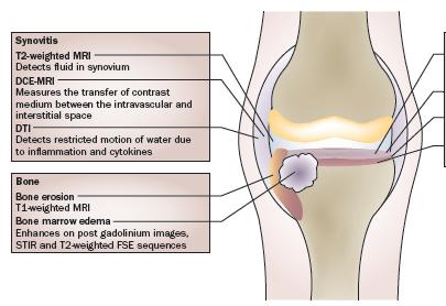
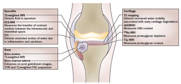
4 MRI investigations of joints Bone erosions Synovitis Bone edema Cartilage?

5 Current MRI investigations of cartilage in Rheumatoid Arthritis

6 Current MRI quantitative investigations of cartilage in Rheumatoid Arthritis N = 15 patients vs 4 controls 5 readers 1 reader twice
7

8 MRI investigations of joints Score related to the JSN what else. More functional / quantitative information?
 T2")
9 Cartilage Matrix Collagen 20% Structural basis Neutral Proteoglycans 5% Negatively charged T1 mapping (Gd) T2 mapping Na +
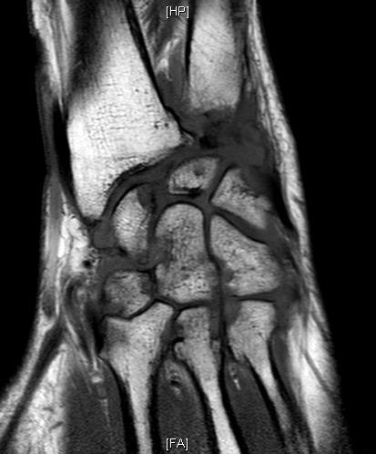
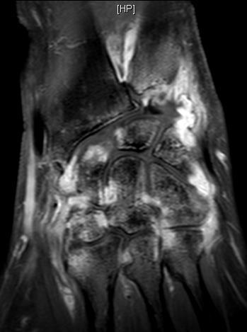
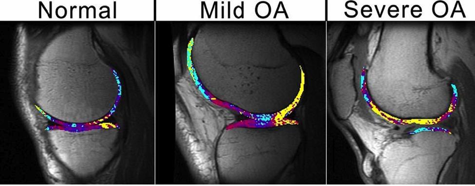
10 Biochemical investigation of cartilage using MRI Normal Partial depletion Total depletion
 2- Map T1 T1 Map")
11 delayed Gadolinium-Enhanced MRI of Cartilage Gd(DTPA) 2- Map T1 T1 Map [GAG] Map
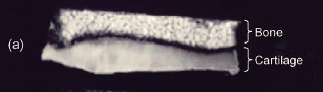
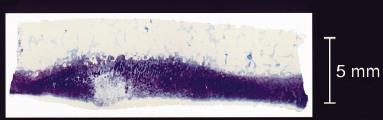

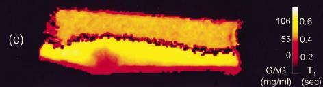
12 delayed Gadolinium-Enhanced MRI of Cartilage Proton density MRI T1-W MRI T1 Map Histological coloration
")

13 delayed Gadolinium-Enhanced MRI of Cartilage From a practical point of view MRI PreC Gd injection Exercise (20m) Rest(60m) MRI Post-C duration : 3 to 15 min according to the pulse sequnce used Résolution: 0.4 mm * 0.4 mm Slice thickness : 3mm

14 Biochemical investigation of cartilage using MRI * *


15 - 42 subjects (45-55 years) with - OA risk factors - right knee pain or not - [WOMAC] pain score >/=5) - (Kellgren/Lawrence score </=1) - 3T MRI of the right knee ± 1.8 ms 34.4 ± 1.8 ms
16 MRI of Cartilage [Na + ] = 320 mm T2 Na+ = 2-10ms [H + ] = mm T2 H+ = 30ms Proteoglycans dgemric 23 Na+ Small SNR (long acquisition time) Dedicated coils HF and UHF (High and Ultra-high fields)

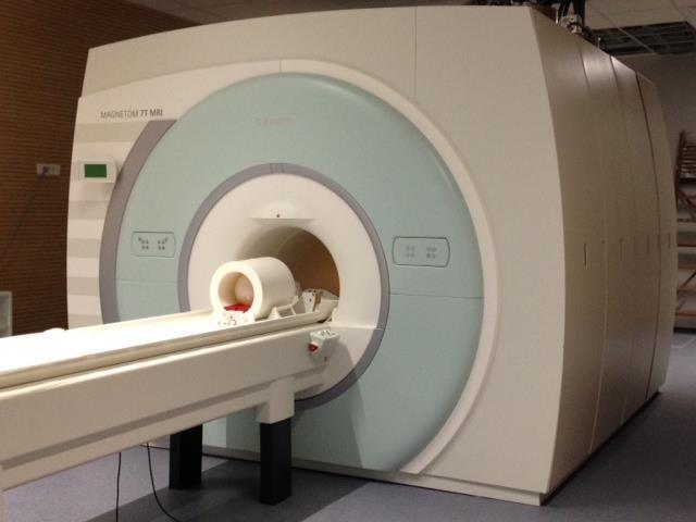
17 MRI «ultra high-field»


18 MRI of the wrist at 7 tesla using an eight-channel array coil combined with parallel imaging: Preliminary results. Chang et al. Volume 31, Issue 3, pages , March min 1min
 [Na + ]")
![= 316 mm [Na + ] = 261 mm. Shapiro, EM.](/docs-images/72/67000995/images/19-2.jpg "Et al. Magn Reson Med.")
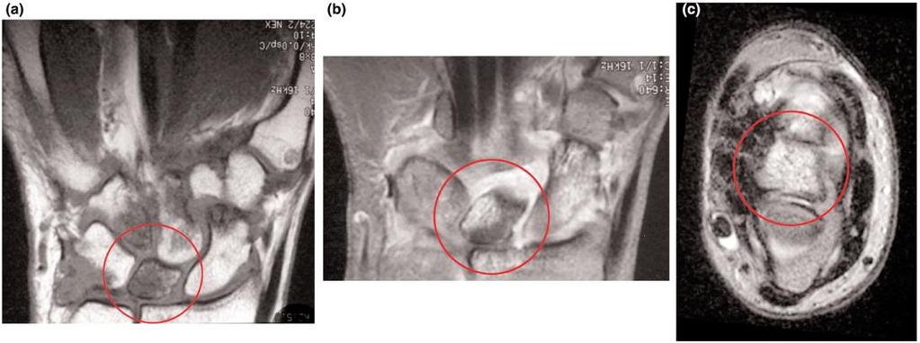
19 23 Na MRI of cartilage (half-degradation experiment) [Na + ] = 316 mm [Na + ] = 261 mm. Shapiro, EM. Et al. Magn Reson Med February; 47(2):

20 Representative sodium maps and proton images. Madelin G et al. Radiology doi: /radiol by Radiological Society of North America


21 Sodium MRI at 3T Resolution Coil availability Cooper et al

22 Coming MRI Quantitative Functional High-field Ultra-high field
 CEMEREM Building 1.")
23 Research Unit 6612 (CRMBM / CEMEREM) CEMEREM Building 1.5 T 3 T 7 T
24 Quantitative MRI of Knee Cartilage in Osteoarthritis Jean-Pierre MATTEI*, David BENDAHAN *Department of Rheumatology, Sainte Marguerite Hospital, Marseille - France Center for Magnetic Resonance in Biology and Medicine, Marseille - France Main Reference : WHAT LESSONS HAVE WE LEARNT FROM RECENT OA IMAGING STUDIES? F. Eckstein, Institute of Anatomy, PMU, Salzburg, Austria
25 The articular cartilage of human adults composed of : cartilage cell and extracellular matrix including water : weight, 65-80% type II collagen weight, 10-20% proteoglycans weight, 5% or less

26 Fibrillar Collagen Network and aggrecan (chondroitine sulfate proteoglycan) interactions with Hyaluronic Acid (HA) CS DS Type IX collagen Decorin core HA G1 Type II collagen fibril Aggrecan G3 CS KS G2 Link Protein Collagen tensile strength Aggrecan compressive stiffness water binding Diagrammatic representation of the macrofibrillar collagen network and aggrecan superaggregate with hyaluronic acid. (Modified with permission from Poole AR: Cartilage in Health and Disease. In Koopman W [ed]. Arthritis and Allied Conditions. A Textbook of Rheumatology. Ed 14. Vol 1. New York, Lippincott Williams & Wilkins , 2001.)
27 Initial Hurts In The Degenerative Osteoarthritis Loss of glycosaminoglycans Increase of hydric contents Degradation of the collagen network
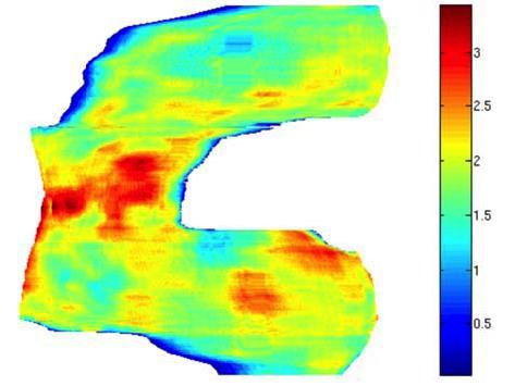
28 Cartilage Magnetic Resonance Imaging Morphological imaging Information on the thickness and the volume Biochemical imaging Information on the biochemical components of cartilage
29 Cartilage Magnetic Resonance Imaging Conventional imaging T1, T2, STIR, SPGR or FLASH Thickness and volume Hydration and biomechanics Biochemical imaging T2 mapping Integrity of the collagen network Hydration T1 Rho mapping Concentration in collagen and proteoglycans DGEMRIC (delayed gadolinium-enhanced MRI of cartilage) Concentration in proteoglycans
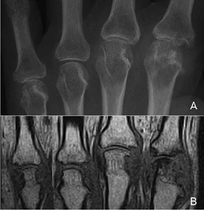
30 Conventional MRI Improving the sequences Cartilage morphology and semiquantitative assessment Optimized Sequences with high spatial resolution T1-weighted SPGR or FLASH (T1-weighted) a relatively long acquisition time Fat-suppressed T2- or intermediate (IM) weighted fast spin-echo (FSE) cartilage surface well depicted steep gradient of signal intensity between the cartilage and the synovial fluid Double echo steady-state imaging (DESS) (T2-weighted ) : SPGR : spoiled gradient echo FLASH : fast low-angle shot
.")
31 Conventional MRI (a) Sagittal fat saturated SPGR image of the knee in a patient with early OA and a cartilage fissure (arrow). (b) Sagittal fat saturated intermediate (IM) weighted image of the same patient

32 Conventional Imaging: Increasing The Magnetic Field Fat suppressed/water excitation gradient echo sequences a et b : 1.5 Tesla c: 1 Tesla d : 3 Tesla
33 Conventional MRI Semi Qantitative Scoring Scores to estimate the severity of cartilage involvement scaled from 0 to 3 or 0 to 4 based on subjective evaluations cartilage lesions of <50% depth, >50% depth and full thickness cartilage lesions MRI scoring system (WORMS whole- organ MRI scoring), cartilage signal and morphology, subchondral bone marrow abnormalities, subarticular cysts, subarticular bone attrition, marginal osteophytes, medial and lateral meniscal integrity, anterior and posterior cruciate ligament integrity, medial and lateral collateral ligament integrity, synovitis/effusion, intraarticular loose bodies and periarticular cysts/bursitis Knee osteoarthritis scoring system (KOSS)
34 Conventional MRI Cartilage Segmentation Methods of semi automatic segmentation Bases: -Region which gets fat -Demarcation of borders -Modelling of the form
35 Segmentation

36 Usual imaging Segmentation
37 Segmentation épaisseur

38 Modeling of the form
39 Performance Segmentation by MRI better than by X-rays: - higher sensitivity and specificity -higher accuracy and precision

40 Biochemical Quantitative Imaging Not only see images, but also quantify the biochemical components of the cartilage
41 T2 mapping T2 : physical quantitative parameter measured in each voxel Construction of a map of the cartilage which reflects the hydration and the integrity of the collagen network Cartilage: weakly hydrated tissue: = > short T2 (30-70 ms) Degenerative osteoarthritis: destruction of the collagen and increase of the hydration = > increase of T2

42 T2 MAP S= S 0 e -TE/T2

43 T2 MaP Takashi Nishii, MD, PhD, Osaka University

44 T2 MAP Takashi Nishii, MD, PhD, Osaka University

45 T2 MAP vs DESS vs Histology
46 T2 MAP
47 dgemeric Imaging Proteoglycans Especially glycosaminoglycans Responsible for the elasticity and for the impact strength of the cartilage Molecules which are negatively loaded and which do not fix the gadolinium Gd-DTPA2-charged negatively Injected by IV (double dose) Binding on the regions where the concentration of glycosaminoglycans falls

48 dgemeric Imaging 90 minutes waiting after the injection of gadolinium

49 dgemric Imaging
50 T1 Rho Imaging T1 slightly different from usual T1 Depends on the concentration in proteoglycans and in collagen When proteoglycans are reduced : T1 Rho increases When the collagen weft gets damaged :T1 Rho increases

51 T1 Rho Imaging

52 Exploring cartilage with MRI Morphology Proteoglycans Proteoglycans and collagen Hydration and collagen
53 Other Techniques Proton imaging : Saturation transfert imaging Diffusion imaging Sodium imaging
54 Quantitative Imaging Advantages Many more information on The volume and the thickness The structure of collagen The contents in proteoglycans The degree of pathological hydration Limits Duration of the examination Dose of Gadolinium Costs
RECENT ADVANCES IN CLINICAL MR OF ARTICULAR CARTILAGE
 In Practice RECENT ADVANCES IN CLINICAL MR OF ARTICULAR CARTILAGE By Atsuya Watanabe, MD, PhD, Director, Advanced Diagnostic Imaging Center and Associate Professor, Department of Orthopedic Surgery, Teikyo
Why Talk About Technique? MRI of the Knee:
 Why Talk About Technique? MRI of the Knee: Part 1 - Imaging Techniques Mark Anderson, M.D. University of Virginia Health Sciences Center Charlottesville, Virginia Always had an interest teach our fellows
Imaging of Articular Cartilage
 Clinical Imaging of Articular Cartilage Imaging of Articular Cartilage Prof. Dr. K. Verstraete Ghent University Introduction : Articular Cartilage Histology and biochemical composition Review of Imaging
This presentation is the intellectual property of the author. Contact them for permission to reprint and/or distribute.
 MRI of the Knee Jennifer Swart, M.D. Musculoskeletal Radiology South Texas Radiology Group Outline Coils, Patient Positioning Acquisition Parameters, Planes and Pulse Sequences Knee Arthrography Normal
This presentation is the intellectual property of the author. Contact them at for permission to reprint and/or distribute.
 MRI of the Knee Jennifer Swart, M.D. Musculoskeletal Radiology South Texas Radiology Group Financial Disclosure Dr. Jennifer Swart has no relevant financial relationships with commercial interests to disclose.
醫用磁振學 MRM 肌肉骨骼磁振造影簡介 肌肉骨骼磁振造影. 本週課程內容 General Technical Considerations 肌肉骨骼磁振造影簡介 盧家鋒助理教授國立陽明大學生物醫學影像暨放射科學系
 本週課程內容 http://www.ym.edu.tw/~cflu 肌肉骨骼磁振造影簡介 醫用磁振學 MRM 肌肉骨骼磁振造影 盧家鋒助理教授國立陽明大學生物醫學影像暨放射科學系 alvin4016@ym.edu.tw MRI of the musculoskeletal system (5th/6th edition) Editor: Thomas H. Berquist MD 2 General
How Much Tesla Is Too Much?
 How Much Tesla Is Too Much? Johnny U. V. Monu, MB, BS; MSc Professor of Radiology and Orthopedics University of Rochester School of Medicine Rochester, New York Historical Timeline Clinical Imaging 1970
MRI Assessments of Cartilage
 SNR IMPACTS THE ACCURACY AND PRECISION OF KNEE ARTICULAR CARTILAGE T2 RELAXATION TIME MEASUREMENTS B.J. Dardzinski 1, E. Schneider 2 1 Merck Sharp & Dohme Corp., West Point, PA USA 2 Imaging Institute,
In vivo diffusion tensor imaging (DTI) of articular cartilage as a biomarker for osteoarthritis
 of articular cartilage as a biomarker for osteoarthritis") In vivo diffusion tensor imaging (DTI) of articular cartilage as a biomarker for osteoarthritis Jose G. Raya 1, Annie Horng 2, Olaf Dietrich 2, Svetlana Krasnokutsky 3, Luis S. Beltran 1, Maximilian F.
In vivo diffusion tensor imaging (DTI) of articular cartilage as a biomarker for osteoarthritis Jose G. Raya 1, Annie Horng 2, Olaf Dietrich 2, Svetlana Krasnokutsky 3, Luis S. Beltran 1, Maximilian F.
dgemric Effectively Predicts Cartilage Damage Associated with Femoroacetabular Impingement
 Riccardo Lattanzi 1,2 Catherine Petchprapa 2 Daniele Ascani 1 Roy I. Davidovitch 3 Thomas Youm 3 Robert J. Meislin 3 Michael. Recht 2 1 The Bernard and Irene Schwartz Center for Biomedical Imaging, New
Quantitative magnetic resonance imaging of osteoarthritis
 For reprint orders, please contact: reprints@futuremedicine.com PERSPECTIVE Quantitative magnetic resonance imaging of osteoarthritis Felix Eckstein Institute of Anatomy & Musculoskeletal Research, Paracelsus
E. ÇAĞLAR, G. ŞAH N 1, T. OĞUR, E. AKTAŞ. Introduction. Abstract. OBJECTIVE: To identify changes in
 European Review for Medical and Pharmacological Sciences Quantitative evaluation of hyaline articular cartilage T2 maps of knee and determine the relationship of cartilage T2 values with age, gender, articular
Analysis of the Osteoarthritis Initiative Incidence Cohort: Patellar Cartilage T2 and Focal Knee Pathology
 TECHNISCHE UNIVERSITÄT MÜNCHEN Klinikum rechts der Isar Institut für Röntgendiagnostik (Direktor: Univ.-Prof. Dr. E. J. Rummeny) Analysis of the Osteoarthritis Initiative Incidence Cohort: Patellar Cartilage
Osteoarthritis. RA Hughes
 Osteoarthritis RA Hughes Osteoarthritis (OA) OA is the most common form of arthritis and the most common joint disease Most of the people who have OA are older than age 45, and women are more commonly
MR imaging of the knee in marathon runners before and after competition
 Skeletal Radiol (2001) 30:72 76 International Skeletal Society 2001 ARTICLE W. Krampla R. Mayrhofer J. Malcher K.H. Kristen M. Urban W. Hruby MR imaging of the knee in marathon runners before and after
Echelon Oval provides a robust suite of leading musculoskeletal imaging capabilities for detailed assessment of all anatomy for your most challenging
 Echelon Oval provides a robust suite of leading musculoskeletal imaging capabilities for detailed assessment of all anatomy for your most challenging cases. Hitachi Medical Systems America, Inc. 1959 Summit
Cartilage Repair Options
 Imaging of Cartilage Repair Carl S. Winalski, MD Imaging Institute Department of Biomedical Engineering Cleveland Clinic Cartilage Repair Options Direct repair Marrow stimulation Autologous transplantation
Richard Magin, Chair and Advisor Mrignayani Kotecha Dieter Klatt
 Proteoglycans Quantification in Tissue Engineered Cartilage using Sodium MRI at 11.7 T BY DAN YU B.S., Tianjin University, 2011 THESIS Submitted as partial fulfillment of the requirements for the degree
FieldStrength. Achieva 3.0T enables cutting-edge applications, best-in-class MSK images
 FieldStrength Publication for the Philips MRI Community Issue 33 December 2007 Achieva 3.0T enables cutting-edge applications, best-in-class MSK images Palo Alto Medical Clinic Sports Medicine Center employs
Tissue-engineered medical products Evaluation of anisotropic structure of articular cartilage using DT (Diffusion Tensor)-MR Imaging
-MR Imaging") Provläsningsexemplar / Preview TECHNICAL REPORT ISO/TR 16379 First edition 2014-03-01 Tissue-engineered medical products Evaluation of anisotropic structure of articular cartilage using DT (Diffusion Tensor)-MR
Provläsningsexemplar / Preview TECHNICAL REPORT ISO/TR 16379 First edition 2014-03-01 Tissue-engineered medical products Evaluation of anisotropic structure of articular cartilage using DT (Diffusion Tensor)-MR
SSSR. 1. Nov Ankle. Postoperative Imaging of Cartilage Repair. and Lateral Ligament Reconstruction
 Ankle Postoperative Imaging of Cartilage Repair and Lateral Ligament Reconstruction Andrea B. Rosskopf, MD University Hospital Balgrist Imaging of Cartilage Repair Why? To assess the technical success
Thanks. As something to help remember. Thanks 07/02/14. Arthroscopy TKR V Uni. Arthroscopy TKR V Uni. Role of arthroscopy to choose operation
 07/02/14 Thanks Role of arthroscopy to choose operation TKR Versus Uni Francois and Philippe Faculty Audience Audiovisual Pierre and Vincent Myles Coolican Val d Isere 2014 Thanks Francois and Philippe
JMSCR Vol 05 Issue 01 Page January
 www.jmscr.igmpublication.org Impact Factor 5.244 Index Copernicus Value: 83.27 ISSN (e)-2347-176x ISSN (p) 2455-0450 DOI: https://dx.doi.org/10.18535/jmscr/v5i1.28 Diagnostic Accuracy of Magnetic Resonance
Case Report: Knee MR Imaging of Haemarthrosis in a Case of Haemophilia A
 Clinical > Pediatric Imaging Case Report: Knee MR Imaging of Haemarthrosis in a Case of Haemophilia A M. A. Weber, J. K. Kloth University Hospital Heidelberg, Department of Diagnostic and Interventional
When (How) MRI Became the Gold Standard Hollis G. Potter, MD
 MRI Became the Gold Standard Hollis G. Potter, MD") When (How) MRI Became the Gold Standard Hollis G. Potter, MD potterh@hss.edu Target audience: Radiologists and imaging scientists interested in assessing MRI of cartilage Outcome/Objectives: 1. To become
When (How) MRI Became the Gold Standard Hollis G. Potter, MD potterh@hss.edu Target audience: Radiologists and imaging scientists interested in assessing MRI of cartilage Outcome/Objectives: 1. To become
High-Resolution 3D Cartilage Imaging with IDEAL SPGR at 3 T
 Siepmann et al. Knee MRI with IDEL SPGR Musculoskeletal Imaging Technical Innovation David. Siepmann 1 Jeff McGovern 2 Jean H. rittain 3 Scott. Reeder 1,4 Siepmann D, McGovern J, rittain JH, Reeder S Keywords:
Discovery of a Small Molecule Inhibitor of the Wnt Pathway (SM04690) as a Potential Disease Modifying Treatment for Knee Osteoarthritis
 as a Potential Disease Modifying Treatment for Knee Osteoarthritis") Discovery of a Small Molecule Inhibitor of the Wnt Pathway (SM469) as a Potential Disease Modifying Treatment for Knee Osteoarthritis Vishal Deshmukh, Ph.D., Charlene Barroga, Ph.D., Yong Hu, Ph.D., John
Discovery of a Small Molecule Inhibitor of the Wnt Pathway (SM469) as a Potential Disease Modifying Treatment for Knee Osteoarthritis Vishal Deshmukh, Ph.D., Charlene Barroga, Ph.D., Yong Hu, Ph.D., John
Original Research JOURNAL OF MAGNETIC RESONANCE IMAGING 22: (2005)
") JOURNAL OF MAGNETIC RESONANCE IMAGING 22:788 793 (2005) Original Research STIR vs. T1-Weighted Fat-Suppressed Gadolinium- Enhanced MRI of Bone Marrow Edema of the Knee: Computer-Assisted Quantitative Comparison
JOURNAL OF MAGNETIC RESONANCE IMAGING 22:788 793 (2005) Original Research STIR vs. T1-Weighted Fat-Suppressed Gadolinium- Enhanced MRI of Bone Marrow Edema of the Knee: Computer-Assisted Quantitative Comparison
Why the dog? Analogy of the anatomy
 Why the dog? Analogy of the anatomy Surgically Induced canine OA models: Anterior (cranial) cruciate ligament transection model Pond MJ, Nuki G. Ann Rheum Dis 1973 (and > 100 others) Meniscal disruption
Non-Invasive Characterization of Cartilage Properties Using MR Imaging
 Non-Invasive Characterization of Cartilage Properties Using MR Imaging by Sophia Natalie Ziemian Department of Biomedical Engineering Duke University Date: Approved: Farshid Guilak, Supervisor Lori A.
MRI KNEE WHAT TO SEE. Dr. SHEKHAR SRIVASTAV. Sr.Consultant KNEE & SHOULDER ARTHROSCOPY
 MRI KNEE WHAT TO SEE Dr. SHEKHAR SRIVASTAV Sr.Consultant KNEE & SHOULDER ARTHROSCOPY MRI KNEE - WHAT TO SEE MRI is the most accurate and frequently used diagnostic tool for evaluation of internal derangement
Sensitivity and Specificity in Detection of Labral Tears with 3.0-T MRI of the Shoulder
 Magee and Williams MRI for Detection of Labral Tears Musculoskeletal Imaging Clinical Observations C M E D E N T U R I C L I M G I N G JR 2006; 187:1448 1452 0361 803X/06/1876 1448 merican Roentgen Ray
Viviane Khoury, MD. Assistant Professor Department of Radiology University of Pennsylvania
 U Penn Diagnostic Imaging: On the Cape Chatham, MA July 11-15, 2016 Viviane Khoury, MD Assistant Professor Department of Radiology University of Pennsylvania Hip imaging has changed in recent years: new
Oak foundation for donating the 3T Siemens Verio scanner. Board of directors BBH and Frh Hospitals for supporting the
 Knee pain and inflammation in the infrapatellar fat pad estimated by conventional and dynamic contrast-enhanced magnetic resonance imaging in obese patients with osteoarthritis: a crosssectional study
High Field MR of the Spine
 Department of Radiology University of California San Diego 3T for MR Applications Advantages High Field MR of the Spine Increased signal-to-noise Better fat suppression Increased enhancement with gadolinium
A Method to Monitor Local Changes in MR Signal Intensity in Articular Cartilage: A Potential Marker for Cartilage Degeneration in Osteoarthritis
 A Method to Monitor Local Changes in MR Signal Intensity in Articular Cartilage: A Potential Marker for Cartilage Degeneration in Osteoarthritis Josephine H. Naish 1, Graham Vincent 2, Mike Bowes 2, Manish
4 2 Osteoarthritis 1
 Osteoarthritis 1 Osteoarthritis ( OA) Osteoarthritis is a chronic disease and the most common of all rheumatological disorders. It particularly affects individuals over the age of 65 years. The prevalence
SPINAL MAGNETIC RESONANCE IMAGING INTERPRETATION
 CLINICAL VIGNETTE 2017; 3:2 SPINAL MAGNETIC RESONANCE IMAGING INTERPRETATION Editor-in-Chief: Idowu, Olufemi E. Neurological surgery Division, Department of Surgery, LASUCOM/LASUTH, Ikeja, Lagos, Nigeria.
KNEE ALIGNMENT SYSTEM (KAS) MRI Protocol
 MRI Protocol") KNEE ALIGNMENT SYSTEM (KAS) MRI Protocol Sample referral sticker Referral Sticker Insert here Corin 17 Bridge Street Pymble NSW Australia 2073 P: +61 (0)2 9497 7400 F: +61 (0)2 9497 7498 E: KAS.customerservice@coringroup.com
KNEE ALIGNMENT SYSTEM (KAS) MRI Protocol Sample referral sticker Referral Sticker Insert here Corin 17 Bridge Street Pymble NSW Australia 2073 P: +61 (0)2 9497 7400 F: +61 (0)2 9497 7498 E: KAS.customerservice@coringroup.com
Osteoarthritis. Dr Anthony Feher. With special thanks to Dr. Tim Williams and Dr. Bhatia for allowing me to use some of their slides
 Osteoarthritis Dr Anthony Feher With special thanks to Dr. Tim Williams and Dr. Bhatia for allowing me to use some of their slides No Financial Disclosures Number one chronic disability in the United States
Knee Articular Cartilage in an Asymptomatic Population : Comparison of T1rho and T2 Mapping
 TR_002 Technical Reports Knee Articular Cartilage in an Asymptomatic Population : Comparison of T1rho and T2 Mapping Min A Yoon 1,*, Suk-Joo Hong 1, Chang Ho Kang 2, Baek Hyun Kim 3 1 Korea University
Musculoskeletal Imaging at 3T with Simultaneous Use of Multipurpose Loop Coils
 Clinical Orthopedic Imaging Musculoskeletal Imaging at 3T with Simultaneous Use of Multipurpose Loop Coils Elena Ferrer 1 ; Rafael Coronado Santos 2 1 Radiology Department, Clínica Creu Blanca, Barcelona,
MRI EVALUATION OF KNEE CARTILAGE
 UPDATING ARTICLE MRI EVALUATION OF KNEE CARTILAGE Marcelo Bordalo Rodrigues 1, Gilberto Luís Camanho 2 ABSTRACT Through the ability of magnetic resonance imaging (MRI) to characterize soft tissue noninvasively,
Assessment and multiparametric functional MRI evaluation of Arthritis
 Assessment and multiparametric functional MRI evaluation of Arthritis 1 2 3 4 T. Martin Noguerol, MD 1 ; A. Luna, MD 1 M. Gomez Cabrera 3, MD; J Vilanova 2, MD, PhD; M. Romero Rivera MD 3 ; F Caro Mateo
ELENI ANDIPA General Hospital of Athens G. Gennimatas
 ELENI ANDIPA General Hospital of Athens G. Gennimatas Technological advances over the last years have caused a dramatic improvement in ultrasound quality and resolution An established imaging modality
Meniscal Tears: Role of Axial MRI Alone and in Combination with Other Imaging Planes
 Nefise Cagla Tarhan 1,2 Christine. Chung 1 urea Valeria Rosa Mohana-orges 1 Tudor Hughes 1 Donald Resnick 1 Received September 30, 2003; accepted after revision February 2, 2004. 1 Department of Radiology,
High-resolution measurements of the multilayer ultra-structure of articular cartilage and their translational potential He et al.
 High-resolution measurements of the multilayer ultra-structure of articular cartilage and their translational potential He et al. He et al. Arthritis Research & Therapy He et al. Arthritis Research & Therapy
T2 Values of Femoral Cartilage of the Knee Joint: Comparison between Pre-Contrast and Post-Contrast Images
 Original Article Musculoskeletal Imaging http://dx.doi.org/.3348/kjr.14.15.1.123 pissn 1229-6929 eissn 05-83 Korean J Radiol 14;15(1):123-129 T2 Values of Femoral Cartilage of the Knee Joint: Comparison
MR Advance Techniques. Vascular Imaging. Class II
 MR Advance Techniques Vascular Imaging Class II 1 Vascular Imaging There are several methods that can be used to evaluate the cardiovascular systems with the use of MRI. MRI will aloud to evaluate morphology
Imaging of Osteoarthritis
 Imaging of Osteoarthritis Ali Guermazi, MD, PhD a, *, Daichi Hayashi, MD, PhD a, Felix Eckstein, MD b, David J. Hunter, MBBS, MSc, PhD, FRACP c, Jeff Duryea, PhD d, Frank W. Roemer, MD a KEYWORDS Osteoarthritis
Comparative study of imaging at 3.0 T versus 1.5 T of the knee
 Skeletal Radiol (2009) 38:761 769 DOI 10.1007/s00256-009-0683-0 SCIENTIFIC ARTICLE Comparative study of imaging at 3.0 T versus 1.5 T of the knee Scott Wong & Lynne Steinbach & Jian Zhao & Christoph Stehling
BioMatrix Tuners: CoilShim
 MAGNETOM Vida special issue Head and Neck Imaging Clinical 11 BioMatrix Tuners: CoilShim Miriam R. Keil, Ph.D.; Jörg Rothard; Carmel Hayes, Ph.D. Siemens Healthineers, Erlangen, Germany A cervical spine
Joint Health Optimum Flex Plus, Hi- Victory, Yucca, Herbal Respond
 Joint Health Optimum Flex Plus, Hi- Victory, Yucca, Herbal Respond May Educational Webinar 2018 Dr Tania Cubitt Performance Horse Nutrition COMPARATIVE ANATOMY 2 JOINT A joint is defined as a structure
1Pulse sequences for non CE MRA
 MRI: Principles and Applications, Friday, 8.30 9.20 am Pulse sequences for non CE MRA S. I. Gonçalves, PhD Radiology Department University Hospital Coimbra Autumn Semester, 2011 1 Magnetic resonance angiography
Figuring out the "fronds"-synovial proliferative disorders of the knee.
 Figuring out the "fronds"-synovial proliferative disorders of the knee. Poster No.: C-1209 Congress: ECR 2014 Type: Educational Exhibit Authors: S. Sivasubramanian; Tamil Nadu/IN Keywords: Imaging sequences,
A MEDICAL IMAGE-BASED COMPUTER-AIDED DIAGNOSIS SYSTEM FOR MUSCULOSKELETAL DISEASE AND DISORDER
 A MEDICAL IMAGE-BASED COMPUTER-AIDED DIAGNOSIS SYSTEM FOR MUSCULOSKELETAL DISEASE AND DISORDER CHUAH TONG KUAN SCHOOL OF CHEMICAL AND BIOMEDICAL ENGINEERING 2012 A MEDICAL IMAGE-BASED COMPUTER-AIDED DIAGNOSIS
Meniscus T2 Relaxation Time at Various Stages of Knee Joint Degeneration
 Meniscus T2 Relaxation Time at Various Stages of Knee Joint Degeneration Richard Kijowski, Michael Fazio, Benjamin Beduhn, and Fang Liu Department of Radiology University of Wisconsin School of Medicine
Available online at
 Original Research Article Evaluation of knee joint by MRI in 65 patients Gulamus sibtain asad *, Himanshu Singla, Ankit vasoya, P. J. Jhala 2 2 nd year Resident, 2 Professor Radiology Department, SBKS
Index. Note: Page numbers of article titles are in boldface type.
 Magn Reson Imaging Clin N Am 12 (2004) 185 189 Index Note: Page numbers of article titles are in boldface type. A Acromioclavicular joint, MR imaging findings concerning, 161 Acromion, types of, 77 79
Magnetic Resonance Angiography
 Magnetic Resonance Angiography 1 Magnetic Resonance Angiography exploits flow enhancement of GR sequences saturation of venous flow allows arterial visualization saturation of arterial flow allows venous
Comparative study of high resolusion ultrasonography and magnetic resonance imaging in diagnosing traumatic knee injuries & pathologies
 Original article: Comparative study of high resolusion ultrasonography and magnetic resonance imaging in diagnosing traumatic knee injuries & pathologies Dr. Rakesh Gujjar*, Dr. R. P. Bansal, Dr. Sandeep
Anatomical and Functional MRI of the Pancreas
 Anatomical and Functional MRI of the Pancreas MA Bali, MD, T Metens, PhD Erasme Hospital Free University of Brussels Belgium mbali@ulb.ac.be Introduction The use of MRI to investigate the pancreas has
Magnetic Resonance Imaging of Superficial Cartilage Lesions: Role of Contrast in Lesion Detection
 JOURNAL OF MAGNETIC RESONANCE IMAGING 10:178 182 (1999) Original Research Magnetic Resonance Imaging of Superficial Cartilage Lesions: Role of Contrast in Lesion Detection Timothy J. Mosher, MD* and Steven
JMSCR Vol 05 Issue 07 Page July 2017
 www.jmscr.igmpublication.org Impact Factor 5.84 Index Copernicus Value: 83.27 ISSN (e)-2347-176x ISSN (p) 2455-0450 DOI: https://dx.doi.org/10.18535/jmscr/v5i7.84 Anatomical Differences Between T2 WI FSE
Speed, Comfort and Quality with NeuroDrive
 Speed, Comfort and Quality with NeuroDrive Echelon Oval provides a broad range of capabilities supporting fast, accurate diagnosis of brain conditions and injuries. From anatomical depiction to vascular
Conservative surgical treatments for osteoarthritis: A Finite Element Study
 Conservative surgical treatments for osteoarthritis: A Finite Element Study Diagarajen Carpanen, BEng (Hons), Franziska Reisse, BEng(Hons), Howard Hillstrom, PhD, Kevin Cheah, FRCS, Rob Walker, PhD, Rajshree
Pain in Osteoarthritis
 Pain in Osteoarthritis By Edward L. Treadwell, MD Professor of Medicine- Rheumatology/Immunology Brody School of Medicine at East Carolina University School of Medicine Greenville, NC 27834 E-mail: treadwelle@ecu.edu
Cover Page. The handle holds various files of this Leiden University dissertation.
 Cover Page The handle http://hdl.handle.net/1887/40654 holds various files of this Leiden University dissertation. Author: Stomp, W. Title: MR imaging in early rheumatoid arthritis : techniques and applications
Meniscal Tears with Fragments Displaced: What you need to know.
 Meniscal Tears with Fragments Displaced: What you need to know. Poster No.: C-1339 Congress: ECR 2015 Type: Authors: Keywords: DOI: Educational Exhibit M. V. Ferrufino, A. Stroe, E. Cordoba, A. Dehesa,
D. Doré 1, C. Ding 1,2, J.P. Pelletier 3, J. Martel-Pelletier 3, F. Cicuttini 2, G. Jones 1.
 Responsiveness of qualitative and quantitative MRI measures over 2.7 years D. Doré 1, C. Ding 1,2, J.P. Pelletier 3, J. Martel-Pelletier 3, F. Cicuttini 2, G. Jones 1. 1 Menzies Research Institute Tasmania,
AIMS We will all come across osteo- and rheumatoid arthritis whatever our clinical practice Overview of pathology of osteoarthritis, its assessment an
 Osteoarthritis and Rheumatoid Arthritis Mr. Guy Barham FY1 & FY2 Orthopaedic Curriculum June 2007 AIMS We will all come across osteo- and rheumatoid arthritis whatever our clinical practice Overview of
ASSOCIATIONS BETWEEN THE BIOLOGICAL, STRUCTURAL, AND MECHANICAL COMPONENTS OF CARTILAGE HEALTH FOLLOWING ACUTE LOADING IN HEALTHY PARTICIPANTS
 ASSOCIATIONS BETWEEN THE BIOLOGICAL, STRUCTURAL, AND MECHANICAL COMPONENTS OF CARTILAGE HEALTH FOLLOWING ACUTE LOADING IN HEALTHY PARTICIPANTS Matthew S. Harkey A dissertation submitted to the faculty
Prevalence of Meniscal Radial Tears of the Knee Revealed by MRI After Surgery
 Downloaded from www.ajronline.org by 46.3.207.114 on 12/22/17 from IP address 46.3.207.114. Copyright RRS. For personal use only; all rights reserved Thomas Magee 1 Marc Shapiro David Williams Received
FAI syndrome with or without labral tear.
 Case This 16-year-old female, soccer athlete was treated for pain in the right groin previously. Now has acute onset of pain in the left hip. The pain was in the groin that was worse with activities. Diagnosis
the new accurate analysis and classification system of knee joint osteoarthritis
 the new accurate analysis and classification system of knee joint osteoarthritis i3a Soft- and Hardware Components created and assembled in Austria Due to the rapidly increasing fraction of aging people
Osteoarthritis What is new? Dr Peter Cheung, Rheumatologist, NUHS
 Osteoarthritis What is new? Dr Peter Cheung, Rheumatologist, NUHS Objective Outline some clinical features that are not well appreciated in OA patients Recent advances in knowledge and management of OA
For more information about how to cite these materials visit
 Author(s): Seetha Monrad, M.D., 2009 License: Unless otherwise noted, this material is made available under the terms of the Creative Commons Attribution Noncommercial Share Alike 3.0 License: http://creativecommons.org/licenses/by-nc-sa/3.0/
Magnetic Resonance Imaging of the Knee: An Overview and Update of Conventional and State of the Art Imaging
 CME ARTICLE Magnetic Resonance Imaging of the Knee: An Overview and Update of Conventional and State of the Art Imaging Nicholas C. Nacey, MD, Matthew G. Geeslin, MD, Grady Wilson Miller, PhD, and Jennifer
MRI of LEFT KNEE. There is a fluid collection seen anterior to and inferior to the superiorly displaced patella.
 MRI of LEFT KNEE Protocol: Multiplanar MRI of the left knee joint performed in the sagittal, coronal and transverse planes using T1 weighted spin echo, T2 and proton-density weighted fast spin echo, fatsaturated
MRI of the sacroiliac joints: what to report and its pitfalls
 MRI of the sacroiliac joints: what to report and its pitfalls Poster No.: C-1920 Congress: ECR 2016 Type: Educational Exhibit Authors: J. Goncalves, A. Y. Aihara, C. Longo, H. Guidorizzi, P. Aguiar, 1
Quantification of Biomechanical Imaging Biomarkers. Sudhakar Tummala
 Quantification of Biomechanical Imaging Biomarkers Sudhakar Tummala February 9, 2012 Contents 1 Introduction 1 1.1 Imaging Osteoarthritis and Biomarkers............ 2 1.1.1 MRI principle.......................
DTI fiber tracking at 3T MR using b-1000 value in the depiction of periprostatic nerve before and after nervesparing prostatectomy
 DTI fiber tracking at 3T MR using b-1000 value in the depiction of periprostatic nerve before and after nervesparing prostatectomy Poster No.: C-2328 Congress: ECR 2012 Type: Scientific Paper Authors:
Degenerative arthritis of Hip Bone Bangalore. Prof Sharath Rao Head, Dept. of Orthopaedics KMC Manipal
 Degenerative arthritis of Hip Prof Sharath Rao Head, Dept. of Orthopaedics KMC Manipal Hip joint Classical Synovial joint Biomechanics of hip Force coincides with trabecular pattern Hip joint Acetabulum
An Overview of RAMRIQ: An Automated MRI Rheumatoid Arthritis Quantitative Assessment System
 Precision. Insight. Innovation. White Paper: An Overview of RAMRIQ: An Automated MRI Rheumatoid Arthritis Quantitative Assessment System At left: Volume of synovitis in the hand and wrist. Areas of enhancement
Functional Chest MRI in Children Hyun Woo Goo
 Functional Chest MRI in Children Hyun Woo Goo Department of Radiology and Research Institute of Radiology Asan Medical Center, University of Ulsan College of Medicine, Seoul, Korea No ionizing radiation
Disclosures: C.B. Raub: None. B.C. Hansen: None. T. Yamaguchi: None. M.M. Temple-Wong: None. K. Masuda: None. R.L. Sah: None.
 En Face Microscopy of Rabbit Knee Articular Cartilage Following Anterior Cruciate Ligament Transection Reveals Early Matrix Damage, Chondrocyte Loss and Cloning Christopher B. Raub, PhD, Bradley C. Hansen,
Cover Page. The handle holds various files of this Leiden University dissertation.
 Cover Page The handle http://hdl.handle.net/1887/35124 holds various files of this Leiden University dissertation. Author: Wokke, Beatrijs Henriette Aleid Title: Muscle MRI in Duchenne and Becker muscular
ORIGINAL ARTICLE. ROLE OF MRI IN EVALUATION OF TRAUMATIC KNEE INJURIES Saurabh Chaudhuri, Priscilla Joshi, Mohit Goel
 ROLE OF MRI IN EVALUATION OF TRAUMATIC KNEE INJURIES Saurabh Chaudhuri, Priscilla Joshi, Mohit Goel 1. Associate Professor, Department of Radiodiagnosis & imaging, Bharati Vidyapeeth Medical College and
Quantitative Comparison of 2D and 3D MRI Techniques for the Evaluation of Chondromalacia Patellae in 3.0T MR Imaging of the Knee
 doi: 10.5505/actamedica.2016.81905 Acta Medica Anatolia Volume 4 Issue 3 2016 Quantitative Comparison of 2D and 3D MRI Techniques for the Evaluation of Chondromalacia Patellae in 3.0T MR Imaging of the
A Comparative Study of Ultrasonographic Findings with Clinical and Radiological Findings of Painful Osteoarthritis of the Knee Joint
 Med. J. Cairo Univ., Vol. 84, No. 3, December: 97-, www.medicaljournalofcairouniversity.net A Comparative Study of Ultrasonographic Findings with Clinical and Radiological Findings of Painful Osteoarthritis
Original Research Article
 Original Research Article Intermediate Weighted Fast Spin Echo (IW FSE) MR Imaging of Hyaline Cartilage Defects of the Knee: Comparison with the Fat Suppressed Three Dimensional Gradient Echo (3D SPGR)
Original Research Article
 Original Research Article Intermediate Weighted Fast Spin Echo (IW FSE) MR Imaging of Hyaline Cartilage Defects of the Knee: Comparison with the Fat Suppressed Three Dimensional Gradient Echo (3D SPGR)
Relaxometric Maps: Sequence Development and Clinical Impact. Initial Observations.
 Relaxometric Maps: Sequence Development and Clinical Impact. Initial Observations. Poster No.: C-0998 Congress: ECR 2011 Type: Scientific Exhibit Authors: E. Soscia, G. Palma, C. Sirignano, D. Iodice,
International Cartilage Repair Society
 Osteoarthritis and Cartilage (2009) 17, 12e18 Published by Elsevier Ltd on behalf of Osteoarthritis Research Society International. doi:10.1016/j.joca.2008.05.016 T1r relaxation time of the meniscus and
Contrast-enhanced Breast MRI RSSA 2013
 Contrast-enhanced Breast MRI RSSA 2013 Prof. dr. Maurice van den Bosch University Medical Center Utrecht, the Netherlands Index 1) Breast cancer 2) Why MRI of the breast 3) Technique 4) Interpretation
COMPARISON OF THREE MAGNETIC RESONANCE IMAGING SEQUENCES FOR MEASURING CARTILAGE THICKNESS IN THE CANINE STIFLE. Lawrence Alexander Brown
 COMPARISON OF THREE MAGNETIC RESONANCE IMAGING SEQUENCES FOR MEASURING CARTILAGE THICKNESS IN THE CANINE STIFLE by Lawrence Alexander Brown A thesis submitted to the Graduate Faculty of Auburn University
Stability of Post Traumatic Osteochondritis Dissecans of the Knee: MR Imaging Findings
 Chin J Radiol 2005; 30: 199-204 199 Stability of Post Traumatic Osteochondritis Dissecans of the Knee: MR Imaging Findings YU-CHUNG HUNG 1 JON-KWAY HUANG 1,2 Department of Radiology 1, Mackay Memorial
6/23/2009. Inversion Recovery (IR) Techniques and Applications. Variations of IR Technique. STIR, FLAIR, TI and TI Null. Applications of IR
 Techniques and Applications. Variations of IR Technique. STIR, FLAIR, TI and TI Null. Applications of IR") The Anatomy of Basic R Pulse Sequences Inversion Recovery () Techniques and Applications Chen Lin, PhD Indiana University School of edicine & Clarian Health Partners agnetization Preparation Section Chemical
The Anatomy of Basic R Pulse Sequences Inversion Recovery () Techniques and Applications Chen Lin, PhD Indiana University School of edicine & Clarian Health Partners agnetization Preparation Section Chemical
Advanced musculoskeletal MRI at ultra-high field (7 T)
") PERSPECTIVE Advanced musculoskeletal MRI at ultra-high field (7 T) MRI at ultra-high field (7 T) offers high signal-to-noise ratio, which can be used beneficially in several different musculoskeletal applications.
PERSPECTIVE Advanced musculoskeletal MRI at ultra-high field (7 T) MRI at ultra-high field (7 T) offers high signal-to-noise ratio, which can be used beneficially in several different musculoskeletal applications.