Enhanced COPD phenotyping to improve treatment strategies
|
|
|
- Clinton Doyle
- 5 years ago
- Views:
Transcription

1 Enhanced COPD phenotyping to improve treatment strategies Proefschrift voorgelegd voor het behalen van de graad van Doctor in de Medische Wetenschappen aan de Universiteit Antwerpen te verdedigen door Bita Hajian Promotor Prof. Dr. W. De Backer Faculteit Geneeskunde en Gezondheidswetenschappen Antwerpen, 2017 Bita Hajian 1
2 Leden van de interne doctoraatscommissie universiteit Antwerpen: Prof. Dr. J Verbraecken Voorzitter Prof. Dr. P Jorens Leden van de externe doctoraatscommissie: Prof. Dr. E.F.M. Wouters Prof. Dr. P. Deron Bita Hajian Enhanced COPD phenotyping to improve treatment strategies/ B. Hajian Universiteit Antwerpen, CDE Universiteitsplein 1, 2610 Wilrijk, Belgium Thesis Universiteit Antwerpen 2017 with summary in Dutch Lay-out & cover art : Sven Wittoeck (sven@nonius.be) 2 Enhanced COPD phenotyping to improve treatment strategies
3 Table of Contents 1. Introduction 1.1.Unmet need for innovation 2. A new approach for phenotyping: Functional imaging as a biomarker 2.1.Measurement methodology of FRI 2.2.Application of FRI in clinical trials 3. A new approach for evaluation of COPD exacerbation and treatment 3.1.A prospective multicenter trial to improve COPD exacerbations phenotyping 3.2.Changes in ventilation-perfusion during and after an COPD exacerbation: An assessment using fluid dynamic modelling 4. Systemic versus inhaled treatment 4.1.Inhaled treatments as the preferred administration route in patients with airway diseases 4.2.Deposition of inhaled compounds 4.3.Systemic treatment in COPD: Functional Respiratory Imaging (FRI) to assess the interaction between inhaled and systemic drugs Roflumilast and ICS/LABA/LAMA in COPD 5. How well do we care for patients with end stage complications? 5.1.Pathophysiological mechanism of long term non-invasive ventilation in stable hypercapnic patients with COPD using functional respiratory imaging 5.2.Pulmonary Vascular effects of pulsed inhaled Nitric oxide in COPD patients with pulmonary hypertension 6. Broncholab: A platform for the clinician who is looking for answers. 6.1.Broncholab case: COPD patient 6.2.Broncholab case: Asthma patient 7. Conclusion: COPD old disease with fresh approach : the path to personalized medicine List of Abbrevations Curriculum Vitae Dankwoord Bita Hajian 3
4 4 Enhanced COPD phenotyping to improve treatment strategies 1


5 Introduction Innovation is not about market timing. It IS about Creation something that fulfils an something that fulfils an Unmet need. 1 Bita Hajian 5
6 6 Enhanced COPD phenotyping to improve treatment strategies
7 1.1. Unmet need for innovation Chronic obstructive pulmonary disease (COPD) is a preventable and treatable disease characterized by progressive airflow limitation and represents one of the most prevalent human health disorders in the world 1. Although mortality associated with cardiovascular disease has been significantly reduced during the last 2 decades, the number of deaths associated with COPD has almost doubled, and COPD is now the fourth leading cause of death globally 2. More than 15 million people have the disease in the United States 3 and more than 210 million globally. Despite significant public health efforts aimed to better understand and prevent the burden of this disease, the World Health Organization (WHO) has predicted that COPD will become the third most common cause of death in the world by Moreover, prevalence estimates suggest that up to a quarter of adults 40 years or older have evidence of airflow obstruction 5. Because of the increase in prevalence, many efforts have been made to measure the epidemiology of COPD at national and international levels. The pathogenesis of COPD 6 is generally thought to involve an abnormal inflammatory response in the lungs to the inhalation of toxic particles and gases, derived from tobacco smoke, air pollution, or occupational exposures. All smokers develop lung inflammation, but this is enhanced and fails to resolve after smoking cessation in those who develop COPD 7.This suggests that, in smokers who develop COPD, there is abnormal regulation of the inflammatory response in the lungs. The susceptibility factors are still poorly understood and likely involve genetic and epigenetic factors, infections, altered immune regulation, or impaired resolution of inflammation and abnormal repair mechanisms 8.However, the relationship between the inflammatory responses in the lungs and the accelerated decline in FEV 1, which characterizes this condition, is far from clear. Moreover, it is now well recognized that COPD is a heterogeneous 5 condition with pathologic changes in the large and small airways (chronic bronchitis and bronchiolitis) and lung parenchyma (emphysema) that vary greatly in their expression among patients. Thus, the mechanisms resulting in these pathogenic changes are also likely to be different. Bita Hajian 7
8 Definition Several definitions of COPD exist, the latest criteria according to GOLD 2017: A clinical diagnosis of COPD should be considered in any patient who has dyspnea, chronic cough or sputum production, and a history of exposure to risk factors for the disease. Spirometry is required to make the diagnosis in this clinical context; the presence of a postbronchodilator FEV 1 /FVC <0.70 confirms the presence of persistent airflow limitation and thus of COPD. GOLD has developed a multidimensional system for the assessment and management of COPD patients that combines (1) the symptoms perceived by the patient, (2) the severity of the airflow limitation, and (3) the previous history of exacerbations 2. In this proposed assessment, COPD patients are classified into four categories or groups (A, B, C, and D) ( fig 1.1) that, together with the assessment of potential comorbidities, can assist clinicians in guiding therapy. Figure Enhanced COPD phenotyping to improve treatment strategies
9 Pathologic Changes in COPD Chronic Bronchitis Chronic bronchitis is defined in clinical terms as the presence of cough and sputum production for most days over 3 months for 2 consecutive years. This clinical definition does not include the presence of airflow limitation. It is thought to result from an innate immune response to inhaled toxic particles and gases, particularly in tobacco smoke. Inflammation is present in the epithelium of the central airways and in the mucus-producing glands in chronic bronchitis 9. This airway inflammation is associated with increased mucus production, reduced mucociliary clearance, and increased permeability of the airspace epithelial barrier. The contribution that mucus hypersecretion makes to the airflow limitation in COPD is still uncertain. In the early stages of COPD, its contribution is small because mucus production in smokers with normal lung function does not appear to predict later development of COPD 10. However, in the later stages of the disease, chronic mucus hypersecretion may accelerate the loss of FEV 1 due to an increased risk of exacerbations 11. Chronic mucus hypersecretion may result from an inflammatory response in the submucosal glands. Inflammatory cells release serine proteases that are potent secretagogues for mucus. Emphysema Emphysema is defined as enlargement of the airspaces distal to the terminal bronchioles, due to destruction of the alveolar walls 12.Distal airspace enlargement with alveolar destruction reduces maximal expiratory airflow by decreasing the lung elastic recoil. The centrilobular or centriacinar form of emphysema results from dilatation or destruction of the respiratory bronchioles, is the type most closely associated with tobacco smoking, and is thought to be more associated with severe small-airway obstruction 13. The panlobular or panacinar form of emphysema, which is associated with α 1 -antitrypsin (α 1 -AT) deficiency, results in a more even dilatation and destruction of the entire acinus. Although one or the other of these types may predominate, there is great heterogeneity. The distribution of these types of emphysema is different with an upper lobe predominance common in centrilobular emphysema and lower lobe predominance in panlobular emphysema. The reason for this is not clear and whether different pathogenic mechanisms are involved is also unknown. Bita Hajian 9
10 There is a relationship between the degree of emphysema and pack-years of smoking, but the relationship is not strong. Around 40% of smokers develop substantial lung destruction from emphysema, and emphysema can be found in some individuals who have normal lung function. Pulmonary Circulation Sustained pulmonary hypertension develops late in the course of COPD 14, although pathologic changes in the pulmonary vasculature with intimal hyperplasia and muscularization of small pulmonary arteries can develop in mild COPD in heavy smokers with normal lung function (figure 1.2). Factors that contribute to pulmonary hypertension include the following: Figure 1.2. Pulmonary arterial constriction as a result of hypoxia Endothelial dysfunction 15 Remodeling (smooth muscle hypertrophy and hyperplasia) of the pulmonary arteries Destruction of the pulmonary capillary bed The development of structural changes in the pulmonary arterioles leads to persistent pulmonary hypertension and right ventricular hypertrophy/enlargement and dysfunction16, 17 (cor pulmonale). 10 Enhanced COPD phenotyping to improve treatment strategies

11 Inflammation, Airway Remodelling, and Airflow Limitation The peripheral airways (bronchioles < 2 mm in diameter) are the major site of increased resistance to airflow in COPD 18. The main pathologic lesions in the peripheral airways include increased number of inflammatory cells 19, 20 and structural changes, such as epithelial goblet cell metaplasia, airway wall fibrosis, and smooth muscle hypertrophy. The increase in the thickness of the airway wall, inflammation 21, 22, fibrosis, and smooth muscle hypertrophy will encroach on the lumen and reduce airway diameter; increased wall thickness may also uncouple the airways and the surrounding lung parenchyma, thereby reducing the elastic force that opposes bronchiolar smooth muscle contraction and promoting airway closure. Airway wall inflammation can also contribute to the destruction of alveolar-bronchiolar attachments 23, producing deformation and narrowing of the airway lumen. This is supported by the observation that, in smokers, the destruction of alveolar attachments correlates with the degree of inflammation 24 in peripheral airways. Clinical Characterization of COPD The clinical presentation of COPD is highly heterogeneous 5. The most common respiratory symptoms (breathlessness, cough, and expectoration), pathologic changes (chronic bronchitis, emphysema), and lung function abnormalities (mostly airflow limitation) vary greatly between patients and are poorly related to disease status and severity. Unmet need: enhanced COPD phenotyping Given the heterogeneous clinical presentation of COPD there is considerable interest in identifying groups of patients with similar prognosis or therapeutic requirements. Strictly speaking, a phenotype is defined by the observable properties (or phenotypic traits ) of an organism, as determined by its genotype and modulated by its environment. In order to provide some clinical perspective, a recent consensus definition has proposed the concept of a clinical phenotype, which is defined as: a single or combination of disease attributes that describe differences between individuals with COPD as they relate to clinically meaningful outcomes (symptoms, exacerbations, response to therapy, rate of disease progression, or death). 25 Bita Hajian 111
12 The identification and validation of novel clinical phenotypes can advance the clinical management of COPD by identifying novel therapeutic targets and determining the efficacy of a pharmacologic treatment/procedure in subgroups with specific attributes. COPD is a complex disease that includes several disease variants. A disease phenotype describes clinically observable characteristics of a disease without direct relationship to an underlying pathophysiology. Endotypes, however, describe subtypes of a disease defined by an intrinsically distinct pathogenetic mechanism. Phenotype is defined by symptoms, radiology, physiology and biomarkers. In COPD, phenotypes describe clinical and morphologic characteristics as well as unique responses to treatment. Phenotypes are clinically relevant in terms of presentation, triggers, and treatment response but do not necessarily relate to or give insights into the underlying pathological mechanism. COPD endotypes, however, describe disease subtypes based on cellular and molecular mechanisms, including the reactivity of structural cells. Understanding these events would allow us to understand and classify COPD endotypes. Endotyping COPD based on disease mechanisms could eventually lead to an individualized management. Disregarding endotypes might lead to two major setbacks: unsuccessful clinical trials because of a bulk patient selection and unequivocal results because of insufficient preselection of patients based on their phenotype and endotype in large cohorts. Therefore, endotype-specific classifications of any participant in clinical studies might be of considerable advantage. The severity of airflow limitation (i.e., FEV1) has been traditionally used as the key variable to assess and guide therapy in patients with chronic obstructive pulmonary disease (COPD) 1. It is now clear that FEV1 is poorly related to other clinically relevant characteristics of the disease and cannot describe its complexity 3, 26, 27. As a result, over the past few years there has been a great deal of interest in characterizing COPD more precisely and to identify homogeneous groups of patients who respond to specific therapeutic interventions (i.e., phenotypes). In the case of COPD, the disease is defined by the presence of persistent airflow limitation that is usually progressive and associated with an enhanced chronic inflammatory response in the airways and the lung to noxious particles or gases. According to this definition, a number of patients commonly encountered in clinical practice cannot be diagnosed with COPD, including smokers with symptoms (chronic bronchitis) and normal spirometry, smokers with 12 Enhanced COPD phenotyping to improve treatment strategies
13 emphysema seen on computed tomography (CT) of the thorax but with normal spirometry, smokers with CT evidence of emphysema and pulmonary fibrosis (a biological conundrum because this situation requires simultaneous tissue loss [emphysema] and tissue gain [fibrosis]), and never smokers with non fully reversible airflow limitation (chronic asthma?). The identification and validation of clinically relevant phenotypes is the way to the future of COPD. We need to identify and validate a number of clinical, radiological, or biological biomarkers 27-29, to respond to the unmet need in the field of COPD. The aim of the present work is to identify such phenotypes and endotypes mainly based on imaging and to relate them to clinical outcomes after a number of specific interventions. Functional Respiratory Imaging (FRI) 30 is a combination of high-resolution, low-dose, volumetric CT scans and Computational Fluid Dynamics (CFD). FRI provides regional details associated with lung structure and function to get a deeper insight in patient health than obtained by the traditional imaging methods. This is the process of extracting patient specific physiological data of the respiratory system from medical images. The purpose of FRI is to assess the influence of a certain biological or mechanical process on the respiratory system of a certain subject. In chapter 2 we will describe the way FRI can function as a biomarker and how it can be used to identify the different phenotypes of COPD. Also how FRI can be used to identify responders and non-responders of a treatment. In chapter 3 the pathophysiological mechanism of a COPD exacerbation will be explored by FRI, leading to new insights for future treatment. During a COPD exacerbation there is per definition ventilation perfusion mismatch, ventilation and perfusion will be looked in detail during and also at the recovery phase of an exacerbation. Phosphodiesterase-4 (PDE4) inhibition provides a novel approach to the treatment of COPD. The role of roflumilast, as an anti-inflammatory compound in combination with ICS/LABA/LAMA inhalation will be studied in chapter 4. Bita Hajian 13
14 Also we will review the state of art in the current inhalation devices and how we can use FRI for making the right decisions in selecting them. There is still a long debate about the usefulness of chronic non-invasive ventilation in COPD patients with persistent hypercapnia. In chapter 5 we try to get more insight in the pathophysiological mechanism of long term NIV therapy during respiratory failure. Another severe complication of end stage COPD will be discussed in chapter 5: pulmonary hypertension due to hypoxic vasoconstriction leading to remodeling of the pulmonary vasculature. Is inhaled nitic oxide, a vasodilator, a potential therapy for patients with pulmonary hypertension? You will read this in chapter 5 also. Broncholab, a platform for the clinicians who are looking for answers? You will find out in Chapter 6. A complex idea can be conveyed with just a single still image 14 Enhanced COPD phenotyping to improve treatment strategies
15 References 1. Rabe KF, Hurd S, Anzueto A, et al. Global strategy for the diagnosis, management, and prevention of chronic obstructive pulmonary disease: GOLD executive summary. Am J Respir Crit Care Med. Sep 2007;176(6): Vestbo J, Hurd SS, Agustí AG, et al. Global strategy for the diagnosis, management, and prevention of chronic obstructive pulmonary disease: GOLD executive summary. Am J Respir Crit Care Med. Feb 2013;187(4): Agusti A, Sobradillo P, Celli B. Addressing the complexity of chronic obstructive pulmonary disease: from phenotypes and biomarkers to scale-free networks, systems biology, and P4 medicine. Am J Respir Crit Care Med. May 2011;183(9): Agusti A, MacNee W. The COPD control panel: towards personalised medicine in COPD. Thorax. Jul 2013;68(7): Agusti A, Calverley PM, Celli B, et al. Characterisation of COPD heterogeneity in the ECLIPSE cohort. Respir Res. 2010;11: Yoshida M, Taguchi O, Gabazza EC, et al. Combined inhalation of nitric oxide and oxygen in chronic obstructive pulmonary disease. Am J Respir Crit Care Med. Feb 1997;155(2): Willemse BW, Postma DS, Timens W, ten Hacken NH. The impact of smoking cessation on respiratory symptoms, lung function, airway hyperresponsiveness and inflammation. Eur Respir J. Mar 2004;23(3): Macnee W. Pathogenesis of chronic obstructive pulmonary disease. Clin Chest Med. Sep 2007;28(3): , v. 9. Russell RE, Culpitt SV, DeMatos C, et al. Release and activity of matrix metalloproteinase-9 and tissue inhibitor of metalloproteinase-1 by alveolar macrophages from patients with chronic obstructive pulmonary disease. Am J Respir Cell Mol Biol. May 2002;26(5): Vestbo J, Lange P. Can GOLD Stage 0 provide information of prognostic value in chronic obstructive pulmonary disease? Am J Respir Crit Care Med. Aug 2002;166(3): Vestbo J, Prescott E, Lange P. Association of chronic mucus hypersecretion with FEV1 decline and chronic obstructive pulmonary disease morbidity. Copenhagen City Heart Study Group. Am J Respir Crit Care Med. May 1996;153(5): Snider GL. Chronic obstructive pulmonary disease: a definition and implications of structural determinants of airflow obstruction for epidemiology. Am Rev Respir Dis. Sep 1989;140(3 Pt 2):S Kim WD, Eidelman DH, Izquierdo JL, Ghezzo H, Saetta MP, Cosio MG. Centrilobular and panlobular emphysema in smokers. Two distinct morphologic and functional entities. Am Rev Respir Dis. Dec 1991;144(6): Barberà JA, Blanco I. Pulmonary hypertension in patients with chronic obstructive pulmonary disease: advances in pathophysiology and management. Drugs. Jun 2009;69(9): Peinado VI, Barbera JA, Ramirez J, et al. Endothelial dysfunction in pulmonary arteries of patients with mild COPD. Am J Physiol. Jun 1998;274(6 Pt 1):L Bita Hajian 15
16 16. Kaushal M, Shah PS, Shah AD, Francis SA, Patel NV, Kothari KK. Chronic obstructive pulmonary disease and cardiac comorbidities: A cross-sectional study. Lung India Jul-Aug 2016;33(4): Voelkel NF, Gomez-Arroyo J, Mizuno S. COPD/emphysema: The vascular story. Pulm Circ Jul-Sep 2011;1(3): Yanai M, Sekizawa K, Ohrui T, Sasaki H, Takishima T. Site of airway obstruction in pulmonary disease: direct measurement of intrabronchial pressure. J Appl Physiol (1985). Mar 1992;72(3): Saetta M, Di Stefano A, Maestrelli P, et al. Activated T-lymphocytes and macrophages in bronchial mucosa of subjects with chronic bronchitis. Am Rev Respir Dis. Feb 1993;147(2): Bleecker ER. Airways hyperreactivity, bronchial inflammation and obstructive lung disease. Agents Actions Suppl. 1990;30: Hogg JC. Pathophysiology of airflow limitation in chronic obstructive pulmonary disease. Lancet Aug ;364(9435): Sorkness RL, Bleecker ER, Busse WW, et al. Lung function in adults with stable but severe asthma: air trapping and incomplete reversal of obstruction with bronchodilation. J Appl Physiol (1985). Feb 2008;104(2): Hogg JC. Airway pathology of functional significance in chronic bronchitis and chronic obstructive airway disease. Agents Actions Suppl. 1990;30: Baraldo S, Saetta M, Cosio MG. Pathophysiology of the small airways. Semin Respir Crit Care Med. Oct 2003;24(5): Han MK, Agusti A, Calverley PM, et al. Chronic obstructive pulmonary disease phenotypes: the future of COPD. Am J Respir Crit Care Med. Sep 2010;182(5): Agustí A, Rennard S, Edwards LD, et al. Clinical and prognostic heterogeneity of C and D GOLD groups. Eur Respir J. Jul 2015;46(1): Segreti A, Stirpe E, Rogliani P, Cazzola M. Defining phenotypes in COPD: an aid to personalized healthcare. Mol Diagn Ther. Aug 2014;18(4): Galbán CJ, Han MK, Boes JL, et al. Computed tomography-based biomarker provides unique signature for diagnosis of COPD phenotypes and disease progression. Nat Med. Nov 2012;18(11): Brightling CE. Chronic obstructive pulmonary disease phenotypes, biomarkers, and prognostic indicators. Allergy Asthma Proc. Nov 2016;37(6): De Backer JW, Vos WG, Vinchurkar SC, et al. Validation of computational fluid dynamics in CT-based airway models with SPECT/CT. Radiology. Dec 2010;257(3): Enhanced COPD phenotyping to improve treatment strategies
17 Bita Hajian 17
18 18 Enhanced COPD phenotyping to improve treatment strategies 2
19 A new approach for phenotyping: Functional imaging as a biomarker 2 Bita Hajian 19
20 20 Enhanced COPD phenotyping to improve treatment strategies
21 2.1. Measurement methodology of FRI Based on: Hajian B, De Backer J, Vos W, et al. Functional respiratory imaging for optimizing therapy development and patient care. Expert review respiratory medicine 2016; Feb;10(2): In recent years, imaging of the thorax has evolved substantially from an experimental tool that was used in only a limited number of centers toward becoming a routine test for clinical assessment of the respiratory system 1-3. In particular, the development of computed tomography (CT) has greatly increased the understanding of the pathophysiology of respiratory diseases such as asthma, chronic obstructive pulmonary disease (COPD) and cystic fibrosis (CF) 4-7. The imaging method itself has been increasingly used in clinical trials, although the absolute number of studies remains limited. Without additional post processing, a CT scan is limited to static information about the respiratory system. This in itself is already very valuable because, due to the high resolution of CT, certain pathologies (e.g. fibrosis and bronchiectasis) can be distinguished very well. Today, CT is the method of choice to determine the extent of emphysema in COPD patients by measuring lung density FRI is a proprietary workflow that was developed and validated by FLUIDDA. The FRI workflow is a combination of several software components and is provided by the company as a service. CT scans are taken using a dose reduction protocol (120 kv, mas; noise factor 28; collimation mm; rotation time 0.6 s and pitch factor 1.375), resulting in an effective dose of 1 2 msv per CT scan 11.This means that the radiation dose is three times less than a classical CT, which usually has a dose of msv. Because of the low radiation dose, repeated scans in clinical follow-up or in clinical studies can be performed without increasing the radiation dose above the exposure that is currently used for routine examinations. The scan resolution is 0.5 mm 3, and the slice increment is 0.6 mm. Scans are taken at two different levels: functional residual capacity (FRC) and total lung capacity (TLC) (Figure 2.1). Bita Hajian 21
22 Figure 2.1. Signal from respiratory gating to ensure adequate total lung capacity (TLC) and functional residual capacity (FRC) levels to reduce clinical variability. To ensure the correct lung volume, CT scans are respiratory volume gated during the moment of scanning using a pneumotach flow signal. Subsequently, CT images are segmented using a segmentation program that has been cleared by the US FDA s Center for Devices and Radiological Health under the 510(k) process (Food and Drug Administration, K073468) and has been CE marked in Europe (Conformité Européenne certificate, BE 05/1191. CE.01). This program converts CT images into patientspecific reconstructions of the lung lobes and the airway tree. By segmenting lung lobes (figure 2.2) at the FRC and TLC, the internal airflow distribution (IAD) can be derived from the relative volume change. 22 Enhanced COPD phenotyping to improve treatment strategies

23 Figure 2.2. Segmentation workflow Bita Hajian 23
24 The airway tree is normally evaluated at the TLC level because doing so eliminates the effects of breathing level. At the TLC, the airway structure can be segmented down to the bronchi, with a diameter of approximately 1 2 mm. Beyond this point, the CT resolution is insufficient to distinguish alveolar and intraluminal air. A typical airway model includes 5 10 generations that depend primarily on the disease state of the individual patient. Distal airway volumes (ivaw) can be assessed at individual airways or in different regions (fig 2.3). 24 Enhanced COPD phenotyping to improve treatment strategies

25 Figure 2.3. FRI-based airway volumes (ivaw) Bita Hajian 25
26 The smoothed airway reconstructions are subsequently trimmed at the trachea and the terminal bronchi to obtain a model that is suitable for flow simulation. Next, these segmentations are exported to a meshing software package, where they are divided into discrete tetrahedral elements. Flow properties are obtained throughout the flow domain by means of Computational Fluid Dynamics (CFD) 12. The out- flow to each lobe is adjusted iteratively for each patient to match the internal flow rate distribution obtained from the CT scans Measures of resistance (iraw) in individual airways or different regions corresponding to the volume measurements are obtained from CFD calculations. Resistance is defined as the total pressure drop (figure 2.4) over an airway divided by the flow rate through that airway. In a typical clinical trial, the imaging procedure will be performed at baseline and after the intervention. 26 Enhanced COPD phenotyping to improve treatment strategies

27 Figure 2.4. FRI-based pressure drop over the airways (green is high pressure, blue is low pressure during inhalation to determine regional airway resistance (iraw) Bita Hajian 27
28 Subsequently, the resulting models are overlaid and compared with one another to establish the changes induced by the therapy. Also airway wall volume can be calculated. The airway wall volume can typically be described to the same generation level as the volume description of the airway lumen: this is where the airway diameter is around 1 2 mm. Blood vessel density can be determined through segmentation and three-dimensional reconstruction of the blood vessels (Figure 2.5). The segmentation is based on a Hounsfield unit (HU) threshold between -600 and 600 and is performed on the TLC scan. The blood vessel volume can be considered a surrogate for perfusion Enhanced COPD phenotyping to improve treatment strategies

 Bita")
29 Figure 2.5. FRI-based blood vessel volume (ivbv) Bita Hajian 29
30 FRI is essentially a composite biomarker because it includes data concerning lobar volumes, airway volumes, airway resistance and aerosol deposition 13-15, 17, 18. With increasing experience with the method, it will become clear what the minimal significant clinical difference for the subsets of parameters will be. For drug development, it has been used to understand the mode of action, whereas in clinical settings, information about regional airflow distribution and related lung structure and vascularization is obtainable. 30 Enhanced COPD phenotyping to improve treatment strategies
31 2.2. Application of FRI in clinical trials Virtually all therapies for lung diseases are aimed at changing lung geometry. Bronchodilators and anti-inflammatory compounds are developed to increase the airway lumen. The former does this through smooth muscle relaxation, and the latter does so through inhibition of inflammation. The resulting improvement in lung geometry is a consequence of ameliorated lung function. Patients should breathe better if the therapy is successful. Eventually, breathing better should result in an improved QOL. Today, QOL is assessed using questionnaires or patient-reported outcome parameters (PROs); lung function is usually analyzed via spirometry and body plethysmography. The geometry can be evaluated using imaging modalities such as CT and FRI. When considering the three categories (geometry, lung function and QOL), the inherent number of confounding factors increases from geometry over lung function to QOL (figure 2.6). For instance, the conventional pulmonary function tests 19, such as spirometry, measure not only the characteristics of the tracheobronchial tree (lower airways, where the intervention is designed to act) but also the extra thoracic airway (upper airway) characteristics, the muscle force and the patient s effort. The questionnaires inherently include the effects of comorbidities and depend on the patient s concentration and willingness to cooperate. With an increased level of confounding factors, the variability of the measured outcome parameter increases, leading to a larger required sample size of the clinical trials. The correlations between the different categories, although significant (p < 0.05), are often weak (low R) due to the large variability in the outcome measure, as described in the next section. By providing information closer to the site of action, imaging and FRI should be able to provide higher signal to noise outcome parameters. FRI focuses on the region of interest and the site of action (usually the tracheobronchial region) and thereby eliminates the noise and variability induced by upper airway resistance and compliance, muscle force and patient effort. Thus, by using FRI outcome parameters, the same statistical significance can be achieved with fewer patients. This in turn makes it possible to use clinical trials as a design tool and not as a mere assessment tool. This offers a rationale for using FRI in clinical studies but also in daily praxis 20, especially in difficult cases where regional information might be helpful in choosing the most adequate treatment approach. Bita Hajian 31
32 Figure 2.6. Cascading effect of lung interventions (from changes in geometry to better lung function and quality of life) and the associated confounding factors and required sample size 32 Enhanced COPD phenotyping to improve treatment strategies
33 References 1. Coxson HO, Rogers RM. New concepts in the radiological assessment of COPD. Semin Respir Crit Care Med. Apr 2005;26(2): Coxson HO, Rogers RM. Quantitative computed tomography of chronic obstructive pulmonary disease. Acad Radiol. Nov 2005;12(11): Coxson HO. Quantitative computed tomography assessment of airway wall dimensions: current status and potential applications for phenotyping chronic obstructive pulmonary disease. Proc Am Thorac Soc. Dec 2008;5(9): Coxson HO, Mayo J, Lam S, Santyr G, Parraga G, Sin DD. New and current clinical imaging techniques to study chronic obstructive pulmonary disease. Am J Respir Crit Care Med. Oct 2009;180(7): Burrowes KS, De Backer J, Smallwood R, et al. Multi-scale computational models of the airways to unravel the pathophysiological mechanisms in asthma and chronic obstructive pulmonary disease (AirPROM). Interface Focus. Apr 2013;3(2): Burrowes KS, Doel T, Brightling C. Computational modeling of the obstructive lung diseases asthma and COPD. J Transl Med. Nov 2014;12 Suppl 2:S5. 7. Roberts HR, Wells AU, Milne DG, et al. Airflow obstruction in bronchiectasis: correlation between computed tomography features and pulmonary function tests. Thorax. Mar 2000;55(3): Han MK, Kazerooni EA, Lynch DA, et al. Chronic obstructive pulmonary disease exacerbations in the COPDGene study: associated radiologic phenotypes. Radiology. Oct 2011;261(1): Bodduluri S, Bhatt SP, Hoffman EA, et al. Biomechanical CT metrics are associated with patient outcomes in COPD. Thorax. Jan Ostridge K, Wilkinson TM. Present and future utility of computed tomography scanning in the assessment and management of COPD. Eur Respir J. Jul 2016;48(1): De Backer JW, Vos WG, Vinchurkar SC, et al. Validation of computational fluid dynamics in CT-based airway models with SPECT/CT. Radiology. Dec 2010;257(3): de Rochefort L, Vial L, Fodil R, et al. In vitro validation of computational fluid dynamic simulation in human proximal airways with hyperpolarized 3He magnetic resonance phase-contrast velocimetry. J Appl Physiol (1985). May 2007;102(5): De Backer LA, Vos W, De Backer J, Van Holsbeke C, Vinchurkar S, De Backer W. The acute effect of budesonide/formoterol in COPD: a multi-slice computed tomography and lung function study. Eur Respir J. Aug 2012;40(2): Vos W, De Backer J, Poli G, et al. Novel functional imaging of changes in small airways of patients treated with extrafine beclomethasone/formoterol. Respiration. 2013;86(5): De Backer J, Vos W, Vinchurkar S, et al. The effects of extrafine beclometasone/formoterol (BDP/F) on lung function, dyspnea, hyperinflation, and airway geometry in COPD patients: novel insight using functional respiratory imaging. J Aerosol Med Pulm Drug Deliv. Apr 2015;28(2): De Backer L, Vos W, Dieriks B, et al. The effects of long-term noninvasive ventilation in hypercapnic COPD patients: a randomized controlled pilot study. Int J Chron Obstruct Pulmon Dis. 2011;6: Bita Hajian 333
34 17. Sheikh K, Coxson HO, Parraga G. This is what COPD looks like. Respirology. Feb 2016;21(2): Grenier PA, Beigelman-Aubry C, Fetita CI, Brillet PY. CT imaging of chronic obstructive pulmonary disease: role in phenotyping and interventions. Expert Opin Med Diagn. Nov 2009;3(6): Cazzola M, Calzetta L, Rogliani P, Matera MG. The Challenges of Precision Medicine in COPD. Mol Diagn Ther. Feb Martinez CH, Chen YH, Westgate PM, et al. Relationship between quantitative CT metrics and health status and BODE in chronic obstructive pulmonary disease. Thorax. May 2012;67(5): Enhanced COPD phenotyping to improve treatment strategies
35 Bita Hajian 35
36 36 Enhanced COPD phenotyping to improve treatment strategies 3
37 A new approach for evaluation of COPD exacerbation and treatment 3 Bita Hajian 37
38 3.1. A prospective multicenter trial to improve COPD exacerbations phenotyping Based on: Functional respiratory imaging: Toward precision medicine in Acute Exacerbations of COPD. Van Geffen W, Hajian B et al. ERJ 2017, submitted March 2017 Background: Exacerbations of COPD are a major burden to patients and economics, and yet little is understood about heterogeneity and phenotypes. This contributes to a persistent one-sizefits all treatment. To replace this by more personalized, precision medicine, new insights are required. We assessed heterogeneity of exacerbations by functional respiratory imaging (FRI) using computational fluid dynamics simulations in three-dimensional models of airways and lungs. Methods: Multicenter trial (NCT ) of patients with an acute exacerbation of COPD who were assessed by functional respiratory imaging, pulmonary function tests and patient reported outcomes, both in the acute stage and during resolution. Results: Forty-seven patients were assessed during and after an exacerbation. FRI analyses showed significant improvements in hyperinflation (a decrease in total volumes at FRC of -0.25±0.61 liter, p=<0.01), airway volume at TLC (+1.70±4.65 liter, p=0.02), and airway resistance both at FRC and TLC level. Large regional differences were documented. As expected, these improvements correlated partially with changes in quality of life and in conventional lung function test parameters. Conclusion: FRI is a useful tool to get a better insight in exacerbations of COPD, and significant improvements in its indices can be demonstrated from the acute phase to resolution even in relatively small groups. FRI clearly visualizes the marked variability within and between individuals in ventilation and resistance during exacerbations and is a promising tool towards better phenotyping of exacerbations. 38 Enhanced COPD phenotyping to improve treatment strategies
39 Introduction Chronic obstructive pulmonary disease (COPD) is a disease with an enormous personal and societal burden. A considerable proportion of the morbidity, mortality and costs of this disease is related to exacerbations of COPD 1. Relatively little is known about exacerbations, and the quest to effectively prevent and manage them continues. Medical treatment of acute exacerbations routinely consists of steroids, bronchodilators, antibiotics, additional oxygen, and sometimes assisted ventilation. Currently, a one-size-fits-all treatment for acute exacerbations is conducted in most clinics. We and many others believe that this should urgently be replaced by more personalized, precision medicine. Some tools to more adequately monitor the start and resolution of an exacerbation have been developed. Progress is definitely being made in the field of patient reported outcome measurements (PROMs), such as the Exact-pro, COPD Assessment Test (CAT), and Clinical COPD Questionnaire (CCQ) 2, 3. Traditionally, COPD patients are monitored by pulmonary function tests, such as the forced expiratory volume in 1 second (FEV1). However, especially during exacerbations, changes in FEV1 are small and correlate poorly with patient reported complaints such as dyspnea and with response to medication 4-6. Both clinicians and researchers struggle with this issue on a daily basis. Therefore, new and more informative correlates of diseases severity and response are eagerly awaited to allow individualized therapy especially during exacerbations. A model for a differential approach depending on the inflammatory status has been developed, where eosinophilic inflammation and bacterial infections help guide therapy. However, this does not address viral infections 7, 8. Furthermore, we believe that hyperinflation, which has been shown to be important in guiding therapy in stable state, should also be addressed during acute exacerbations 1, Hyperinflation is caused by trapping of air during expiration, due to peripheral airway obstruction. Hyperinflation increases functional residual capacity in such a way that inspiratory capacity decreases, resulting in increased dyspnea and limitation of exercise capacity 5, 10, Some data about hyperinflation during exacerbations is available, showing that it increases 5, 15. Whether the increase in hyperinflation during exacerbations is specifically caused by changes in the peripheral airways and decreased expiratory time at times of greater demand remains unknown. Hyperinflation during exacerbations has been Bita Hajian 39
40 studied mostly by inspiratory capacity, yielding limited data, on aggregated level only. The result of this measurement is driven by several more regional factors, contributing differently between patients. Insightful knowledge of changes in hyperinflation during exacerbations is currently lacking, which is most mechanistically unsatisfactory, and hampers the development of personalized treatment. Next to hyperinflation, increased airway resistance has been observed in exacerbations 17. Airway resistance correlates with dyspnea and recovery and stable state bronchodilator response. Until this point, heterogeneity of airway resistance has not been measured in exacerbations, although techniques to measure this, have been developed in stable state of COPD. Clinical consequences of regional differences in airway resistance thus far are not available, perhaps since this technique has been developed only recently. A promising new computational technique to better understand the mechanisms of acute exacerbations is called functional respiratory imaging (FRI) 18. Based on high resolution computed tomography (HRCT) scans, three-dimensional models of airways and lung models are extracted and used for computational fluid dynamics simulations. The technique has been extensively tested subsequently in stable COPD and other diseases and is clinically validated 19. One of the core features of FRI is that local measurements of lobar volumes, airway volumes and airway resistances can be performed while for instance the FEV1 incorporates the whole respiratory system into a single number. The usefulness of FRI has been proven in assessing hyperinflation, airway resistance, and airway diameter during stable state of COPD, but no data are available in the acute setting This study was designed to test whether FRI has additional value on top of assessing conventional lung function tests and patient reported outcomes in the monitoring of patients during exacerbations of COPD. We hypothesized that it is feasible to measure functional respiratory imaging also in the setting of acute exacerbations of COPD, and that changes in FRI parameters correlate with changes in lung function parameters and patient reported outcomes during resolution of COPD exacerbations, especially for FEV1 and hyperinflation. However, the inherent regional aspects of FRI should allow for a more personalized approach in selecting the optimal treatment in exacerbating COPD patients. 5, 16, 40 Enhanced COPD phenotyping to improve treatment strategies
41 Methods The trial was designed as an international multicenter prospective cohort study. The study was funded by FLUIDDA NV Belgium and by GlaxoSmithKline with an unrestricted educational grant. Patients were recruited from 3 hospitals in Belgium, Italy and the Netherlands. Protocols and patient information were all approved by the designated medical ethic committees. The trial is registered as NCT on Patients were recruited at the start of an exacerbation, when written informed consent was obtained. Treatment of the exacerbation was applied as usual care, according to the 2010 global initiative on obstructive lung disease (GOLD) guidelines, and consisted at least of additional bronchodilation and systemic corticosteroids 23. A patient was eligible for inclusion only if all of the following criteria applied; 40 years old, COPD with post-bronchodilator FEV1/FVC < 70% and post-bronchodilator FEV1<80%pred as documented in the last 5 years, a COPD exacerbation defined as an acute change in the patient s baseline dyspnea, cough, and/or sputum that is beyond normal day to day variations, and that necessitated the administration or doubling of systemic corticosteroid treatment. Patients were excluded from participation if any of the following criteria applied: pregnant or lactating females, patients diagnosed with asthma, patients with pneumonia as defined radiologically at the start of the exacerbation, a history of or presence of lung cancer and an indication for non-invasive ventilation, patients who were unlikely to comply with the protocol or unable to understand the nature, scope and possible consequences of the study were excluded as well, as were patients who had received any investigational new drug within the last 4 weeks prior to visit 1. Due to the recommendations of the medical ethics committee in the Netherlands, patients with a weight more than 110 kg were excluded, in the Netherlands only. Measurements: Patient reported outcomes All measurements detailed below were obtained within 5 days of the start of the exacerbation, and repeated at stable state, i.e. day 42 provided the exacerbation had resolved. Otherwise, the stable state measurements were postponed. Bita Hajian 41
42 Health related quality of life and dyspnea score were measured by validated patient reported outcome measures (PROMs): the Saint George s Respiratory Questionnaire (SGRQ), Clinical COPD Questionnaire (CCQ), COPD Assessment Test (CAT), and Modified Medical Research Council (MMRC) Dyspnea scale 3, 24. Lung function Forced expiratory volume in 1 second (FEV1), inspiratory capacity (IC), forced expiratory vital capacity (FVC), total lung capacity (TLC) by plethysmography, functional residual capacity (FRC) and airway resistance (Raw) were measured at both visits, according to the recommendations of the ERS guidelines with the accompanying reference values 25. All lung function tests were repeated, until three technically acceptable measurements had been made, with a maximum of 8 measurements. There was no withholding of bronchodilator treatment for the lung function measurements. Computed tomography: During the exacerbation, and in a clinical stable state, HRCT scans at functional residual capacity (FRC) and total lung capacity (TLC) during breath hold were performed. In order to make sure that the scans were taken at the correct lung function level, FRC and TLC were ensured by lung function technician coaching with the aid of a spirometer. Post-processing of the CT data included segmentation of the airway tree (Mimics, Materialise, Leuven, Belgium). Three-dimensional models of airways and lung models were extracted from scan and were used for computational fluid dynamics simulations. Functional Respiratory Imaging (FRI), a clinically validated computational workflow was used to perform measurements of lobar volumes (ivlobe), a parameter for lung hyperinflation. FRI was also used to asses airway volumes (ivaw) and airway resistances (iraw)(figure 3.1). FRI has the potential to measure all these parameters at all lobes, different airway generations and time points. We predefined the most parameters: changes from exacerbation to stable state in the total score of both lungs for the parameters ivlobe, ivaw, and iraw. Based on the clinical relevance for each FRI parameter, measurements at FRC or TLC level were selected. 42 Enhanced COPD phenotyping to improve treatment strategies

43 Figure 3.1. An overview of the concept of FRI. Patients are first scanned. Second a 3d model of the lung is developed. Third flow is simulated in this model using CFD techniques. Bita Hajian 43
44 Analyses Study outcomes: The primary goal was to assess, during the resolution of an exacerbation, the strength of the association between change in lung function and change in parameters measured by Functional Respiratory Imaging. The secondary goal was to assess the strength of the association between the changes in the same functional respiratory imaging parameters and SGRQ, CAT, MMRC and CCQ. For explanations of these abbreviations, see the measurement section. Statistical analyses: A power calculation was based on an estimated correlation coefficient of 0.7 for the primary objectives with 95% confidence intervals The number needed to achieve 80% power with alpha 0.05 was 50 subjects (2-sided sample size calculation for a Pearson correlation analysis derived by Power and Precision 4.0 (Biostat, USA)). Analyses were performed using R (The R Foundation for Statistical Computing, Vienna, Austria). All continuous variables (imaging based volumes and CFD based resistances, lung function parameters and PROMs) were tested for normality using Shapiro-Wilk W tests and transformed if necessary and successful. Paired Student`s t-tests were used to evaluate the changes of these parameters from exacerbation to recovery. These tests were also used to check for the segmentation reproducibility of the FRI measurements. Two-sided testing was performed. Correlations between the continuous variables (the changes) were examined using multiple regression analysis techniques. ANOVA was used to fit the model, and goodness-of-fit statics were performed to calculate an R-squared. A p-value smaller then 0.05 was defined as statistically significant. Parameters are reported as mean ± standard deviation for descriptive statistics and +/- standard error of the mean for evaluative statistics. 44 Enhanced COPD phenotyping to improve treatment strategies
45 Results: Between October 2012 and August 2014, a total of 54 patients were enrolled in this trial, 38 in Belgium, 6 in the Netherlands, and 10 in Italy. The main patient characteristics of the recruited patients are presented in Table 1. Forty-seven were evaluable due to 7 dropouts (3 participants died, 3 withdrew consent, 1 was lost to follow up). All participants were Caucasian. Table 1. Patient characteristics Patient characteristics Sex M/F 27M/20F Age (years) 68.0±9.9 Obstructive GOLD stage II/III/IV 19/22/6 Current smokers / Ex-smokers / Never smokers 13/33/1 Pack years 46.5±22.2 Treatment in outpatient setting / hospital setting 9/38 Days between exacerbation and stable state measurements 55.1±22.7 Results are shown as mean ± SD. Demographics presented were measured during screening and the lung function data presented in this stable was measured at stable state Bita Hajian 45
46 Changes from exacerbation to stable state Parameters of FRI improved from the acute phase to resolution of the exacerbation (Table 2). A significant improvement was seen in hyperinflation, i.e. a decrease in total volumes (ivlobe, total) at FRC of -0.25±0.61 liter (p=<0.01, Figure 3.2). Similarly, airway volumes (ivaw, total) at TLC increased, 1.70±4.65 liter (p=0.02), though not at FRC. The airway resistance (iraw) both at FRC and TLC level decreased significantly during recovery (Table 2 and Figure 3.3). Figure 3.2. Variability in changes from exacerbation to stable state in ivlobe volumes at TLC in individual patients (8 patients randomly selected) 46 Enhanced COPD phenotyping to improve treatment strategies
47 Table 2. Change from exacerbation to stable state in lung function and functional respiratory imaging parameters Parameter At exacerbation At stable state Mean change Significance Functional Respiratory Imaging parameters ivlobe FRC (L) 5.01± ± ±0.61 <0.01* ivlobe TLC (L) 6.47± ± ± ivaw FRC (ml) 34.78± ± ± ivaw TLC (ml) 54.79± ± ± * sivaw FRC (ml) 6.90± ± ±1.62 <0.01* sivaw TLC (ml) 8.45± ± ± iraw FRC (kpa*s/l) 0.11± ± ± * iraw TLC (kpa*s/l) 0.04± ± ± * siraw FRC (kpa*s/l) 0.49± ± ± * siraw TLC (kpa*s/l) 0.27± ± ± * Pulmonary Function Tests FEV 1 (liter) 1.15± ± ±0.25 <0.01 * FEV 1 (% predicted) 45.92± ± ±9.15 <0.01 * IC (liter) 1.96± ± ±0.27 <0.01 * FRC (liter) 4.83± ± ± * TLC (liter) 6.72± ± ± RV (liter) 3.87± ± ± RAW (kpa*s/l) 0.71± ± ± * TCO(mmol/min/kPa) 3.52± ± ± Patient Reported Outcomes SGRQ 62.18± ± ±18.88 <0.01 * CCQ 3.40± ± ±1.45 <0.01 * CAT 24.06± ± ±11.28 <0.01* mmrc 3.11± ± ±1.23 <0.01* Values are shown as mean ± SD. * denotes p-value <0.05 FRI: Measurements by Functional Respiratory Imaging, ivlobe: volumes, ivaw: airway volumes, iraw: airway resistances, sivaw: specific airway volumes corrected for volumes. siraw: specific airway resistances corrected for volumes, FEV1: Forced expiratory volume in 1 second, IC: inspiratory capacity, TLC: total lung capacity, FRC: Bita Hajian 47
48 functional residual capacity, RV: residual volume, RAW: airway resistance, TCO: transfer factor for carbon monoxide, SGRQ: the Saint George s Respiratory Questionnaire, CCQ: Clinical COPD Questionnaire, CAT: COPD Assessment Test, mmrc: modified medical research council dyspnea scale. Correlations are reported as p and r. * denotes p-value <0.05 Figure 3.3. Variability in changes from exacerbation to stable state in siraw at TLC in individual patients (8 patients randomly selected) Significant improvements were also seen in FEV1, IC, FRC (Table 2), as well as in airway resistance measured by plethysmography. Patients reported significant improvements in health related quality of life (SGRQ, CCQ, and CAT). All improvements were higher than the minimal clinical important difference, and therefore also clinically significant. Correlations of FRI parameters with lung function and patient reported outcome measurements The strength of the association between the changes from exacerbation to stable state in conventional lung function parameters (FEV1, and IC and RAW) and changes in parameters measured by functional respiratory imaging (change ivaw, ivlobe and iraw) was assessed. Change in FEV1 correlated with sivaw (FRC) (p=0.02, R=0.34). The change in RAW measured by plethysmography correlated with change in RAW measured by FRI (iraw, p=0.04). The changes in FEV1, sraw and IC were not correlated with the changes ivaw, siraw and ivlobe. The changes in CCQ and CAT correlated with changes in FRI based specific airway resistance (siraw), Changes in mmrc correlated with sivaw. By contrast, the changes in PRO s did not correlate significantly with changes in ivlobe or ivaw (Table 3). 48 Enhanced COPD phenotyping to improve treatment strategies
49 Table 3. Correlation between change in functional respiratory imaging parameters and change in classical pulmonary function tests and in quality of life, from exacerbation to stable state Change in Pulmonary function parameter or Patient reported outcome Change in Functional respiratory imaging parameter (level of measurement) R p-value Primary endpoints FEV 1 (liter) ivlobe (FRC) FEV 1 (liter) ivaw (TLC) FEV 1 (liter) sivaw (FRC) * IC (liter) ivlobe (FRC) IC (liter) ivaw (TLC) IC (liter) sivaw (FRC) RAW iraw(frc) * sraw siraw(tlc) Secondary endpoints SGRQ FEV 1 (liter) SGRQ ivlobe (FRC) SGRQ ivaw (TLC) SGRQ sivaw (FRC) SGRQ siraw (TLC) CCQ FEV 1 (liter) CCQ ivlobe (FRC) CCQ ivaw (TLC) CCQ sivaw (FRC) CCQ siraw(tlc) * CAT FEV 1 (liter) * CAT ivlobe (FRC) CAT ivaw (TLC) CAT sivaw (FRC) CAT siraw(tlc) 0.42 <0.01* mmrc FEV 1 (liter) * mmrc ivlobe (FRC) mmrc ivaw (TLC) mmrc sivaw (FRC) * mmrc siraw(tlc) * FEV1: Forced expiratory volume in 1 second, IC: inspiratory capacity, Raw: airway resistance, SGRQ: the Saint George s Respiratory Questionnaire, CCQ: Clinical COPD Questionnaire, CAT: COPD Assessment Test, FRI: Measurements by Functional Respiratory Imaging, ivlobe: volumes, ivaw: airway volumes. iraw: airway resistances. sivaw: specific airway volumes corrected for volumes. siraw: specific airway resistances corrected for volumes. Correlations are reported as p and r.* denotes p-value <0.05 Bita Hajian 49
50 FRI measures versus global patient changes We have taken a deeper look at two patients from within this trial. Both were female GOLD Stage III COPD patients, age 57 and 62 years and a smoking history of 35 and 20 pack-years (from now on we will refer to these as patient 1 and patient 2, respectively). Both patients present similar FEV1 values at baseline (39.0%pred and 40.5%pred) as well as a similar change in FEV1 after recovering from an exacerbation (3.0%pred and 2.1%pred). Even though the spirometry measurements are the same for both patients, FRI parameters show clear differences between the two patients. A detailed overview of the FRI parameters can be found in Table 4 and Figure 3.4. Table 4. Changes in functional respiratory imaging parameters from exacerbation to stable state in 2 individual patients with similar changes in classical pulmonary function tests Parameter Patient 1 Patient 2 Baseline Change Baseline Change ivlobe FRC %pred % %pred % IVlobe TLC %pred % %pred % ivaw FRC 7.37 ml % ml % ivaw TLC ml % 42.2 ml +3.06% iraw FRC 0.17 KPas/L % 0.07 KPas/L % iraw TLC 0.04 KPas/L % 0.03 KPas/L +1.08% Measurements by Functional Respiratory Imaging, ivlobe: volumes, ivaw: airway volumes. iraw: airway resistances. TLC: total lung capacity, FRC: functional residual capacity. At baseline, patient 1 is clearly hyperinflated, with a total lung volume of 140.6%pred at TLC and 201.1%pred at FRC in opposition to patient 2 (95.4%pred at TLC and 118.5%pred at FRC. When looking at changes in the FRI parameters, the increase in FEV1 is mainly driven by a decrease in lobar volume (6.1% at TLC and 28.3% at FRC) for patient 1. On the other hand, patient 2 has a slight increase in lobar volume (1.4% at TLC and 4.9% FRC), particularly in the upper lobes, but here the increase in FEV can be linked to larger airway volumes at FRC (+95.72%) and a corresponding drop in resistance (-90.80%). These results suggest that functional respiratory imaging is a more sensitive technique, being able to differentiate between patients with comparable spirometry measurements. 50 Enhanced COPD phenotyping to improve treatment strategies


51 Therefore, FRI may result in more precise diagnosis and treatment, particularly in heterogeneous diseases such as COPD. Figure 3.4. Variability in changes in lobe volumes from exacerbation to stable state in 2 patients using FRI Discussion: We show that functional respiratory imaging is feasible in the acute setting of COPD exacerbations. It is able to demonstrate significant differences between exacerbation and stable state (figure 3.5). The changes in functional respiratory imaging correlate partially with changes in conventional lung function parameters and quality of life. It clearly visualizes regional heterogeneity within patients and large differences between patients. Bita Hajian 51


52 FRI allows individualized measurements of patients with an AECOPD and among others visualizes the marked differences between patients. It does not measure a pulmonary function test with CT, but it measures different parameters. The parameters, hyperinflation, airway diameter and air resistance, specifically of the lungs, and segments within the lungs, are measured without bias from the oropharynx, e.g. cough and oxygen masks. Importantly, the FRI parameters improve during recovery of the exacerbation. Since a CT scan can be performed during an acute event, even in patients in severe respiratory distress when conventional pulmonary function testing is not well possible, these findings might influence treatment decisions in the future, both for the group of acute exacerbations, and for individuals. COPD Healthy Figure 3.5. FRI based air trapping measured at FRC in a COPD patient and in a healthy volunteer 52 Enhanced COPD phenotyping to improve treatment strategies
53 Thus far, no specific therapies for local differences in resistance have made it into daily clinical practice. During exacerbations, the presence and magnitude of effects of bronchodilators on resistance is not routinely assessed at all. FRI allows for measurement of airway resistance at several levels, and has the potential to demonstrate and visualize differences between severity and location of individual patients. Differences between the individuals can be expected to be high, and cluster analysis of larger patient groups will help to identify the best strategy based on airway resistance during exacerbations. This study shows the feasibility of measuring FRI parameters during exacerbations and compared the results to stable state. Especially relevant are the parameters of hyperinflation, which increased markedly during the exacerbation. We propose that its measurement should aid in individualizing treatment, above all of the bronchodilators. In patients who after initial bronchodilation show persistent hyperinflation, the effect of additional bronchodilation should be examined. Based on the distribution of the hyperinflation, bronchodilator device selecting may be performed. For instance, regular nebulizers in case of central hyperinflation, and PMDI s with spacer in more peripheral hyperinflation. Another potential strategy could be the selection of the type of bronchodilator. In stable COPD long-acting bronchodilators have replaced short-acting bronchodilators in patients with a more severe hyperinflation. We speculate that during exacerbations, FRI can be used to treat patients with more hyperinflation with long-acting bronchodilators compared with short-acting bronchodilators in less hyperinflated patients. Additionally, physiotherapeutic strategies can be improved for this group. And, in the long run, perhaps non-invasive lung volume reduction techniques, currently tested only in stable, though severely obstructed patients, could perhaps one day be attempted, as guided by FRI. It would be of great clinical importance if FRI measurements can also aid in delineating the absence of hyperinflation and small airways dysfunction, a markedly different phenotype of exacerbation. Some of these patients suffer predominantly from fear and should be reassured and treated for their anxiety, as opposed to more bronchodilators. The latter, by inducing tachycardia and giving false hopes might even augment fear. We speculate that FRI especially in combination with other biomarkers like eosinophilia, or viral and bacterial diagnosis will allow more precise measurements in acute exacerbations, potentially unlocking precision treatment. Bita Hajian 53
54 This study has several strengths and weaknesses that should be mentioned. It has been performed in different centers, in different countries each with their own local radiologic and pulmonary function equipment making the applicability of its results broader: it is feasible in several different clinical settings. The correlations between the standard pulmonary function measurements and the FRI measurements were relatively low, which should not be a surprise given the difficulty of assessing them during exacerbations. At current, the variability in all these tests, including spirometry, during exacerbations is largely unknown and should be examined. The usefulness of all tests to adequately guide clinical decisions should be assessed, as much of the FRI measurements as of for instance FEV1, which is not routinely performed during acute exacerbations, probably for good reason. Survival, hospital re-admission and length of stay should be considered as endpoint in future trials assessing treatment strategies. In summary, we have shown for the first time that functional respiratory imaging based on HRCT measurements is feasible also in patients with an acute exacerbation of COPD. During the resolution of the exacerbation, the parameters indeed improve considerably. The changes do correlate to some degree with changes in conventionally measured parameters like change in FEV1, resistance, and hyperinflation, as well as with patient reported outcomes, but also provide additional information. The technique clearly visualizes the marked variability within and between individuals in ventilation and resistance during exacerbations. Information about regional differences patients can now be obtained. We believe a future lies ahead towards personalized, precision medicine at first for hyperinflation. 54 Enhanced COPD phenotyping to improve treatment strategies
55 References 1. Global Initiative for Chronic Obstructive LD. From the Global Strategy for the Diagnosis, Management and Prevention of COPD, Global Initiative for Chronic Obstructive Lung Disease (GOLD) GOLD; Leidy NK, Wilcox TK, Jones PW, et al. Standardizing measurement of chronic obstructive pulmonary disease exacerbations. Reliability and validity of a patientreported diary. Am J Respir Crit Care Med. Feb ;183(3): Kocks JW, van den Berg JW, Kerstjens HA, et al. Day-to-day measurement of patientreported outcomes in exacerbations of chronic obstructive pulmonary disease. Int J Chron Obstruct Pulmon Dis. 2013;8: Aaron SD. Management and prevention of exacerbations of COPD. BMJ. Sep ;349:g Parker CM, Voduc N, Aaron SD, Webb KA, O'Donnell DE. Physiological changes during symptom recovery from moderate exacerbations of COPD. Eur Respir J. Sep 2005;26(3): van Geffen WH, Douma WR, Slebos DJ, Kerstjens HA. Bronchodilators delivered by nebuliser versus pmdi with spacer or DPI for exacerbations of COPD. Cochrane Database Syst Rev. Aug (8):CD Lopez-Campos JL, Agusti A. Heterogeneity of chronic obstructive pulmonary disease exacerbations: a two-axes classification proposal. Lancet Respir Med. 9/ ;3(9): van Geffen WH, Bruins M, Kerstjens HA. Diagnosing viral and bacterial respiratory infections in acute COPD exacerbations by an electronic nose: a pilot study. J Breath Res ;10(3): van Geffen WH, Slebos DJ, Kerstjens HA. Hyperinflation in COPD exacerbations. Lancet Respir Med. 12/ ;3(12):e43-e Mahler DA, O'Donnell DE. Recent advances in dyspnea. Chest. 1/ ;147(1): O'Donnell DE, Laveneziana P. The clinical importance of dynamic lung hyperinflation in COPD. COPD. 12/ ;3(4): Shah PL, Herth FJ, van Geffen WH, Deslee G, Slebos DJ. Lung volume reduction for emphysema. Lancet Respir Med. 9/29/ ( (Electronic)). 13. Cooper CB. The connection between chronic obstructive pulmonary disease symptoms and hyperinflation and its impact on exercise and function. Am J Med. 10/ ;119(10 Suppl 1): Rossi A, Aisanov Z, Avdeev S, et al. Mechanisms, assessment and therapeutic implications of lung hyperinflation in COPD. Respir Med. Jul 2015;109(7): Stevenson NJ, Walker PP, Costello RW, Calverley PM. Lung mechanics and dyspnea during exacerbations of chronic obstructive pulmonary disease. Am J Respir Crit Care Med. Dec ;172(12): Taube C, Lehnigk B, Paasch K, Kirsten DK, Jorres RA, Magnussen H. Factor analysis of changes in dyspnea and lung function parameters after bronchodilation in chronic obstructive pulmonary disease. Am J Respir Crit Care Med. 7/ ;162(1): Santus P, Radovanovic D, Henchi S, et al. Assessment of acute bronchodilator effects from specific airway resistance changes in stable COPD patients. Respir Physiol Neurobiol. 6/15/ ;197: Bita Hajian 555
56 18. De Backer JW, Vos WG, Gorle CD, et al. Flow analyses in the lower airways: patientspecific model and boundary conditions. Med Eng Phys. Sep 2008;30(7): De Backer JW, Vos WG, Vinchurkar SC, et al. Validation of computational fluid dynamics in CT-based airway models with SPECT/CT. Radiology. Dec 2010;257(3): Hajian B, De BJ, Vos W, Van HC, Clukers J, De BW. Functional respiratory imaging (FRI) for optimizing therapy development and patient care. Expert Rev. Respir Med. 2/ ;10(2): Vos W, Hajian B, De BJ, et al. Functional respiratory imaging to assess the interaction between systemic roflumilast and inhaled ICS/LABA/LAMA. Int. J Chron. Obstruct Pulmon Dis ;11: De Backer LA, Vos W, De Backer J, Van Holsbeke C, Vinchurkar S, De Backer W. The acute effect of budesonide/formoterol in COPD: a multi-slice computed tomography and lung function study. Eur Respir J. Aug 2012;40(2): GOLD. Global Strategy for the Diagnosis, Management and Prevention of COPD. Global Initiative for Chronic Obstructive Lung Disease [ Jones PW, Beeh KM, Chapman KR, Decramer M, Mahler DA, Wedzicha JA. Minimal clinically important differences in pharmacological trials. Am J Respir Crit Care Med. 2/1/ ;189(3): Miller MR, Hankinson J, Brusasco V, et al. Standardisation of spirometry. Eur Respir J. Aug 2005;26(2): Enhanced COPD phenotyping to improve treatment strategies
57 3.2. Changes in ventilation-perfusion during and after an COPD exacerbation Based on Changes in ventilation-perfusion during and after an COPD exacerbation: An assessment using fluid dynamic modelling Hajian B et al, COPD 2017, submitted March 2017 Introduction Exacerbation frequency has been increasingly used as an endpoint in clinical trials for obstructive lung diseases such as asthma and chronic obstructive pulmonary disease (COPD). While an exacerbating episode is, without a doubt, clinically relevant, the use of exacerbation frequency as a (primary) endpoint in clinical studies remains problematic. For starters, the definition of a respiratory exacerbation, per GOLD guidelines, is very general, described as an acute event characterized by a worsening of the patient s respiratory symptoms that is beyond normal day-to-day variations and leads to a change in medication 1. In practice, this implies that a COPD exacerbation is typically diagnosed by the physician based on patientreported changes in his or her symptoms rather than an objective threshold defined by changes in lung function (e.g. spirometry) or by image-based parameters derived from computed tomography (CT) or magnetic resonance imaging (MRI). Confounding factors such as physical and psychological co-morbidities can also influence the signal of this endpoint. Consequently, the signal-to-noise ratio is quite low and requires large clinical trials to ensure adequate statistical power. Another downside is that this endpoint requires a reliable baseline of the patient s exacerbation rate, typically defined as at least 2 exacerbations per year. Often the observed improvement in exacerbation rate, even in large clinical trials, is very limited in absolute value (e.g. reduction of exacerbations from 1.2 to 1 per year). Further, a relatively long study duration is required to observe significant and relevant treatment effects. Taking the above into consideration, having a more objective assessment of exacerbations would be highly beneficial in new drug development. In addition, objective parameters could also add prognostic value in clinical practice by predicting future exacerbating episodes. The latter Bita Hajian 57
 has potential to be such a parameter.")
58 could be achieved if parameters detect clinically relevant changes in structure and function in lungs that remain sub-symptomatic. We believe that the lobar ventilation-perfusion ratio (V/Q) has potential to be such a parameter. The main function of the lung, after all, is to transfer oxygen in the surrounding air to blood circulation. This process can only be effective if proper matching exists between regional lung ventilation and regional lung perfusion. A hallmark feature of COPD patients, especially among those with an emphysematous phenotype, is the destruction of lung tissue, resulting in altered vascularization and internal flow distribution, with potentially less airflow going to still well perfused areas (Figure 3.6). Figure 3.6 FRI can describe vascular structures and blood vessel density as well as lobe and lung expansion and therefore indicate the imaged Ventilation-Perfusion Ratio (ivq) This mismatch in V/Q has been associated with worsening of patient-reported outcomes and exercise tolerance but, to our knowledge, has not been confirmed during exacerbation periods in sufficient detail. A method that could therefore assess patient-specific V/Q ratio in a straightforward manner, preferably on a lobar basis, has high potential to be a relevant parameter and potentially a more objective descriptor of the pathophysiological changes during an exacerbation. It also has the potential to be a prognostic factor. Lobar level seems 58 Enhanced COPD phenotyping to improve treatment strategies
59 relevant as many diseases exhibit patterns where either the upper lobes (e.g. emphysema in COPD) or lower lobes (e.g. emphysema in alpha 1 antitrypsin deficiency or fibrosis in idiopathic pulmonary fibrosis) are initially affected followed by a spreading of the disease to the other lobes. Our group has developed a method called Functional Respiratory Imaging (FRI) that addresses the call for more objective parameters. FRI is a combination of high-resolution, low-dose, volumetric CT scans and Computational Fluid Dynamics (CFD). FRI provides regional details associated with lung structure and function. In previous studies, we demonstrated the capabilities of FRI to provide accurate parameters related to ventilation, perfusion, and tissue characteristics 2-4. As such, FRI has emerged as a powerful tool to assess bronchodilation, vasodilation, and even inhaled aerosol deposition. The aim of the current study was to examine exacerbations in COPD patients using FRI to better understand alterations in lung structure and function due to the exacerbating episode. Special attention was given to the FRI-based V/Q ratio considering as an objective marker for COPD exacerbations. Materials and Methods A total of 42 patients with COPD participating in a multi-center trial (NCT ) were included in this study. COPD was defined by the Global Initiative for Chronic Obstructive Lung Disease (GOLD). Patients had documented histories of post-bronchodilator FEV1/FVC < 70% and post-bronchodilator FEV1 < 80% predicted in the last 5 years and smoking histories of at least 10 pack-years. At entry into the study, experiencing an exacerbation was defined as an acute change in a patient's baseline dyspnea, cough, and/or sputum that was beyond normal day to day variations and that necessitates the administration or doubling of systemic corticosteroid treatment. Patients diagnosed with asthma, pneumonia as defined radiologically at the start of the exacerbation, and/or a history of or presence of lung cancer were excluded. Patients with an indication for non-invasive ventilation were also excluded, as we know that this can alter airway geometries. Bita Hajian 59
60 Patients underwent two low-dose HRCT scans, one during an exacerbation and one 6 to 8 weeks after the recovery from the episode. During the scan sessions, one scan was taken at total lung capacity (TLC), the lung level attained after deep inspiration, and another was taken at functional residual capacity (FRC), the lung volume attained after a normal expiration. A handheld pneumotach was used for volume gating purposes to ensure scans were taken at the correct lung level. The HRCT scans were then processed using the FRI approach which quantifies lobar volumes (ivlobes), airway volumes (ivaw), airway resistance (iraw), and blood vessel volumes (ivbv). In addition to the scans, spirometry was performed and patients completed a range of patient-reported outcomes (PROs), including the COPD Assessment Test (CAT) and Modified Medical Research Council (MMRC) Dyspnea scale. Arterial blood gases were obtained at baseline in a subset of patients not taking oxygen and in all patients during recovery without oxygen. The study protocol was approved by the Ethical Committee of the University Hospital of Antwerp, (14/35/361) and written informed consent was given by each patient at the time of entry to the study. All procedures performed in this study were in accordance with the ethical standards of the institutional research committee and with the 1964 Helsinki declaration and its later amendments or comparable ethical standards. The clinical trial registration number for this study is NCT Functional Respiratory Imaging (FRI) HRCT images were imported into Mimics, a commercial, FDA approved, medical image processing software package (Materialise, Leuven, Belgium, Food and Drug Administration, K073468; CE certificate, BE 05/1191 CE01). This software package converts the HRCT images into patient-specific, three-dimensional (3D) computer models of the lung lobes and the airway tree, which can be segmented down to bronchi with a diameter of around 1-2 mm. The 3D models were converted in a computational grid using a commercial software package. Mesh size was around cells. Steady flow simulations on these grids were performed using a commercial CFD solver. The airway tree was evaluated at TLC and at FRC. A mass flow of 30 L/min was specified as a boundary condition at the trachea to reflect normal tidal breathing. At the outlets, the static pressure was defined using an iterative process to reflect the internal mass flow distribution matching the lobar expansion as derived from the CT data. 60 Enhanced COPD phenotyping to improve treatment strategies
61 The flow was considered laminar, incompressible, and adiabatic. The local airway resistance (iraw) was obtained from the CFD data and defined as iraw = Δp/ F, where Δp equals the total pressure drop over a certain region and F equals the mass flow rate of air through this region. FRI Assessment of Blood Vessel Density Blood vessel density can be determined through segmentation and three-dimensional reconstruction of the blood vessels. Segmentation is based on a Hounsfield unit (HU) threshold of [-600; 600] and is performed on the TLC scan. Blood vessel density can be considered a surrogate for perfusion, as outlined in the study by De Backer et al 5. These insights allow us to study the localization of emphysema in COPD patients and their ventilation-perfusion match (V/Q). We calculated perfusion and ventilation separately for each lobe. Image-based perfusion (iq) was calculated by blood vessel density at TLC multiplied by image volume at TLC. Image-based ventilation (iv) was calculated by imaged volume at TLC minus the image-based volume at FRC. This was calculated for the 5 lobes separately. The mean value of image-based ventilation-perfusion match (iv/q) for each individual patient was calculated. Statistical Analysis Statistical analysis was performed using the Statistica (Statistica 13, Statsoft, Dell). Spearman rank order correlations were performed between parameters that were measured on a lobar basis. Significance level was set at p <0.05. Results A total of 54 patients were enrolled (intention to treat population) in the clinical trial (NCT ) between October 2012 and August 2014 in Belgium (N=38), the Netherlands (N= 6), and Italy (N=10). Forty-two patients were evaluated in the current study in a per protocol analysis. This sample was 55% male (N=23), with a mean age of 68 years and a mean FEV1 of 1.13 liters. Descriptive data are summarized in Table 1. Bita Hajian 61
62 The V/Q ratio derived from the HRCT scans significantly correlated with the alveolar arterial gradient (AaDO2; p = ; Figure 3.7). It was found that V/Q significantly improved after recovering from an exacerbation (p <0.05). Improvement in V/Q correlated with increased lung expansion (p = , Figure 3.8). Oxygen Gradient & Imaged based Ventilation-Perfusion Oxygen gradient Ventilation-Perfusion Figure 3.7. There is a significant correlation (Spearman Rank Order R= , p=0.015) between imaged Ventilation-Perfusion (ivq)(at visit 2) and the alveolar-arterial oxygen gradient (AaDO2) (at visit 2) indicating that ivq truly reflects ventilation-perfusion mismatch 62 Enhanced COPD phenotyping to improve treatment strategies
63 Improved lung expansion & Ventilation-Perfusion after recovery from exacerbation Improvement in Ventilation Perfusion after recovery Improvement in lung expansion after recovery Figure 3.8. There is a significant correlation between the improvement in lung expansion (as a sum of lobar expansion) and the improvement in Ventilation-Perfusion (ivq) (Spearman Rank R=0.508, p=0.000) indicating that the improvement in ivq is due to improved ventilation at the lobar level. Bita Hajian 63
64 Improvement in lung expansion and distal airway resistance at recovery 0.07 Distal airway resistance at recovery (kpa.s/l) Improvement in lung expansion after recovery (L) Figure 3.9. Correlation between the improvement in lung expansion (as a sum of lobar expansion) (Spearman Rank Order R= , p=0.028) after recovery and distal airway resistance (measured at TLC) after recovery (at visit 2) 64 Enhanced COPD phenotyping to improve treatment strategies
65 Improvement in lung expansion and central airway resistance at recovery Central airway resistance at recovery (kpa.s/l) Improvement in lung expansion after recovery (L) Figure Correlation between lung expansion (Spearman Rank Order R=-0.531, p=0.000) after recovery and central airway resistance (at TLC) after recovery (at visit 2). Central airway resistance and distal airway resistance both declined after an exacerbation. The increased lung expansion significantly correlated with the improved distal airway resistance (Figure 3.9, p=0.03) and with the improved central airway resistance (Figure 3.10, p=0.00). Improvement in iv/q appeared to be mainly driven by reduction in hyperinflation related to the decline of the distal airway resistance because changes in FRI-based distal airway resistance (iraw distal) measured at FRC significantly correlated with changes in the PROs mainly CAT (Figure 3.11, p=0.04) and mmrc p=0.002). No association was found between patients improvement in FEV1 and their PROs (Figure 3.12). Bita Hajian 65
66 Improvement in CAT score and distal airway resistance after recovery 0.10 Improvement in distal airway resistance after recovery (kpa.s /L) Improvement in CAT score after recovery Figure Correlation between the improvement in CAT score (Spearman Rank Order R=0.32, p=0.04) and the drop in distal airway resistance (at FRC) after recovery. Improvement in CAT score and FEV1 after recovery 1.0 Improvement in FEV1 (L) after recovery Improvement in CAT score after recovery Figure Absence of correlation between the improvement in FEV1 and the improvement in CAT score (Spearman Rank Order R=-0.25, p=0.12). 66 Enhanced COPD phenotyping to improve treatment strategies

67 Discussion For the first time, HRCT data and CFD has been used to understand the pathophysiological changes that take place during a COPD exacerbation. FRI provided deeper insight into regional changes in distal and central airway resistance and the ventilation-perfusion ratio during an exacerbation and in the recovery phase. We observed that the increase in distal airway resistance mainly leads to hyperinflation and V/Q mismatch during the exacerbation. Further, changes in perfusion only partially contribute to the observed worsening in V/Q. Small airways are usually defined as non-cartilaginous conducting airways with an internal diameter <2 mm (corresponding to approximately eight generations of airways down to the terminal and respiratory bronchioles) and alveoli 6, 7. We looked at distal airways (starting from the 4 th generation until the 7 th to 8 th generation) since these airways include almost all of the airway resistance (Figure 3.13). Figure Relationship between airway resistance and airway generation. The largest part of the airway resistance is located before the 9 th generation Bita Hajian 67
68 A characteristic finding in COPD subjects with severe airflow limitation (GOLD stage III and IV) is increased epithelial thickness, probably related to squamous metaplasia and goblet cell hyperplasia 8. An increase in airway resistance can be caused by a range of mechanisms such as respiratory infection, cardiac failure, or emotional distress and is not well-defined by traditional lung function measurements 9. A retrospective review of patients who underwent spirometry and static lung volume measurements before and after the administration of 200μg salbutamol showed that FEV1 is an insensitive marker for bronchodilator responsiveness and especially unable to predict improvement in symptoms or exercise tolerance 10. Lung hyperinflation correlates better with dyspnea and health status than with FEV1 and is responsible for excessive loading and functional weakness of inspiratory muscles Our findings using FRI indicate that treatment of an exacerbation should be focused on improvement of distal airway resistance. However, measurement of distal airway resistance in patients with COPD have proven difficult to explore. An invasive technique that involves an esophageal balloon to measure pleural pressure can be used to calculate the total pulmonary resistance. Forced oscillation techniques and multiple nitrogen washout tests have the disadvantage of inconsistent reliability 14, 15. FRI provides deeper insight as the static images obtained with CT can be made more functional by means of CFD. CFD allows for patient-specific, high-quality 3D modeling with accurate boundary conditions. The process is also relatively simply and significantly less invasive. Patients undergo two low-dose HRCT scans with respiratory gating, performed at FRC and TLC. A combination of increased speed and reduced slice thickness makes it possible to scan the patient during one breath hold and subsequently reconstruct the airway geometry in a 3D computer model up to the distal airways. FRI provides details of the ventilation, perfusion, and deposition of inhaled drugs 3, 5, 13, Airway Resistance Regional airway resistance and impedance can also be measured with FRI. The airways can be segmented up to the point where no distinction can be made between the intraluminal and alveolar air. This is where the airway diameter is around 1 2 mm, typically around the 5th to 68 Enhanced COPD phenotyping to improve treatment strategies
69 10th bifurcation, depending mainly on the disease state of the individual patient. From the resulting model, the central and distal airway volumes (ivaw) can be assessed at individual airways or within different regions. The distal airway volume is defined as the segmented airway volume starting from the third bifurcation 24. For the boundary conditions, a steady normal inspiratory flow is chosen, typically at approximately l/min to simulate tidal breathing. The outflow to each lobe is adjusted iteratively for each patient to match the internal flow rate distributions obtained from the segmentation of the CT scans. The airflow distribution in the computer model reflects the airflow distribution as derived from the expansion of the lung lobes from FRC to TLC. This way, airway resistance (iraw), defined as the total pressure drop over an airway, divided by the flow rate through that airway, in individual airways or different regions can be obtained from the CFD calculations (Figure 8). Distal airways abnormalities are found early during COPD and this provides a rationale for early therapy targeting these airways. Yanai et al showed that distal airway resistance was significantly increased in patients with chronic airflow obstruction 25. Despite recognition that distal airways are important in the pathophysiology of COPD, current inhaled therapies mostly deposit in proximal airways 26. In our patients, there was significant improvement in distal resistance in the recovery phase of an exacerbation. Better treatment strategies for patients with COPD include the delivery of oral medications or inhaled medication using extra-fine particles, which are known to deposit in both proximal and distal airways, as opposed to nonextra-fine particles, which deposit predominantly in proximal airways. Other possible methods to improve distal airway resistance are systematic effective antiinflammatory therapies. Roflumilast is a novel phosphodiesterase 4 inhibitor that has been shown to induce a modest increase in FEV1 and to reduce COPD exacerbations in specific subgroups of patients with COPD 23, In a previous study, we demonstrated that orally administered roflumilast supported the reduction of regional hyperinflation in areas previously undertreated by inhalation medication 23. The local reduction in hyperinflation induced a redistribution of ventilation and aerosol deposition leading the enhanced efficacy of the concomitant ICS/LABA/LAMA therapy. Reducing the airway resistance could also be established by increasing the intraluminal airway pressure with non-invasive ventilation (NIV). NIV effects in chronic hypercapnic COPD patients were studied using FRI to identify the mechanism behind the beneficial effect and to investigate if there were different responder Bita Hajian 69
70 groups 21. A mass flow redistribution occurred in patients treated with long-term NIV over 6 months, providing a better VQ match and, hence, better blood gases and lung function in patients with a blood vessel density of >9%. Control patients improved homogeneously in ivaw and iraw, but no changes were found in gas exchange, and so no increase in ventilation/perfusion ratio or alveolar ventilation was seen. This led us to conclude that only patients with localized small airway disease may be good candidates for long-term NIV treatment. In our opinion, all currently available inhaled drugs may have effects on distal airways if they can reach them. Opening the distal airways by systemic therapy, inhaled therapy, and/or airway clearance techniques is the adequate treatment for a COPD exacerbation and maintenance therapy. Pulmonary Arteries Structural abnormalities are not limited to small conducting airways and alveoli, but also extend to small pulmonary arteries (diameter <500 μm). Abnormalities in pulmonary arteries can also be found in patients with moderate COPD and in smokers with normal lung function, suggesting that these abnormalities may originate at an early stage in cigarette smokeinduced respiratory disease 30. Abnormalities in pulmonary arteries may result in gas exchange impairment and, in a minority of patients, in pulmonary hypertension 3, 31. HRCT can also determine vasculature by calculating the percentage of voxels in a lobe that lie in the (-600, 600) HU range, a measure of the electron density of the tissue. Furthermore, the severity of emphysema can also be correlated with a decrease in local HU, indicating a destruction in pulmonary lung tissue. These insights in perfusion allow us to study the localization of emphysema in COPD patients 31 and their ventilation-perfusion match (V/Q). In our study, there was a significant correlation between iv/q and the alveolar-arterial oxygen gradient, indicating that the iv/q truly represents VQ mismatch. Improvement of the iv/q at recovery of an exacerbation appears mainly driven by lobar expansion, indicative of better ventilation with minimal change in perfusion. No significant changes in the blood vessel density were observed. 70 Enhanced COPD phenotyping to improve treatment strategies
71 Patient-Reported Outcomes Improvement in the PROs did not correlate with changes in FEV1. However, distal airway resistance was significantly associated with patients self-reported symptoms (Figure 6). Change in FRI based distal airway resistance at FRC correlated with the change in CAT and mmrc dyspnea scores. We can conclude from these results that distal airways are a major site of airflow limitation in COPD patients during an exacerbation and have a direct impact on their perceived symptoms. Conclusion Therapeutic interventions should be aimed at improving distal airway resistance, especially during COPD-associated exacerbations. Image-based ventilation-perfusion helps us to better understand the pathophysiology of COPD exacerbations and other effects of complex pulmonary diseases. FRI is a compatible tool to determine the underlying mechanisms that lead to exacerbations as well as the potential effectiveness of a treatment by providing a highly sensitive and detailed description of airway resistance. Bita Hajian 71
72 References 1. Vestbo J, Hurd SS, Rodriguez-Roisin R. The 2011 revision of the global strategy for the diagnosis, management and prevention of COPD (GOLD)--why and what? Clin Respir J. Oct 2012;6(4): Hajian B, De Backer J, Vos W, Van Holsbeke C, Clukers J, De Backer W. Functional respiratory imaging (FRI) for optimizing therapy development and patient care. Expert Rev Respir Med. Feb 2016;10(2): Hajian B, De Backer J, Vos W, et al. Pulmonary vascular effects of pulsed inhaled nitric oxide in COPD patients with pulmonary hypertension. Int J Chron Obstruct Pulmon Dis. 2016;11: Hajian B, De Backer J, Vos W, Aerts J, Cluckers J, De Backer W. Efficacy of inhaled medications in asthma and COPD related to disease severity. Expert Opin Drug Deliv. Jun : De Backer W, Vos W, Van Holsbeke C, et al. The effect of roflumilast in addition to LABA/LAMA/ICS treatment in COPD patients. Eur Respir J. May Burgel PR, Bourdin A, Chanez P, et al. Update on the roles of distal airways in COPD. Eur Respir Rev. Mar 2011;20(119): Burgel PR, de Blic J, Chanez P, et al. Update on the roles of distal airways in asthma. Eur Respir Rev. Jun 2009;18(112): Hogg JC, Chu F, Utokaparch S, et al. The nature of small-airway obstruction in chronic obstructive pulmonary disease. N Engl J Med. Jun 2004;350(26): Richardson A, Tolley E, Hartmann J, et al. "Evaluation of chronic obstructive pulmonary disease (COPD) and reduced ejection fraction heart failure (HFrEF) discharge medication prescribing: Is drug therapy concordant with national guidelines associated with a reduction in 30-day readmissions?" [Respir. Med. 119 (October 2016) ]. Respir Med. Dec Newton MF, O'Donnell DE, Forkert L. Response of lung volumes to inhaled salbutamol in a large population of patients with severe hyperinflation. Chest. Apr 2002;121(4): Garcia-Rio F, Lores V, Mediano O, et al. Daily physical activity in patients with chronic obstructive pulmonary disease is mainly associated with dynamic hyperinflation. Am J Respir Crit Care Med. Sep ;180(6): O'Donnell DE, Bertley JC, Chau LK, Webb KA. Qualitative aspects of exertional breathlessness in chronic airflow limitation: pathophysiologic mechanisms. Am J Respir Crit Care Med. Jan 1997;155(1): De Backer J, Vos W, Vinchurkar S, et al. The effects of extrafine beclometasone/formoterol (BDP/F) on lung function, dyspnea, hyperinflation, and airway geometry in COPD patients: novel insight using functional respiratory imaging. J Aerosol Med Pulm Drug Deliv. Apr 2015;28(2): Goldman MD, Saadeh C, Ross D. Clinical applications of forced oscillation to assess peripheral airway function. Respir Physiol Neurobiol. Aug 2005;148(1-2): Verbanck S, Schuermans D, Meysman M, Paiva M, Vincken W. Noninvasive assessment of airway alterations in smokers: the small airways revisited. Am J Respir Crit Care Med. Aug ;170(4): Enhanced COPD phenotyping to improve treatment strategies
73 16. De Backer JW, Vos WG, Vinchurkar SC, et al. Validation of computational fluid dynamics in CT-based airway models with SPECT/CT. Radiology. Dec 2010;257(3): Vinchurkar S, Backer LD, Vos W, Holsbeke CV, Backer JD, Backer WD. A case series on lung deposition analysis of inhaled medication using functional imaging based computational fluid dynamics in asthmatic patients: effect of upper airway morphology and comparison with in vivo data. Inhal Toxicol. 2012;24(2): De Backer LA, Vos W, De Backer J, Van Holsbeke C, Vinchurkar S, De Backer W. The acute effect of budesonide/formoterol in COPD: a multi-slice computed tomography and lung function study. Eur Respir J. Aug 2012;40(2): De Backer LA, Vos WG, Salgado R, et al. Functional imaging using computer methods to compare the effect of salbutamol and ipratropium bromide in patient-specific airway models of COPD. Int J Chron Obstruct Pulmon Dis. 2011;6: De Backer J, Vos W, Van Holsbeke C, et al. Effect of high-dose N-acetylcysteine on airway geometry, inflammation, and oxidative stress in COPD patients. Int J Chron Obstruct Pulmon Dis. 2013;8: De Backer L, Vos W, Dieriks B, et al. The effects of long-term noninvasive ventilation in hypercapnic COPD patients: a randomized controlled pilot study. Int J Chron Obstruct Pulmon Dis. 2011;6: De Backer W, Devolder A, Poli G, et al. Lung deposition of BDP/formoterol HFA pmdi in healthy volunteers, asthmatic, and COPD patients. J Aerosol Med Pulm Drug Deliv. Jun 2010;23(3): Vos W, Hajian B, De Backer J, et al. Functional respiratory imaging to assess the interaction between systemic roflumilast and inhaled ICS/LABA/LAMA. Int J Chron Obstruct Pulmon Dis. 2016;11: De Backer JW, Vos WG, Devolder A, et al. Computational fluid dynamics can detect changes in airway resistance in asthmatics after acute bronchodilation. J Biomech. 2008;41(1): Yanai M, Sekizawa K, Ohrui T, Sasaki H, Takishima T. Site of airway obstruction in pulmonary disease: direct measurement of intrabronchial pressure. J Appl Physiol (1985). Mar 1992;72(3): Leach C, Colice GL, Luskin A. Particle size of inhaled corticosteroids: does it matter? J Allergy Clin Immunol. Dec 2009;124(6 Suppl):S De Backer W, Vos W, Van Holsbeke C, et al. The effect of roflumilast in addition to LABA/LAMA/ICS treatment in COPD patients. Eur Respir J. Aug 2014;44(2): Calverley PM, Rabe KF, Goehring UM, et al. Roflumilast in symptomatic chronic obstructive pulmonary disease: two randomised clinical trials. Lancet. Aug 2009;374(9691): Fabbri LM, Calverley PM, Izquierdo-Alonso JL, et al. Roflumilast in moderate-tosevere chronic obstructive pulmonary disease treated with longacting bronchodilators: two randomised clinical trials. Lancet. Aug 2009;374(9691): Santos S, Peinado VI, Ramirez J, et al. Characterization of pulmonary vascular remodelling in smokers and patients with mild COPD. Eur Respir J. Apr 2002;19(4): Magee F, Wright JL, Wiggs BR, Paré PD, Hogg JC. Pulmonary vascular structure and function in chronic obstructive pulmonary disease. Thorax. Mar 1988;43(3): Bita Hajian 73
74 74 Enhanced COPD phenotyping to improve treatment strategies 4
75 Systemic versus inhaled treatment 4 Bita Hajian 75
76 4.1. Inhaled treatments as the preferred administration route in patients with airway diseases Based on: Efficacy of inhaled medications in asthma and COPD related to disease severity Hajian B et al, Expert Opin Drug Deliv. 2016; Dec 13(12): The administration of medication 1, 2 by inhalation has become the most important route in treating airway diseases. The efficacy of this route depends on several factors like correct inhalation techniques, compliance and the size of the particles. The flow properties and internal flow distribution contribute to the deposition pattern. Drugs are directly delivered to the target organ, which allows for a lower dose than is necessary with systemic delivery; Therefore, there are fewer and less severe adverse effects. Aerosol doses are generally smaller than systemic doses, and the onset of the effect is faster with inhalation than with oral administration. For example, the onset of the effect of inhaled salbutamol is approximately 5 min, whereas the onset of the effect of oral salbutamol is 30 min 3. Grimwood et al 4 performed a study to determine the most effective method of giving salbutamol. Seventeen children with severe asthma received active salbutamol (4 mg via a nebulizer, 400 μg as an inhalational powder, or a 4 mg as tablet) together with complementary placebo on a doubleblind, triple-dummy randomly allocated basis. The bronchodilation effect was greatest when the patients received nebulized salbutamol (p < 0.05) but lasted the longest when they received the tablet (p<0.0001). Dulfano and Glass 5 examined the effect of terbutaline as a bronchodilator by the subcutaneous, oral, and aerosol routes in 56 patients. The inhalation route had the fastest onset of action, maximal response, and longest duration (a larger change in the forced expiratory volume in 1 s [FEV1] occurred with 0.75 mg of inhaled terbutaline than with a 5.0-mg oral dose). This was followed by the subcutaneous route, and the slowest onset and smallest effect was observed with the oral route. The analysis of the results did not indicate a definite dose response dependency within the aerosol or oral routes. However, the incidence of side effects was clearly dose-dependent. Comparing the dose response effects with terbutaline 6 given intravenously versus as an aerosol on lung function (FEV1), heartrate, blood pressure, and skeletal muscle tremor shows that a dose of 8.0 mg given by inhalation 76 Enhanced COPD phenotyping to improve treatment strategies
77 compared with 0.34 mg terbutaline given via infusion resulted in significantly less adverse effects with the inhalation therapy despite a much larger dose. Terbutaline inhalation produced the same bronchial relaxation without any effect on heart rate, blood pressure, or tremor. By placing the drug directly in the target organ through inhalation, there is maximum concentration in the target tissue areas to provide the greatest potential therapeutic effect. This delivery route minimizes the systemic concentration of the drug, thereby minimizing the risk of systemic adverse effects. Available devices: metered-dose inhalers vs. dry-powder inhalers vs. nebulization Inhaled bronchodilators and corticosteroids are the mainstay of treatment for asthma and chronic obstructive pulmonary disease (COPD). These medications are delivered via jet or ultrasonic nebulizer, metered-dose inhaler (MDI), or dry-powder inhaler (DPI). The ideal inhaler does not exist. General features and the advantages and disadvantages of each individual inhalation system have to be considered. There is a high rate of errors in device use with all these devices, especially the MDI. In choosing a drug device combination for a patient, the clinician must take into account several factors, including the cognitive and physical ability of the patient, ease of use, convenience, costs, and patient preferences. Clinicians should also have an understanding of aerosol principles to be able to teach appropriate use of aerosol devices to their patients. The increasing number of inhaler devices could be very confusing to patients and doctors. There are advantages and disadvantages of each type of aerosol device for various patient populations and clinical settings. Once a device is chosen for a patient, the patient must be able to use the device appropriately to ensure adequate lung delivery and maximize benefit from the drug. Proper use of most current devices is not intuitive and requires education and reeducation of the patient. To choose the right device, there are some questions to consider (see Table 1): which inhaler is the easiest to use correctly in a various age groups? Another factor to consider in choosing an inhaler is the reproducibility of delivering the highest fraction of the dose to the intrapulmonary airways when using the device optimally. Additionally, the deposition in the intrapulmonary airways might vary from one drug to another within the same device 7, 8. Bita Hajian 777
78 Table 1. Advantages and disadvantages of inhalation devices. Nebulizer Dry-powder inhalers (DPIs) Pressurized metered-dose inhalers Advantages Delivers larger doses Portable and easy to use Portable and can be used very quickly Can be used at any age and for any disease severity or acuity Multidoses and reproducible dosing Multidose and reproducible dosing Is possible to mix more than one medication Inexpensive Inexpensive Confidence in the patient (or parent) because Do not contain propellants High lung deposition fraction it is visible Require very little teaching in clinic Spacers are not necessary with DPIs It is sealed Disadvantages Nonportable Powder aggregation by humidity High oral deposition Drug wastage Adequate inspiratory flow Limited drugs available Poor delivery efficiency Requires cleaning for infection control Cognitive ability to use the device Most difficult of all the aerosol devices: requires time for teaching well Expensive Adequate lung volume Requireshand mouthcoordination (spacer and holding chamber) Different models have wide performance variation Most of the DPIs contain lactose 78 Enhanced COPD phenotyping to improve treatment strategies
79 Nebulizer Nebulizers could be used at any age and for any disease severity. In some cases, it is possible to mix more than one medication in a nebulizer and to deliver the medications simultaneously, although this combination delivery lengthens the administration time. One of the benefits of nebulizers with diseases other than asthma and COPD is the ability to use very high drug doses. An example is inhalable tobramycin (300 mg) for pseudomonas infections in cystic fibrosis. Patients are more confident getting the medication because the nebulizer generates a visible mist for several minutes. Finally, nebulizers contain no propellants and require very little teaching in the clinic. It may be useful to add that the increase in administration time is caused by an increase in nebulizer volume fill, rather than by the mixing of drugs per se. It would be useful to state the usual nominal dose of tobramycin, which is 300 mg. Lung deposition from nebulizers is not inevitably low some vibrating mesh nebulizers achieve better lung deposition than jet nebulizers, and this fact should be commented upon. There are several disadvantages to nebulizers. The treatment is time-consuming; the increase in administration time is caused by an increase in nebulizer volume fill, rather than by the mixing of drugs. Drug wastage is high (50% loss with continuously operated nebulizers) because of drug remaining in the nebulizer or releasing into the environment during expiration. Therefore, most of the prescribed drug never reaches the lung with nebulization. The efficiency of different nebulizers is variable, depending on the driving gas flow, drug, fill volume, and other factors. The amount of lung deposition is not predictable because of these factors 9. Some vibrating mesh nebulizers achieve better lung deposition than jet nebulizers. MDI The MDI is the first portable outpatient inhalation device; it is small, portable, and easy to use. Because of this, the MDI is the most widely used inhalation device today. The MDI is driven by propellants such as hydrofluoroalkane solutions. Because of the high velocity and large particle size, there is a high deposition in the oropharyngeal region. Most MDIs do not have incorporated dose counters. The delivery efficiency of an MDI depends on different factors: hand mouth coordination, inspiratory flow rate, and breathing pattern. It is important to inhale slowly so that the deposition in the peripheral regions is enhanced. The required Bita Hajian 79
80 technique and coordination make the MDI the most difficult device to use. Teaching to use an MDI device correctly is essential and could be time-consuming. To improve coordination and reduce the oropharyngeal deposition of the high-velocity sprays from MDIs, an assortment of holding chambers has been developed. Antistatic chambers have been developed to reduce the deposition of the particles on the walls of the chamber. Not all MDIs have high spray velocity and result in high oropharyngeal deposition. DPI There are different DPI devices available: single-dose devices that use drug contained in a capsule, multidose devices with bulk drug and a dosing chamber, and multidose devices with individual doses inside. DPIs are a type of aerosol delivery. They do not contain propellants, are very portable, and are quick to use. Spacers are not necessary. DPIs are easier to use because they are breath actuated. The patient s inhalation disaggregates the powder into smaller particles. The multidose DPIs incorporate dose counters. Some patients, especially very young children, are unable to use DPIs because a higher inspiratory flow is necessary to operate. DPIs are generally recommended for patients 5 years and older who have adequate inspiratory flow, cognitive ability, and adequate lung volume. However, Borgström 10 showed that asthmatic children, patients with acute asthma, and patients with advanced COPD are able to generate the inhalation flows needed to generate an efficient drug aerosol from a DPI. Patients with acute asthma who use a DPI in an efficient way had a better clinical effect with the DPI than with a pressurized MDI (pmdi) with a spacer. Dolovich et al. 11 reviewed clinical trials comparing different inhaler devices and published evidence-based guidelines for the selection of aerosol devices. Fifty-nine randomized controlled trials were included. Over 20 separate meta-analyses were conducted across different populations and settings for the various delivery devices. The authors stated that several factors should be considered when selecting a delivery device for individuals with asthma and COPD. The end points were FEV1, peak flow, mechanics, symptoms and physical findings, forced vital capacity, forced expiratory flow 25 75%, blood gas, adrenergic use of beta2 agonists, technique or preference, heart rate, blood pressure, and electrocardiogram. The availability of the device, the clinical setting, the age of the patient and their ability to use the device appropriately, the use of multiple medications, costs and reimbursement, drug administration time, convenience and 80 Enhanced COPD phenotyping to improve treatment strategies
81 durability, and patient and physician preference are driving the choice of the device were also considered. Correct use of inhaled therapies The correct choice of the device, proper inhalation technique, and patient adherence are the key factors for the effectiveness of inhaled therapy. New devices with extrafine particle have been designed for better drug depositions along the respiratory tract 3, The clinician must select devices that patients are capable of using and also devices for which they are able to provide instruction. Patients need to understand how the devices work and what the drugs actually do for a better compliance with the therapy 12, 14, 15. Once a device is chosen for a patient, the patient must be able to use it appropriately to ensure adequate lung delivery and to maximize benefit from the drug. Education and reeducation of the patient for the proper use of the device is very important to ensure the appropriate inhalation technique Molimard et al. 21 evaluated inhaler handling in 3811 patients for at least 1 month using the Aerolizer, Autohaler, Diskus, pmdi, or Turbuhaler devices. Approximately half of the subjects made at least one error when using the Aerolizer, Autohaler, Diskus, or Turbuhaler, and 76% made at least one error with an MDI. The incorrect use of inhaler devices remains an obstacle for respiratory diseases control. All of these studies highlight the difficulties in using inhaler devices, but especially the MDI. Device continuity is important to maintain disease control in asthma and COPD. It is recommended by the European Respiratory Society and the International Society for Aerosols in Medicine that patients with stable disease remain on their current device rather than switching 22, although this continuity is difficult to achieve in a real-life setting. Inhaler technique checks and training need to be an integral part of the routine management of any patient with asthma or COPD. Bita Hajian 81
82 Particle deposition Particle deposition in the lower airways is highly dependent on the size of the particles, which is expressed as median mass aerodynamic diameter (MMAD) and fine particle fractions (FPFs). Many clinical studies have indicated that the use of inhaled therapies formulated with lower MMAD and higher FPF improve distal deposition and airway caliber of the distal airways (see Section 5.0). However, other factors are also important. First, the internal flow distribution is crucial in the deposition of inhaled medications. If disease progression goes along with redistribution of flow towards badly perfused zones and with a stealing effect of flow from wellperfused zones, then patients deteriorate clinically with more hypoxia and lower exercise tolerance. The anatomy of the upper airways also is an important factor. We could demonstrate that patients with an abnormal upper airway 23, depicted by 3D upper airway reconstruction, have significant lower lung deposition. Furthermore, the velocities of the particles and corresponding flow patterns are important. With turbulent flow, particles tend to deposit in the pharyngeal area. The use of spacers improves coordination and reduces the speed of the flow and introduces more laminar flow pattern. The latter flow pattern results in more distal particle deposition. Internal flow distribution and particle deposition could be well described using functional respiratory imaging (FRI), which further analyzes a lung image (mostly computed tomography [CT] scan) by using segmentation algorithms and computational methods that describe flow (computational fluid dynamics [CFD]). 82 Enhanced COPD phenotyping to improve treatment strategies
83 References 1. Rau JL. The inhalation of drugs: advantages and problems. Respir Care. Mar 2005;50(3): Labiris NR, Dolovich MB. Pulmonary drug delivery. Part II: the role of inhalant delivery devices and drug formulations in therapeutic effectiveness of aerosolized medications. Br J Clin Pharmacol. Dec 2003;56(6): Louridas G, Kakoura M, Galanis N, Patakas D, Kastritsi K. Bronchodilatory effect of inhaled versus oral salbutamol in bronchial asthma. Respiration. 1983;44(6): Grimwood K, Johnson-Barrett JJ, Taylor B. Salbutamol: tablets, inhalational powder, or nebuliser? Br Med J (Clin Res Ed). Jan 1981;282(6258): Dulfano MJ, Glass P. The bronchodilator effects of terbutaline: route of administration and patterns of response. Ann Allergy. Nov 1976;37(5): Thiringer G, Svedmyr N. Comparison of infused and inhaled terbutaline in patients with asthma. Scand J Respir Dis. 1976;57(1): Matthys H. Inhalation delivery of asthma drugs. Lung. 1990;168 Suppl: Pedersen S. Inhalers and nebulizers: which to choose and why. Respir Med. Feb 1996;90(2): Geller DE. Comparing clinical features of the nebulizer, metered-dose inhaler, and dry powder inhaler. Respir Care. Oct 2005;50(10): ; discussion Borgström L. On the use of dry powder inhalers in situations perceived as constrained. J Aerosol Med. 2001;14(3): Dolovich MB, Ahrens RC, Hess DR, et al. Device selection and outcomes of aerosol therapy: Evidence-based guidelines: American College of Chest Physicians/American College of Asthma, Allergy, and Immunology. Chest. Jan 2005;127(1): Fromer L, Goodwin E, Walsh J. Customizing inhaled therapy to meet the needs of COPD patients. Postgrad Med. Mar 2010;122(2): Stevens N. Inhaler devices for asthma and COPD: choice and technique. Prof Nurse. Jul 2003;18(11): Hanania NA, Wittman R, Kesten S, Chapman KR. Medical personnel's knowledge of and ability to use inhaling devices. Metered-dose inhalers, spacing chambers, and breath-actuated dry powder inhalers. Chest. Jan 1994;105(1): Melani AS, Bonavia M, Cilenti V, et al. Inhaler mishandling remains common in real life and is associated with reduced disease control. Respir Med. Jun 2011;105(6): Chorão P, Pereira AM, Fonseca JA. Inhaler devices in asthma and COPD--an assessment of inhaler technique and patient preferences. Respir Med. Jul 2014;108(7): Takemura M, Kobayashi M, Kimura K, et al. Repeated instruction on inhalation technique improves adherence to the therapeutic regimen in asthma. J Asthma. Mar 2010;47(2): Fink JB, Rubin BK. Problems with inhaler use: a call for improved clinician and patient education. Respir Care. Oct 2005;50(10): ; discussion Gold PM. The 2007 GOLD Guidelines: a comprehensive care framework. Respir Care. Aug 2009;54(8): Bateman ED, Hurd SS, Barnes PJ, et al. Global strategy for asthma management and prevention: GINA executive summary. Eur Respir J. Jan 2008;31(1): Bita Hajian 83
84 21. Molimard M. How to achieve good compliance and adherence with inhalation therapy. Curr Med Res Opin. 2005;21 Suppl 4:S Mitchell JP. What the pulmonary specialist should know about the new inhalation therapies. Eur Respir J. Apr 2012;39(4): ; authhor reply Vinchurkar S, Backer LD, Vos W, Holsbeke CV, Backer JD, Backer WD. A case series on lung deposition analysis of inhaled medication using functional imaging based computational fluid dynamics in asthmatic patients: effect of upper airway morphology and comparison with in vivo data. Inhal Toxicol. 2012;24(2): Enhanced COPD phenotyping to improve treatment strategies
85 4.2. Deposition of inhaled compounds Based on: Assessment of lung deposition and functional analysis of the effect of fluticasone/salmeterol HFA pmdi in stable persistent asthma patients using functional respiratory imaging De Backer J, Van Holsbeke C, Vos W, Vinchurkar S, Dorinsky P, Rebello J, Mangale M, Hajian B, De Backer W Expert review Respir Medicine 2016; Aug;10(8): Background: Unambiguously for inhaled products, pharmacokinetic measures are best suited for ensuring that the total systemic exposure is equivalent for two products but cannot provide regional information about lung deposition and structural changes. Functional respiratory imaging (FRI) has been demonstrated to be sensitive for distinguishing small but imperative differences related to a single treatment. Methods: In this study FRI is used in 16 asthmatic patients to assess equivalence in regional deposition for two products (fluticasone/salmeterol, test and reference) by directly measuring regional functional and structural changes within the lungs following its administration. Results: No differences were observed between the lung deposition patterns and the effects on lung structure and function of two products, having the same formulation and manufactured by different organizations using FRI. Conclusions: Results using FRI complement PK assessments. The added value of this approach to the conventional clinical methods could be significant. Introduction Asthma is a chronic inflammatory disease characterized by reversible airway obstruction and bronchial hyperresponsiveness. This chronic inflammatory process leads to airway infiltration by inflammatory cells, subepithelial deposition of collagen, and hyperplasia of smooth-muscle cells, goblet cells, and submucosal glands. Chronic inflammation in the airways may also lead to permanent changes in the airways, a process referred to as airway remodeling. The goal of therapy is to control asthma by reducing impairment and reducing risk for future loss of control 1. Many treatment options exist for asthma. Several studies have shown the benefits Bita Hajian 85
86 of adding a long-acting beta-2 agonist (LABA) to an inhaled corticosteroid (ICS) for the treatment of asthma in patients who remain symptomatic, despite the use of low-to-medium doses of an ICS and as needed shortacting beta 2 agonists 2-6. The rationale for combination therapy of an ICS with a LABA in the treatment of asthma is scientifically sound with LABAs counteracting smooth-muscle constriction and ICSs treating the underlying airway inflammation. Combination therapy is now a mainstay of persistent asthma treatment and is a recommended option in current asthma treatment guidelines for patients with persistent asthma 1. Moreover, clinical programs conducted with currently marketed combination products (Advair Diskus, Advair hydrofluoroalkane [HFA] pressurized metered dose inhaler [pmdi], and Symbicort HFA pmdi) have demonstrated that ICS/LABA combination products provide greater improvement in pulmonary function and other measures of asthma control than either individual component alone in children, adolescents, and adults with persistent asthma 7-9. Functional respiratory imaging (FRI) applications based on computerized tomography (CT) analysis and computational fluid dynamics (CFD) emerge as a novel technique that can simulate different outcomes such as ventilation, lung deposition, and perfusion of airway blood vessels. The static images obtained with CT are made functional by means of CFD. Previous studies have demonstrated the value of CT in the assessment of lung diseases The advantage of FRI is mainly the enhanced sensitivity to detect clinically relevant changes on a regional level, potentially leading to smaller, more cost-effective clinical trials 14, 15. The technique, thus, emphasizes understanding of the lung pathology and simulation of inhaled drug delivery methods. Since particle deposition is significantly dependent on flow characteristics, which, in turn, are reliant on the geometrical configuration of airways and regional ventilation of the lungs, it is desirable to conduct subject-specific CFD with an anatomically realistic model of airway geometry. Numerous reports have already demonstrated the feasibility of FRI to accomplish just that Cipla has recently developed a Salmeterol/Fluticasone Cipla pmdi product that was designed to be equivalent to the marketed product, Seretide Evohaler. The scope of the study is to combine a new technique of using FRI in evaluating differences between lung deposition, airway resistance (iraw), and airway volume (ivaw) parameters of Seretide (reference product) and Salmeterol/Fluticasone Cipla pmdi (test product) in asthma patients after the administration of a single dose of each product in a crossover manner, using FRI. 86 Enhanced COPD phenotyping to improve treatment strategies
87 Patient population & methods Male or female subjects 18 years of age, with a documented diagnosis of persistent asthma that was stable and treated according to the GINA guidelines 1, were eligible for the study. Eligible subjects also had to be nonsmokers or exsmokers who had stopped smoking at least 1 year prior to screening (visit 1) and had a smoking history of <10 pack years. Subjects were assessed for inclusion/exclusion criteria based on their demographic data, medical/surgical history, prior medication records (prior 3 months), physical examination, safety laboratory investigations, spirometry, body plethysmography and 6-min walk test (6MWT). All participants provided written informed consent and the study was approved by the ethics committee of the Antwerp University Hospital (ref: 13/5/50). Subjects were also assessed for the correctness of their inhaler usage technique and had to demonstrate correct inhaler technique in order to be eligible to participate in the study. Unstable patients with an exacerbation in the 8 weeks prior to the study and patients with an upper or lower airway infection or those unable to perform pulmonary function testing were not included in the study. Patients who received oral corticosteroids within the last 4 weeks prior to screening (visit 1), or who received any investigational new drug within the last 4 weeks prior to visit 1 or twice the duration of the biological effect of any drug (whichever was longer) were excluded. Patients with a history of alcohol or substance abuse that could be of clinical significance, major surgery in the last 12 weeks before visit 1 or a planned major surgery before the end of the trial were also excluded. Study design This study was a randomized, double-blind, double-dummy, two-period crossover study in stable persistent asthma subjects (Figure 4.1). Bita Hajian 87
88 Figure 4.1. : Study design A total of 16 stable persistent asthma subjects, treated in accordance with the GINA guidelines were included. The run-in period ranged from a minimum of 7 days to a maximum of 11 days. On the first dosing day, asthma stability (lung function parameters, body plethysmography parameters, and 6MWT) was assessed during the pre-dose measurements and was evaluated for current pharmacologic and non-pharmacologic treatment. The stable asthma patients were then randomized into the study, allocated to a treatment sequence and received a single dose of two puffs of either the test product; Salmeterol/Fluticasone propionate HFA pmdi 25/ 250 μg per actuation (FS HFA Cipla Ltd) or the reference product; Salmeterol/Fluticasone propionate 25/250 μg per actuation (Seretide Evohaler pmdi; Allen and Hanburys Ltd., UK), under the supervision of the investigator or authorized site personnel. Additionally, patients also received a matching placebo of the alternate treatment as a dummy inhaler. The patient inhaled two puffs from both treatment and placebo inhalers without the aid of a spacer device. Predose and 2-h post dose, lung images as well as lung function were measured as described below. The patient inhaled two puffs from both inhalers. There was a washout period of at least 3 days (not more than 7 days) between dosing days. The inhalation profiles were measured during inhalation maneuvers for all patients for the test product and reference product, separately. 88 Enhanced COPD phenotyping to improve treatment strategies
89 Computed tomography High-resolution computed tomography (HRCT) scans were obtained at two different lung volumes (i.e. total lung capacity [TLC] and functional residual capacity [FRC]) before and after the administration of a single dose of either the test or the reference product. During the first treatment visit (visit 2), an additional scan of the upper airway was taken for deposition analyses prior to dosing. The HRCT scans were taken with a multi-slice scanner with 64 detectors (GE VCT LightSpeed) and the lung levels were controlled using spirometry during the HRCT procedure. The HRCT data for the asthmatic subjects were utilized for constructing computer models which were analyzed numerically using CFD for iraw and particle deposition, and for bronchodilation comparison in terms of ivaw and surface area (isaw) for the test and reference products. Deposition is determined using CFD. CFD requires boundary conditions related to the flow domain (in this case the respiratory system), the inhalation profile, and the formulation characteristics such as median aerodynamic diameter (MMAD), and geometric standard deviation (GSD), fine particle fraction, emitted dose (figure 4.2). The flow domain is derived from the HRCT scans and hence is highly patient specific, the inhalation profile is measured during the study and the aerosol characteristics are derived using the Anderson Cascade Impactor which yields the required parameters. Figure 4.2. FRI-based aerosol deposition in central, distal and peripheral airways Bita Hajian 89
90 Model development The HRCT scans were loaded into the Mimics 15.0 software suite (Materialise, Leuven, Belgium). This validated package (FDA K073468, Conformité Européenne Certificate BE 05/ 1191.CE.01) was used to generate three-dimensional representations of the airways and lobes. Airway geometries were extracted from the HRCT scan at TLC using a semi-automatic algorithm of the airways up to the point where no distinction could be made between the intra-luminal and alveolar air (7 10th airway generation). The segmentation and threedimensional reconstruction were performed, and subsequently, the segmented airway model was smoothed with a volume compensation algorithm. Additionally, the lung lobes at both FRC and TLC were extracted. Simulation methodology The three-dimensional airway models were converted to tetrahedral 3D volume meshes using TGrid 14.0 (Ansys Inc., Canonsburg, PA). CFD flow simulations were performed in Fluent 14.0 (Ansys Inc., Canonsburg, PA). As boundary conditions, the percentage of flow exiting the model toward a lobe was used as this equals the relative lobar expansion as obtained from the patient-specific inspiratory and expiratory scans. This method has been described previously in De Backer et al. and Vinchurkar et al. 19, 20. Deposition characteristics of both LABA and ICS can be assessed using the respective MMAD and GSD of both products that were measured using an Anderson Cascade Impactor (MMAD ± GSD in µm: fluticasone test: 3.5 ± 1.6, salmeterol test 3.6 ± 1.5, fluticasone reference: 3.4 ± 1.6, salmeterol reference 3.5 ± 1.6). Outcomes measured The primary end points estimated in this study were the changes in isaw, ivaw, iraw, and lung deposition after acute bronchodilator, which reflects the effect of the LABA component. Statistical methods Due to the novel nature of the FRI technology, limited data exist to perform a comprehensive sample size calculation. Hence, the current study design and associated sample size is judged to be adequate to demonstrate the potential of FRI to describe the effect of two drugs in the same patient population. However, the intent of this study is not to make explicit claims about bio-equivalence based on the results of this study alone. The non-parametric Wilcoxon 90 Enhanced COPD phenotyping to improve treatment strategies
91 matched pair test was used for the statistical analysis. The changes from baseline (i.e. pre- vs. post-intervention) for the various end points were analyzed statistically for the test product and the reference product. The differences between the test product and reference product for the changes from baseline for these end points were then compared. Results Demographic and other baseline characteristics A total of 16 stable persistent asthma patients were enrolled in the study and received both treatments (Table 1). Bita Hajian 91
92 Mean Min Max Age [y] (8.70) Height [cm] (9.46) Weight [kg] (16.25) BP Sys [mmhg] (7.70) BP Dia [mmhg] (9.65) HR [bpm] (12.73) FVC [L] 4.23 (1.25) FVC [%p] (13.20) FEV1 [L] 2.97 (0.91) FEV1 [%p] (18.87) FEV1/FVC [%] (9.61) PEF [L/s] 8.21 (2.71) MEF50 [L/s] 2.70 (2.71) FEF75 [L/s] 0.73 (2.71) FEF25 [L/s] 5.54 (2.71) RV [L] 2.50 (0.75) RV [%p] (23.77) TLC [L] 6.84 (1.88) TLC [%p] (13.79) FRC [L] 3.45 (0.95) FRC [%p] (18.39) Raw [kpas/l] (0.130) sraw [kpas] (0.533) MWT [m] (75.15) MWT [%p] (11.96) Table 1: Demographic data at baseline 92 Enhanced COPD phenotyping to improve treatment strategies
93 ivaw and iraw ivaw, isaw, and iraw were measured using patient-specific anatomical images from CT scans. The differences in treatment effects between the test product and reference product were not statistically significant for any of the imaging end points. At 2 h post-dose, a significant increase in ivaw was observed following administration of FS HFA pmdi 25/250 mcg (55.66 ± ml) and Seretide HFA pmdi 25/250 mcg (55.82 ± ml) compared with their respective baselines (51.93 ± ml and ± ml, respectively, p = ). Two hours posttreatment, the total iraw decreased from ± to ± kpa/l/s with FS HFA and from ± to ± kpa/l/s with Seretide HFA pmdi (p = ) (Figure 4.3). Two hours posttreatment, the isaw increased from ± to ± and ± to ± cm2, for FS HFA pmdi and Seretide HFA pmdi, respectively (p = ) (Table 2). Table 2. Imaging results for (a) changes post treatment for the test product and (b) changes post treatment for the reference product. Imaging tests Pre test Post test Change (test) Pre reference Post reference Change reference P value change vqluedd Mean(SD) Mean(SD) Mean (SD) Mean (SD) Mean (SD) Mean (SD) (reference-test) ivaw [cm 3 ] Total (23.59) (25.85) 3.72 (3.36)* (24.10) (25.16) 3.23 (1.82)* Central (18.26) (19.26) 1.39 (1.61)** (18.92) (19.26) 1.03 (1.17)*** Distal (6.23) (7.61) 2.34 (2.12)* (6.04) (6.67) 2.20 (1.37)* isaw [cm 2 ] Total (93.13) (103.03) (17.37)* (89.34) (94.06) (12.02)* Central (32.93) (33.92) 3.23 (2.68)* (34.29) (34.53) 2.92 (3.12)* Distal (65.29) (74.42) (15.73)* (60.75) (64.96) (12.00)* iraw [kpa/l/s] Total (0.024) (0.016) (0.013)*** (0.023) (0.013) (0.013)* Central (0.009) (0.007) (0.003)*** (0.010) (0.007) (0.003)** Distal (0.017) (0.011) (0.011)** (0.017) (0.007) (0.011)* *p < 0.001, **p < 0.01, ***p < 0.05, p > 0.05 for all comparisons. Bita Hajian 93
94 Figure 4.3. Difference in the effects of test and reference product on ivaw and iraw two hours post administration of study drug Lung deposition imaging The test and reference products were not statistically different for deep lung deposition (distal + peripheral airways) for the fluticasone propionate/salmeterol combination (Figures 4.4 and 4.5). While there are no currently available techniques for separately assessing the deposition of the salmeterol and fluticasone propionate, salmeterol and fluticasone propionate are codeposited in the airways when administered from the HFA pmdi. Based on the in vitro profiles of salmeterol and fluticasone propionate in the combination products, CFD estimates the percent deposition of salmeterol with Seretide HFA pmdi to be 18.7 ± 6.6% and 18.6 ± 5.7% for FS HFA pmdi (p = for Seretide HFA pmdi vs. FS HFA pmdi) and 18.4 ± 6.6% with Seretide HFA pmdi and 18.2 ± 5.7% with FS HFA pmdi for fluticasone propionate (p = for Seretide HFA pmdi vs. FS HFA pmdi). 94 Enhanced COPD phenotyping to improve treatment strategies
 and the reference (b) product. Safety assessments Overall, both treatments were well tolerated.")

95 Figure 4.4.: Lung deposition estimates for SM and FP for the reference and the test product Figure 4.5. Lung deposition of inhaled salmeterol and fluticasone for the test (a) and the reference (b) product. Safety assessments Overall, both treatments were well tolerated. There were a total of 18 adverse events reported, of which 1 was serious. The single serious adverse event (pneumonia) was reported with the test product. However, it was not considered to be related to the study treatment. None of the reported AE s were related to the study treatment except for one incident of heart palpitation (FS HFA) and one incident of headache (Seretide ). Bita Hajian 95
96 Discussion The current study was designed to evaluate whether or not there were differences between Seretide and FS HFA in asthma patients after the administration of a single dose of each treatment in a crossover manner, using FRI. While the study was not designed to assess bioequivalence, the study demonstrated that there were no appreciable differences between the two products as assessed by airway volume, resistance, and deposition. It is well known that evaluating inhaled products for bioequivalence has particular challenges compared with oral drugs. The delivered dose to the lung is a function of, for example, the delivery device, the aerosol formulation, the patient s upper airway morphology, and inhalation maneuver. Drug absorption is determined by the regional deposition and local bioavailability. In this regard, pharmacokinetic testing can evaluate systemic exposure, but this may not relate directly to whether or not two inhaled products are functionally equivalent. Additionally, conventional pulmonary function tests such as FEV1 do not provide information about regional drug deposition and lack the sensitivity to distinguish clearly between doses. Hence, the assessment of bioequivalence of inhaled products requires either large multicenter clinical trials which have limited sensitivity for distinguishing small but important differences between products or very complex study designs such as the 4-way crossover design specified in US FDA s Albuterol guidance. The current study showed that FRI has the capability to provide regional information both in terms of actual bronchodilation and deposition. Furthermore, in previous studies, it has been demonstrated that FRI has enhanced sensitivity (better signalto-noise ratio) compared with conventional spirometry, thereby suggesting that products that are not different in terms of FRI end points are most likely therapeutically equivalent. Future studies with more elaborate sample size calculations need to be performed to confirm this. While the current study focused on the bronchodilating effect of salmeterol, it is also possible to evaluate ICS-specific end points using FRI. The effect of ICS was determined in an asthmatic population 7 and COPD population 21 by studying the changes in FRI parameters after 6 months of treatment with the study drug and after washout of bronchodilation. Importantly, the results using FRI complement PK assessments. Specifically, PK measures are best suited for ensuring that the total systemic exposure is equivalent for two products, while FRI provides the confirmation that regional deposition and associated in-vivo effect is equivalent for two products, by directly measuring regional functional and structural changes within the lungs 96 Enhanced COPD phenotyping to improve treatment strategies
97 following administration of an inhaled product. The current study had a number of limitations: the average FEV1 at baseline was >100% in the current study, potentially limiting the ability to distinguish between treatments. However, it is important to note that changes in iraw/ivaw following bronchodilator administration can be detected in healthy subjects and in patients with airway disease even when changes in FEV1, for example, cannot be detected 22. This was the reason for selecting these outcome measures to be sure that changes following treatment administration could be detected. Additionally, FRI by itself has some limitations. The number of scans is limited due to radiation exposure and the resolution of the HRCT does not allow direct measurement of the very small airways (diameter < 2 mm). However, this is also not believed to be a significant limitation of FRI since the latter can be inferred from assessing lobe expansion from expiration to inspiration. One final consideration is that the sample size of this study is relatively small. However, the results of this study will be used for sample size calculations for future comparative studies that use FRI end points. Finally, variation in inhalation technique could alter efficacy outcomes independent of actual differences between the test and reference products. However, care was taken to be sure each patient was able to inhale correctly from the MDI in order to minimize variability due to inhalation technique. This was further substantiated by inhalation profiles which were measured during inhalation maneuvers for all subjects and were not appreciably different for the test product and reference product Conclusion FRI using CFD emerges as a potential tool for assessing clinical equivalence of two products. It acts as an efficient biomarker for respiratory diseases and adds value to both drug and device development. The geometry of the respiratory system differs for every individual and this can be analyzed using FRI technique which subsequently helps evaluate the pulmonary function and the aerosol deposition. This study demonstrated that there were no appreciable differences between the two products as measured by using FRI. The measurements of ivaw and resistance were not statistically different for the test and reference products. The lung deposition (both distal and peripheral) with the two products was also not statistically different. While the current study was not designed to assess bioequivalence, the added value of this approach to conventional clinical methods could be significant, especially since FRI is sensitive for distinguishing small but important differences between products with small Bita Hajian 97
98 sample sizes. Expert commentary The advances in pulmonary medicine is a fundamental requirement, considering the composite dynamics of the pulmonary organization and the lung s response to a disease. However, the global measurements obtained via clinical pulmonary function tests do not sufficiently capture lung complexity and may only be marginally transformed by significant local disease. Quantifiable image-based measurements, including assessment of the static and dynamic structure and function, are now acknowledged as very sensitive markers of localized disease and appear to designate intricate lung processes much better than clinical measurements. FRI is a novel approach that shows alterations in respiratory functions that associate with the classical clinical outcome parameters and consequently provide a deeper insight into the physiological aspects of bronchodilation. The imaging helps in determining ivaw and iraw along with the lung functions like forced expiratory volume and peak expiratory flow rate. However the technique has a few limitations like number of scans is restricted due to radiation exposure and the resolution of the uninterrupted measurement of the very small airways (diameter<2mm). The drawbacks, nevertheless, can be tackled and imaging can become a viable tool for simulation of airflow in the human pulmonary system. 98 Enhanced COPD phenotyping to improve treatment strategies
99 References 1. Bateman ED, Hurd SS, Barnes PJ, et al. Global strategy for asthma management and prevention: GINA executive summary. Eur Respir J. Jan 2008;31(1): De Backer LA, Vos W, De Backer J, Van Holsbeke C, Vinchurkar S, De Backer W. The acute effect of budesonide/formoterol in COPD: a multi-slice computed tomography and lung function study. Eur Respir J. Aug 2012;40(2): Kelsen SG, Church NL, Gillman SA, et al. Salmeterol added to inhaled corticosteroid therapy is superior to doubling the dose of inhaled corticosteroids: a randomized clinical trial. J Asthma. Dec 1999;36(8): Mitchell C, Jenkins C, Scicchitano R, Rubinfeld A, Kottakis J. Formoterol (Foradil) and medium-high doses of inhaled corticosteroids are more effective than high doses of corticosteroids in moderate-to-severe asthma. Pulm Pharmacol Ther. 2003;16(5): Strand AM, Luckow A, treatment DIfA. Initiation of maintenance treatment of persistent asthma: salmeterol/fluticasone propionate combination treatment is more effective than inhaled steroid alone. Respir Med. Oct 2004;98(10): Papi A, Paggiaro PL, Nicolini G, Vignola AM, Fabbri LM, Group ICATvSISS. Beclomethasone/formoterol versus budesonide/formoterol combination therapy in asthma. Eur Respir J. Apr 2007;29(4): Vos W, De Backer J, Poli G, et al. Novel functional imaging of changes in small airways of patients treated with extrafine beclomethasone/formoterol. Respiration. 2013;86(5): Halpin DM. Symbicort: a pharmacoeconomic review. J Med Econ. 2008;11(2): Ringdal N, Eliraz A, Pruzinec R, et al. The salmeterol/fluticasone combination is more effective than fluticasone plus oral montelukast in asthma. Respir Med. Mar 2003;97(3): Bhatt SP, Bodduluri S, Newell JD, et al. CT-derived Biomechanical Metrics Improve Agreement Between Spirometry and Emphysema. Acad Radiol. Oct 2016;23(10): Bordas R, Lefevre C, Veeckmans B, et al. Development and Analysis of Patient-Based Complete Conducting Airways Models. PLoS One. 2015;10(12):e Thomson NC, Chaudhuri R, Spears M, et al. Poor symptom control is associated with reduced CT scan segmental airway lumen area in smokers with asthma. Chest. Mar 2015;147(3): Diaz AA, Hardin ME, Come CE, et al. Childhood-onset asthma in smokers. association between CT measures of airway size, lung function, and chronic airflow obstruction. Ann Am Thorac Soc. Nov 2014;11(9): Berger WE. New approaches to managing asthma: a US perspective. Ther Clin Risk Manag. Apr 2008;4(2): De Backer JW, Vos WG, Devolder A, et al. Computational fluid dynamics can detect changes in airway resistance in asthmatics after acute bronchodilation. J Biomech. 2008;41(1): Inthavong K, Choi LT, Tu J, Ding S, Thien F. Micron particle deposition in a tracheobronchial airway model under different breathing conditions. Med Eng Phys. Dec 2010;32(10): Bita Hajian 999
100 17. Zhang Z, Kleinstreuer C, Kim CS. Effects of curved inlet tubes on air flow and particle deposition in bifurcating lung models. J Biomech. May 2001;34(5): De Backer W, Vos W, Van Holsbeke C, et al. The effect of roflumilast in addition to LABA/LAMA/ICS treatment in COPD patients. Eur Respir J. Aug 2014;44(2): De Backer JW, Vos WG, Vinchurkar SC, et al. Validation of computational fluid dynamics in CT-based airway models with SPECT/CT. Radiology. Dec 2010;257(3): Vinchurkar S, Backer LD, Vos W, Holsbeke CV, Backer JD, Backer WD. A case series on lung deposition analysis of inhaled medication using functional imaging based computational fluid dynamics in asthmatic patients: effect of upper airway morphology and comparison with in vivo data. Inhal Toxicol. 2012;24(2): De Backer J, Vos W, Vinchurkar S, et al. The effects of extrafine beclometasone/formoterol (BDP/F) on lung function, dyspnea, hyperinflation, and airway geometry in COPD patients: novel insight using functional respiratory imaging. J Aerosol Med Pulm Drug Deliv. Apr 2015;28(2): De Backer LA, Vos WG, Salgado R, et al. Functional imaging using computer methods to compare the effect of salbutamol and ipratropium bromide in patient-specific airway models of COPD. Int J Chron Obstruct Pulmon Dis. 2011;6: Enhanced COPD phenotyping to improve treatment strategies
101 4.3. Systemic treatment in COPD Based on: Functional Respiratory Imaging (FRI) to assess the interaction between inhaled and system drugs Roflumilast and ICS/LABA/LAMA in COPD Vos W, Hajian B, De Backer J, Van Holsbeke C, Vinchurkar S, Claes R, Hufkens A, Parizel P, Bedert L, De Backer W, Int J Chron Obstruct Pulmon Dis. 2016; Feb;11: Background: Patients with COPD show a significant reduction of the lobar hyperinflation at the functional residual capacity level in the patients who improved 120 ml in forced expiratory volume in 1 second (FEV1) after 6 months of treatment with roflumilast in addition to inhaled corticosteroids (ICSs)/long-acting beta-2 agonists (LABAs)/long-acting muscarinic antagonists (LAMAs). Methods: Functional respiratory imaging was used to quantify lobar hyperinflation, blood vessel density, ventilation, aerosol deposition, and bronchodilation. To investigate the exact mode of action of roflumilast, correlations between lobar and global measures have been tested using a mixed-model approach with nested random factors and Pearson correlation respectively. Results: The reduction in lobar hyperinflation appears to be associated with a larger blood vessel density in the respective lobes (t=-2.2, P=0.04). Lobes with a higher percentage of blood vessels reduce more in hyperinflation in the responder group. Subsequently, it can be observed that lobes that reduce in hyperinflation after treatment are better ventilated (t=-5.37, P,0.001). Functional respiratory imaging (FRI)-based aerosol deposition showed that enhanced ventilation leads to more peripheral particle deposition of ICS/LABA/LAMA in the better-ventilated areas (t=2.40, P=0.02). Finally, the study showed that areas receiving more particles have increased FRI-based bronchodilation (t=2.56, P=0.02), leading to an increase in FEV1 (R=0.35, P=0.03). Conclusion: The study demonstrated that orally administered roflumilast supports the reduction of regional hyperinflation in areas previously undertreated by inhalation medication. The local reduction in hyperinflation induces a redistribution of ventilation and aerosol deposition Bita Hajian 1011
102 leading the enhanced efficacy of the concomitant ICS/LABA/LAMA therapy. FRI appears to be a sensitive tool to describe the mode of action of novel compounds in COPD. Future studies need to confirm the enhanced sensitivity and the potential of FRI parameters to act as surrogates for clinically relevant, but more difficult to measure endpoints, such as exacerbations. Introduction Today the standard of care for patients suffering from chronic obstructive pulmonary disease (COPD) usually consists of long acting beta 2 agonists (LABA), long acting muscarinic antagonists (LAMA), inhaled corticosteroids (ICS), or a combination of the above. Recently, a PD4 inhibitor has been added as a therapeutic option, but some open questions remain. While the LABA and LAMA relax the smooth muscle, the ICS component and PDE4 inhibitor is targeted at reducing the chronic inflammation associated with COPD. While ICS combined with LABA remains an established standard, several monoclonal antibodies, a new class of anti-inflammatory agents, are under development to treat respiratory diseases 1,2. Often these products are administered systemically either orally or intravenously. During the clinical trials investigating the efficacy of these novel compounds, it is of paramount importance to understand the interaction between the systemic and the inhaled drugs as it is expected that the systemic drugs will only gradually replace the inhaled therapy, if at all. This implies that patients in clinical practice will be treated with both inhaled and systemic drugs, which is already often the case. So far, traditional endpoints such as the forced expiratory volume in one second (FEV1) and exacerbations have been typically used to assess the efficacy of novel drugs. However, for both of these endpoints significant challenges arise when applied to the study of anti-inflammatory drugs. The FEV1 alone lacks the sensitivity to describe regional effects of the novel therapy, which for an anti-inflammatory product is a crucial aspect of the mechanism of action. While exacerbations are clinically relevant endpoints, the lack of a specific definition introduces significant variability, especially in multi-center trials. In addition, in order to detect a beneficial effect on exacerbations, the investigators first have to establish a baseline exacerbation rate to assess potential improvements by the new drug. Both establishing the baseline and assessing the therapeutic effect requires long and large clinical trials. Therefore, the development of novel drugs would benefit from additional endpoints that yield regional information and could potentially act as surrogates for exacerbations in 102 Enhanced COPD phenotyping to improve treatment strategies
103 early phase II trials. The latter would increase the probability of success of the, often expensive, phase III trials. Functional respiratory imaging or FRI is a novel tool consisting of a combination of high-resolution CT (HRCT) and computational fluid dynamics (CFD). The HRCT provides regional information of the lung, airway and vascular structure, while the CFD subsequently provides the functional component in terms of airway resistance and aerosol deposition characteristics. The regional ventilation and deposition was previously validated on a lobar basis, using SPECT CT 3, gamma scintigraphy 4 and hyperpolarized gases 5. Subsequently, the method has been used to investigate the mechanism of action of LABA 6, LABA/ICS 7, SABA/SAMA 8 and anti-oxidant therapy 9. In a recent study 10 our group assessed the effect of the PDE4 inhibitor roflumilast as add-on therapy to ICS/LABA/LAMA in COPD GOLD III and IV patients. roflumilast is a selective phosphodiesterase type 4 (PDE4) inhibitor 11. PDE4 regulates cyclic adenosine monophosphate (camp) in most of the cell types that are involved in inflammatory processes. Inhibition of PDE4 reduces the breakdown of camp, which in turn down-regulates the inflammatory process. In vitro and in vivo studies have confirmed the anti-inflammatory capabilities of the compound in terms of reducing the circulating TNF-α, the TNF-α in bronchoalveolar lavage (BAL), which acts as a pro-inflammatory mediators and increased interleukin 10 (IL-10), an anti-inflammatory mediator 12,13. Two large-scale clinical trials 14,15 have assessed the clinical efficacy of roflumilast in terms of improving or stabilizing FEV1 and reducing exacerbations. Our study using a combination of classical endpoints and FRI demonstrated a superior effect of roflumilast over placebo. At the same time, the trial demonstrated that especially patients suffering from dynamic hyperinflation at baseline (8 out of 23 patients, 35%), determined using the 6-minute walk test, benefited from roflumilast with a post-treatment improvement in FEV1 >120ml. In the discussion of the manuscript, we speculated that this was caused by the fact that the orally administered roflumilast reached areas in the lung previously undertreated by ICS. The current paper aimed to extend the research by further investigating this hypothesis in the responder group. Specifically, the regional vasculature and the deposition patterns of the inhaled triple therapy were analyzed in more detail using FRI. The hypothesis for the mode of action of roflumilast as outlined in figure 4.6 will be assessed. The current paper therefore focuses on assessing regional Bita Hajian 103
104 hyperinflation, internal airflow distribution, regional aerosol deposition patters and imagebased bronchodilation. Figure 4.6. Hypothesis for the mode of action of Roflumilast Materials and methods The study design can be found in De Backer et al. 10 and is summarized in the consort diagram in figure 4.7. Baseline measurement consisted of spirometry, body plethysmography, 6- minute walk test, patient reported outcome parameters and functional respiratory imaging (FRI). All measurements were repeated after 6 months of treatment. Both baseline and 6 month measurements were performed after bronchodilation. The remainder of this paper will mainly discuss the FEV1 parameter, which is measured using spirometry and a set of FRI based parameters that are extensively described below. FRI parameters are derived from HRCT scans taken at FRC and TLC at baseline and after 6 months of treatment. Patient characteristics and demographics can be found in table Enhanced COPD phenotyping to improve treatment strategies
105 Figure 4.6. CONSORT diagram of the clinical trial Bita Hajian 105
106 Roflumilast placebo total Responders Non-responders Length (cm) 166.2± ± ± ±10.56 Packyears (years) 53.13± ± ± ±30.24 Age (years) 63.61± ± ± ±6.76 Weight (kg) 84.08± ± ± ±26.18 FVC (%pred) 79.76± ± ± ±13.43 FEV1 (%pred) 41.3± ± ± ±11.19 FEV1/FVC (%) 42.5± ± ± ±10.41 RV (%pred) ± ± ± ±28.26 TLC (%pred) ± ± ± ±11.02 FRC (%pred) ± ± ± ±25.07 Raw (kpas/l) 0.77± ± ± ±0.35 sraw (kpas) 3.97± ± ±2.30 3±1.26 LCI (-) 8.87± ± ± ±1.94 N2 washout time (min) 5.48± ± ± ±1.16 6MWT (m) ± ± ± ± Table 1 Patient characteristics and demographics after randomization Note: Data presented as mean ± standard deviation Abbreviations:6MWT, 6-minute walk test; % pred, percentage of predicted; FEV1, forced expiratory volume in 1 second; FRC, functional residual capacity; Raw, airways resistance; RV, residual volume, sraw, specific airway resistance; TLC, total lung capacity 106 Enhanced COPD phenotyping to improve treatment strategies
107 CT image acquisition The level of inspiration during CT was monitored with an commercially available spirometry system (Spirostick, Geratherm, Germany) that enables real-time monitoring of the breathing cycle. This ensured that the examinations were performed at the correct lung volume. The CT settings were as follows: tube voltage, 100 kv; tube current, mas; noise factor, 45; collimation, mm ; rotation time, 0.6 sec; and pitch factor, FRI assessment of blood vessel density Blood vessel density can be determined through segmentation and three-dimensional reconstruction of the blood vessels. The segmentation is based on a Hounsfield unit (HU) threshold of [-600;600] and is performed on the TLC scan. The blood vessel density can be considered a surrogate for perfusion as outlined in De Backer et al 10. Since Roflumilast is orally administered, the regional perfusion determines regional delivery of the PDE4 inhibitor. FRI assessment of regional hyperinflation The FRI process includes segmentation of the lung volumes at functional residual capacity (FRC) and total lung capacity (TLC) from the HRCT images by using a HU threshold of [-1024;- 400]. In addition, the fissures that separate the individual lung lobes are identified. By using the fissure lines as cutting planes, the individual lobe volumes can be determined and expressed as percent predicted. FRI assessment of internal airflow distribution The patient specific internal airflow distribution can be determined by subtracting the FRC lobe volume from the TLC lobe volume. Bita Hajian 107
108 Figure 4.7. Regional hyperinflation at FRC level for a COPD patient from the roflumilast trial and a healthy volunteer (both female, both 1m70 in height) FRI assessment of regional aerosol deposition Regional aerosol deposition is determined by simulating the flow in the patient specific geometries using patient specific boundary conditions by means of CFD. CFD essentially solves the Navier-Stokes equations numerically on a computational grid. While solving the flow equations, simultaneously particles are released in the flow and the force mass balance of the individual particles is determined through additional discrete phase computations. More information on the method can be found in De Backer et al 16. The patient-specific HRCT based airway geometries are the basis for the aerosol deposition assessments. The threedimensionally reconstructed airway geometries were transformed into computational grids using TGrid 14.0 (Ansys Inc, Canonsburg, PA). High quality, unstructured grids consisting of tetrahedral elements were created. Several grids were tested to assess the mesh sensitivity and a typical grid of cells was selected for the analyses to ensure good resolution at all high gradient regions. 108 Enhanced COPD phenotyping to improve treatment strategies

109 Flow simulations were performed using Fluent 14.0 (Ansys Inc, Canonsburg, PA). The air in the lower airway was considered to be homogeneous, incompressible and Newtonian. The simulations were performed unsteady and patient specific inhalation profiles were used. These profiles were determined in the clinical center by means of a pneumotach. The pressure-based solver was used with a node-based Green-Gauss gradient treatment. A second order pressure discretization scheme was selected for the pressure calculation and a second order upwind scheme was used for the momentum equations. The pressure velocity coupling was solved using the SIMPLE scheme. Turbulence was evaluated through large eddy simulations with a turbulent kinetic subgrid model to assess the possible influence of turbulence on the flow pattern and aerosol deposition. Aerosol transport was modeled by an implicit Runge-Kutta Lagrangian discrete particle model, with a one-way coupling of the forces from the flow to the particle and taking into account the Saffman lift forces. Particles were considered deposited the moment they hit the airway wall. The aerosol characteristics used for the simulations include the mass mean aerodynamic diameter (MMAD), the geometric standard deviation (GDS) and the fine particle fraction (FPF) of the respective ICS/LABA and LAMA products. Please refer to Table 2 for an overview of the aerosol parameters. Aerosols were injected over a range of diameter to represent the above mentioned particle size distribution. Inhalation regimens in the trial were: Spiriva 18 (100% of the patients), Seretide Diskus 50/500 and 50/250 (54% and 4%), Symbicort 9/320 and 4.5/160 (18% and 7%), Inuvair 12/200 and 6/100 (11% and 4%), Pulmicort 400 and 200, Oxis 9, Onbrez 150 and Seretide MDI 25/250 (all 4%). Bita Hajian 109
110 Table 2 Aerosol characteristics of LAMA and LABA/ICS used in the FRI deposition calculations Inhaler MMAD GSD FPF [%] Spiriva (LAMA) Onbrez (LABA) Oxis (LABA) Pulmicort (ICS) Seretide Diskus (ICS/LABA) 29 ICS LABA Symbicort (ICS/LABA) 29 ICS LABA Inuvair MDI (ICS/LABA) 30 ICS LABA Seretide MDI (ICS/LABA) 31 ICS LABA FRI assessment of bronchodilation Segmentation of the airways or image-based airway volume (ivaw) can be performed down to the level of the smaller airways with a diameter of 1-2mm using a HU threshold of [-1024;- 824]. By performing airway segmentation at baseline and at 6 months, the patient specific three-dimensional geometry of the airways can be extracted at both time points. The two geometries can be subsequently placed on top of each other and regional measurements of changes in airway volume (i.e. bronchodilation or bronchoconstriction) can be performed. FRI typically reports changes through color-coded figures. 110 Enhanced COPD phenotyping to improve treatment strategies
111 Statistics Statistical analysis is performed using the open-source statistical environment R Correlations between parameters that are measured on a lobar basis are assessed using a using a mixed model approach where the different lobes are considered nested random factors. Correlations between global measurements are determined using Pearson correlation. Significance level is set at p<0.05. A responder group was a priori defined based on the measurement error of FEV1 which was recently determined to be 120ml 17. Results De Backer et al 10 previously reported a significant reduction of the lobar hyperinflation (ilobes_frc) at the FRC level in the patients who improved 120 ml in FEV1, the so-called responder group. The reduction in lobar hyperinflation appears to be associated with a larger blood vessel density in the respective lobes (Figure 4.8; t=-2.154, P=0.040): lobes with a higher percentage of blood vessels reduce more during hyperinflation in the responder group. Figure 1.8. Negative correlation between the lobar blood vessel density and the change in lobar hyperinflation in the Roflumilast responder group after 6 months of treatment: better perfused lobes experience larger reduction in hyperinflation. Bita Hajian 1111
112 Figure 4.9. No correlation between the lobar blood vessel density and the change in lobar hyperinflation in the placebo group No correlation between blood vessel density and change in hyperinflation is found in the placebo group (Figure 4.9; t=-0.760, P=0.453), indicating that the reduction in hyperinflation might be caused by PDE4 inhibition. Subsequently, it can be observed that lobes that reduce during hyperinflation after treatment are better ventilated (Figure 4.10; t=-5.368, P,0.001), where ventilation is determined by the difference in TLC and FRC lobe volumes. FRI-based aerosol deposition shows that enhanced ventilation also leads to more particle deposition of ICSs/LABAs/ LAMAs in the better-ventilated areas (Figure 4.11; t=2.407, P=0.024). Finally, the study shows that areas receiving more particles have increased FRI-based bronchodilation (Figure 4.12; t=2.564, P=0.017), leading to an increase in FEV Enhanced COPD phenotyping to improve treatment strategies
113 Figure Negative correlation between the change in lobar hyperinflation and the change in lobar ventilation in the roflumilast responder group after 6 months of treatment: lobes that experience a larger reduction in hyperinflation are better ventilated Figure Positive correlation between the change in lobar ventilation and the change in lobar aerosol deposition in the roflumilast responder group after 6 months of treatment: lobes that are better ventilated consequently experience more drug deposition. Bita Hajian 113
114 Figure Positive correlation between the change in lobar aerosol deposition and the change in lobar CT based bronchodilation (ivaw) in the roflumilast responder group after 6 months of treatment: lobes with higher deposition had more bronchodilation Discussion This report provides additional analyses of the study reported by De Backer et al. 10, with the main focus on regional changes in ventilation, aerosol deposition, bronchodilation, and blood vessel density. Lobes with a high percentage of blood vessels reduce more during hyperinflation in the responder group. For the first time, HRCT data and CFD are used to understand the interaction between systemic and inhaled drugs. The study appears to support the hypothesis that roflumilast reaches, via the vasculature, areas that were previously undertreated by the inhaled medication. Opening smaller airways or preventing airway collapse, presumably by reducing inflammation and edema, results in a reduction of regional hyperinflation, eventually cascading into enhanced efficacy of the concomitant ICS/LAMA/LABA treatment. Even though this appears to be the mode of action of roflumilast, it is likely that the PDE4 inhibitor by itself also has a positive, but potentially smaller effect, on the larger airways in terms of bronchodilation and reduction of inflammation. This can be concluded from the differences observed between the treatment and placebo arms. The latter remains a topic of ongoing research and can be assessed by studying inhaled PDE4 inhibitors currently under development 18, 19 and comparing the results with those from orally 114 Enhanced COPD phenotyping to improve treatment strategies
115 administered roflumilast. Overall, the results of this study raise several challenging research questions. A previous study by Vestbo et al. 32 on inhaled PDE4 inhibitor showed no effect of treatment on spirometric measures and symptoms score in patients with moderate to-severe COPD. However, we think this needs further investigation: It may have no effect on spirometric values, but we can get better insight of the local responses with functional respiratory imaging to fully evaluate the regional hyperinflation, internal airflow distribution, regional aerosol deposition patterns, and image-based bronchodilation. So, inhaled roflumilast could be a therapeutic option for COPD patients without the systemic side effects, but needs further investigation. First of all, one can wonder whether inhalation medication by itself adequately treats COPD patients. Today s standard of care in lung diseases is usually LABA, LAMA, ICS, or a combination thereof. However, if it is really true that areas in the lungs are chronically undertreated due to either ineffective deposition (insufficient amount of particles) or ineffective exposure (insufficient dose), then even the most potent compounds will not be able to demonstrate high efficacy. Giembycz and Newton 20 recently argued that roflumilast is beneficial in severe COPD patients who frequently show exacerbation, because these patients are prone to bacterial colonization, infection, and a high level of inflammation. Moreover, Yu et al. 21 concluded that patients at high risk of severe exacerbations have a net benefit from roflumilast. On the basis of these findings and the results of our study, we would suggest that frequent exacerbations in patients are a result of inadequate control of the disease using inhalation medication, due to low regional exposure of the drug in poorly ventilated areas. The systemic delivery route of roflumilast, and particularly earlier start of the treatment, therefore could mitigate this under treatment and provide a benefit to these severe patients. While the results of this study provide initial evidence of the hypothesis, additional confirmatory trials are needed. Considering this, the recent publication of the REACT trial results 22 strengthens the belief of the authors that the proposed hypothesis might indeed be correct. The second interesting research topic relates to the pathophysiology of COPD exacerbations. The label of roflumilast currently indicates that the product can be used to reduce exacerbations, and the studies mentioned herein also support this indication. Our trial demonstrated that the mode of action of roflumilast is probably related to regional hyperinflation. This finding triggers the following hypothesis: COPD exacerbations are associated with regional hyperinflation. The association of exacerbations with overall hyperinflation measured using body plethysmography has been described in recent studies Bita Hajian 115
116 23,24. However, these aggregate measures might not fully reflect the changes in hyperinflation due to the interdependence between the lobes. It could be that certain lobes are hyperinflated, but when an intervention reduces the hyperinflation in one lobe, the neighboring lobe(s) might re-inflate, thereby reducing the overall signal in the conventional black box parameters. Imaging, and in particular FRI, provides highly detailed, quantifiable information, which makes it a valuable tool in early clinical development of novel compounds. The fact that FRI end points can be assessed on a lobar level already provides higher statistical power, because one patient now yields five measurement points, compared to conventional outcome parameters such as FEV1, whereby one patient only yields one black box measure. However, a correction for the lobe interdependence needs to be included in the statistical analysis (as described in the preceding text, via the generalized estimating equation with an autoregressive covariance). In a recent review, Coxson et al. 25 confirmed the potential of imaging to go beyond FEV1 in COPD. Especially in early clinical phases, it is particularly useful to have a surrogate for exacerbations. Having a surrogate would allow the researcher to assess the effect of the novel drug on this surrogate, thereby de-risking the late phase of drug development. At present, the usefulness of FRI to better characterize COPD exacerbations is speculative, and additional studies using FRI need to be performed to confirm the correlation between regional hyperinflation and exacerbations. A clinical trial with the latter topic (NCT ) has been recently concluded, and the results will be available shortly. Often, in clinical trials with FRI and spirometry, one raises the following question: which end points are measurements and which end points are calculations. While the easy answer is to state that all parameters derived from the HRCT are calculated, the topic becomes more complex when we take one step back and realize that virtually all measurements are in fact calculations. The FEV1, for instance, is a calculation of a pressure drop over a known resistance integrated between 0 second and 1 second; the HRCT is regarded as a measurement or even a picture, while it is inherently a calculation (the C stands for computed). The problem we experienced using terms such as calculation, model, simulation etc is that these end points are often dismissed as not real or idealized, while the opposite is true. FRI parameters are advanced quantifications of patient specific measures. In our view, the difference between a measurement and a calculation is merely the level of general acceptance of the method used to obtain the parameter. The study demonstrated that orally administered roflumilast supports the reduction of regional hyperinflation in areas previously undertreated by 116 Enhanced COPD phenotyping to improve treatment strategies
117 inhalation medication. The local reduction in hyperinflation induces a redistribution of ventilation and aerosol deposition, leading to enhanced efficacy of the concomitant ICS/LABA/LAMA therapy. FRI appears to be a sensitive tool to describe the mode of action of novel compounds in COPD. Future studies need to confirm the enhanced sensitivity and the potential of FRI parameters to act as surrogates for clinically relevant, but more difficult to measure, end points such as exacerbations. Bita Hajian 117
118 References 1. Solèr M: Omalizumab for Severe Allergic Asthma: 7 Years and Open Questions. Respir. Int. Rev. Thorac. Dis Wenzel S, Ford L, Pearlman D, Spector S, Sher L, Skobieranda F, Wang L, Kirkesseli S, Rocklin R, Bock B, Hamilton J, Ming JE, Radin A, Stahl N, Yancopoulos GD, Graham N, Pirozzi G: Dupilumab in persistent asthma with elevated eosinophil levels. N. Engl. J. Med. 2013;368(26): De Backer JW, Vos WG, Vinchurkar SC, Claes R, Drollmann A, Wulfrank D, Parizel PM, Germonpré P, De Backer W: Validation of Computational Fluid Dynamics in CT-based Airway Models with SPECT/CT. Radiology 2010;257(3): Vinchurkar S, Backer L De, Vos W, Holsbeke C Van, Backer J De, Backer W De: A case series on lung deposition analysis of inhaled medication using functional imaging based computational fluid dynamics in asthmatic patients: effect of upper airway morphology and comparison with in vivo data. Inhal. Toxicol. 2012;24(2): De Rochefort L, Vial L, Fodil R, Maître X, Louis B, Isabey D, Caillibotte G, Thiriet M, Bittoun J, Durand E, Sbirlea-Apiou G: In vitro validation of computational fluid dynamic simulation in human proximal airways with hyperpolarized 3He magnetic resonance phase-contrast velocimetry. J. Appl. Physiol. Bethesda Md ;102(5): De Backer JW, Vos WG, Devolder A, Verhulst SL, Germonpré P, Wuyts FL, Parizel PM, De Backer W: Computational fluid dynamics can detect changes in airway resistance in asthmatics after acute bronchodilation. J. Biomech. 2008;41(1): De Backer LA, Vos W, De Backer J, Van Holsbeke C, Vinchurkar S, De Backer W: The acute effect of budesonide/formoterol in COPD: a multi-slice computed tomography and lung function study. Eur. Respir. J. Off. J. Eur. Soc. Clin. Respir. Physiol. 2012;40(2): De Backer LA, Vos WG, Salgado R, De Backer JW, Devolder A, Verhulst SL, Claes R, Germonpré PR, De Backer WA: Functional imaging using computer methods to compare the effect of salbutamol and ipratropium bromide in patient-specific airway models of COPD. Int. J. Chron. Obstruct. Pulmon. Dis. 2011;6: De Backer J, Vos W, Van Holsbeke C, Vinchurkar S, Claes R, Parizel PM, De Backer W: Effect of high-dose N-acetylcysteine on airway geometry, inflammation, and oxidative stress in COPD patients. Int. J. Chron. Obstruct. Pulmon. Dis. 2013;8: De Backer W, Vos W, Van Holsbeke C, Vinchurkar S, Claes R, Hufkens A, Parizel PM, Bedert L, De Backer J: The effect of roflumilast in addition to LABA/LAMA/ICS treatment in COPD patients. Eur. Respir. J Chong J, Poole P, Leung B, Black PN: Phosphodiesterase 4 inhibitors for chronic obstructive pulmonary disease. Cochrane Database Syst. Rev. Online 2011;(5):CD Bateman ED, Rabe KF, Calverley PMA, Goehring UM, Brose M, Bredenbröker D, Fabbri LM: Roflumilast with long-acting β2-agonists for COPD: influence of exacerbation history. Eur. Respir. J. 2011;38(3): Rabe KF: Roflumilast for the treatment of chronic obstructive pulmonary disease. Expert Rev. Respir. Med. 2010;4(5): Fabbri LM, Calverley PMA, Izquierdo-Alonso JL, Bundschuh DS, Brose M, Martinez FJ, Rabe KF: Roflumilast in moderate-to-severe chronic obstructive pulmonary disease 118 Enhanced COPD phenotyping to improve treatment strategies
119 treated with longacting bronchodilators: two randomised clinical trials. Lancet 2009;374(9691): Calverley PMA, Rabe KF, Goehring U-M, Kristiansen S, Fabbri LM, Martinez FJ: Roflumilast in symptomatic chronic obstructive pulmonary disease: two randomised clinical trials. Lancet 2009;374(9691): De Backer JW, Vos WG, Vinchurkar SC, Claes R, Drollmann A, Wulfrank D, Parizel PM, Germonpré P, De Backer W: Validation of Computational Fluid Dynamics in CT-based Airway Models with SPECT/CT. Radiology 2010;257(3): Janssens W, Liu Y, Liu D, Kesten S, Tashkin DP, Celli BR, Decramer M: Quality and reproducibility of spirometry in COPD patients in a randomized trial (UPLIFT( )). Respir. Med Franciosi LG, Diamant Z, Banner KH, Zuiker R, Morelli N, Kamerling IMC, de Kam ML, Burggraaf J, Cohen AF, Cazzola M, Calzetta L, Singh D, Spina D, Walker MJA, Page CP: Efficacy and safety of RPL554, a dual PDE3 and PDE4 inhibitor, in healthy volunteers and in patients with asthma or chronic obstructive pulmonary disease: findings from four clinical trials. Lancet Respir. Med. 2013;1(9): Watz H, Mistry SJ, Lazaar AL: Safety and tolerability of the inhaled phosphodiesterase 4 inhibitor GSK in moderate COPD. Pulm. Pharmacol. Ther. 2013;26(5): Giembycz MA, Newton R: How phosphodiesterase 4 inhibitors work in patients with chronic obstructive pulmonary disease of the severe, bronchitic, frequent exacerbator phenotype. Clin. Chest Med. 2014;35(1): Yu T, Fain K, Boyd CM, Singh S, Weiss CO, Li T, Varadhan R, Puhan MA: Benefits and harms of roflumilast in moderate to severe COPD. Thorax Martinez FJ, Calverley PMA, Goehring U-M, Brose M, Fabbri LM, Rabe KF: Effect of roflumilast on exacerbations in patients with severe chronic obstructive pulmonary disease uncontrolled by combination therapy (REACT): a multicentre randomised controlled trial. The Lancet 2015;385(9971): Wedzicha JA, Brill SE, Allinson JP, Donaldson GC: Mechanisms and impact of the frequent exacerbator phenotype in chronic obstructive pulmonary disease. BMC Med. 2013;11: Wedzicha JA, Decramer M, Seemungal TAR: The role of bronchodilator treatment in the prevention of exacerbations of COPD. Eur. Respir. J. 2012;40(6): Coxson HO, Leipsic J, Parraga G, Sin DD: Using Pulmonary Imaging to Move COPD Beyond FEV1. Am. J. Respir. Crit. Care Med Chapman KR, Fogarty CM, Peckitt C, Lassen C, Jadayel D, Dederichs J, Dalvi M, Kramer B: Delivery characteristics and patients handling of two single-dose dry-powder inhalers used in COPD. Int. J. Chron. Obstruct. Pulmon. Dis. 2011;6: Weuthen T, Roeder S, Brand P, Müllinger B, Scheuch G: In Vitro Testing of Two Formoterol Dry Powder Inhalers at Different Flow Rates. J. Aerosol Med. 2002;15(3): Hoe S, Traini D, Chan H-K, Young PM: Measuring charge and mass distributions in dry powder inhalers using the electrical Next Generation Impactor (engi). Eur. J. Pharm. Sci. 2009;38(2): Tarsin WY, Pearson SB, Assi KH, Chrystyn H: Emitted dose estimates from Seretide Diskus and Symbicort Turbuhaler following inhalation by severe asthmatics. Int. J. Pharm. 2006;316(1 2): Bita Hajian 119
120 30. De Backer W, Devolder A, Poli G, Acerbi D, Monno R, Herpich C, Sommerer K, Meyer T, Mariotti F: Lung Deposition of BDP/Formoterol HFA pmdi in Healthy Volunteers, Asthmatic, and COPD Patients. J. Aerosol Med. Pulm. Drug Deliv. 2010;23(3): Trukhacheva LA, Gorpinchenko NV, Dementyev SP: Comparative analysis of the in vitro equivalence of the metered aerosol inhalers Seretide and Tevacomb conducted by the new generation impactor Next. Pediatric pharmacology 2012;9(5): Vestbo J, Tan L, Atkinson G, Ward J; UK-500,001 Global Study Team. A controlled trial of 6-weeks with a novel inhaled phosphodiesterase-type-4 inhibitor in COPD. Eur Respir J. 2009;33(5): Enhanced COPD phenotyping to improve treatment strategies
121 Bita Hajian 1211
122 122 Enhanced COPD phenotyping to improve treatment strategies 5
123 How well do we care for patients with end stage complications? 5 Bita Hajian 123
124 5.1. Pathophysiological mechanism of long term non-invasive ventilation in stable hypercapnic patients with COPD using functional respiratory imaging Based on: Pathophysiological mechanism of long term non-invasive ventilation in stable hypercapnic patients with COPD using functional respiratory imaging; Hajian B et al., Accepted June 2017, Int J Chron Obstruct Pulmon Dis Introduction: Patients with severe chronic obstructive pulmonary disease (COPD) often develop chronic hypercapnic respiratory failure. Their prognosis worsens and they are more likely to develop exacerbations. This has major influence on the health-related quality of life. Currently, there is no information about the success of long-term non-invasive ventilation (NIV) among patients who received NIV in acute settings. Also, little is known about the pathophysiological mechanism of non-invasive ventilation. Method: Ten GOLD stage III and IV COPD patients with respiratory failure and hospitalized following acute exacerbation were treated with NIV using the Synchrony BiPAP device (Respironics, Inc, Murrsville, PA) for 6 months. Arterial blood gasses and lung function parameters were measured. Low-dose computed tomography of the thorax was performed and used for segmentation. Further analyses provided lobe volume (ivlobe), airway volume (ivaw),, and airway resistance (iraw, giving an overall functional description of the separate airways and lobes. Ventilation perfusion (VQ) was calculated. Patient-reported outcomes were evaluated. Results: PaCO2 significantly improved from mmhg at baseline to mmhg after one month and to mmhg after 6 months (p=0.006). Subjects showed improvement in the 6- minute walk tests by an average of 51 meters (from 332m at baseline to 359 meters at month 1 to 383 meters at 6 months). Patients demonstrated improvement in self-reported anxiety (p=0.018). The improvement in imaged-based VQ was positively associated with the 6MWT and the Anxiety domain of the Severe Respiratory Insufficiency Questionnaire (SRIQ). Conclusion: Though previous studies of long-term non-invasive ventilation have shown conflicting results, this study demonstrates that patients can benefit from long-term NIV treatment, resulting in improved VQ, gas exchange, and exercise tolerance. 124 Enhanced COPD phenotyping to improve treatment strategies
125 INTRODUCTION Chronic obstructive pulmonary disease (COPD) is a progressive disease that affects an increasing number of patients due to aging of the population 1. Some patients become especially handicapped with pronounced respiratory failure, often with both severe hypoxia and hypercapnia. Pharmacological treatments in this population remain controversial. Systemic steroids have shown to be beneficial by enhancing recovery during acute exacerbations and reducing hospital stays 2, 3. They are, however, not suited for long-term treatment because of severe side effects 4. Inhaled steroids do not slow FEV1 decline but may prevent exacerbations in some patients, although the latter has recently been questioned again 5. For these reasons, clinicians often have to rely on non-pharmacological treatments 6. Among these treatments, non-invasive ventilation (NIV) stands out as a possible effective therapy. Convincing evidence has been given over the years for its use during acute exacerbations 7. Predicting factors, like the severity of the hypercapnia and acidosis, have been defined 8. However the chronic use of NIV in stable hypercapnic COPD patients, remains very controversial. Initial studies and reviews have given overall negative results, likely due to a lack of standardization of patients, different ventilator settings, and not well standardized concomitant treatments 9, 10, 11. Nevertheless, with increasing experience and technological advances, it has become clear that some patients may benefit from this treatment. Moreover, NIV seems to be widely used even without clear evidence for its effectiveness 12, 13. During the last decade, some groups have indicated that the lack of response may be due to the use of inspiratory positive airway pressure (IPAP). Higher pressures are shown to be more effective but limit the use because of intolerance 14, 15. Given the high unmet need in severe COPD patients and the clear indication that patients can benefit, we are urged to make significant progress in this field. A different approach to solve the outstanding question of defining responders is needed. For this, a better understanding of the pathophysiological effects induced by NIV, and its influence on patient reported outcomes (PROs), is necessary. Among relevant pathophysiological parameters are regional changes in airway caliber, perfusion, and ventilation-perfusion ratio. The latter determines gas exchange, mainly hypoxemia, and has the potential to correlate with clinical markers, such as exercise tolerance and quality of life. In a previous study, we showed that NIV can significantly redirect internal airflow presumably by opening partially or completely occluded airways, especially distal airways 16. When airflow Bita Hajian 125
126 is redirected towards well-perfused areas, the ventilation perfusion ratio may improve. In this study, arterial PO2 improved in some patients, but regional changes in VQ were not measured directly. The aim of the present study is to measure the ventilation and perfusion at the lobar level directly using FRI 17. The latter method directly measures airway caliber, airway resistance, regional flow, regional perfusion, and regional VQ. This study examined the most advanced FRI parameters to see whether these parameters changed during NIV and, more importantly, whether they correlated with PROs and exercise tolerance. This information sets the stage for algorithms that predict changes in these physiological parameters and can help identify responding patients. PATIENTS AND METHODS Ten patients with Global Initiative on Obstructive Lung Disease (GOLD) stages III and IV COPD (FEV1 <50%, Tiffeneau <70%) participated in this study. Patients were included if they were years of age, had a diagnosis of COPD GOLD III or IV (FEV1 <50%, Tiffeneau <70%), were hospitalized due to an exacerbation, and had developed persistent hypercapnia (PaCO2>45 mmhg) on day 5 12 under maximal pharmacological treatment. They might have stopped smoking and might not have had any treatment with home NIV before admission. Patients who were invasively ventilated and/or had been diagnosed with asthma, restrictive lung diseases, malignancy, heart failure, or obstructive sleep apnea syndrome were excluded. The approval of the local ethical committee was obtained. Primary outcome variables were arterial blood gas values and functional imaging of the lungs. Secondary outcome variables were lung function tests (static and dynamic lung volumes, diffusion) and exercise tolerance. NIV was supplied with a Synchrony BiPAP device (Respironics, Inc, Murrsville, PA), for >5 hours a day, with full face mask, starting 5 12 days after admission. Modes were adapted until O2 saturation was >90% during 90% of the time and PaCO2 was decreased 5% in 1 hour. Patients were ventilated during at least 6 months and were in follow-up for 12 months. Arterial blood gases were taken before starting NIV and repeated after 1 and 6 months. Between day 5 and 12, lung function tests and 6-minute walk tests [6MWT]) were performed and then repeated after 1 and 6 months. Patients completed two disease-specific measures of quality of life, the Saint George s Respiratory Questionnaire (SGRQ) and Severe Respiratory 126 Enhanced COPD phenotyping to improve treatment strategies
127 Insufficiency (SRI) Questionnaire, and the Visual Analogue Scale (VAS) Measure of Clinical Dyspnea, and the BiPAP questionnaire UZA Low dose high-resolution computed tomography (HRCT) scans were performed at baseline, after 1 month, and after 6 months. One scan was taken at total lung capacity (TLC), the lung level attained after deep inspiration, and another was taken at functional residual capacity(frc), the lung volume attained after a normal expiration. A handheld pneumotach was used for volume gating purposes to ensure scans were taken at the correct lung level. The HRCT scans were then processed using the functional respiratory imaging (FRI) approach which quantifies lobar volumes (ivlobes), airway volumes (ivaw), airway resistance (iraw), and blood vessel volumes (ivbv). FRI provides (by FluidDA, Kontich, Belgium) more information on regional lung function characteristics HRCT images were imported into Mimics, a commercial, FDA-approved, medical image processing software package (Materialise, Leuven, Belgium, Food and Drug Administration, K073468; CE certificate, BE 05/1191 CE01). This software package converts the HRCT images into patient-specific, threedimensional (3D) computer models of the lung lobes and the airway tree, which can be segmented down to the bronchi with a diameter of around 1-2 mm. The 3D models were converted in a computational grid using a commercial software package. Perfusion and ventilation were calculated separately for each lobe. Blood vessel density can be considered a surrogate for perfusion. Image-based perfusion (iq) was calculated by blood vessel density at TLC multiplied by image volume at TLC. Image-based ventilation (iv) was calculated by imaged volume at TLC subtracted from image-based volume at FRC. This was calculated for the 5 lobes separately. The mean value of image-based ventilation-perfusion match (ivq) for each individual patient was calculated. Statistical Analysis All statistical analyses were performed in R version (The R Foundation for Statistical Computing, Vienna, Austria). The statistical significance threshold was set to p<0.05 for all analyses. Differences between the baseline, 1 month, and 6 month visits where assessed using linear mixed models, setting the visit as the fixed effect and the patient as the random effect. A posteriori t-test was performed to obtain the p-values between each 2 visits separately. These Bita Hajian 127
128 results are visualized by means of boxplots. In these figures, the extremes of the box represent the quartiles and the black line gives the median. The whiskers extend to the most extreme data point which is no more than 1.5 times the interquartile range from the box. All data points outside this range (outliers) are visualized as individual points. Asterisks indicate significant changes from baseline. Correlations between clinical and FRI parameters were also assessed by linear mixed models. These results are visualized by standard x-y plots where each dot represents a single measurement. The fitted line represents the fixed effect of the linear mixed model. RESULTS Patients were treated with NIV using a maximal inspiratory pressure (IPAP) of 16,3 ±2,4 cm H20 and a minimal inspiratory pressure of 12,0± 2,4 cmh20 (AVAPS system used) and a mean expiratory pressure of 5,0± 0,9 cm H20 (Table 1). Patient Vt bf IPAP max IPAP min EPAP (ml) (BPM) (cm H2O) (cm H2O) (cm H2O) Mean ,1 16,3 12,3 5 (SD) (42,5) (1,2) (2,4) (2,4) (0,9) Tabel 1. IPAP and EPAP pressures used during the study. 128 Enhanced COPD phenotyping to improve treatment strategies
129 Patients showed significant improvement in hypercapnia, with PaCO2 ranging from a mean of mmhg at baseline to mmhg after 1 month to mmhg after 6 months (Fig. 5.1; p=0.006). Changes in PaCO2 during NIV Figure 5.1. Patients treated with NIV saw significant improvements in PaCO2. There is an important variability between the individual responses. Bita Hajian 129
130 Most patients who improved after one month also showed improvement after 6 months. Changes in PaCO2 were not accompanied by significant changes in lung volumes. Both FVC and TLC remained almost unchanged (Fig. 5.2; p=0.108). Lung volumes during NIV Figure 5.2. No significant changes in forced vital capacity (FVC) were seen at 1 and 6 months of NIV treatment. Although improvement in the 6-minute walk test was not statistically significant, the mean increase trend of 50 meters (from 332 at baseline to 359 meters at month 1 to 383 meters at 6 months) is clinically meaningful (Fig. 5.3). Statistically significant changes were found in the Anxiety domain of the SRI questionnaire (Fig. 5.4; p=0.018). 130 Enhanced COPD phenotyping to improve treatment strategies
131 Exercise tolerance (6 minute walk test) during NIV Figure 5.3. Changes in the Six-Minute Walk Test (6MWT) were not statistically significant, but a clinically meaningful mean increase trend of 51 meters was found. Bita Hajian 1311
132 Patient-reported anxiety during NIV Figure 5.4. Patient-reported anxiety significantly improved after NIV treatment. 132 Enhanced COPD phenotyping to improve treatment strategies
133 Changes in imaged airway volume and resistance during NIV Figure 5.5. A trend towards an increase in imaged airway volume (ivaw) and a drop in imaged airway resistance (iraw) was seen when measured at Total lung capacity (TLC). Bita Hajian 1333
134 Correlation between drop in PaCO2 and decrease in hyperinflation Figure 5.6. A significant correlation was found between a decrease in arterial PCO2 (ABGpCO2) and increase in inspiratory capacity (IC). While the changes in airway resistance and airway volume were not statistically significant, results demonstrate a trend towards an increase airway volume and decrease in airway resistance at 6 months (Fig 5.5). There was a significant negative correlation between the drop in PaCO2 and inspiratory capacity (Fig. 5.6; p=0.01). A similar correlation was seen between oxygen saturation at the end of the 6MWT and inspiratory capacity, indicating that hyperinflation goes along with more oxygen desaturation during exercise (Fig. 5.7; p=0.03). The SRI Anxiety domain score was positively associated with an improvement in distal airway volume (Fig. 5.8; p<0.001). Improvement in VQ was also positively correlated with the 6MWT and the SRI Anxiety domain score (Fig. 5.9; p<0.001; p=0.04). 134 Enhanced COPD phenotyping to improve treatment strategies
135 Exercise tolerance (6 min walking test) and drop in hyperinflation Figure 5.7. A significant correlation was found between an increase in oxygen saturation (SaO2) during the Six-Minute Walk Test (6MWT) and inspiratory capacity (IC). Bita Hajian 135
136 Improvement in imaged airway volume and SRI anxiety score Figure 5.8. The Severe Respiratory Insufficiency (SRI) Questionnaire Anxiety domain score correlated with improvement in distal imaged airway volume (Distal ivaw TLC) measured at Total Lung Capacity. 136 Enhanced COPD phenotyping to improve treatment strategies
137 Changes in imaged based VQ and exercise (6 minute walking test) Changes in imaged based VQ and anxiety score Figure 5.9. Improvement in imaged-based VQ (ivq) correlated significantly with improvement in the Six- Minute Walk Test (6MWT) (upper panel) and Severe Respiratory Insufficiency (SRI) Questionnaire Anxiety domain score (lower panel) Bita Hajian 137
138 DISCUSSION In this study, we demonstrated that non-invasive ventilation (NIV) given to COPD patients following an hypercapnic exacerbation lowers their PaCO2 months later without requiring adjustments to pharmacological treatments. Exercise tolerance also appears to improve with NIV therapy, especially as associated with ventilation-perfusion (VQ). A drop in hyperinflation is associated with improvement in blood gases. Therefore, improvement in VQ is probably due to increased ventilation. It has been previously hypothesized that NIV can improve VQ relationships through recruitment of poorly ventilated lung units 18. However, this has not been confirmed in followup clinical studies with relevant intermediate outcome parameters 19. FRI technology has made it possible to measure VQ at the lobar level in patients with hypercapnic COPD using long term NIV, demonstrating tangible changes in VQ and its associated impact on relevant clinical outcomes like exercise tolerance and quality of life. In an earlier controlled pilot study, we randomized 15 patients to a pharmacological treatment or to standard of care and NIV during 6 month 12. We assessed arterial blood gasses, lung function parameters, and performed a low-dose computed tomography of the thorax with segmentation. Regional airway resistance and internal flow distribution was calculated using FRI. Blood gasses, both hypoxia and hypercapnia, improved. There was a significant improvement in exercise tolerance as measured by the 6MWT. FRI showed a remodelling after 6 months of treatment, with a redistribution of airflow towards well perfused lobes in some patients, presumably due to the opening of partially or completely occluded airways, especially distally 16. It is hypothesized that the NIV ventilator settings may be important as higher inspiratory pressures will have more capacity to open up the distal airways. Specifically, mean inspiratory pressure and backup frequencies must be high enough to improve the alveolar ventilation and thereby reduce chronic hypercapnia 14, 20. Köhnlein et al. 21 came to the same conclusion. They performed a large randomized controlled trial of 150 patients treated with NIV in an acute setting. They were randomized to usual standard of care or to continuing non-invasive ventilation for 1 year. They showed that with effective ventilator strategies, it is possible to reduce significantly the hypercapnia. This was associated with significant improvements in overall mortality (12 % vs. 33 % for the controls), quality of life, and exercise capacity. In a recent study of 42 COPD patients with an acute exacerbation, FRI 138 Enhanced COPD phenotyping to improve treatment strategies
139 technology demonstrated that an increase in distal airway resistance mainly leads to hyperinflation and V/Q mismatch during the acute phase of an exacerbation 22. In the recovery phase, there was significant improvement in distal resistance and improvement in VQ. Collectively, these studies suggest that exacerbation treatment should therefore be focused on improvement of distal airway resistance. By increasing the intraluminal airway pressure with non-invasive ventilation, distal airway resistance will reduce, providing a better VQ match, especially due to an increase in the ventilation related to a drop in distal airway resistance. Opening the distal airways with systemic therapy 23, inhaled therapy, and/or airway clearance techniques are adequate treatments for COPD exacerbation and persistent respiratory failure due to COPD. Lastly, improvement in distal airway resistance can also lead to improved patient reported outcomes and can therefore be considered as a useful surrogate clinical marker. In this study, patients showed a trend to improvement their exercise capacity with an average increase of 51 m in the 6MWT. Self-reported anxiety also improved over the course of treatment. These findings demonstrate that the observed changes in physiological parameters have a clinical meaning. There has been a long debate about the usefulness of NIV in the chronic treatment of COPD. While some overviews argue against the chronic use of NIV in these patients, this study demonstrates that patients can benefit from treatment via improved VQ, gas exchange and exercise tolerance, but although not all patients respond. However this is no reason to withhold the treatment from all patients, especially when we now are realizing that the responders can have significant benefit. FRI allows us to measure relevant surrogate physiological markers, like VQ, and correlate them with clinical outcomes. It offers the possibility of detecting responders in early phases of treatment and establishing stopping rules. Bita Hajian 139
140 References 1. Ambrosino N, Simonds A. The clinical management in extremely severe COPD. Respir Med. Aug 2007;101(8): Walters JA, Tan DJ, White CJ, Wood-Baker R. Different durations of corticosteroid therapy for exacerbations of chronic obstructive pulmonary disease. Cochrane Database Syst Rev. Dec 2014(12):CD Burgel PR. Toward Personalized Prescription of Systemic Steroids for Patients Hospitalized With COPD Exacerbations. Chest. Aug 2016;150(2): Garvey C. Recent updates in chronic obstructive pulmonary disease. Postgrad Med. 2016;128(2): Calverley PM, Anderson JA, Celli B, et al. Salmeterol and fluticasone propionate and survival in chronic obstructive pulmonary disease. N Engl J Med. Feb 2007;356(8): Elliott MW. Non-invasive ventilation for acute respiratory disease. Br Med Bull. 2004;72: Pisani I, Comellini V, Nava S. Noninvasive ventilation versus oxygen therapy for the treatment of acute respiratory failure. Expert Rev Respir Med. Jul 2016;10(7): Pastaka C, Kostikas K, Karetsi E, Tsolaki V, Antoniadou I, Gourgoulianis KI. Non-invasive ventilation in chronic hypercapnic COPD patients with exacerbation and a ph of 7.35 or higher. Eur J Intern Med. Nov 2007;18(7): Wijkstra PJ, Lacasse Y, Guyatt GH, et al. A meta-analysis of nocturnal noninvasive positive pressure ventilation in patients with stable COPD. Chest. Jul 2003;124(1): Struik FM, Lacasse Y, Goldstein RS, Kerstjens HA, Wijkstra PJ. Nocturnal noninvasive positive pressure ventilation in stable COPD: a systematic review and individual patient data meta-analysis. Respir Med. Feb 2014;108(2): Struik FM, Sprooten RT, Kerstjens HA, et al. Nocturnal non-invasive ventilation in COPD patients with prolonged hypercapnia after ventilatory support for acute respiratory failure: a randomised, controlled, parallel-group study. Thorax. Sep 2014;69(9): Roberts CM, Lopez-Campos JL, Pozo-Rodriguez F, Hartl S, European CAt. European hospital adherence to GOLD recommendations for chronic obstructive pulmonary disease (COPD) exacerbation admissions. Thorax. Dec 2013;68(12): Crimi C, Noto A, Princi P, et al. Domiciliary Non-invasive Ventilation in COPD: An International Survey of Indications and Practices. COPD. Aug 2016;13(4): Windisch W, Storre JH, Kohnlein T. Nocturnal non-invasive positive pressure ventilation for COPD. Expert Rev Respir Med. Jun 2015;9(3): Kohnlein T, Windisch W, Kohler D, et al. Non-invasive positive pressure ventilation for the treatment of severe stable chronic obstructive pulmonary disease: a prospective, multicentre, randomised, controlled clinical trial. Lancet Respir Med. Sep 2014;2(9): Kinsella JP, Abman SH. Inhaled nitric oxide in the premature infant: animal models and clinical experience. Semin Perinatol. Oct 1997;21(5): Hajian B, De Backer J, Vos W, Van Holsbeke C, Clukers J, De Backer W. Functional respiratory imaging (FRI) for optimizing therapy development and patient care. Expert Rev Respir Med. Feb 2016;10(2): Enhanced COPD phenotyping to improve treatment strategies
141 18. Diaz O, Iglesia R, Ferrer M, et al. Effects of noninvasive ventilation on pulmonary gas exchange and hemodynamics during acute hypercapnic exacerbations of chronic obstructive pulmonary disease. Am J Respir Crit Care Med. Dec 1997;156(6): Struik FM, Lacasse Y, Goldstein R, Kerstjens HM, Wijkstra PJ. Nocturnal non-invasive positive pressure ventilation for stable chronic obstructive pulmonary disease. Cochrane Database Syst Rev. Jun (6):CD Dreher M, Storre JH, Schmoor C, Windisch W. High-intensity versus low-intensity noninvasive ventilation in patients with stable hypercapnic COPD: a randomised crossover trial. Thorax. Apr 2010;65(4): Kohnlein T, Criee CP, Kohler D, Welte T, Laier-Groeneveld G. [Multicenter study on "non-invasive ventilation in patients with severe chronic obstructive pulmonary disease and emphysema(copd)"]. Pneumologie. Aug 2004;58(8): Hajian B DBJ, Vos W, Van Holsbeke C, Van Geffen W, Ferreira F, Aerts J, De Winter P, Usmani O, CahnT, Kerstjens H, Pistolesi M, De Backer W. Changes in ventilationperfusion during and after COPD exacerbation: an assessment using functional respiratory imaging. COPD. 2017;In press. 23. Vos W, Hajian B, De Backer J, et al. Functional respiratory imaging to assess the interaction between systemic roflumilast and inhaled ICS/LABA/LAMA. Int J Chron Obstruct Pulmon Dis. 2016;11: Hajian B, De Backer J, Vos W, Aerts J, Cluckers J, De Backer W. Efficacy of inhaled medications in asthma and COPD related to disease severity. Expert Opin Drug Deliv. Dec 2016;13(12): De Backer W, Vos W, Van Holsbeke C, et al. The effect of roflumilast in addition to LABA/LAMA/ICS treatment in COPD patients. Eur Respir J. Aug 2014;44(2): Burgel PR, Bourdin A, Chanez P, et al. Update on the roles of distal airways in COPD. Eur Respir Rev. Mar 2011;20(119): Burgel PR, de Blic J, Chanez P, et al. Update on the roles of distal airways in asthma. Eur Respir Rev. Jun 2009;18(112): Ides K, Vos W, De Backer L, et al. Acute effects of intrapulmonary percussive ventilation in COPD patients assessed by using conventional outcome parameters and a novel computational fluid dynamics technique. Int J Chron Obstruct Pulmon Dis. 2012;7: Vissers D, Baeyens JP, Truijen S, Ides K, Vercruysse CC, Van Gaal L. The effect of whole body vibration short-term exercises on respiratory gas exchange in overweight and obese women. Phys Sportsmed. Oct 2009;37(3): Bita Hajian 1411
142 5.2. Pulmonary Vascular effects of pulsed inhaled Nitric oxide in COPD patients with pulmonary hypertension Pulmonary hypertension Pulmonary hypertension often complicates patient care in the surgical units and intensive care units (ICUs). Severe acute pulmonary hypertension may produce right ventricular (RV) failure. Pulmonary hypertension and RV failure may reduce left ventricular (LV) filling, decrease cardiac output, and lead to systemic hypotension. Decreased arterial blood pressure may compromise RV coronary perfusion at a time when RV end-diastolic pressures and RV myocardial oxygen consumption are increased as a result of increased RV wall tension, thereby leading to RV ischemia. 1 RV ischemia will exacerbate RV failure, causing a further reduction in cardiac output and blood pressure ( figure 5.10 ). This vicious cycle may continue unless the pulmonary artery pressure (PAP) is reduced, permitting an increased RV ejection fraction. Unfortunately, treatment of pulmonary hypertension with intravenous vasodilators may worsen the systemic hypotension. Inhalation of nitric oxide (NO) produces selective pulmonary vasodilation without reducing the systemic arterial pressure in patients with acute or chronic pulmonary hypertension. Figure Vicious cycle of right ventricular (RV) failure triggered by pulmonary hypertension. CO, Cardiac output; LVEF, left ventricular ejection fraction 142 Enhanced COPD phenotyping to improve treatment strategies

143 Inhaled Nitric Oxide Since the recognition of NO as a key endothelial-derived vasodilator molecule in 1987, the field of NO research has expanded to encompass many areas of biomedical research. NO is an important signaling molecule throughout the body. Endogenous NO is produced from oxygen and l -arginine by a group of enzymes called nitric oxide synthases (NOSs) with l -citrulline as a by-product. Most of the effects of NO in the cardiovascular system are mediated by the activation of the enzyme soluble guanylate cyclase (sgc), which catalyzes the formation of the second messenger cyclic guanosine monophosphate (cgmp) from guanosine-5 -triphosphate (GTP). NO may directly modulate other signaling systems. In 1991, Frostell 2 and colleagues demonstrated a therapeutic potential for inhaled NO to act as a selective pulmonary vasodilator; they showed that breathing NO gas produced rapid and profound pulmonary vasodilation in an awake lamb model of pulmonary hypertension. Because NO rapidly binds to hemoglobin (Hb) with a high affinity, the vasodilatory effect of inhaled NO is limited to the lung, which is in sharp contrast to intravenously infused vasodilators that often cause systemic vasodilation and can lead to systemic arterial hypotension. This unique ability of inhaled NO to cause highly selective pulmonary vasodilation has prompted a large number of preclinical and clinical studies. The early pilot studies also demonstrated that inhaled NO therapy rapidly improves oxygenation without causing systemic hypotension in newborns who are critically ill with acute pulmonary hypertension. 3 4 Subsequently, the efforts of many research groups studying animals and patients led to the approval of inhaled NO by the U.S. Food and Drug Administration in 1999, by the European Medicine Evaluation Agency and European Commission in 2001, and by the Ministry of Health, Labour and Welfare in Japan in Enhancement of Ventilation-Perfusion Matching by Nitric Oxide Inhalation The intrapulmonary distribution of blood flow and ventilation (ventilation-perfusion) is a major determinant of the efficiency of transpulmonary oxygenation and determines the partial arterial pressure of oxygen (PaO2 ). In the normal, healthy lung, the majority of ventilated areas are well perfused. The shunt from the right to the left side of the circulation is mainly extrapulmonary (e.g., bronchial veins) and is less than 5% to 8% of cardiac output. 5 Bita Hajian 143
144 In the normal lung, local alveolar hypoxia constricts the vascular bed adjacent to hypoxic regions and redistributes blood flow toward lung regions with better ventilation and a higher intra-alveolar partial pressure of oxygen (PAO2 ). Inhaled NO may amplify this mechanism by increasing blood flow to well-ventilated lung areas, which may, in some diseases, have an increased vasomotor tone. This inhaled vasodilatory effect is in marked contrast to the effect of intravenously administered vasodilators, which produce diffuse dilation of the pulmonary vasculature, thereby increasing blood flow to areas of nonventilated lung and increasing intrapulmonary shunting and reducing the PaO2. In contrast, inhaled NO should selectively improve the perfusion of ventilated regions, thereby reducing intrapulmonary shunting and improving arterial oxygenation. These beneficial effects of inhaled NO on intrapulmonary shunting and oxygenation have been demonstrated in some adult and pediatric patients with acute respiratory distress syndrome (ARDS) 6, 7. Unfortunately, this effect can, at times, be transient 8. After inhalation, NO rapidly diffuses across the alveolar-capillary membrane into the subjacent smooth muscle of pulmonary vessels. NO stimulates sgc to synthesize cgmp that, in turn, activates cgmp-dependent protein kinase G (PKG), leading to vascular relaxation. Prolonged inhalation of low levels of NO appears to be safe. The major clinical toxicity is due to the formation of NO2 and methemoglobinemia. Nitrogen dioxide is rapidly converted to nitric acid in aqueous solution (e.g., acid rain) and is highly toxic to the respiratory tract. Increased airway reactivity in humans occurs after exposures to as low as 1.5 ppm NO2. 9 At higher inhaled NO2 doses, pulmonary edema is the major toxicologic effect and can result in rapid death after inhalation exposure. 10 We must also be aware of the possibility that inhaled NO can produce pulmonary vasodilation and may overwhelm a failing LV, thereby producing pulmonary edema. 144 Enhanced COPD phenotyping to improve treatment strategies
145 References 1. Vlahakes GJ, Turley K, Hoffman JI. The pathophysiology of failure in acute right ventricular hypertension: hemodynamic and biochemical correlations. Circulation. Jan 1981;63(1): Frostell C, Fratacci MD, Wain JC, Jones R, Zapol WM. Inhaled nitric oxide. A selective pulmonary vasodilator reversing hypoxic pulmonary vasoconstriction. Circulation. Jun 1991;83(6): Roberts JD, Fineman JR, Morin FC, et al. Inhaled nitric oxide and persistent pulmonary hypertension of the newborn. The Inhaled Nitric Oxide Study Group. N Engl J Med. Feb 1997;336(9): Kinsella JP, Neish SR, Shaffer E, Abman SH. Low-dose inhalation nitric oxide in persistent pulmonary hypertension of the newborn. Lancet. Oct 1992;340(8823): Steudel W, Hurford WE, Zapol WM. Inhaled nitric oxide: basic biology and clinical applications. Anesthesiology. Oct 1999;91(4): Rossaint R, Pison U, Gerlach H, Falke KJ. Inhaled nitric oxide: its effects on pulmonary circulation and airway smooth muscle cells. Eur Heart J. Nov 1993;14 Suppl I: Sheridan RL, Zapol WM, Ritz RH, Tompkins RG. Low-dose inhaled nitric oxide in acutely burned children with profound respiratory failure. Surgery. Nov 1999;126(5): Michael JR, Barton RG, Saffle JR, et al. Inhaled nitric oxide versus conventional therapy: effect on oxygenation in ARDS. Am J Respir Crit Care Med. May 1998;157(5 Pt 1): Frampton MW, Morrow PE, Cox C, Gibb FR, Speers DM, Utell MJ. Effects of nitrogen dioxide exposure on pulmonary function and airway reactivity in normal humans. Am Rev Respir Dis. Mar 1991;143(3): Clutton-Brock J. Two cases of poisoning by contamination of nitrous oxide with higher oxides of nitrogen during anaesthesia. Br J Anaesth. May 1967;39(5): Bita Hajian 145
146 Effects of pulsed inhaled Nitric oxide in COPD patients with pulmonary hypertension Based on: Pulmonary Vascular effects of pulsed inhaled Nitric oxide in COPD patients with pulmonary hypertension Hajian B et al., Int J Chron Obstruct Pulmon Dis 2016; Jul 5;11: ) Introduction: A frequent complication of severe COPD is pulmonary hypertension (PH), which is associated with worse prognosis. Inhalation of nitric oxide (NO) with oxygen could be a promising treatment in patients with chronic obstructive disease (COPD) and pulmonary hypertension. NO is an important mediator in vascular reactions especially in pulmonary circulation. Oral compounds can act by NO mediated pathways, but delivering pulsed inhaled NO (ino) directly to the airways and pulmonary vasculature could result in additional patient benefit. Therefore, a proof of concept study was performed to quantify blood vessel caliber changes after ino administration using CT based functional respiratory imaging (FRI). Method: 6 patients with secondary pulmonary hypertension due to COPD received pulsed NO in combination with oxygen for 20 minutes via a nasal cannula. Patients underwent a HRCT scan with contrast before and after ino. Using FRI, blood vessel and associated lobe volumes were quantified. Oxygen saturation and blood pressure were monitored and patients were asked about their subjective feelings. Results: Blood vessel volume increased by 7.06±5.37% after ino. A strong correlation (Ω 2 0=0.32, p=0.002) was obtained between ventilation and vasodilation, suggesting that using the pulsed system, ino is directed towards the ventilated zones and consequently the better ventilated regions experience more vasodilation. Patients did not develop oxygen desaturation, remained normotensive and perceived an improvement in their dyspnea sensation. Conclusion: Inhalation of pulsed nitric oxide with oxygen may safely and effectively be used for the treatment of pulmonary hypertension in patients with severe COPD. ino has a profound effect on the pulmonary vessels caliber and FRI can detect these changes. ino causes significant vasodilation in COPD patients with PH. Vasodilation occurs in well ventilated areas. A high degree of heterogeneity was found in the level of vasodilation. Patients tend to feel better after the treatment. Chronic use trials are warranted. 146 Enhanced COPD phenotyping to improve treatment strategies
147 INTRODUCTION An estimated 30% to 70% of patients with chronic obstructive pulmonary disease (COPD) also have pulmonary hypertension (PH) 1. Pulmonary hypertension associated with COPD is classified as World Health Organization (WHO) Group 3 PH that is associated with lung disease and/or hypoxemia 2. Even though the relevance of a vascular pathology for the pathogenesis of COPD is still somewhat unresolved 3, 4, there is clear evidence that PH plays an important role in morbidity and mortality associated with COPD both from a biological and clinical perspective. The prognosis is particularly poor in COPD with severe PH and a resting PAP > mm Hg 5, 6. Inhaled NO, a prescription pharmaceutical drug under the brand name of inomax (nitric oxide) for inhalation, is available commercially as a gaseous mixture of NO and nitrogen (N2). inomax is approved in the US (December 1999), European Union (August 2001) and other national authorities, for neonates (> 34 weeks old) with hypoxic respiratory failure associated with clinical or echocardiographic evidence of PH when used in conjunction with ventilatory support and other appropriate agents The recommended dose for this neonate population is 20 ppm for up to 14 days of treatment. Inhaled NO is currently under development for other potential clinical indications because it is a powerful endothelium-derived relaxing factor Pepke-Zaba 13 looked at the acute effects of inhaled nitric oxide (NO) (40 ppm in air) on pulmonary (PVR) and systemic (SVR) vascular resistance. In the patients with pulmonary hypertension, PVR fell significantly after inhaled NO and after prostacyclin. After inhaled NO, there was no effect on SVR in any patient. Therefor inhaled NO therefore seems to be both a selective and effective pulmonary vasodilator. The aim of this study is to investigate the feasibility, safety, and therapeutic effect of pulsed nitric oxide in oxygen dependent COPD patients with pulmonary hypertension. METHODS Patients included in the study had a confirmed diagnosis of COPD by the Global initiative for chronic Obstructive Lung Disease (GOLD) criteria (a post-bronchodilator FEV1/FVC < 0.7 and a FEV1 < 60% predicted) and were on long term oxygen therapy for more than 10 hours a day. Bita Hajian 147
148 Pulmonary hypertension was determined by one of the following procedures performed within the past 12 months: a right heart catheterization with an mpap 25 mmhg, or an echocardiogram with a TRV 2.9 m/s or spap 38 mmhg. Patients had to be between 40 and 80 years old. Subjects were excluded from the study if they had any of the following: an exacerbation during the last month requiring the start of or increase in systemic oral corticosteroid therapy and/or hospitalization, left ventricular dysfunction (as measured by screening echocardiographic evidence of left ventricular systolic dysfunction (left ventricular ejection fraction [LVEF] < 40%). Patients with moderate left ventricular diastolic dysfunction (i.e., > Grade 2), or any history of pulmonary capillary wedge pressure (PCWP), left atrial pressure (LAP) or left ventricular end diastolic pressure (LVEDP) > 18 mmhg as measured during cardiac catheterization within the past 6 months (unless documented to have resolved by a subsequent cardiac catheterization) were also excluded. Also patients with renal impairment (i.e. an estimated GFRMDRD < 60 ml/min/1.73 m2) or history of renal failure, clinically significant valvular heart disease that may contribute to PH, and patients using currently or within 30 days of screening an approved PH medication, such as sildenafil or bosentan, were excluded. At screening, treatment visit and post treatment, the following were measured: vital signs (HR, RR, NIBP and SpO2), methemoglobin (via pulse oximeter). After NO treatment, spirometry was also performed. 148 Enhanced COPD phenotyping to improve treatment strategies
 is being developed as a drug/device combination product to be used with the investigational INOpulse DS-C delivery device")
149 Figure InOpulse DS-C delivery device and cartridge. For the indication of COPD-associated PH, inhaled nitrogen oxide (ino) is being developed as a drug/device combination product to be used with the investigational INOpulse DS-C delivery device (Bellerophon Therapeutics, Warren, NJ, USA). The investigational INOpulse DS-C delivery system is made up of a device that uses 0.16 L mini cylinders containing 3.0 mg/l (2,440 ppm) or 6.0 mg/l (4,880 ppm) of NO gas (Figure 5.11). The device is lightweight, portable, and can be used in an ambulatory setting. The main advantage of the INOpulse DS- C device for spontaneously breathing patients lies in its ability to deliver precise, preset ino doses over time, independent of the patients respiration rate and tidal volume. Prescribed doses of ino are delivered through INOpulse DS-C device according to the ideal body weight (IBW) per hour (micrograms/kilogram IBW/hour). The ino dose is pulsed during the beginning of the subject s inspiration, rather than throughout the entire inspiratory period, and the hourly set dose is accurately delivered each hour. Because the amount of drug delivered by the INOpulse DS-C device is independent of the subject s respiratory frequency and tidal volume, the dose of ino is tightly controlled. A direct comparison between pulsatile delivery and constant concentration delivery is difficult as the delivered dose varies with the breath rate for constant concentration delivery, while it is constant for the pulsatile delivery of the INOpulse device. The level of exposure between Bita Hajian 149
150 pulsatile and constant concentration deliveries can be compared by making assumptions regarding respiration rate and tidal volume, as well as the IBW, for the INOpulse device. Patients received 30 µg/kg IBW/hour doses (INOpulse DS-C setting of mg/kg IBW/hour). One advantage of pulsed delivery of ino early in inspiration is that it presumably delivers the drug selectively to the healthiest well ventilated lung segments by using a short pulse width. Study protocol and patient characteristics The objective of this exploratory study was to assess the effect of pulsed ino on vascular geometry in subjects with COPD-related PH who are on LTOT. The primary end point was the change in lobar blood volume at total lung capacity (TLC), measured by functional respiratory imaging (FRI), after dosing with pulsed ino. Additional end points were internal airflow distribution (to link regional vasodilation with regional ventilation) and patient feeling of dyspnea and exercise tolerance. The study protocol (Figure 5.12) was approved by the Ethical Committe of the University Hospital of Antwerp, (14/35/361) and informed consent was given by each patient at the time of entry to the study. The clinical trial registration number for this study is NCT Six patients (three males and three females) with adequate renal function (estimated GFRMDRD < 60 ml/min/1.73 m2) and meeting all inclusion and none of the exclusion criteria were enrolled (Table 1). Figure Study protocol. Note: LTOT is represented in L/min. Abbreviations: FRC, functional residual capacity; IBW, ideal body weight; ino, inhaled nitric oxide; LTOT, long-term oxygen therapy; NO, nitric oxide; TLC, total lung capacity. 150 Enhanced COPD phenotyping to improve treatment strategies
151 Table 1 Patient characteristics Pat1 Pat2 Pat3 Pat4 Pat5 Pat6 Age (years) Gender F M F M M F Length (cm) Weight (kg) FEV1(l) pre NO FEV1(l) post NO FVC (l) pre No FVC (l) post NO FEV1/FVC (%) pre NO FEV1/FVC (%) post NO LTOT (l) Hours LTOT/day Smoking history (pack years) SpO2 (%) pre NO SpO2 (%) post NO PAPs(mm Hg) screening Bloodpressure(mm 135/80 110/60 116/62 130/70 122/70 152/80 Hg) pre NO Bloodpressure 140/86 134/74 170/80 170/80 120/70 158/80 (mmhg) post NO MethHb pre NO 0, MethHb post NO 0, Abbreviations: %pred, percentage of predicted; F, female; FEV1, forced expiratory volume in 1 second; FEV1/FVC, Tiffeneau Pinelli index; FVC, forced vital capacity; LTOT, long-term oxygen therapy; M, male; NO, nitric oxide; post-no, post-nitric oxide inhalation; pre-no, prenitric oxide inhalation; SpO2, oxygen partial pressure; spap, systolic pulmonary arterial pressure; methhb, methemoglobin. At screening, treatment visit, and posttreatment, the following were measured: vital signs [heart rate (HR), respiratory rate (RR), noninvasive blood pressure (NIBP) and oxygen partial pressure (SpO2)] and methemoglobin (methb) (via pulse oximeter). After NO treatment also, Bita Hajian 1511
152 spirometry was performed. Four low-dose high-resolution computed tomography (HRCT) scans were obtained while the patient was on LTOT and room air for at least 20 minutes (baseline phase). Two baseline TLC scans were taken quickly after each other with contrast, given before the first TLC scan. These two baseline HRCT scans were performed at TLC to demonstrate baseline variability and/or repeatability. Next, one baseline functional residual capacity (FRC) scan and one upper airway CT scan was performed. After the patient received 30 µg/kg IBW/hour, the patient remained lying down, not removed from the motorized bed of the CT scanner, starting at least 20 minutes before the baseline CT scans were taken. MetHb measurement and vital signs were obtained before discontinuing dosing with ino. The baseline TLC and FRC scans (preceding the ino dose) were used to compute the baseline values for the primary end point (ie, blood vessel density) and the secondary end points (ie, internal airflow distribution). Functional respiratory imaging In the FRI workflow, CT images are converted into three-dimensional computer measurement of the lung lobes and the airway tree and vessels By segmenting lung lobes at FRC and TLC (fig 5.13), the internal airflow distribution can be derived from the relative volume change. The airway tree is evaluated at TLC level. At TLC the airway structure can be segmented down to bronchi with a diameter of about 1-2 mm. Beyond this point, the CT resolution is insufficient to distinguish alveolar and intraluminal air. A typical airway model includes 5-10 generations, depending mainly on the disease state of the individual patient. Distal airway volumes (ivaw) can be assessed at individual airways or in different regions. Outcome parameters include lung vessels, lung volumes, lobar volumes, and local airway volumes. Based on this, we can describe regional flow distribution. The contrast CT scans make it possible to extract the blood vessels by performing Hounsfield thresholding combined with region growing starting from the central vessels. The HRCT parameters are extracted using a semi-automated tool (Mimics 15.0, Materialise N.V., Belgium, Food and Drug Administration, K073468; Conformité Européenne certificate, BE 05/1191.CE.01) that identify the fissures separating the lung lobes. For the calculations of vasodilation, only blood vessels that are present in both scans will be considered. 152 Enhanced COPD phenotyping to improve treatment strategies

153 Statistical analysis All statistical analyses were conducted using R version (The R Foundation for Statistical Computing, Vienna, Austria). The significance level was set at The global differences before and after the treatment phase were assessed using a paired t-test. The data were also analyzed on a lobar level using linear mixed-effects models, whereby different subjects are combined in a random factor. In this mixed effect approach, goodness of fit is described using Ω20, as proposed by Xu.22. Results Changes in blood vessel caliber could be observed in all patients, as shown in figure The color code indicates the changes from baseline. The vasodilation is heterogeneous (Figure 5.14), but the overall effect is that there is big improvement in the volume of the vessels. In some areas, the blood vessel volume increased 20% compared to the baseline value. Figure Segmented blood vessel model can be colored to display the regional change in volume after 20 minutes of ino treatment. Abbreviations: FRC, functional residual capacity; LLL, left lower lobe; LUL, left upper lobe; RLL, right lower lobe; RML, right middle lobe; RUL, right upper lobe; TLC, total lung capacity; pre- NO, pre-nitric oxide inhalation; post-no, post-nitric oxide inhalation. Bita Hajian 153

154 Figure Regional changes in blood vessel volume. Abbreviations: TLC, total lung capacity; FRC, functional residual capacity. Figure 5.15 shows the vascular changes for the total lung and for the individual lobes. In addition, the figure depicts the variability of the FRI measurement (-1.56%±3.57%) relative to the treatment effect (7.06%±5.88%). It can be observed that the variability of the measurement, as determined using the test retest scenario, is small compared to the ino treatment effect. There is a significant vasodilation in almost all of the lung lobes. The changes in the volume of the blood vessels correlate well with regional ventilation, also derived from the functional CT based on lobe expansion. Vasodilation occurs in areas that are well ventilated. Because of this, we can anticipate that the ventilation perfusion ratio is preserved (Figure 5.16). 154 Enhanced COPD phenotyping to improve treatment strategies
155 Figure Repeatability of two TLC scans and total lung vasodilation (%) and lobar vasodilation (%). Notes: (A) Vasodilation for the total lung: repeatability of the two TLC scans is 1.5%, there is a total increase of 7% in the blood vessel volume using paired t-test (P=0.016). (B) Lobar vasodilation: repeatability of the two TLC scans is 1.5%, there is a total increase of 6.8% of the blood vessel volume using paired t-test (P,0.001). Gray lines: improvement in lobar blood volume; red lines: lobar blood volume remains stable or decreases. Abbreviations: lobar ivbv, image-based blood vessel volume of the lobes; TLC, total lung capacity; total ivbv, image-based blood vessel volume of the total lung. There was no decrease in the oxygen saturation (Figure 5.17), which confirms the fact that the vasodilation is in line with the ventilation. Therefore, this presumably reflects a preserved ventilation perfusion ratio. The patients improved in their subjective feelings of dyspnea and exercise tolerance 24 hours after treatment (Figure 5.18). Bita Hajian 1555
156 Figure Correlation between ventilation and vasodilation. Notes: Ω2 is similar to R2 but for mixed models. Mixed models correct for the interdependence of lobes (five lobes in one patient). Figure The SpO2 did not decrease after ino. Abbreviations: ino, inhaled nitric oxide; SpO2, oxygen partial pressure. 156 Enhanced COPD phenotyping to improve treatment strategies
157 Figure Patients subjective feelings of dyspnea and exercise tolerance. Abbreviation: CI, confidence interval. Discussion Combined inhalation of nitric oxide and oxygen with a pulsed device during 20 minutes in patients with chronic pulmonary hypertension due to COPD caused a significant vasodilation in a majority of the vessels visualized by FRI. Imaging, combined with segmentation provides sensitive information about the changes in the blood vessel volume. Changes in the blood volume seems to occur in well ventilated areas of the lung. In this study, we didn t see a drop in the oxygen saturation, so we concluded that there is a preserved ventilation perfusion ratio. The mechanism of NO-mediated vasodilation occurs via the activation of soluble guanylate cyclase, the production of cyclic guanosine monophosphate (cgmp), and subsequent relaxation of vascular smooth muscle. Inhaled NO produces pulmonary vasodilation with minimal effect on systemic vascular beds, due to its high affinity for hemoglobin and rapid inactivation. Patients with end stage COPD have an impaired NO-mediated endothelium pulmonary artery relaxation 18 13, because of a reduced expression of endothelial NO synthase. 19, 20 We and others have shown that short term use of nitric oxide can improve the hemodynamics in COPD patients. However the effect on the PaO2 is controversial. In some studies NO, lowers PaO2 26, in other it improves 24,and in some there seems to be no effect. 25 So there are probably several phenotypes for NO response in COPD. Finding the right dose to reduce the mpap and PVR while maintaining PaO2 is challenging In a number of animal species and under several vasoconstrictive stimuli 21, 22, ino produced rapid and effective Bita Hajian 157
158 pulmonary vasodilation at concentrations between 5 and 80 ppm. Evaluations in the newborn lamb demonstrated that ino selectively reverses hypoxic pulmonary vasoconstriction, with maximal pulmonary vasodilation produced by NO concentrations of 80 ppm. In an animal model of persistent pulmonary hypertension of the newborn (PPHN), neonatal lambs displayed a marked and rapid pulmonary vasodilation that improved oxygenation at ino concentrations of 100 ppm. High doses of nitric oxide 27 may decrease the PaO2 due to worsening the ventilation perfusion distributions, as shown by a greater dispersion of the blood-flow distribution. Taking into account these earlier studies, our patients received 30 mcg/kg IBW/hr doses. Beyond finding the right dose of nitric oxide, determining the way of administration of nitric oxide is also essential. Vonbank 25 did a randomized controlled trial of forty COPD patients with pulmonary hypertension. They were treated during 3 months with pulsed inhaled nitric oxide combined with oxygen. Compared with oxygen alone, the combined inhalation caused a significant decrease in mean pulmonary arterial pressure and pulmonary arterial resistance index, without decreasing arterial oxygenation. There was an increase in cardiac output, with preserved systemic haemodynamics. This was the first controlled randomized controlled trial showing that inhalation of nitric oxide with oxygen can safely been used for treating pulmonary hypertension in patients with COPD. They concluded that a NO pulsed device is the best way to treat these patients. With the pulsed system, it is possible to give a small concentration of NO at the beginning of inspiration. As the exposure of the lung to NO is reduced to a very small volume, the systemic overall toxicity of NO would be less. Advantage of delivering in pulsed doses early in inspiration is that it delivers the drug selectively to the healthiest lung segments by using a short pulse width. One can also hypothesize that the severity of the pulmonary hypertension may play an essential role in the response to nitric oxide treatment. Our patients had relatively high severity of pulmonary hypertension, the mean systolic pulmonary artery pressure was 49 mmhg. This was also confirmed by Katayama. 28 They looked at the effect of nitric oxide in nine patients with chronic obstructive disease, eleven patients with severe pulmonary hypertension and 14 healthy volunteers. The patients were randomised for inhalation of 40 ppm nitric oxide or air during 20 minutes. There was a fall in transcutaneous arterial oxygen tension in normal subjects and in patients with COPD. There was no change in arterial oxygen 158 Enhanced COPD phenotyping to improve treatment strategies
159 tension in patients with severe pulmonary hypertension. The responding phenotype of COPD patients had relatively high pulmonary artery pressure. In our study vasodilation occurred in most of the lobes, after 20 minutes of inhaled pulsed Nitric oxide, which is significant in COPD patients with pulmonary hypertension. It occurs in well ventilated areas because it follows the ventilation. Although there is a degree of heterogeneity in vasodilation, because it follows ventilation, there was no worsening in the VQ mismatch and no oxygen desaturation. Finally, and probably the most important observation was that the patients did feel better after. Our hypotheses is that heterogeneous, ventilation driven, vasodilation by pulsed ino will lead to a reduction of the cardiac afterload (=larger cardiac output) and better oxygenation (preserved V/Q ratio), resulting in improvement of exercise tolerance in patients with combined COPD/PH. We expect that pulsed nitric oxide could also be a promising treatment in other lung diseases leading to pulmonary hypertension, such as pulmonary fibrosis. This was confirmed by Channick. 30 They showed in a patient with pulmonary fibrosis and pulmonary hypertension that nitric oxide inhalation induced a significant improvement of arterial oxygenation and reduction in pulmonary vascular resistance. So, in chronic lung disease in combination with severe pulmonary hypertension, inhaled nitric oxide could have a potential therapeutic role as a selective pulmonary vasodilator. In conclusion, FRI is a sensitive tool to detect therapy induced vasodilation. With FRI, we were able to show a significant vasodilation of most of the vessels. In our patients, there was an average increase of blood volume of 7.06% after inhalation of pulsed nitric oxide, without a drop in oxygen saturation, and patients tended to feel better after the treatment, in contrast to systemic treatment of pulmonary hypertension in patients with COPD. These treatments showed no significant elevated oxygen partial pressure and improved health related life quality. The usefulness of vasodilator agents are limited due to their side effects and poor tolerance Short term treatment with pulsed nitric oxide in combination with oxygen could be a promising treatment for patients with COPD and pulmonary hypertension. Further studies are needed to determine the effect of long-term ambulatory breathing of pulsed NO and oxygen in patients with COPD and pulmonary hypertension, especially looking at exercise tolerance and quality of life. There is also a need to investigate the effect of pulsed nitric oxide in other chronic lung diseases complicated by pulmonary hypertension, such as pulmonary fibrosis. Bita Hajian 159
160 References 1. Minai OA, Chaouat A, Adnot S. Pulmonary hypertension in COPD: epidemiology, significance, and management: pulmonary vascular disease: the global perspective. Chest. 2010;137(6 Suppl): 39S-51S. 2. McLaughin VV, Hoeper MM. Pulmonary arterial hypertension: the race for the most effective treatment. Am J Respir Crit Care Med. 2005;171(11): Peinado VI, Barbera JA, Ramirez J, et al. Endothelial dysfunction in pulmonary arteries of patients with mild COPD. Am J Physiol. 1998;274(6 Pt 1): L Peinado VI, Pizarro S, Barbera JA. Pulmonary vascular involvement in COPD. Chest. 2008;134(4): Weitzenblum E, Mammosser M, Ehrhart M. Evolution and prognosis of pulmonary hypertension in chronic obstructive pulmonary diseases. Herz. 1986;11(3): Weitzenblum E, Chaouat A, Kessler R. Pulmonary hypertension in chronic obstructive pulmonary disease. Pneumonol Alergol Pol. 2013;81(4): Abman SH, Chatfield BA, Hall SL, McMurtry IF. Role of endothelium-derived relaxing factor during transition of pulmonary circulation at birth. Am J Physiol. 1990;259(6 Pt 2): H Shaul PW, Yuhanna IS, German Z, Chen Z, Steinhorn RH, Morin FC. Pulmonary endothelial NO synthase gene expression is decreased in fetal lambs with pulmonary hypertension. Am J Physiol. 1997;272(5 Pt 1): L Kinsella JP, Abman SH. Inhaled nitric oxide in the premature infant: animal models and clinical experience. Semin Perinatol. 1997;21(5): Gross I. Recent advances in respiratory care of the term neonate. Ann N Y Acad Sci. 2000;900: Palmer RM, Ferrige AG, Moncada S. Nitric oxide release accounts for the biological activity of endothelium-derived relaxing factor. Nature. 1987;327(6122): Frostell C, Fratacci MD, Wain JC, Jones R, Zapol WM. Inhaled nitric oxide. A selective pulmonary vasodilator reversing hypoxic pulmonary vasoconstriction. Circulation. 1991;83(6): Pepke-Zaba J, Higenbottam TW, Dinh-Xuan AT, Stone D, Wallwork J. Inhaled nitric oxide as a cause of selective pulmonary vasodilatation in pulmonary hypertension. Lancet. 1991;338(8776): Pepke-Zaba J, Morrell NW. Pulmonary hypertension in patients with COPD: NO treatment? Thorax. 2003;58(4): De Backer JW, Vos WG, Vinchurkar SC, et al. Validation of computational fluid dynamics in CT-based airway models with SPECT/CT. Radiology. 2010;257(3): De Backer W, Vos W, Van Holsbeke C, et al. The effect of roflumilast in addition to LABA/LAMA/ICS treatment in COPD patients. Eur Respir J. 2014;44(2): De Backer J, Vos W, Vinchurkar S, et al. The effects of extrafine beclometasone/formoterol (BDP/F) on lung function, dyspnea, hyperinflation, and airway geometry in COPD patients: novel insight using functional respiratory imaging. J Aerosol Med Pulm Drug Deliv. 2015;28(2): Dinh-Xuan AT, Higenbottam TW, Clelland CA, et al. Impairment of endotheliumdependent pulmonary-artery relaxation in chronic obstructive lung disease. N Engl J Med. 1991;324(22): Enhanced COPD phenotyping to improve treatment strategies
161 19. Giaid A, Saleh D. Reduced expression of endothelial nitric oxide synthase in the lungs of patients with pulmonary hypertension. N Engl J Med. 1995;333(4): Yang Q, Shigemura N, Underwood MJ, et al. NO and EDHF pathways in pulmonary arteries and veins are impaired in COPD patients. Vascul Pharmacol. 2012;57(2-4): Zayek M, Cleveland D, Morin FC. Treatment of persistent pulmonary hypertension in the newborn lamb by inhaled nitric oxide. J Pediatr. 1993;122(5 Pt 1): Bland RD, Albertine KH, Carlton DP, MacRitchie AJ. Inhaled nitric oxide effects on lung structure and function in chronically ventilated preterm lambs. Am J Respir Crit Care Med. 2005;172(7): Germann P, Ziesche R, Leitner C, et al. Addition of nitric oxide to oxygen improves cardiopulmonary function in patients with severe COPD. Chest. 1998;114(1): Yoshida M, Taguchi O, Gabazza EC, et al. Combined inhalation of nitric oxide and oxygen in chronic obstructive pulmonary disease. Am J Respir Crit Care Med. 1997;155(2): Vonbank K, Ziesche R, Higenbottam TW, et al. Controlled prospective randomised trial on the effects on pulmonary haemodynamics of the ambulatory long term use of nitric oxide and oxygen in patients with severe COPD. Thorax. 2003;58(4): Roger N, Barberà JA, Roca J, Rovira I, Gómez FP, Rodriguez-Roisin R. Nitric oxide inhalation during exercise in chronic obstructive pulmonary disease. Am J Respir Crit Care Med. 1997;156(3 Pt 1): Barberà JA. Nitric oxide in chronic obstructive pulmonary disease. Monaldi Arch Chest Dis. 1996;51(6): Katayama Y, Higenbottam TW, Diaz de Atauri MJ, et al. Inhaled nitric oxide and arterial oxygen tension in patients with chronic obstructive pulmonary disease and severe pulmonary hypertension. Thorax. 1997;52(2): Moinard J, Manier G, Pillet O, Castaing Y. Effect of inhaled nitric oxide on hemodynamics and VA/Q inequalities in patients with chronic obstructive pulmonary disease. Am J Respir Crit Care Med. 1994;149(6): Channick RN, Hoch RC, Newhart JW, Johnson FW, Smith CM. Improvement in pulmonary hypertension and hypoxemia during nitric oxide inhalation in a patient with end-stage pulmonary fibrosis. Am J Respir Crit Care Med. 1994;149(3 Pt 1): Whyte KF, Flenley DC. Can pulmonary vasodilators improve survival in cor pulmonale due to hypoxic chronic bronchitis and emphysema? Thorax. 1988;43(1): Mookherjee S, Ashutosh K, Smulyan H, Vardan S, Warner R. Arterial oxygenation and pulmonary function with Saralasin in chronic lung disease. Chest. 1983;83(6): Blanco I, Santos S, Gea J, et al. Sildenafil to improve respiratory rehabilitation outcomes in COPD: a controlled trial. Eur Respir J. 2013;42(4): Bita Hajian 1611
162 162 Enhanced COPD phenotyping to improve treatment strategies 6
163 Broncholab: A platform for the clinical practice 6 Bita Hajian 163
164 6. Broncholab: A platform for the clinical practice Recently a genius web platform has been made by Fluidda, wich is available to all clinicians who are interested to have a deeper insight in the true pathophysiology of the lungs in an individual patient. On this platform, it is possible to get a view into the dynamics of the airways, lung vasculature and deposition of inhaled particles. Especially for the patients with a not well controlled disease, broncholab guides the clinicians to choose the right treatment strategy. Today in medicine, it is common that physicians often use a trial and error strategy until they find the treatment therapy that is most effective for their patient. With personalized medicine, these treatments can be more specifically tailored to an individual and give insight into how their airways will respond to the drug. Broncholab is accessible for all health care professionals ( Therapy with the right drug at the right dose in the right patient Here we present some illustrative broncholab cases: A case of severe COPD: FRI was used to choose the right inhalation medication This patient is a 65 years old male, diagnosed since years with severe GOLD 3 COPD. He also had mild aortic valve disease. At the time of his first visit, he was treated with an ICS/LABA, digoxin, diuretic and a beta-blocker. He used 1.5 liter of oxygen at rest and 3 liters during exercise. The patient has a FEV1 of 0.86 l (25%), residual volume of 5.51 l (208 %); total lung capacity of 9,07 l (130%), diffusion capacity is 1.77 l (20 %). So, there is very severe obstruction with a significant increase in TLC and RV due to hyperinflation. Also a severe reduction in the diffusion capacity due to severe emphysema. The patient had to use often his rescue medication of short acting anti-cholinergic or short acting beta agonist. To study the effect of these short acting bronchodilators, FRI was used pre and post bronchodilator application. The differences in FEV1 improvement between anticholinergic and beta-2 mimetics in this patient were minimal. However, FRI did show a clearly different improvement in ivaw (imaged airway volume) and iraw (imaged airway resistance) favoring responsiveness towards the anticholinergic. 164 Enhanced COPD phenotyping to improve treatment strategies
165 This finding caused a switch towards triple therapy (ICS/LABA and SAMA) in this patient, resulting in a permanent increase in FEV1. FRI was much more sensitive to detect the changes induced by treatment than FEV1. Airway resistance FRC at baseline Bita Hajian 165
166 Internal airflow distribution at baseline 166 Enhanced COPD phenotyping to improve treatment strategies
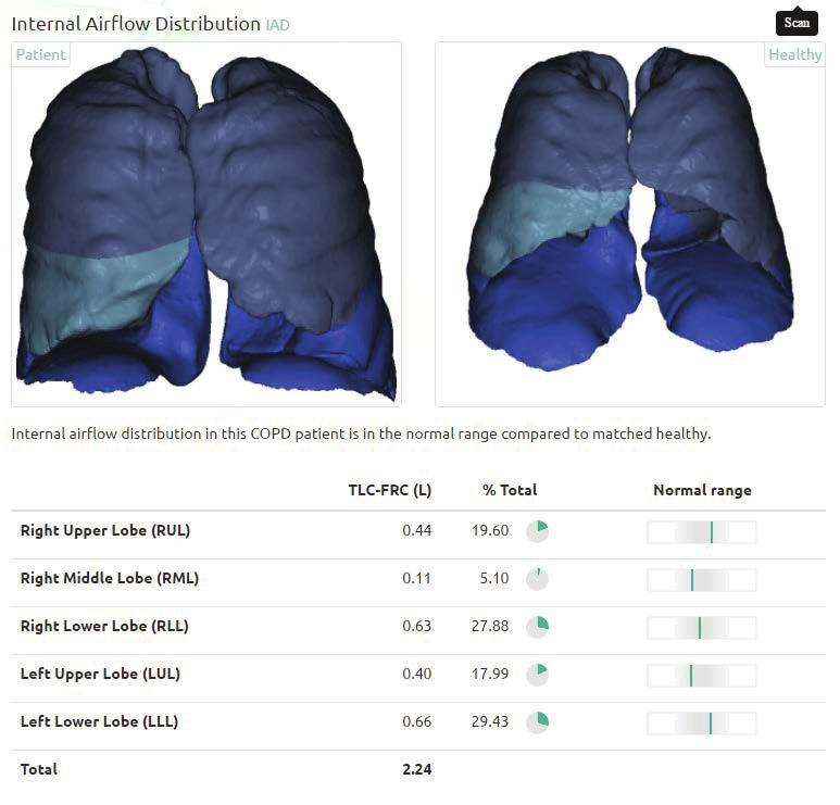
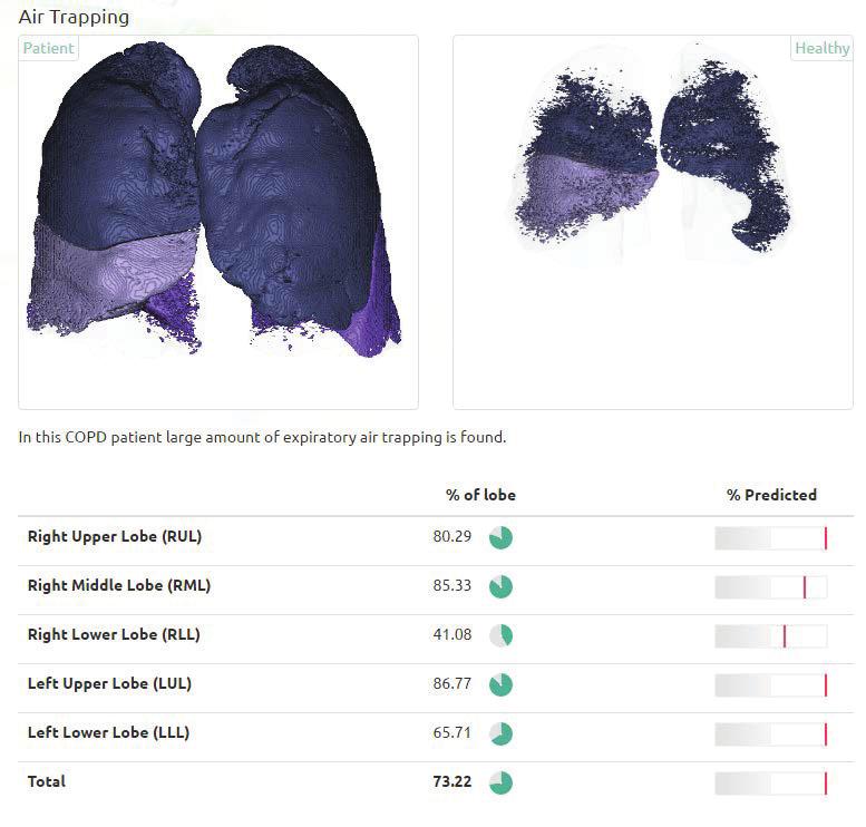
167 Airtrapping at baseline Bita Hajian 167
168 Airway resistance FRC after bronchodilation with salbutamol 168 Enhanced COPD phenotyping to improve treatment strategies
169 Internal airflow distribution after bronchodilation with salbutamol Bita Hajian 169
170 Airtrapping after bronchodilation with salbutamol 170 Enhanced COPD phenotyping to improve treatment strategies

171 Airway resistance FRC after bronchodilation with ipratropium Bita Hajian 1711
172 Internal airflow distribution after bronchodilation with ipratropium 172 Enhanced COPD phenotyping to improve treatment strategies


173 Airtrapping after bronchodilation with ipratropium Bita Hajian 173
174 Aerosol deposition comparing distribution between two particle sizes: 4 µg versus 2µg 174 Enhanced COPD phenotyping to improve treatment strategies
; Tiff index 0.7; TLC 8.")
175 A case of uncontrolled asthma: FRI revealed the true disease severity This moderate severe asthma patient had been treated for years with a conventional therapy of ICS/LABA and LAMA. The patient's asthma state was well controlled under its current treatment. Although there was a stable spirometry: FEV l (116 %); Tiff index 0.7; TLC 8.61 l (118 %); the patient complained about recurring symptoms. An FRI analysis was conducted to detect the underlying mechanisms. Based on the disease state, the physician realized that the patient was uncontrolled, which went undetected by the FEV1. He noticed that some regions of the lungs probably were undertreated by the current therapy. Therefore he proposed to change the therapy by another ICS/LABA with different particle size. To follow up the effect of the drug intervention, an analysis was performed acutely after the administration and after 6 months of treatment. The patient showed a strong acute reversibility of its lung function, which was confirmed by a large bronchodilation as seen by FRI. After 6 months of this new treatment, an increase in pre-bronchodilator FEV1 was observed and FRI demonstrated that the airway dimension did increase more than 15% as compared to baseline. This clinical case demonstrates that FRI is more sensitive than the conventional lung function tests and that it can be used to assess the disease state and the effect of drug intervention. Bita Hajian 175
176 Airway resistance at baseline 176 Enhanced COPD phenotyping to improve treatment strategies

177 Air trapping at baseline Bita Hajian 1777
178 Airway resistance after 6 month inhalation with beclomethasone/formoterol 178 Enhanced COPD phenotyping to improve treatment strategies
179 Air trapping after 6 month inhalation with beclomethasone/formoterol Bita Hajian 179
180 Aerosol deposition comparing distribution between two particle sizes: 4 µg versus 2µg 180 Enhanced COPD phenotyping to improve treatment strategies

181 Bita Hajian 1811
182 182 Enhanced COPD phenotyping to improve treatment strategies 7
183 Conclusion 7 Bita Hajian 183
184 COPD old disease with fresh approach : the path to personalized medicine Chronic obstructive pulmonary disease (COPD) is a heterogeneous disease associated with significant morbidity and mortality. Current diagnostic criteria are based on lung function impairment, symptoms, and exacerbation frequency but do not capture the heterogeneity of the disease. Recognition of the importance of defining the heterogeneity of COPD has led to an unmet need for identification of different COPD phenotypes with specific biomarkers. Pheno-/endotyping COPD is really important because it provides patients with precise and personalized medicine. So, we can provide the patients with the right treatment at the right dose at the right time. This was described in the Introduction. In Chapter 2, we discussed functional imaging techniques and why FRI is a very sensitive biomarker for phenotyping COPD patients. FRI adds functionality to HRCT images, using segmentation and Computational Fluid Dynamics. This gives detailed information about the loco-regional pathology in COPD patients. Local volumes of airways, lobes and lungs, local resistance, emphysema score and also recently ventilation and perfusion ratios can be measured. All this correlate with the conventional lung function, so these parameters from FRI can be used for a better phenotyping of the COPD patients. With FRI, we are informed precisely about the internal flow distribution and particle deposition. Early and small changes in the lung structure including lung vasculature can be detected by FRI, with much more sensitivity than the classical lung function. The technique can highlight changes in the airways after treatment, hereby it is possible to identify responders and non-responders of a treatment. There is also a very good correlation with the patient reported outcomes and clinical measurements like 6MWT. In Chapter 3, we used FRI for the first time for understanding the true pathophysiology of a COPD exacerbation. With FRI it is possible to identify the different phenotype of patients during an exacerbation. Patients with hyperinflation should be treated differently than the one without the characteristics of hyperinflation and small airways dysfunction, a markedly different phenotype of exacerbation. FRI can be used to choose the right treatment strategy during an exacerbation, a patient with loco-regional distal airway inflammation should be 184 Enhanced COPD phenotyping to improve treatment strategies
185 treated differently (with another device/ other particle size) ) than a patient with central airway inflammation (regular bronchodilators as nebulizer or DPI in case of central airway inflammation, and PMDI s with spacer and small particles in more peripheral airway inflammation). FRI showed a good correlation with the patient reported outcomes. Also there was special attention for the pathophysiological mechanism causing ventilation/perfusion mismatch during an exacerbation. We were able to show that mainly the ventilation part is responsible for this, the role of the perfusion was here limited. We concluded that COPD exacerbations are mainly caused by an increase in the distal airway resistance. Opening the distal airways by systemic therapy, inhaled therapy, and/or airway clearance techniques are adequate treatments for a COPD exacerbation and subsequent maintenance therapy. In Chapter 4, we used FRI to evaluate pharmacological treatment of inhaled compounds and systemic anti-inflammatory compounds. First we started with a review about the administration of medication by inhalation in treating airway diseases. We concluded that the efficacy of this route depends on several factors, like correct inhalation techniques, compliance and the size of the particles. The flow properties and internal flow distribution also significantly contribute to the deposition pattern. FRI can describe internal flow distribution and associated particle tracking. With this regional information, clinicians can choose the most adequate treatment approach. The mode of action of systemic anti-inflammatory compounds was also discussed in chapter 4. Roflumilast reduces the inflammatory response in the small airways, hereby improving dynamic hyperinflation, in the areas previously undertreated by inhalation medication. The improved ventilation leads to improved deposition of ICS/LABA/LAMA inhalation. With the roflumilast study, we were able to identify responders to this anti-inflammatory compound. Especially, patients suffering from dynamic hyperinflation at baseline, determined using the 6-minute walk test, benefited from roflumilast. We concluded that the responders can be identified by the FRI (lobes with higher percentage of blood vessels reduce more in hyperinflation = systemic effect), by PRO s and also clinical tests like 6MWT. The role of vascular compounds in treatment of patients with COPD and pulmonary hypertension was studied in chapter 5. These patients are frequent exacerbators with a poor prognosis, are very limited treatment options. Bita Hajian 185
186 Inhalation of pulsed nitric oxide, a vasodilator was studied is these patients. We concluded that inhalation of pulsed NO with oxygen has a profound effect on the pulmonary vessels caliber. With FRI, it was possible to detect the changes in pulmonary vasculature under treatment with NO. Most of the vessels dilated, especially the vessels in the more ventilated zones. Inhaled NO could be a promising treatment for this end stage complication of COPD. The role of chronic treatment with noninvasive ventilation in patients with hypercapnic respiratory failure was studied as well in Chapter 5. Literature study showed that chronic use of NIV is still controversial. FRI technology made it possible to measure ventilation and perfusion at the lobar level in patients with respiratory failure using long term NIV. The true pathophysiological mechanism was visualized: the right treatment strategy reduces the distal airway resistance, hereby a redistribution of flow to well perfused areas by opening the occluded airways. This gives an indication for the selection of patients for this treatment: patients with only localized emphysema seem to benefit most of NIV. In our population most of the patients improved with this treatment, there was a significant reduction in hypercapnia and also symptom score. Broncholab, a platform for the clinicians to get a deeper insight in the pathophysiology of the lungs. In chapter 6 a few cases were described using FRI for better phenotyping. So it was possible to choose the right treatment strategy (particle size, NO) I hope that the present work will provide our patients a fresh approach towards the best health care possible, tailoring the therapeutic approach for each patient: a step forward to personalized medicine in the treatment of COPD, especially in patients with an end stage disease. 186 Enhanced COPD phenotyping to improve treatment strategies
187 Bita Hajian 187
188 188 Enhanced COPD phenotyping to improve treatment strategies
189 Een frisse kijk op COPD : de weg naar gepersonaliseerde geneeskunde Chronisch obstructieve longziekte is een heterogene ziekte die geassocieerd is met een significante morbiditeit en mortaliteit. Huidige diagnostische criteria zijn gebaseerd op verminderde longfunctie, symptomen en exacerbatie frequentie. De heterogeniteit van het ziektebeeld wordt hiermee onvoldoende beschreven. De erkenning van het belang om de heterogeniteit van COPD te definiëren, heeft geleid tot een grote behoefte ten aanzien van identificatie van verschillende COPD-fenotypes met specifieke biomarkers. Fenotyperen en endotyperen van COPD is van groot belang voor onze patiënten, zodat onze patiënten een gepersonaliseerde behandeling kunnen krijgen. Hierdoor zijn we in staat de juiste behandeling met een juiste dosering op de juiste tijd te geven. Dit werd beschreven in de introductie. In hoofdstuk 2 werd functionele imaging techniek besproken en werd besproken waarom FRI een gevoelige biomarker is voor fenotypering van de COPD-patiënten. FRI voegt functionaliteit toe aan de HRCT-beelden, door gebruik te maken van segmentatie methode en Computational Fluid Dynamics. Dit geeft gedetailleerde informatie over de locoregionale pathologie bij COPD-patiënten. Met deze techniek kan het lokale volume van de luchtwegen en longkwabben, de weerstand van de luchtwegen, emfyseem score en recent ook ventilatie perfusie ratio s berekend worden. Al deze parameters correleren met de conventionele longfunctie, zodat deze parameters afkomstig uit FRI gebuikt kunnen worden voor beter fenotypering van de COPD-patiënten. Met FRI worden we precies geïnformeerd over de interne flowdistributie en partikel depositie. Kleine veranderingen in de long structuur en long vasculatuur kunnen vroeg worden opgespoord, met een grotere sensitiviteit in vergelijking met de klassieke longfunctie. Met deze techniek kunnen veranderingen in de luchtwegen na een behandeling in kaart worden gebracht waardoor we in staat zijn om responders en niet responders van een behandeling te identificeren. Er is tevens een goede correlatie met de patiënt reported outcomes en met klinische metingen zoals het zes minuten loop test. In hoofdstuk 3 wordt FRI voor de eerste keer gebruikt om de pathofysiologie van COPDexacerbaties te begrijpen. Met FRI is het mogelijk de verschillende fenotypes van patiënten Bita Hajian 189
190 tijdens een exacerbatie te identificeren. Patiënten met hyperinflatie moeten anders behandeld worden dan patiënten zonder karakteristieken van hyperinflatie en kleine luchtweg dysfunctie, dit betreft een ander fenotype van een exacerbatie. FRI kan worden gebruikt om de juiste behandelstrategie te kiezen tijdens een exacerbatie. Een patiënt met locoregionale distale luchtweginflammatie moet anders behandeld worden (met een andere device/ andere partikel grootte) in vergelijking met een patiënt met een centrale luchtweginflammatie: reguliere bronchodilatatoren als nebuliser of DPI bij centrale luchtweginflammatie en PMDI s met een voorzetkamer en smalle partikels bij meer perifere luchtweginflammatie. Ook hebben we gekeken naar de pathofysiologische mechanisme van het ontstaan van ventilatie/perfusie mismatch gedurende een COPD-exacerbatie. Wij waren in staat aan te tonen dat met name de verandering in de ventilatie verantwoordelijk is hiervoor, en dat de rol van perfusie verandering beperkt is. We concludeerden dat COPD-exacerbaties met name veroorzaakt worden door een toename in de distale luchtweg weerstand. Openen van de distale luchtwegen door systemische therapie, inhalatie therapie, en/of vrijmaken van de luchtwegen door fysiotherapie zijn adequate behandelingen bij een COPD-exacerbatie en ook als onderhoudstherapie. In hoofdstuk 4 werd FRI gebruikt voor evaluatie van farmacologische behandeling van inhalatie compounds en systemische anti-inflammatoire compounds. Eerst volgde een review over het toedienen van inhalatie medicatie voor het behandelen van luchtweg ziekten. We concludeerden dat de efficiëntie van inhalatie afhangt van verschillende factoren, zoals correcte inhalatietechniek, compliantie en de grootte van de partikels. De flow eigenschappen en interne flow distributie zijn tevens in belangrijke mate verantwoordelijk voor het depositie patroon. Met FRI kan interne flow distributie in kaart gebracht worden, en hiermee is het ook mogelijk partikels op te sporen. Met deze regionale informatie, kunnen clinici de meest adequate behandelstrategie toepassen. Het werkingsmechanisme van systemisch anti-inflammatoire compounds werd tevens besproken in hoofdstuk 4. Roflumilast vermindert het inflammatoire respons in de kleine luchtwegen, hierdoor ontstaat er een afname van dynamische hyperinflatie in de regionen die 190 Enhanced COPD phenotyping to improve treatment strategies
191 eerder onvoldoende werden behandeld door inhalatie medicatie. Het verbeterde ventilatie leidt tot verbeterde depositie van ICS/LABA/LAMA inhalaties. Met de roflumilast studie waren we in staat om de responders voor anti-inflammatoire compounds te identificeren. Met name de patiënten die op baseline tijdens het 6 minuten looptest last hadden van dynamische hyperinflatie, hadden het meeste profijt van roflumilast. We concludeerden dat de responders geïdentificeerd kunnen worden met FRI (kwabben met hoger percentage van bloedvaten nemen meer af in hyperinflatie = systemisch effect), door patiënt reported outcomes en tevens klinische onderzoeken zoals het 6 minuten looptest. De rol van vasculaire compounds bij behandeling van patiënten met COPD en pulmonale hypertensie werd in hoofdstuk 5 bestudeerd. Deze patiënten hebben een slechte prognose en ze hebben frequente exacerbaties met weinig behandelmogelijkheden. Inhalatie van gepulseerd nitric oxide, een vasodilatator werd onderzocht bij deze patiënten. Inhalatie van gepulseerde nitric oxide in combinatie met zuurstof heeft een groot effect op het kaliber van de bloedvaten. Met FRI was het mogelijk om de veranderingen in de pulmonale vasculatuur tijdens behandeling met NO te detecteren. De meeste bloedvaten vasodilateren, met name de bloedvaten in de goed geventileerde zones van de long. Inhalatie met NO zou een veelbelovend therapie kunnen zijn voor patiënten met pulmonale hypertensie als eindstadium complicatie van COPD. De rol van chronische behandeling met non-invasieve ventilatie bij patiënten met hypercapnisch respiratoir falen werd tevens bestudeerd in hoofdstuk 5. Literatuurstudie laat zien dat de rol van chronisch gebruik van NIV nog altijd controversieel is. Met FRI-technologie is het mogelijk om ventilatie perfusie op kwab niveau te berekenen bij patiënten met respiratoir falen die langdurig NIV gebruiken. Het pathofysiologische mechanisme van deze behandeling werd gevisualiseerd: de juiste behandelstrategie vermindert de weerstand in de distale luchtwegen, hierdoor ontstaat er een distributie van de flow naar de goed geperfundeerde zones door het openen van de geoccludeerde luchtwegen. Dit geeft een indicatie voor de selectie van de patiënten voor deze behandeling: patiënten met enkel gelokaliseerde emfyseem schijnen het meeste baat te hebben van de NIV. In onze populatie verbeterden de meeste patiënten met deze behandeling, er was een significante reductie in hypercapnie en tevens de symptomen score. Bita Hajian 1911
192 Broncholab, een platform voor clinici om meer inzicht te krijgen in de pathofysiologie van de longen. In hoofdstuk 6 werden een aantal casussen beschreven, waarbij FRI gebruikt werd voor de juiste phenotypering van de patiënten, hierdoor was het mogelijk de juiste behandelstrategie toe te passen. Ik hoop dat het huidig werk een frisse benadering is voor de beste gezondheidszorg voor onze patiënten, therapie op maat voor elke patiënt: een stap vooruit naar gepersonaliseerde geneeskunde in de behandeling van COPD, met name bij patiënten met eindstadium complicaties. 192 Enhanced COPD phenotyping to improve treatment strategies
193 Bita Hajian 193
194 List of abbreviations 6MWD AaDO2 ABG AECOPD ATS BAL BIPAP camp CCQ CFD CI COPD CPAP CAT CT scan DLCO DPI ERS ERV F FEV1 FEV1/FVC FPFs FRC FPF FRI FVC six minute walking distance arterial alveolar oxygen gradient arterial blood gasses acute exacerbation of chronic obstructive pulmonary disease American thoracic society Broncho alveolar lavage bi-level positive pressure ventilation cyclic adenosine monophosphate clinical COPD questionnaire computational fluid dynamics confidence interval chronic obstructive pulmonary disease continuous positive airway pressure chronic obstructive disease assessment test computer tomography scans diffusion capacity for carbon monoxide dry-powder inhaler european respiratory society expiratory reserve volume female forced expiratory volume in 1 second tiffeneau-pinelli index fine particle fractions functional residual capacity fine particle fraction functional respiratory imaging functional vital capacity 194 Enhanced COPD phenotyping to improve treatment strategies
195 GDS HCO3 - HR HRCT HRQol ICU iraw iq isaw iv ivaw ivbv LABA LAMA LCI Lobar ivbv LLL LUL LTOT SABA SAMA LTOT LV M geometric standard deviation bicarbonate ion hart rate high-resolution computer tomography health related quality of life intensive care unit image based airway resistance image based perfusion image based surface area image based ventilation image based airway volume image based blood vessel volume long-acting beta-2-agonist long-acting muscarinic antagonist lung clearance index lobar image based blood vessel volume left lower lobe left upper lobe long-term oxygen therapy short-acting beta-2-agonist short-acting muscarinic antagonist long-term oxygen therapy left ventricular male MEF25 maximal expiratory flow at 25% MEF50 maximal expiratory flow at 50% MEF75 maximal expiratory flow at 75% methhb MMAD MMEF75/25 methhemoglobin mean aerodynamic diameter maximum midexpiratory flow Bita Hajian 195
196 MRC msv MVV NIBP NIPPV NIV NO NS OSAS Post-NO Pre-NO SpO2 spap sraw PaCO 2 PaO 2 PEF PEFR PH PO2 PRO R Raw RCT RL RLL RML RR RUL RV medical research questionnaire millisievert maximal voluntary ventilation non-invasive blood pressure non-invasive positive pressure ventilation non-invasive ventilation nitric oxide not significant obstructive sleep apnea syndrome post-nitric oxide inhalation pre-nitric oxide inhalation oxygen partial pressure systolic pulmonary artery pressure specific airway resistance partial carbondioxyde pressure partial oxygen pressure peak expiratory flow peak expiratory flow rate Pondus hydrogenii or acidity oxygen pressure patient reported outcome resistance airway resistance randomized controlled trials right lung right lower lobe right middle lobe respiratory rate right upper lobe right ventricular 196 Enhanced COPD phenotyping to improve treatment strategies
197 RV SaO 2 SD SGRQ SRI SRIQ Tiff TLC Total ivbv ULL V/Q VA VAS VC residual volume oxygen saturation standard deviation Saint George respiratory questionnaire severe respiratory insufficiency severe respiratory insufficiency questionnaire Tiffeneau index total lung capacity total image based blood vessel volume upper left lung ventilation perfusion alveolar volume visual analogue scale vital capacity Bita Hajian 197
198 198 Enhanced COPD phenotyping to improve treatment strategies
 Elodie (27-5-2013) Olivia (29-11-2014)")
199 Bita Hajian Curriculum Vitae Personal data Name: Bita Hajian Date of birth: 12 December Phone: Partner: Edwin Herskovic, Master of the Laws Children: Bibi ( ) Elodie ( ) Olivia ( ) Bita Hajian 1999
200 Education Secondary Education: : VWO, Eemland College South University: : Master of Medicine, University of Utrecht 2002: USMLE ( United States Medical License Exam) : Internal Medicine, University of Utrecht : Pulmonology, University of Amsterdam 2010: Board certified Pulmonologist 200 Enhanced COPD phenotyping to improve treatment strategies
201 Residencies : Fellow Internal Medicine/ cardiology/ ICU, University of California Los Angeles : Fellow Cardiology, University of Utrecht : Fellow Internal Medicine, University of Utrecht : Fellow Pulmonology, University of Amsterdam : Pulmonologist Academical Medical Center, Amsterdam : Pulmonologist Spaarne Hospital, Hoofddorp ( fellow Oncology ) 2014 present: Senior Pulmonologist, University Hospital of Antwerp 2017 present: Fellow Pulmonary Rehabilitation, University Hospital of Antwerp and Affiliated Hospitals Sep present: Member LRPC European Respiratory Society, assembly 4 (clinical physiology, sleep and pulmonary circulation) Bita Hajian 201
202 Presentations 1. Thematic poster: Pulmonary vascular effects after pulsed inhaled NO evaluated by functional respiratory imaging; ATS May Poster discussion: Functional respiratory imaging in a COPD Gene Cohort; ATS May Thematic poster: Functional respiratory imaging as a tool for phenotyping asthmatic patients; ATS May Oral presentation: Pulmonary vascular effects after pulsed inhaled NO evaluated by functional respiratory imaging; ERS Sep Thematic poster: Changes in FEV1 after recovery from COPD exacerbations are driven by heterogenous regional changes in airway caliber and hyperinflation; ERS Sep Thematic Poster: Improvement in FEV1 after acute COPD exacerbations is driven by changes in hyperinflation than by changes in proximal airway volume; ERS Sep Thematic poster: Changes in FEV1 after recovery from COPD exacerbation are driven by heterogenous regional changes in airway caliber and hyperinflation; ERS Sep Thematic poster: Functional respiratory imaging as a sensitive biomarker to assess therapeutic interventions in lung diseases; ERS Sep Thematic poster: Assessment of the effect of pulsed nitric oxide in COPD patients with pulmonary hypertension using functional respiratory imaging; ATS May Thematic poster: Unravelling the Mode of Action of Pulsed Inhaled Nitric Oxide in Severe IPF using Functional Respiratory Imaging (FRI); ATS May Thematic poster: Pulsed Inhaled Nitric Oxide Has the Potential to Improve Exercise Tolerance in Severe COPD Subjects with Pulmonary Hypertension); ATS May Thematic poster: Dose-efftect of inhaled NO in patients with IPF using right heart catherization; submitted ERS Enhanced COPD phenotyping to improve treatment strategies
203 Publications 1. Hajian B, De Backer J, Vos W, Aerts J, Cluckers J, De Backer W. Efficacy of inhaled medications in asthma and COPD related to disease severity. Expert Opin Drug Deliv. Dec 2016;13(12): Vos W, Hajian B, De Backer J, Van Holsbeke C, Vinchurkar S, Claes R, Hufkens A, Parizel P, Bedert L, De Backer W. Functional respiratory imaging to assess the interaction between systemic roflumilast and inhaled ICS/LABA/LAMA. Int J Chron Obstruct Pulmon Dis. 2016;11: De Backer J, Van Holsbeke C, Vos W, Vinchurkar S, Dorinsky P, Rebello J, Mangale M, Hajian B, De Backer W. Assessment of lung deposition and analysis of the effect of fluticasone/salmeterol hydrofluoroalkane (HFA) pressurized metered dose inhaler (pmdi) in stable persistent asthma patients using functional respiratory imaging. Expert Rev Respir Med. Aug 2016;10(8): Hajian B, De Backer J, Vos W, Van Holsbeke C, Ferreira F, Quinn D, Hufkens A, Claes R, De Backer W. Pulmonary vascular effects of pulsed inhaled nitric oxide in COPD patients with pulmonary hypertension. Int J Chron Obstruct Pulmon Dis. 2016;11: Hajian B, De Backer J, Vos W, Van Holsbeke C, Clukers J, De Backer W. Functional respiratory imaging (FRI) for optimizing therapy development and patient care. Expert Rev Respir Med. Feb 2016;10(2): Hajian B, De Backer J, Sneyers C, Ferreira F, Barboza K, Leemans G, Vos W, De Backer W. Pathophysiological mechanism of long term non- invasive ventilation in stable hypercapnic patients with COPD using functional respiratory imaging. Accepted June 2017, Int J Chron Obstruct Pulmon Dis. 7. Hajian B, De Backer J, Vos W, Van Holsbeke C, Van Geffen W, Ferreira F, Aerts J, De Winter P, Usmani O, Cahn T, Kerstjens H, Pistolesi M, De Backer W. Changes in ventilation-perfusion during and after an COPD exacerbation: An assessment using functional respiratory imaging. In revision May 2017, COPD 8. Van Geffen W, Hajian B, Van Holsbeke C, Vos W, De Backer J, Cahn A, Usmani O, Pistolesi M, Kerstjens H, De Backer W. Functional respiratory imaging: Toward precision medicine in Acute exacerbation of COPD. Submitted March 2017, ERJ. Bita Hajian 203
204 204 Enhanced COPD phenotyping to improve treatment strategies


205 Dankwoord Het is eindelijk zover! Ik mag mijn dankwoord schrijven. Dit proefschrift is tot stand gekomen dankzij het werk en de steun van velen. Alle mensen die deze periode voor mij zo onvergetelijk hebben gemaakt, wil ik hierbij dan ook graag bedanken. Eerst en vooral wil ik mijn promotor, professor de Backer bedanken. Ik betrapte mijzelf er vrij vaak op dat ik al in een pril stadium van mijn proefschrift bezig was met mijn dankwoord aan u: zonder u was dit nooit gelukt en zonder u was ik de uitdaging Nederland-Antwerpen niet aangegaan. Oneindig veel dank voor u enthousiasme, begeleiding en flexibiliteit. Onze gesprekken over research, innovatie en goede patiëntenzorg zal ik nooit vergeten. Ondanks de nodige tegenslagen af en toe, stond u echt altijd klaar met u advies. Ik had me tijdens mijn promotie geen betere begeleider kunnen wensen! Bedankt, bedankt, bedankt. The SKY IS THE LIMIT Bita, dat zal ik onthouden. De leden van de doctoraatscommissie, Prof Verbraecken en Prof Jorens wil ik hartelijk bedanken voor hun tijd om het manuscript te lezen en te beoordelen. Gelijkaardige appreciatie gaat uit naar Prof Wouters en Prof Deron. Beste Jan, jij bent een grote voorbeeld voor mij. Fluidda is jouw kindje, inmiddels uitgegroeid tot een wereldbedrijf door al jouw inspanningen. Het grote succes achter al het fantastische research is jouw passie en verbondenheid met het kliniek. Daardoor is Fluidda uniek te noemen. Marijke door jouw warme persoonlijkheid heb ik mij direct welkom gevoeld in het Antwerpse. Ontzettend veel dank daarvoor. Ook de rest van het Fluidda team wil ik ontzettend bedanken. Zonder jullie was dit niet gelukt! Sven zonder jouw, was dit boekje nooit zo mooi geworden. Het was een plezier om samen met jou, dit fantastisch onderzoek in een prachtig jasje te steken. Er werd vooral veel gelachen hierbij! Bedankt. Annemie, Rita en Ilse, jullie zijn de beste research verpleegkundigen die een promovendi en een patiënt kunnen treffen. We hebben hard moeten werken samen, om onze deadlines te halen. Het is allemaal perfect gelukt dankzij jullie inspanningen. Waarvoor mijn dank. Ook wil ik alle medewerkers van onze longfunctieafdeling, de polikliniek pneumologie en de verpleging van dienst pneumologie bedanken. Ik kon altijd rekenen op jullie medewerking tijdens de afgelopen jaren. Jullie zijn goud waard. Annemie, bedankt voor je voortdurende interesse en relativeringsvermogen. Je was altijd beschikbaar voor overleg. Mijn speciale dank gaat uit naar de afdeling cardiologie, in het bijzonder professor Shivalkar. Bedankt voor u voortdurende enthousiasme en interesse voor mijn proefschrift en de fantastische echocardiografieën. Lieve Mia, wij hebben samengewerkt in het AMC. Ik heb van jou mogen leren wat echte betrokkenheid voor je patiënten betekent en nog zoveel meer Ook de afgelopen jaren heb Bita Hajian 205
Understanding the mode of action of a drug using Functional Respiratory Imaging (FRI) Roflumilast Study. Jan De Backer, MSc, PhD, MBA CEO
 Roflumilast Study. Jan De Backer, MSc, PhD, MBA CEO") Understanding the mode of action of a drug using Functional Respiratory Imaging (FRI) Roflumilast Study Jan De Backer, MSc, PhD, MBA CEO IPAC RS/UF Orlando Inhalation Conference March 20, 2014 OVERVIEW
Understanding the mode of action of a drug using Functional Respiratory Imaging (FRI) Roflumilast Study Jan De Backer, MSc, PhD, MBA CEO IPAC RS/UF Orlando Inhalation Conference March 20, 2014 OVERVIEW
2/4/2019. GOLD Objectives. GOLD 2019 Report: Chapters
 GOLD Objectives To provide a non biased review of the current evidence for the assessment, diagnosis and treatment of patients with COPD. To highlight short term and long term treatment objectives organized
Functional Respiratory Imaging
 Functional Respiratory Imaging (FRI) to assess the bioequivalence of inhaled medication Jan De Backer, MSc, PhD, MBA CEO BABE 2014 Functional Respiratory Imaging (FRI) FRI outcome parameters Added value
COPD: From Phenotypes to Endotypes. MeiLan K Han, M.D., M.S. Associate Professor of Medicine University of Michigan, Ann Arbor, MI
 COPD: From Phenotypes to Endotypes MeiLan K Han, M.D., M.S. Associate Professor of Medicine University of Michigan, Ann Arbor, MI Presenter Disclosures MeiLan K. Han Consulting Research support Novartis
COPD. Dr.O.Paknejad Pulmonologist Shariati Hospital TUMS
 IN THE NAME OF GOD COPD Dr.O.Paknejad Pulmonologist Shariati Hospital TUMS Definition of COPD* COPD is a preventable and treatable chronic lung disease characterized by airflow limitation that is not fully
TORCH: Salmeterol and Fluticasone Propionate and Survival in COPD
 TORCH: and Propionate and Survival in COPD April 19, 2007 Justin Lee Pharmacy Resident University Health Network Outline Overview of COPD Pathophysiology Pharmacological Treatment Overview of the TORCH
9/22/2015 CONFLICT OF INTEREST OBJECTIVES. Understanding COPD - Recent Research and the Evolving Definition of COPD for MNACVPR
 Understanding COPD - Recent Research and the Evolving Definition of COPD for MNACVPR by Scott Cerreta, BS, RRT Director of Education www.copdfoundation.org scerreta@copdfoundation.org CONFLICT OF INTEREST
Understanding COPD - Recent Research and the Evolving Definition of COPD for MNACVPR
 Understanding COPD - Recent Research and the Evolving Definition of COPD for MNACVPR by Scott Cerreta, BS, RRT Director of Education www.copdfoundation.org scerreta@copdfoundation.org CONFLICT OF INTEREST
Supplementary Online Content
 Supplementary Online Content Regan EA, Lynch DA, Curran-Everett D, et al; Genetic Epidemiology of COPD (COPDGene) Investigators. Clinical and radiologic disease in smokers with normal spirometry. Published
COPD, Asthma, Or Something In Between? Sharon R. Rosenberg Assistant Professor of Medicine Northwestern University December 4, 2013
 COPD, Asthma, Or Something In Between? Sharon R. Rosenberg Assistant Professor of Medicine Northwestern University December 4, 2013 None Disclosures Definitions Asthma Asthma is a chronic inflammatory
Update on heterogeneity of COPD, evaluation of COPD severity and exacerbation
 Update on heterogeneity of COPD, evaluation of COPD severity and exacerbation Yung-Yang Liu, MD Taipei Veterans General Hospital Aug 29, 2015 G O lobal Initiative for Chronic bstructive L D ung isease
Pathophysiology of COPD 건국대학교의학전문대학원
 Pathophysiology of COPD 건국대학교의학전문대학원 내과학교실 유광하 Rate per 100 0,000 population 550 500 450 400 350 300 250 200 150 100 50 0 Heart disease Cancer Stroke 1970 1974 1978 1982 1986 1990 1994 1998 2002 Year of
Life-long asthma and its relationship to COPD. Stephen T Holgate School of Medicine University of Southampton
 Life-long asthma and its relationship to COPD Stephen T Holgate School of Medicine University of Southampton Definitions COPD is a preventable and treatable disease with some significant extrapulmonary
OPTIMIZING MANAGEMENT OF COPD IN THE PRACTICE SETTING 10/16/2018 DISCLOSURES I have no financial or other disclosures
 OPTIMIZING MANAGEMENT OF COPD IN THE PRACTICE SETTING J. Michael Fuller, MD, MEd, FACP, FCCP Associate Professor of Medicine University of South Carolina Greenville DISCLOSURES I have no financial or other
The role of lung function testing in the assessment of and treatment of: AIRWAYS DISEASE
 The role of lung function testing in the assessment of and treatment of: AIRWAYS DISEASE RHYS JEFFERIES ARTP education Learning Objectives Examine the clinical features of airways disease to distinguish
COPD is a syndrome of chronic limitation in expiratory airflow encompassing emphysema or chronic bronchitis.
 1 Definition of COPD: COPD is a syndrome of chronic limitation in expiratory airflow encompassing emphysema or chronic bronchitis. Airflow obstruction may be accompanied by airway hyper-responsiveness
ASTHMA-COPD OVERLAP SYNDROME 2018: What s All the Fuss?
 ASTHMA-COPD OVERLAP SYNDROME 2018: What s All the Fuss? Randall W. Brown, MD MPH AE-C Association of Asthma Educators Annual Conference July 20, 2018 Phoenix, Arizona FACULTY/DISCLOSURES Randall Brown,
COPD: Change in Definition. COPD Pathology with 3D Interactive. COPD: Definitions of 21st Century 1. COPD Includes Chronic Bronchitis 2
 COPD: Change in Definition COPD Pathology with 3D Interactive by Scott Cerreta, BS, RRT Director of Education www.copdfoundation.org COPD used to include 5 disease processes 1 1 Chronic Bronchitis 2 Emphysema
RESPIRATORY PHYSIOLOGY Pre-Lab Guide
 RESPIRATORY PHYSIOLOGY Pre-Lab Guide NOTE: A very useful Study Guide! This Pre-lab guide takes you through the important concepts that where discussed in the lab videos. There will be some conceptual questions
Small Airways Disease. Respiratory Function In Small Airways And Asthma. Pathophysiologic Changes in the Small Airways of Asthma Patients
 Small Airways Disease Respiratory Function In Small Airways And Relevant Questions On Small Airway Involvement In How can small airway disease be defined? What is the link between small airway abnormalities
Clinical and radiographic predictors of GOLD-Unclassified smokers in COPDGene
 Clinical and radiographic predictors of GOLD-Unclassified smokers in COPDGene Emily S. Wan, John E. Hokanson, James R. Murphy, Elizabeth A. Regan, Barry J. Make, David A. Lynch, James D. Crapo, Edwin K.
SGRQ Questionnaire assessing respiratory disease-specific quality of life. Questionnaire assessing general quality of life
 SUPPLEMENTARY MATERIAL e-table 1: Outcomes studied in present analysis. Outcome Abbreviation Definition Nature of data, direction indicating adverse effect (continuous only) Clinical outcomes- subjective
Lecture Notes. Chapter 4: Chronic Obstructive Pulmonary Disease (COPD)
") Lecture Notes Chapter 4: Chronic Obstructive Pulmonary Disease (COPD) Objectives Define COPD Estimate incidence of COPD in the US Define factors associated with onset of COPD Describe the clinical features
Lecture Notes Chapter 4: Chronic Obstructive Pulmonary Disease (COPD) Objectives Define COPD Estimate incidence of COPD in the US Define factors associated with onset of COPD Describe the clinical features
DISEASES OF THE RESPIRATORY SYSTEM 2018 DR HEYAM AWAD LECTURE 3: CHRONIC BRNCHITIS AND BRONCHIECTASIS
 DISEASES OF THE RESPIRATORY SYSTEM 2018 DR HEYAM AWAD LECTURE 3: CHRONIC BRNCHITIS AND BRONCHIECTASIS INTRDUCTION In the last lecture we discussed the difference between restrictive and obstructive lung
Parametric response mapping
 Parametric response mapping Utility of a novel imaging biomarker in pulmonary disease Dharshan Vummidi MD, Lama VN MD, Yanik G MD, Kazerooni EA MD, Meilan Han MD, Galban C PhD Radiology, Pulmonary & Critical
The Journal Club: COPD Exacerbations
 252 The Journal Club: COPD Exacerbations Chronic Obstructive Pulmonary Diseases: Journal of the COPD Foundation Journal Club The Journal Club: COPD Exacerbations Ron Balkissoon, MD, MSc, DIH, FRCPC 1 Abbreviations:
How does COPD really work?
 How does COPD really work? by Alex Goodell View online Where does COPD fit in the mix of respiratory diseases? I ve made a map of the major pathologies outlined in Robbins and First Aid (obviously these
Pulmonary Function Testing: Concepts and Clinical Applications. Potential Conflict Of Interest. Objectives. Rationale: Why Test?
 Pulmonary Function Testing: Concepts and Clinical Applications David M Systrom, MD Potential Conflict Of Interest Nothing to disclose pertinent to this presentation BRIGHAM AND WOMEN S HOSPITAL Harvard
Chronic Obstructive Pulmonary Disease (COPD) Clinical Guideline
 Clinical Guideline") Chronic Obstructive Pulmonary Disease (COPD) Clinical These clinical guidelines are designed to assist clinicians by providing an analytical framework for the evaluation and treatment of patients. They
Chronic Obstructive Pulmonary Disease (COPD) Clinical These clinical guidelines are designed to assist clinicians by providing an analytical framework for the evaluation and treatment of patients. They
Overview of Obstructive Diseases of the Lung, Lung Physiology and Imaging Modalities
 Overview of Obstructive Diseases of the Lung, Lung Physiology and Imaging Modalities Jann Mortensen Final Phil meeting, 29 april 2004 Obstructive disease Definition Epidemiologi Mechanisms Natural history
Differential diagnosis
 Differential diagnosis The onset of COPD is insidious. Pathological changes may begin years before symptoms appear. The major differential diagnosis is asthma, and in some cases, a clear distinction between
Airways Disease MDT - 6th May 2014
 Airways Disease MDT - 6th May 2014 The inaugural AD-MDT was held on 6/5/14. The AIM of the meeting is to develop the skills and knowledge to be able to run an AD-MDT - the time frame from the start to
Corporate Medical Policy
 Corporate Medical Policy File Name: Origination: Last CAP Review: Next CAP Review: Last Review: lung_volume_reduction_surgery 4/1996 3/2018 3/2019 3/2018 Description of Procedure or Service Emphysema is
THE CHALLENGES OF COPD MANAGEMENT IN PRIMARY CARE An Expert Roundtable
 THE CHALLENGES OF COPD MANAGEMENT IN PRIMARY CARE An Expert Roundtable This activity is supported by an educational grant from Sunovion Pharmaceuticals Inc. COPD in the United States Third leading cause
APSR RESPIRATORY UPDATES
 APSR RESPIRATORY UPDATES Volume 4, Issue 7 Newsletter Date: July 2012 APSR EDUCATION PUBLICATION Inside this issue: Quantitative imaging of airways Small-Airway Obstruction and Emphysema in Chronic Obstructive
Defining COPD. Georgina Grantham Community Respiratory Team Leader/ Respiratory Nurse Specialist
 Defining COPD Georgina Grantham Community Respiratory Team Leader/ Respiratory Nurse Specialist Defining COPD Chronic Obstructive Pulmonary Disease (COPD) is a common, preventable and treatable disease
COPD. Breathing Made Easier
 COPD Breathing Made Easier Catherine E. Cooke, PharmD, BCPS, PAHM Independent Consultant, PosiHleath Clinical Associate Professor, University of Maryland School of Pharmacy This program has been brought
Small Airways : How To Confident Diagnosis and Management?
 Small Airways : How To Confident Diagnosis and Management? Assistant Prof. Siwasak Juthong. M.D. Division of Respiratory and Respiratory Critical Care Medicine Faculty of Medicine Prince of Songkla University
COPD or not COPD, that is the question.
 COPD or not COPD, that is the question. Asthma-COPD Overlap Syndrome: ACOS Do we really need this? Michelle Harkins Disclosure Slide Slide help - William Busse, MD Organizational Interests ATS, ACCP, ACP
Turning Science into Real Life Roflumilast in Clinical Practice. Roland Buhl Pulmonary Department Mainz University Hospital
 Turning Science into Real Life Roflumilast in Clinical Practice Roland Buhl Pulmonary Department Mainz University Hospital Therapy at each stage of COPD I: Mild II: Moderate III: Severe IV: Very severe
Basic mechanisms disturbing lung function and gas exchange
 Basic mechanisms disturbing lung function and gas exchange Blagoi Marinov, MD, PhD Pathophysiology Department, Medical University of Plovdiv Respiratory system 1 Control of breathing Structure of the lungs
The development of chronic obstructive pulmonary
 THE PATHOGENESIS AND PATHOLOGY OF COPD: IDENTIFYING RISK FACTORS AND IMPROVING MORBIDITY AND MORTALITY * Paul D. Scanlon, MD ABSTRACT Chronic obstructive pulmonary disease (COPD) is a chronic inflammatory
Chronic Obstructive Pulmonary Disease (COPD) KAREN ALLEN MD PULMONARY & CRITICAL CARE MEDICINE VA HOSPITAL OKC / OUHSC
 KAREN ALLEN MD PULMONARY & CRITICAL CARE MEDICINE VA HOSPITAL OKC / OUHSC") Chronic Obstructive Pulmonary Disease (COPD) KAREN ALLEN MD PULMONARY & CRITICAL CARE MEDICINE VA HOSPITAL OKC / OUHSC I have no financial disclosures Definition COPD is a preventable and treatable disease
Chronic Obstructive Pulmonary Disease (COPD) KAREN ALLEN MD PULMONARY & CRITICAL CARE MEDICINE VA HOSPITAL OKC / OUHSC I have no financial disclosures Definition COPD is a preventable and treatable disease
Functional Respiratory Imaging
 Functional Respiratory Imaging Your solution for DELIVERY OPTIMIZATION DISEASE CHARACTERIZATION THERAPY ANALYSIS MARKETING EXCELLENCE www.fluidda.com We can help. W e know that the respiratory field is
COPD: early detection, screening and case-finding: what is the evidence? Prof. Jan-Willem Lammers, Md PhD Department of Respiratory Diseases
 COPD: early detection, screening and case-finding: what is the evidence? Prof. Jan-Willem Lammers, Md PhD Department of Respiratory Diseases «If you test one smoker with cough every day You will diagnose
Advances in the management of chronic obstructive lung diseases (COPD) David CL Lam Department of Medicine University of Hong Kong October, 2015
 David CL Lam Department of Medicine University of Hong Kong October, 2015") Advances in the management of chronic obstructive lung diseases (COPD) David CL Lam Department of Medicine University of Hong Kong October, 2015 Chronic obstructive pulmonary disease (COPD) COPD in Hong
Advances in the management of chronic obstructive lung diseases (COPD) David CL Lam Department of Medicine University of Hong Kong October, 2015 Chronic obstructive pulmonary disease (COPD) COPD in Hong
Mai ElMallah,MD Updates in Pediatric Pulmonary Care XII: An Interdisciplinary Program April 13, 2012
 Mai ElMallah,MD Updates in Pediatric Pulmonary Care XII: An Interdisciplinary Program April 13, 2012 Recognize the importance of Pulmonary Function Testing in Cystic Fibrosis Be aware of different types
UNDERSTANDING COPD MEDIA BACKGROUNDER
 UNDERSTANDING COPD MEDIA BACKGROUNDER What is COPD? Chronic Obstructive Pulmonary Disease (COPD) also called emphysema and/or chronic obstructive bronchitis* is a preventable lung disease caused by the
Referring for specialist respiratory input. Dr Melissa Heightman Consultant respiratory physician, UCLH,WH, CNWL
 Referring for specialist respiratory input Dr Melissa Heightman Consultant respiratory physician, UCLH,WH, CNWL Respiratory Specialist- who? GPSI Community Team Secondary Care Respiratory physician and
Asthma COPD Overlap (ACO)
") Asthma COPD Overlap (ACO) Dr Thomas Brown Consultant Respiratory Physician Thomas.Brown@porthosp.nhs.uk Dr Hitasha Rupani Consultant Respiratory Physician Hitasha.rupani@porthosp.nhs.uk What is Asthma
Asthma COPD Overlap (ACO) Dr Thomas Brown Consultant Respiratory Physician Thomas.Brown@porthosp.nhs.uk Dr Hitasha Rupani Consultant Respiratory Physician Hitasha.rupani@porthosp.nhs.uk What is Asthma
COPD/ Asthma. Dr Heather Lewis Honorary Clinical Lecturer
 COPD/ Asthma Dr Heather Lewis Honorary Clinical Lecturer Objectives To understand the pathogenesis of asthma/ COPD To recognise the clinical features of asthma/ COPD To know how to diagnose asthma/ COPD
COPD Bronchiectasis Overlap Syndrome.
 COPD Bronchiectasis Overlap Syndrome. John R Hurst 1, J Stuart Elborn 2, and Anthony De Soyza 3 on Behalf of the BRONCH-UK Consortium (D Bilton, J Bradley, JS Brown, J Duckers, F Copeland, A Floto, J Foweraker,
Chronic obstructive lung disease. Dr/Rehab F.Gwada
 Chronic obstructive lung disease Dr/Rehab F.Gwada Obstructive lung diseases Problem is in the expiratory phase Lung disease Restrictive lung disease Restriction may be with, or within the chest wall Problem
Asthma. - A chronic inflammatory disorder which causes recurrent episodes of wheezing, breathlessness, cough and chest tightness.
 Obstructive diseases Asthma - A chronic inflammatory disorder which causes recurrent episodes of wheezing, breathlessness, cough and chest tightness. - Characterized by Intermittent and reversible (the
What s new in COPD? Apichart Khanichap MD. Department of Medicine, Faculty of Medicine, Thammasat university
 What s new in COPD? Apichart Khanichap MD. Department of Medicine, Faculty of Medicine, Thammasat university Management stable COPD Relieve symptoms Improve exercise tolerance Improve health status Prevent
Management of Acute Exacerbations of COPD
 MiCMRC Educational Webinar Management of Acute Exacerbations of COPD August 22, 2018 MiCMRC Educational Webinar Management of Acute Exacerbations of COPD Expert Presenter: Catherine A. Meldrum PhD RN MS
RESPIRATORY BLOCK. Bronchial Asthma. Dr. Maha Arafah Department of Pathology KSU
 RESPIRATORY BLOCK Bronchial Asthma Dr. Maha Arafah Department of Pathology KSU marafah@ksu.edu.sa Jan 2018 Objectives Define asthma (BA) Know the two types of asthma 1. Extrinsic or atopic allergic 2.
an inflammation of the bronchial tubes
 BRONCHITIS DEFINITION Bronchitis is an inflammation of the bronchial tubes (or bronchi), which are the air passages that extend from the trachea into the small airways and alveoli. Triggers may be infectious
DISEASES OF THE RESPIRATORY SYSTEM 2018 DR HEYAM AWAD LECTURE 2: ATELECTASIS AND EMPHYSEMA
 DISEASES OF THE RESPIRATORY SYSTEM 2018 DR HEYAM AWAD LECTURE 2: ATELECTASIS AND EMPHYSEMA INTRODUCTION In this lecture we will discuss atelectasis which is a complication of several medical and surgical
CARE OF THE ADULT COPD PATIENT
 CARE OF THE ADULT COPD PATIENT Target Audience: The target audience for this clinical guideline is all MultiCare providers and staff including those associated with our Clinically Integrated Network. The
Chronic obstructive pulmonary disease. The acute effect of budesonide/formoterol in COPD: a multi-slice computed tomography and lung function study
 Eur Respir J 212; 4: 298 35 DOI:.1183/931936.72511 CopyrightßERS 212 The acute effect of budesonide/formoterol in COPD: a multi-slice computed tomography and lung function study Lieve A. De Backer*, Wim
4.6 Small airways disease
 4.6 Small airways disease Author: Jean-Marc Fellrath 1. INTRODUCTION Small airways are defined as any non alveolated and noncartilaginous airway that has an internal diameter of 2 mm. Several observations
Assessment of Pulmonary Artery Pressure in Chronic Obstructive Pulmonary Disease Patients without Resting Hypoxemia
 The Egyptian Journal of Hospital Medicine (July 2018) Vol. 73 (2), Page 6021-6027 Assessment of Pulmonary Artery Pressure in Chronic Obstructive Pulmonary Disease Patients without Resting Hypoxemia Muhammed
COMPARISON BETWEEN INTERCOSTAL STRETCH AND BREATHING CONTROL ON PULMONARY FUNCTION PARAMETER IN SMOKING ADULTHOOD: A PILOT STUDY
 COMPARISON BETWEEN INTERCOSTAL STRETCH AND BREATHING CONTROL ON PULMONARY FUNCTION PARAMETER IN SMOKING ADULTHOOD: A PILOT STUDY Shereen Inkaew 1 Kamonchat Nalam 1 Panyaporn Panya 1 Pramook Pongsuwan 1
Outline FEF Reduced FEF25-75 in asthma. What does it mean and what are the clinical implications?
 Reduced FEF25-75 in asthma. What does it mean and what are the clinical implications? Fernando Holguin MD MPH Director, Asthma Clinical & Research Program Center for lungs and Breathing University of Colorado
COPD: Current Medical Therapy
 COPD: Current Medical Therapy Angela Golden, DNP, FNP-C, FAANP Owner, NP from Home, LLC Outcomes As a result of this activity, learners will be able to: 1. List the appropriate classes of medications for
Coexistence of confirmed obstruction in spirometry and restriction in body plethysmography, e.g.: COPD + pulmonary fibrosis
 Volumes: IRV inspiratory reserve volume Vt tidal volume ERV expiratory reserve volume RV residual volume Marcin Grabicki Department of Pulmonology, Allergology and Respiratory Oncology Poznań University
THE PHARMA INNOVATION - JOURNAL Clinical Characteristics of Chronic Obstructive Pulmonary Disease
 Received: 09012014 Accepted: 30032014 ISSN: 2277 7695 CODEN Code: PIHNBQ ZDBNumber: 26630382 IC Journal No: 7725 Vol. 3 No. 2. 2014 Online Available at www.thepharmajournal.com THE PHARMA INNOVATION JOURNAL
COPD: Preventable and Treatable. Lecture Outline. Diagnosis of COPD. COPD: Defining Terms
 COPD: Preventable and Treatable Christopher H. Fanta, M.D. Partners Asthma Center Pulmonary and Critical Care Division Brigham and Women s Hospital Harvard Medical School Lecture Outline I. Diagnosis and
Advancing COPD treatment strategies with evidencebased. 17:15 19:15 Monday 11 September 2017 ERS 2017, Milan, Italy
 Advancing COPD treatment strategies with evidencebased approaches 17:15 19:15 Monday 11 September 2017 ERS 2017, Milan, Italy Increasing understanding of COPD and the effect on guideline evolution. GOLD
What do pulmonary function tests tell you?
 Pulmonary Function Testing Michael Wert, MD Assistant Professor Clinical Department of Internal Medicine Division of Pulmonary, Critical Care, and Sleep Medicine The Ohio State University Wexner Medical
Chronic Obstructive Pulmonary Disease (COPD).
.") Chronic Obstructive Pulmonary Disease (COPD). Linde: Living healthcare 02 03 Chronic Obstructive Pulmonary Disease (COPD). A pocket guide for healthcare professionals. COPD the facts Moderate to severe
Chronic Obstructive Pulmonary Disease (COPD). Linde: Living healthcare 02 03 Chronic Obstructive Pulmonary Disease (COPD). A pocket guide for healthcare professionals. COPD the facts Moderate to severe
WHAT ARE THE PHYSIOLOGICAL DETERMINANTS OF EXPIRATORY FLOW?
 Lung Volume the physiology of breathing: OBSTRUCTIVE LUNG DISORDERS Dr. Andrew Fon MBBS FRACP Respiratory and Sleep Medicine Physician PhD Research Fellow Dept. of Thoracic Medicine, ROYAL ADELAIDE HOSPITAL
Course Handouts & Disclosure
 COPD: Disease Trajectory and Hospice Eligibility Terri L. Maxwell PhD, APRN VP, Strategic Initiatives Weatherbee Resources Hospice Education Network Course Handouts & Disclosure To download presentation
Pulmonary Pathophysiology
 Pulmonary Pathophysiology 1 Reduction of Pulmonary Function 1. Inadequate blood flow to the lungs hypoperfusion 2. Inadequate air flow to the alveoli - hypoventilation 2 Signs and Symptoms of Pulmonary
Study No.: Title: Rationale: Phase: Study Period Study Design: Centres: Indication: Treatment: Objectives : Primary Outcome/Efficacy Variable:
 The study listed may include approved and non-approved uses, formulations or treatment regimens. The results reported in any single study may not reflect the overall results obtained on studies of a product.
Roflumilast (Daxas) for chronic obstructive pulmonary disease
 for chronic obstructive pulmonary disease") Roflumilast (Daxas) for chronic obstructive pulmonary disease August 2009 This technology summary is based on information available at the time of research and a limited literature search. It is not intended
Roflumilast (Daxas) for chronic obstructive pulmonary disease August 2009 This technology summary is based on information available at the time of research and a limited literature search. It is not intended
People with asthma who smoke. The combination of asthma, a chronic airway disease, and smoking increases the risk of COPD even more.
 COPD Chronic obstructive pulmonary disease (COPD) is a chronic inflammatory lung disease that causes obstructed airflow from the lungs. Symptoms include breathing difficulty, cough, sputum (phlegm) production
Yuriy Feschenko, Liudmyla Iashyna, Ksenia Nazarenko and Svitlana Opimakh
 2018; 7(1): 74-78 ISSN (E): 2277-7695 ISSN (P): 2349-8242 NAAS Rating: 5.03 TPI 2018; 7(1): 74-78 2018 TPI www.thepharmajournal.com Received: 11-11-2017 Accepted: 12-12-2017 Yuriy Feschenko Liudmyla Iashyna
PDF of Trial CTRI Website URL -
 Clinical Trial Details (PDF Generation Date :- Wed, 31 Oct 2018 11:13:48 GMT) CTRI Number Last Modified On 17/01/2015 Post Graduate Thesis Type of Trial Type of Study Study Design Public Title of Study
Respiratory Disease. Dr Amal Damrah consultant Neonatologist and Paediatrician
 Respiratory Disease Dr Amal Damrah consultant Neonatologist and Paediatrician Signs and Symptoms of Respiratory Diseases Cardinal Symptoms Cough Sputum Hemoptysis Dyspnea Wheezes Chest pain Signs and Symptoms
Interventional procedures guidance Published: 20 December 2017 nice.org.uk/guidance/ipg600
 Endobronchial valve insertion to reduce lung volume in emphysema Interventional procedures guidance Published: 20 December 2017 nice.org.uk/guidance/ipg600 Your responsibility This guidance represents
The Aging Lung. Sidney S. Braman MD FACP FCCP Professor of Medicine Brown University Providence RI
 The Aging Lung Sidney S. Braman MD FACP FCCP Professor of Medicine Brown University Providence RI Is the respiratory system of the elderly different when compared to younger age groups? Respiratory Changes
Study No.: Title: Rationale: Phase: Study Period: Study Design: Centres: Indication: Treatment: Objectives: Primary Outcome/Efficacy Variable:
 The study listed may include approved and non-approved uses, formulations or treatment regimens. The results reported in any single study may not reflect the overall results obtained on studies of a product.
BPCO: dalle novità patogenetiche alla terapia
 BPCO: dalle novità patogenetiche alla terapia Gianna Camiciottoli Dip. Di Scienze Biomediche, Sperimentali e Cliniche Mario Serio Università degli Studi di Firenze Firenze, 11 novembre 2016 Conflict of
This is a cross-sectional analysis of the National Health and Nutrition Examination
 SUPPLEMENTAL METHODS Study Design and Setting This is a cross-sectional analysis of the National Health and Nutrition Examination Survey (NHANES) data 2007-2008, 2009-2010, and 2011-2012. The NHANES is
Chronic Obstructive Pulmonary Disease
 Chronic Obstructive Pulmonary Disease 07 Contributor Dr David Tan Hsien Yung Definition, Diagnosis and Risk Factors for (COPD) Differential Diagnoses Goals of Management Management of COPD THERAPY AT EACH
Decramer 2014 a &b [21]
![Decramer 2014 a &b [21]](/thumbs/90/101390504.jpg "Decramer 2014 a &b [21]") Buhl 2015 [19] Celli 2014 [20] Decramer 2014 a &b [21] D Urzo 2014 [22] Maleki-Yazdi 2014 [23] Inclusion criteria: Diagnosis of chronic obstructive pulmonary disease; 40 years of age or older; Relatively
Buhl 2015 [19] Celli 2014 [20] Decramer 2014 a &b [21] D Urzo 2014 [22] Maleki-Yazdi 2014 [23] Inclusion criteria: Diagnosis of chronic obstructive pulmonary disease; 40 years of age or older; Relatively
Endobronchial valve insertion to reduce lung volume in emphysema
 NATIONAL INSTITUTE FOR HEALTH AND CARE EXCELLENCE Interventional procedure consultation document Endobronchial valve insertion to reduce lung volume in emphysema Emphysema is a chronic lung disease that
GOLD UPDATE on COPD and the Importance of Accurate Dyspnea Evaluation
 GOLD UPDATE on COPD and the Importance of Accurate Dyspnea Evaluation George Anderson, PhD Sr. Medical Science Liaison MA-AZAP ML-4006-US-0212 Approved 11/16 Glob al Strategy for the Diagnosis, Management,
Chronic Obstructive Pulmonary Disease (COPD) Copyright 2014 by Mosby, an imprint of Elsevier Inc.
 Copyright 2014 by Mosby, an imprint of Elsevier Inc.") Chronic Obstructive Pulmonary Disease () 8.18.18 Copyright 2014 by Mosby, an imprint of Elsevier Inc. Description Airflow limitation not fully reversible progressive Abnormal inflammatory response of lungs
Chronic Obstructive Pulmonary Disease () 8.18.18 Copyright 2014 by Mosby, an imprint of Elsevier Inc. Description Airflow limitation not fully reversible progressive Abnormal inflammatory response of lungs
Respiratory Pathology. Kristine Krafts, M.D.
 Respiratory Pathology Kristine Krafts, M.D. Normal lung: alveolar spaces Respiratory Pathology Outline Acute respiratory distress syndrome Obstructive lung diseases Restrictive lung diseases Vascular
NATIONAL INSTITUTE FOR HEALTH AND CLINICAL EXCELLENCE SCOPE
 NATIONAL INSTITUTE FOR HEALTH AND CLINICAL EXCELLENCE 1 Guideline title SCOPE Chronic obstructive pulmonary disease: the management of adults with chronic obstructive pulmonary disease in primary and secondary
Supplementary appendix
 Supplementary appendix This appendix formed part of the original submission and has been peer reviewed. We post it as supplied by the authors. Supplement to: Calverley P M A, Anzueto A R, Carter K, et
The role of blood eosinophil level in acute exacerbation of Chronic Obstructive Pulmonary Disease
 112 Original Article The role of blood eosinophil level in acute exacerbation of Chronic Obstructive Pulmonary Disease Department of Pulmonology and Critical Care Medicine, Tribhuvan University Teaching
11.3 RESPIRATORY SYSTEM DISORDERS
 11.3 RESPIRATORY SYSTEM DISORDERS TONSILLITIS Infection of the tonsils Bacterial or viral Symptoms: red and swollen tonsils, sore throat, fever, swollen glands Treatment: surgically removed Tonsils: in
How to treat COPD? What is the mechanism of dyspnea? Smoking cessation
 : The Increasing Role of the FP Alan Kaplan, MD, CCFP(EM) Presented at the Primary Care Today: Education Conference and Medical Exposition, Toronto, Ontario, May 2006. Chronic obstructive pulmonary disease
PULMONARY FUNCTION. VOLUMES AND CAPACITIES
 PULMONARY FUNCTION. VOLUMES AND CAPACITIES The volume of air a person inhales (inspires) and exhales (expires) can be measured with a spirometer (spiro = breath, meter = to measure). A bell spirometer
Update on COPD John Hurst PhD FRCP
 Update on COPD John Hurst PhD FRCP Reader / Consultant in Respiratory Medicine UCL / Royal Free London NHS Foundation Trust Director, UCL Royal Free COPD Centre j.hurst@ucl.ac.uk Questions 1. What is the
Computed Tomography-Based Centrilobular Emphysema Subtypes Relate with Pulmonary Function
 Send Orders of Reprints at reprints@benthamscience.net 54 The Open Respiratory Medicine Journal, 213, 7, 54-59 Computed Tomography-Based Centrilobular Emphysema s Relate with Pulmonary Function Mamoru