The use of advanced imaging in the diagnosis of TB. Pierre Goussard, Robert Gie Tygerberg Children`s Hospital and University of Stellenbosch
|
|
|
- Thomasina Palmer
- 5 years ago
- Views:
Transcription


1 The use of advanced imaging in the diagnosis of TB Pierre Goussard, Robert Gie Tygerberg Children`s Hospital and University of Stellenbosch
2 Imaging Bronchoscopy Tracheo-bronchograms Chest CT-scan Ultrasound TBNA FDG scan MRI Virtual bronchoscopy
3 TB airway obstruction model
4 TB airway obstruction : The We we were able to demonstrate present that bronchoscopy has an important role to play in accurately assessing the site and degree of airway narrowing due to TB lymph node compression. The correct assessment is essential in determining whether the affected child requires surgical or medical treatment. There was a statistically significant correlation between bronchoscopic and Chest CT scan findings with both of these investigations identifying bronchus intermedius and the left main bronchus as the most common sites as well as the most severely compressed regions of the airways. One third of children with clinical and radiologically significant airway narrowing will need surgical intervention. Outcome of surgery was excellent resulting in significant improvement in airway size with low morbidity and no mortality. The diagnostic yield during bronchoscopy was been improved by the application of TBNA and Xpert MTB/RIF, although there is still significant room for improvement.
5 TB airway obstruction : The past Well known that TB cause airway obstruction Locations and cause of airway obstruction was not well defined Imaging of airway obstruction was lacking Bronchoscopy was mostly used to collect sample for culture Rigid bronchoscopy was the only instrument available Natural history of TB airway obstruction was not well documented Which children and the timing of surgical intervention was not sorted out
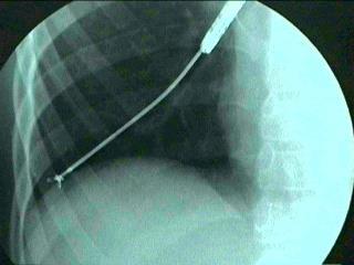
6 Tracheo-bronchogram
7 Bronchoscopy Diagnostic Samples for cultures
8 Indications for bronchoscopy Not indicated for routine specimen collection as the yield from BAL is less than gastric lavage To confirm the diagnosis of PTB. Bronchial obstruction may be present in the absence of visibly enlarged hilar and mediastinal lymph nodes on a chest radiograph. To determine the degree of airway obstruction in complicated disease. For the endoscopic enucleation of glands that have ulcerated into the airway, and for the removal of granulation tissue and caseous material causing airway obstruction. To evaluate the response to treatment in children with complicated airway disease

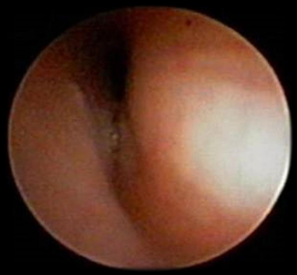
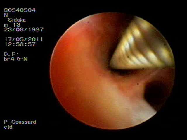
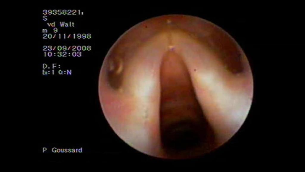
9 Gland herniating into airway

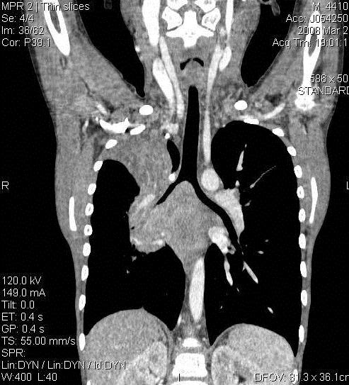
10 Bronchus intermedius obstruction
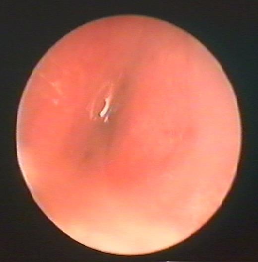
11 tracheal compression
12 Bronchus intermedius obstruction
13 Samples for culture BAL TBNA Endobronchial tissue Transbronchial biopsy tissue

14 Endobronchial lumen biopsy
15 TBNA
16
17
18
19 TBNA

20 Excamples of diagnosis made by TBNA Ziehl Neelsen stain showing numerous acid fast bacilli. ZN 1,000.
21 TBB


22 Bronchus intermedius obstruction
23
24 Chest CT-scan
25

26 Lymphadenopathy was common, but only 46 patients had lymph nodes greater than 1 cm. Enhancement characteristics of tuberculous adenopathy differ from that described previously. Typical enhancement was ghost-like rather than discreet ring enhancing with a low-density centre. The subcarinal region is most frequently involved and is also the site of the largest lymph-node masses. The presence of lymph-node groups at other recognised sites adds confidence when there is doubt, as multifocal involvement is common. 25% of patients with hilar adenopathy may have bronchial compression in childhood
27 Chest CT--scan should be done in children with clinically and radiological significant airway compression to determine the location of glandular involvement and the relationship of these glands to the airways. CT scan is useful to determine the nature of mediastinal glands and to demonstrate the typical ghost like enhancement of the rims of the lymph glands after contrast administration. The most common location for tuberculosis lymph gland enlargement in children with tuberculosis: subcarinal (90%) right hilar (74%) left hilar (72%), bilateral hilar (61%) anterior mediastinum (79%) The largest group of nodes is located in the subcarinal area (87%). The most common site of airway compression was the left main bronchus (21%), right main bronchus (14%) and bronchus intermedius (8%) Andronikou S et al. CT scanning for the decection of tuberculous mediastinal and hilar lymphadenopathy in hildren. Ped Radiology 2004;34:


28 Ghostlike enhancement Ring-enhancing with low-density centres.

29 Lymphobronchial TB 1 2 Obstruction Consolidation 3 4 Necrosis Cavitation
30 Age 0-12 months years years years years Number of patients
31 Why so young? It is well documented that children under 5 are more likely to have nodal disease Younger airways are softer and more compressible Younger airways are narrower; BI in a 1 year old is only 3 mm in diameter! However, not all children with PTB will develop LBTB; incidence in the literature varies from 28% (1) to 38% (2) 1.) Weber AL, Bird KT, Janower ML. Primary tuberculosis in childhood with particular emphasis on changes affecting the tracheobronchial tree. Am J Roentgenol Radium Ther Nucl Med 1968; 103(1): ) Theart AC, Marais BJ, Gie RP, Hesseling AC, Beyers N. Criteria used for the diagnosis of childhood tuberculosis at primary health care level in a high-burden, urban setting. Int J Tuberc Lung Dis 2005; 9(11):

32 Sites of nodes Subcarinal Right paratracheal Right hilar Right azygooesophageal Left hilar Right paracardiac

33 Sites of Obstructions Trachea LMB LUL LLL RMB BI RLL

34 Number of compressions by site and severity Site Mild Moderate Severe Complete Total BI LMB Trachea RLL RMB LLL LUL All sites
35 Tracheal compressions mostly mild Airway most likely to be compressed is BI Airway most likely to show severe or complete compression is BI Severe and complete compressions made up almost half of the BI compressions
36 Number of compressions by age and severity Age N Mild Moderate Severe Complete Total Comp/pt 0-1 yrs yrs yrs yrs yrs All Ages
37
38

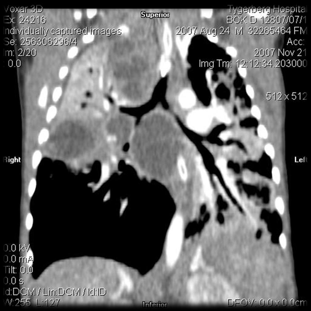
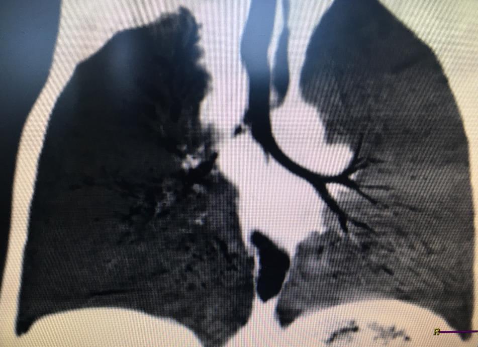
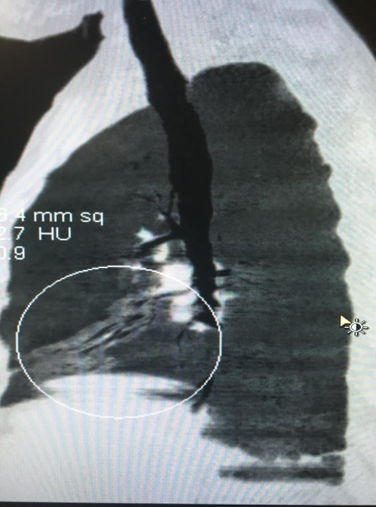
39 Coronal minimum intensity projection (MiniP) image showing compression of the bronchus intermedius in a nutcracker fashion. (arrows) by lymphadenopathy (black stars)
 (white arrow) and moderate compression of the left main bronchus against the left pulmonary")
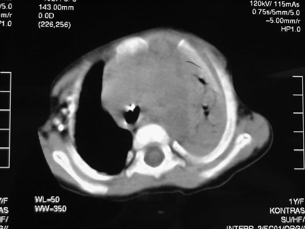
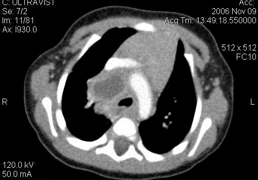
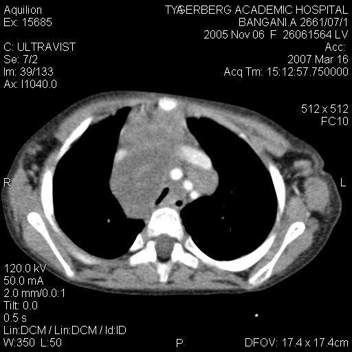
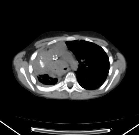
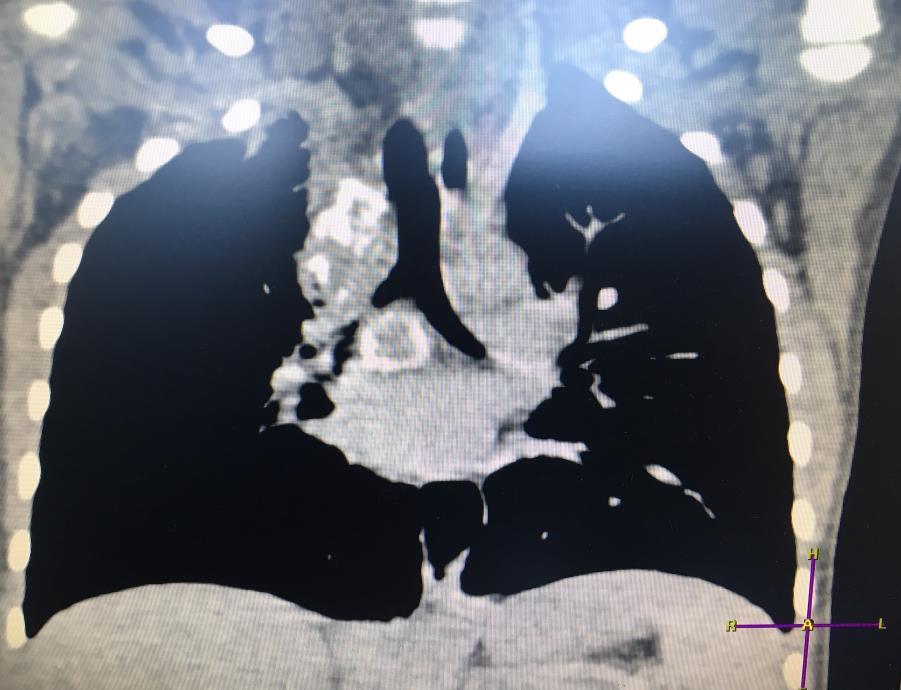
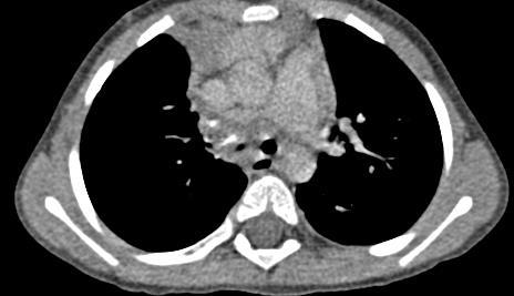
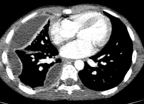
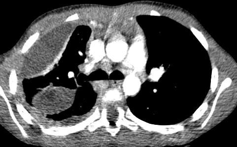
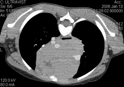
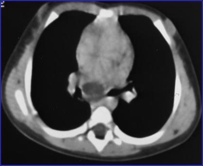
40 Axial CT scan post contrast demonstrates right hilar and subcarinal lymphadenopathy resulting in severe circumferential compression of the bronchus intermedius (collaring) (white arrow) and moderate compression of the left main bronchus against the left pulmonary artery

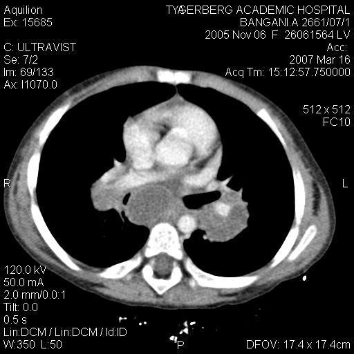
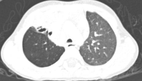
41 Axial CT scan on lung windows demonstrates air-trapping of the right lung as a result of mild to moderate compression of the bronchus intermedius

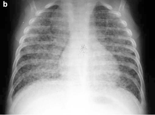
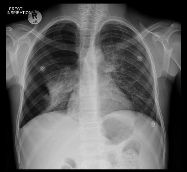
42 Milliary TB

43

44 BEF

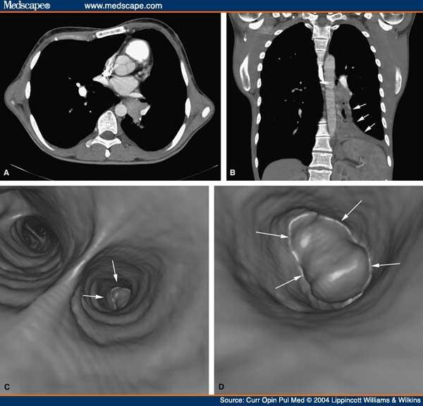
45 Contrast study done with water-soluble contrast medium : demonstrating BOF too the left main bronchus. BOF demonstrated in 88% of cases (n =15)

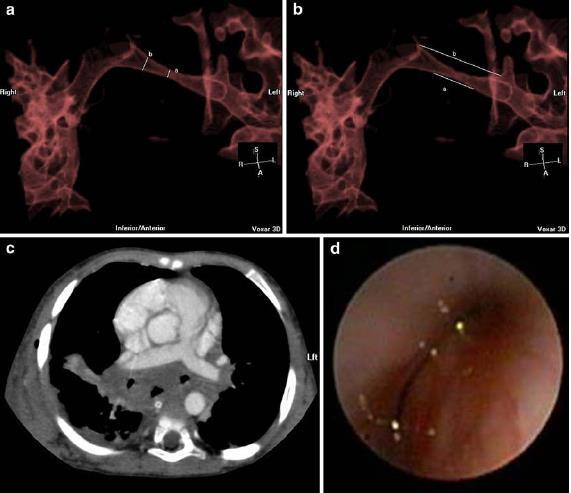
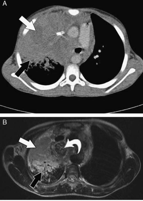
46 Figure 2 a and b: CT scan chest: A direct communication is demonstrated between the oesophagus and the left main bronchus.


47 Figure 1 a and b: Axial CT scans in a child with BOF involving the left main bronchus Contrast enhanced axial CT immediately caudal to the carina demonstrates a calcified subcarinal lymph node that abuts the left main bronchus and has eroded into the oesophagus and projects into the lumen. In addition there is compression of the bronchus intermedius between the subcarinal node and a large right hilar node. Lung window more caudally demonstrates a large amount of air in the distal third of the oesophagus. This axial slcie also demonstrates a solitary parenchymal nodule representing a Ghon focus at the right cardiophrenic angle.


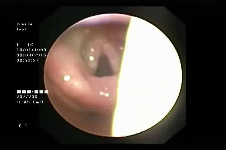
48 Bronchoscopy demonstrates left sided fistula just below the carina Bronchoscopy demonstrates methylene blue in the airway after instillation in oesophagus demonstrating the BOF
49 Which is better CT-scan or scope?

50 Conclusions: Flexible bronchoscopy remains a very relevant tool in the diagnostic and therapeutic management of childhood pulmonary tuberculosis but resulted in treatment modifi cation or microbiological proof in a minority of our patients. We propose that fl exible bronchoscopy in children with pulmonary tuberculosis be limited to those who show tracheobronchial luminal narrowing on an initial CT scan. Arch Dis Child 2010;95:
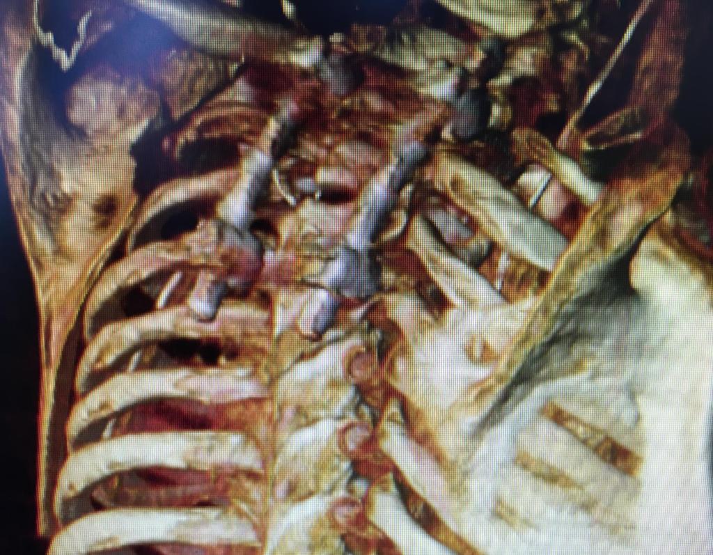
51 Conclusion 3-D VR demonstrates a very good correlation with FTB in determining airway compression caused by TB lymphadenopathy in children. In combination with FTB, 3- D VR adds confidence to the bronchoscopy findings and complements FTB by adding additional information on the status of the airway distal to severe obstructions unreachable by FTB.
52
53 FDG scan

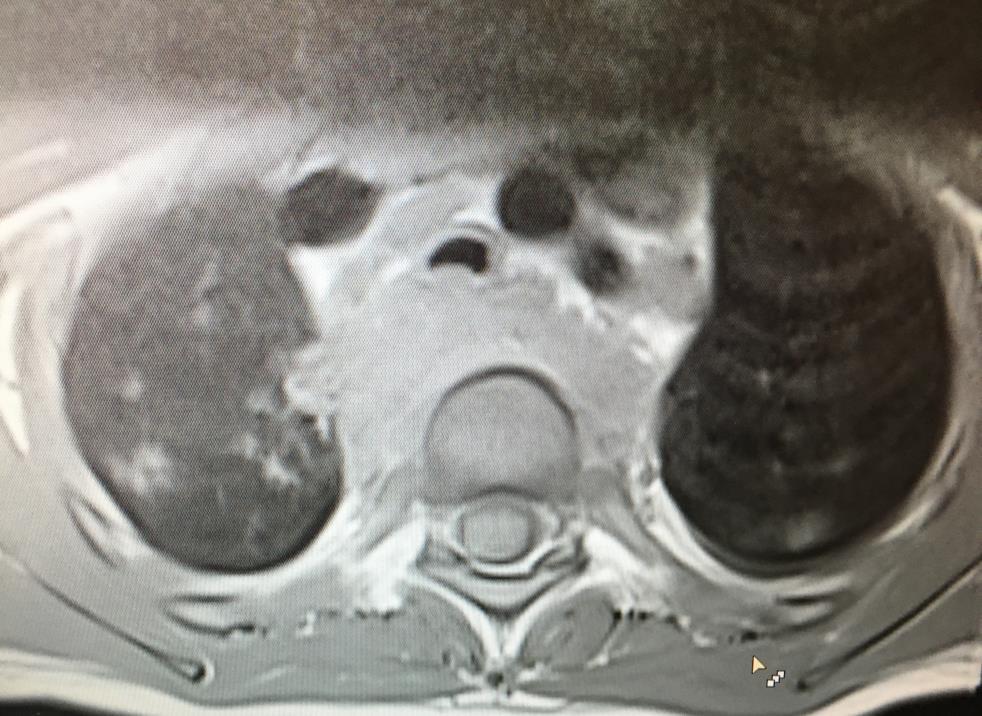
54 MR Conclusion: Lung parenchymal necrosis in primary pulmonary TB in children may be of low signal intensity on T2 and STIR magnetic resonance imaging. This may be distal to lymphobronchial obstruction and is probably due to the caseating necrosis. J Thorac Imaging 2011;00:

55 Postcontrast CT scan of the lungs demonstrating central areas of low density (arrows) within an area of airspace consolidation. Note the lack of vessels, bronchograms, and enhancement in the areas of low density, representing lung necrosis. In contrast, the airspace consolidation demonstrates enhancement, vessels, and air bronchograms.
 presumably due to different stages of")
56 STIR MRI demonstrates that one area of CT low density demonstrates high signal (white arrow) intensity, whereas the other demonstrates low signal intensity (black arrow) presumably due to different stages of necrosis.
, whereas the airspace consolidation")
57 Postgadolinium T1-weighted MRI demonstrates that the areas of CT low density lack enhancement (arrows), whereas the airspace consolidation enhances.
58
59 Why should we treat TB correctly... Most get better anyway Airway related complications Broncheal stenosis Balloon Dilatations Parenchymal complications Cystic lung disease and volume loss lobectomy / pneumonectomy Bronchiectasis HIV/ BOF related asymptomatic / symptomatic Fibrosing mediastinitis: Lethal disease

60 Complications of PTB 1. Bronchial stenosis 2. Bronchiectasis 3. Hemoptysis 4. Lung cysts 5. Fibrosing mediastinitis
61
62
63
64

65
66
67 Fibrosing mediastinatis
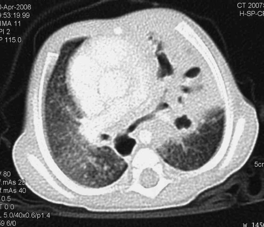
68 TB empyema Pleural involvement due to TB is believed to result from direct spread of caseous material from a sub-pleural parenchymal or lymph node focus, or from haematogenous spread. The degree of pleural pathology depends on the dose and virulence of bacilli that enter the pleural space and the immune status of the patient. The presence of caseous material in the pleural space triggers a hypersensitivity inflammatory response which leads to the accumulation of serous straw-colored fluid containing few tuberculosis bacilli. If there is active caseation in the pleural space, thick loculated pus, containing many tuberculosis bacilli will develop. The chest CT scan picture includes pleural thickening with enhancement, fluid collections with mediastinal lymph nodes and parenchymal lesions. The presence of caseating empyema is normally indicated by a persistent high swinging fever and aspiration of pus can be very difficult due to the fact that it is very thick.
69 A B
70

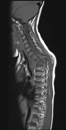
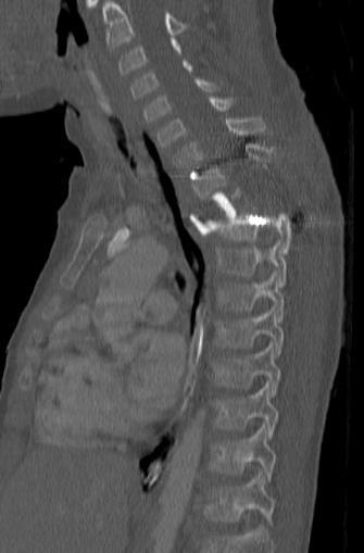
71 Tuberculosis of the spine
72
73

74
75 Why should we treat TB correctly... Most get better anyway Airway related complications Broncheal stenosis Balloon Dilatations Parenchymal complications Cystic lung disease and volume loss lobectomy / pneumonectomy Bronchiectasis HIV/ BOF related asymptomatic / symptomatic Fibrosing mediastinitis: Lethal disease
76 Lung biopsy for TB diagnosis

77

78 5/51 cases = 9.8%
79 The present study is one of the first to describe TB as a cause of DLD in infants and children. Our study emphasises that, in a high TB burden country, TB pneumonia cannot be excluded using conventional investigations. In the one child treated for pulmonary TB before the OLB, MDR-TB was isolated, necessitating a change of treatment. Few studies have described the clinical picture of acute pneumonia caused by M. tuberculosis. In a study from Uganda, risk factors associated with severe pneumonia caused by M. tuberculosis were children aged,5 years (odds ratio [OR] 2.4, 95%CI ) and contact with a smear-positive TB case (OR 3.0, 95%CI ).14

80
81 The Future PC models for predicting PTB Virtual bronchoscopy EBUS Electromagnetc navigation system to biopsy peripheral lung nodules
82 Presents two methods to analyse airway shape and detect airway pathology from CT images. Features were extracted using (1) the principal components of the airway surface mesh and (2) branch radius and orientation features. These methods were applied to a dataset of 61 TB and non-tb paediatric patients. Nested cross-validation of the support vector classifier found the sensitivity of detecting TB to be 86% and a specificity of 91% for the first 10 PCA modes while radius based features had a sensitivity of 86% and a specificity of 94%. These methods show the potential of computer assisted detection of TB and other airway pathology from airway shape deformation.
83

84 Virtual bronchoscopy
85 Not PTB Lymphoma Foreign body aspiration Vascular and cardiac abnormalities Bronchogenic cyst Cryptococcus neoformans

86
87
88
89

90 Fig 1 Fig 2 Chest CT-scan: Fig 1 anterior mediastinal lymphnodes and para tracheal lymphnodes causing tracheal compression from the right side. Fig 2 Subcarinal lymphnodes and anterior mediastinal glands causing compression of carina and right main bronchus


91 Bronchoscopy Fig a Fig b Bronchoscopy: Fig a demonstrates tracheal compression and fig b external compression of both Right upper lobe bronchus and bronchus intermedius

92 Enucleation of lymph glands Paratracheal and Subcarinal Histology Granulomatous inflammation Stain positive for yeast cells Cytology Cryptococcus neoformans Tuberculous Zn and culture negative Special investigations
COMPUTED TOMOGRAPHY DEMONSTRATION OF THE COMPLICATIONS AND ASSOCIATIONS OF LYMPHOBRONCHIAL TUBERCULOSIS IN CHILDREN
 COMPUTED TOMOGRAPHY DEMONSTRATION OF THE COMPLICATIONS AND ASSOCIATIONS OF LYMPHOBRONCHIAL TUBERCULOSIS IN CHILDREN Dr Susan Lucas A research report submitted to the Faculty of Health Sciences, University
Intrathoracic tuberculous lymphadenopathy in children: a guide to chest radiography
 Pediatr Radiol (2017) 47:1277 1282 DOI 10.1007/s00247-017-3890-1 MINISYMPOSIUM: IMAGING OF CHILDHOOD TUBERCULOSIS Intrathoracic tuberculous lymphadenopathy in children: a guide to chest radiography Anthony
PULMONARY TUBERCULOSIS RADIOLOGY
 PULMONARY TUBERCULOSIS RADIOLOGY RADIOLOGICAL MODALITIES Medical radiophotography Radiography Fluoroscopy Linear (conventional) tomography Computed tomography Pulmonary angiography, bronchography Ultrasonography,
Diagnosis of tuberculosis in children
 Diagnosis of tuberculosis in children H Simon Schaaf Desmond Tutu TB Centre Department of Paediatrics and Child Health, Stellenbosch University, and Tygerberg Children s Hospital (TCH) Estimated TB incidence
TB Intensive Houston, Texas
 TB Intensive Houston, Texas October 15-17, 17 2013 Diagnosis of TB: Radiology Rosa M Estrada-Y-Martin, MD MSc FCCP October 16, 2013 Rosa M Estrada-Y-Martin, MD MSc FCCP, has the following disclosures to
TB Radiology for Nurses Garold O. Minns, MD
 TB Nurse Case Management Salina, Kansas March 31-April 1, 2010 TB Radiology for Nurses Garold O. Minns, MD April 1, 2010 TB Radiology for Nurses Highway Patrol Training Center Salina, KS April 1, 2010
Chest Radiology Interpretation: Findings of Tuberculosis
 Chest Radiology Interpretation: Findings of Tuberculosis Get out your laptops, smart phones or other devices pollev.com/chestradiology Case #1 1 Plombage Pneumonia Cancer 2 Reading the TB CXR Be systematic!
Diagnostic Value of EBUS-TBNA in Various Lung Diseases (Lymphoma, Tuberculosis, Sarcoidosis)
") Diagnostic Value of EBUS-TBNA in Various Lung Diseases (Lymphoma, Tuberculosis, Sarcoidosis) Sevda Sener Cömert, MD, FCCP. SBU, Kartal Dr.Lütfi Kırdar Training and Research Hospital Department of Pulmonary
Diagnostic Value of EBUS-TBNA in Various Lung Diseases (Lymphoma, Tuberculosis, Sarcoidosis) Sevda Sener Cömert, MD, FCCP. SBU, Kartal Dr.Lütfi Kırdar Training and Research Hospital Department of Pulmonary
TB Intensive San Antonio, Texas November 29-December 2, 2011
 TB Intensive San Antonio, Texas November 29-December 2, 2011 Diagnosis of TB: Radiology Michael McCarthy, MD, FACR November 30, 2011 Michael McCarthy, MD, FACR has the following disclosures to make: No
An Introduction to Radiology for TB Nurses
 An Introduction to Radiology for TB Nurses Garold O. Minns, MD September 14, 2017 TB Nurse Case Management September 12 14, 2017 EXCELLENCE EXPERTISE INNOVATION Garold O. Minns, MD has the following disclosures
New Horizons in the Imaging of the Lung
 New Horizons in the Imaging of the Lung Postprocessing. How to do it and when do we need it? Peter M.A. van Ooijen, MSc, PhD Principal Investigator, Radiology, UMCG Discipline Leader Medical Imaging Informatics
INTRATHORACIC TUBERCULOSIS IN CHILDREN
 DIAGNOSTIC ATLAS OF INTRATHORACIC TUBERCULOSIS IN CHILDREN A Guide For Low Income Countries 2003 International Union Against Tuberculosis and Lung Disease DIAGNOSTIC ATLAS OF INTRATHORACIC TUBERCULOSIS
TB in Children. Rene De Gama Block 10 Lectures 2012
 TB in Children Rene De Gama Block 10 Lectures 2012 Contents Epidemiology Transmission and pathogenesis Diagnosis of TB TB and HIV Management Epidemiology The year 2000 8.3 million new TB cases diagnosed
The Surgical Treatment of Tracheobronchial Tuberculosis. The Thoracic Department of Beijing Chest Hospital, Capital Medical University
 The Surgical Treatment of Tracheobronchial Tuberculosis ) The Thoracic Department of Beijing Chest Hospital, Capital Medical University Named also: endobronchial tuberculosis,ebtb defined as tuberculous
10/17/2016. Nuts and Bolts of Thoracic Radiology. Objectives. Techniques
 Nuts and Bolts of Thoracic Radiology October 20, 2016 Carleen Risaliti Objectives Understand the basics of chest radiograph Develop a system for interpreting chest radiographs Correctly identify thoracic
Tuberculosis: The Essentials
 Tuberculosis: The Essentials Kendra L. Fisher, MD, PhD THORACIC TUBERCULOSIS: THE BARE ESSENTIALS Kendra Fisher MD, FRCP (C) Department of Radiology Loma Linda University Medical Center TUBERCULOSIS ()
Diagnosis of tuberculosis in children H Simon Schaaf
 Diagnosis of tuberculosis in children H Simon Schaaf Desmond Tutu TB Centre Department of Paediatrics and Child Health, Stellenbosch University, and Tygerberg Children s Hospital (TCH) Estimated TB incidence
Pediatric TB Intensive Houston, Texas
 Pediatric TB Intensive Houston, Texas November 13, 2009 Radiographic Manifestations of Pediatric TB Susan D. John, MD, FACR November 13, 2009 Radiologic Presentation of Childhood TB Susan D. John, MD,
Subject Index. Bacterial infection, see Suppurative lung disease, Tuberculosis
 Subject Index Abscess, virtual 107 Adenoidal hypertrophy, features 123 Airway bleeding, technique 49, 50 Airway stenosis, see Stenosis, airway Anaesthesia biopsy 47 complications 27, 28 flexible 23 26
Interventional Pulmonology
 Interventional Pulmonology The Division of Thoracic Surgery Department of Cardiothoracic Surgery New York Presbyterian/Weill Cornell Medical College p: 212-746-6275 f: 212-746-8223 https://weillcornell.org/eshostak
Bronchial syndrome. Atelectasis Draining bronchus Bronchiectasis
 Bronchial syndrome Atelectasis Draining bronchus Bronchiectasis Etienne Leroy Terquem Pierre L Her SPI / ISP Soutien Pneumologique International / International Support for Pulmonology Atelectasis Consequence
Bronchogenic Carcinoma
 A 55-year-old construction worker has smoked 2 packs of ciggarettes daily for the past 25 years. He notes swelling in his upper extremity & face, along with dilated veins in this region. What is the most
Pediatric TB Radiology: It s Not Black and White Part 2
 Experiencing technical difficulties? Please call Adobe Connect for technical assistance at 1-800-422-3623 Pediatric TB Radiology: It s Not Black and White Part 2 June 18, 2018 A National Webinar June 18,
Pediatric TB Intensive Houston, Texas October 14, 2013
 Pediatric TB Intensive Houston, Texas October 14, 2013 Radiologic Presentation of Childhood TB Susan D. John, MD, FACR October 14, 2013 Disclosures I have no disclosures or conflicts of interest to report
Tuberculosis - clinical forms. Dr. A.Torossian,, M.D., Ph. D. Department of Respiratory Diseases
 Tuberculosis - clinical forms Dr. A.Torossian,, M.D., Ph. D. Department of Respiratory Diseases 1 TB DISEASE Primary Post-primary (Secondary) Common primary forms Primary complex Tuberculosis of the intrathoracic
Overview of airway involvement in tuberculosis
 bs_bs_banner Journal of Medical Imaging and Radiation Oncology 57 (2013) 576 581 RADIOLOGY PICTORIAL ESSAY Overview of airway involvement in tuberculosis Arundeep Arora, Ashu Seith Bhalla, Manisha Jana
GUIDELINES FOR CANCER IMAGING Lung Cancer
 GUIDELINES FOR CANCER IMAGING Lung Cancer Greater Manchester and Cheshire Cancer Network Cancer Imaging Cross-Cutting Group April 2010 1 INTRODUCTION This document is intended as a ready reference for
Lung. 10/24/13 Chest X-ray: 2.9 cm mass like density in the inferior lingular segment worrisome for neoplasm. Malignancy cannot be excluded.
 Lung Case Scenario 1 A 54 year white male presents with a recent abnormal CT of the chest. The patient has a history of melanoma, kidney, and prostate cancers. 10/24/13 Chest X-ray: 2.9 cm mass like density
Undergraduate Teaching
 Prof. James F Meaney Undergraduate Teaching Chest X-Ray Understanding the normal anatomical by reference to cross sectional imaging Radiology? It s FUN! Cryptic puzzle Sudoku (Minecraft?) It s completely
Diagnosis of TB: Radiology David Finlay, MD
 TB Intensive Tyler, Texas June 2-4, 2010 Diagnosis of TB: Radiology David Finlay, MD June 3, 2010 2stages stages- Tuberculosis 1. primary infection 2. reactivation, or post primary disease 2 1 Primary
The Various Methods to Biopsy the Lung PROF SHITRIT DAVID HEAD, PULMONARY DEPARTMENT MEIR MEDICAL CENTER, ISRAEL
 The Various Methods to Biopsy the Lung PROF SHITRIT DAVID HEAD, PULMONARY DEPARTMENT MEIR MEDICAL CENTER, ISRAEL Conflict of Interest This presentation is supported by AstraZeneca Two main steps before
Atypical radiologic appearances of pulmonary tuberculosis in non-hiv adult patients
 Atypical radiologic appearances of pulmonary tuberculosis in non-hiv adult patients Poster No.: R-0200 Congress: RANZCR-AOCR 2012 Type: Educational Exhibit Authors: K. N. Jeon, K. Bae, M. J. Park Keywords:
Radiology Pathology Conference
 Radiology Pathology Conference Sharlin Johnykutty,, MD, Cytopathology Fellow Sara Majewski, MD, Radiology Resident Friday, August 28, 2009 Presentation material is for education purposes only. All rights
Case of the Day Chest
 Case of the Day Chest Darin White MDCM FRCPC Department of Radiology, Mayo Clinic 76 th Annual Scientific Meeting Canadian Association of Radiologists Montreal, QC April 26, 2013 2013 MFMER slide-1 Disclosures
Slide 1. Slide 2. Slide 3. Investigation and management of lung cancer Robert Rintoul. Epidemiology. Risk factors/aetiology
 Slide 1 Investigation and management of lung cancer Robert Rintoul Department of Thoracic Oncology Papworth Hospital Slide 2 Epidemiology Second most common cancer in the UK (after breast). 38 000 new
An Image Repository for Chest CT
 An Image Repository for Chest CT Francesco Frajoli for the Chest CT in Antibody Deficiency Group An Image Repository for Chest CT he Chest CT in Antibody Deficiency Group is an international and interdisciplinary
Radiological Features of Mycobacterium tuberculosis TUBERCULE BACILLUS TUBERCULE BACILLUS DIAGNOSIS. Guy Richards. PATHOGENESIS of TUBERCULOSIS
 Radiological Features of Guy Richards Department of critical care Charlotte Maxeke Johannesburg Academic Hospital University of the Witwatersrand, Johannesburg, South Africa TUBERCULE BACILLUS Discovery
Pulmonary TB Clinical Diagnosis
 Pulmonary TB Clinical Diagnosis Dr Onn Min Kon TB Clinics St Mary s Hospital + Hammersmith Hospital History back to basics Symptoms Ethnicity/ age Recent arrival/ travel history Contact history BCG history
TB & HIV CO-INFECTION IN CHILDREN. Reené Naidoo Paediatric Infectious Diseases Broadreach Healthcare 19 April 2012
 TB & HIV CO-INFECTION IN CHILDREN Reené Naidoo Paediatric Infectious Diseases Broadreach Healthcare 19 April 2012 Introduction TB & HIV are two of the leading causes of morbidity & mortality in children
Sectional Anatomy Quiz - III
 Sectional Anatomy - III Rashid Hashmi * Rural Clinical School, University of New South Wales (UNSW), Wagga Wagga, NSW, Australia A R T I C L E I N F O Article type: Article history: Received: 30 Jun 2018
The indications and role of paediatric bronchoscopy in a developing. country, with high prevalence of pulmonary tuberculosis and HIV
 The indications and role of paediatric bronchoscopy in a developing country, with high prevalence of pulmonary tuberculosis and HIV I Webster, FC Paed 1 P. Goussard, Phd (Paed), 1 R.P. Gie, FC Paed, 1
Introduction to Interventional Pulmonology
 Introduction to Interventional Pulmonology Alexander Chen, M.D. Director, Interventional Pulmonology Assistant Professor of Medicine and Surgery Divisions of Pulmonary and Critical Care Medicine and Cardiothoracic
Respiratory Interactive Session. Elaine Borg
 Respiratory Interactive Session Elaine Borg Case 1 Respiratory Cytology 55 year old gentleman Anterior mediastinal mass EBUS FNA Case 1 Respiratory Cytology 55 year old gentleman with anterior mediastinal
Use of Integrated PET CT in the Clinical Staging of Non Small Cell Lung Cancer
 November 2010 Use of Integrated PET CT in the Clinical Staging of Non Small Cell Lung Cancer Laura Myers, Harvard Medical School, Year III Clinical Presentation 79yo woman with cough productive of green
Surgical indications: Non-malignant pulmonary diseases. Punnarerk Thongcharoen
 Surgical indications: Non-malignant pulmonary diseases Punnarerk Thongcharoen Non-malignant Malignant as a pathological term: Cancer Non-malignant = not cancer Malignant as an adjective: Disposed to cause
Radiological Aspects of Pulmonary Tuberculosis in Immunocompetent Hosts
 Nov 2003 Radiological Aspects of Pulmonary Tuberculosis in Immunocompetent Hosts Josh Rempell, Harvard Medical School Year III Tuberculosis: the captain of all (wo)men of death Overall, one third of the
Right infrahilar nodule
 Right infrahilar nodule Search Infrahilar nodule Nov 9, 2015.. CT chest showed a right infrahilar mass 3.5 2.5 cm along with multiple bilateral lung nodules of size 9 to 11 mm. Bronchoscopy. Jun 13, 2015.
American College of Radiology ACR Appropriateness Criteria
 American College of Radiology ACR Criteria Radiologic Management of Thoracic Nodules and Masses Variant 1: Middle-aged patient (35 60 years old) with an incidental 1.5-cm lung nodule. The lesion was smooth.
UERMMMC Department of Radiology. Basic Chest Radiology
 UERMMMC Department of Radiology Basic Chest Radiology PHYSICS DENSITIES BONE SOFT TISSUES WATER FAT AIR TELEROENTGENOGRAM Criteria for an Ideal Chest Radiograph 1. Upright 2. Posteroanterior View 3. Full
Tuberculosis. By: Shefaa Q aqa
 Tuberculosis By: Shefaa Q aqa Tuberculosis is a communicable chronic granulomatous disease caused by Mycobacterium tuberculosis. It usually involves the lungs but may affect any organ or tissue in the
Chief Complain. For chemotherapy
 Chief Complain For chemotherapy Present Illness 93.12 Progressive weakness of R t arm for 1 year X-ray: peneative lesion over right proximal humorous Bone scan: multiple increased intake Biopsy of distal
OBJECTIVES. Solitary Solid Spiculated Nodule. What would you do next? Case Based Discussion: State of the Art Management of Lung Nodules.
 Organ Imaging : September 25 2015 OBJECTIVES Case Based Discussion: State of the Art Management of Lung Nodules Dr. Elsie T. Nguyen Dr. Kazuhiro Yasufuku 1. To review guidelines for follow up and management
MEDIASTINAL STAGING surgical pro
 MEDIASTINAL STAGING surgical pro Paul E. Van Schil, MD, PhD Department of Thoracic and Vascular Surgery University of Antwerp, Belgium Mediastinal staging Invasive techniques lymph node mapping cervical
GROUP 1: Peripheral tumour with normal hilar and mediastinum on staging CT with no disant metastases. Including: Excluding:
 GROUP 1: Including: Excluding: Peripheral tumour with normal hilar and mediastinum on staging CT with no disant metastases Solid pulmonary nodules 8mm diameter / 300mm3 volume and BROCK risk of malignancy
WORLD TUBERCULOSIS DAY 2018
 WORLD TUBERCULOSIS DAY 2018 Bernard F. Laya, MD, DO, FPCR LOWER RESPIRATORY INFECTIONS } Most common cause of illness in children and is a significant cause of morbidity and mortality } 156 million cases
David E. Griffith, MD has the following disclosures to make:
 Diagnosis of TB: Radiology David E. Griffith, MD March 13, 2015 TB for Pulmonologist March 13, 2015 Phoenix, AZ EXCELLENCE EXPERTISE INNOVATION David E. Griffith, MD has the following disclosures to make:
Mediastinal Mysteries: What can be solved with EBUS?
 Mediastinal Mysteries: What can be solved with EBUS? W. Graham Carlos MD Pulmonary & Critical Care Fellow Indiana University School of Medicine Disclosures None Objectives Introduce you to the technique
Endoscopy. Pulmonary Endoscopy
 Pulmonary 1 Direct visualization of TB tree Developed in 1890 s to remove foreign bodies - rigid metal tube Advances added light system, Sx Flexible fiberoptic scopes introduced in early 1960 s 2 Used
Larry Tan, MD Thoracic Surgery, HSC. Community Cancer Care Educational Conference October 27, 2017
 Larry Tan, MD Thoracic Surgery, HSC Community Cancer Care Educational Conference October 27, 2017 To describe patient referral & triage for the patient with suspected lung cancer To describe the initial
Pictorial essay of unusual radiologic manifestations of pulmonary and airway metastasis at initial presentation of lung cancer
 Pictorial essay of unusual radiologic manifestations of pulmonary and airway metastasis at initial presentation of lung cancer Poster No.: C-2297 Congress: ECR 2012 Type: Educational Exhibit Authors: Y.
Thoracic Sarcoidosis Imaging Updated: Jul 19, 2013
 Thoracic Sarcoidosis Imaging Updated: Jul 19, 2013 Overview Radiography Computed Tomography Magnetic Resonance Imaging Nuclear Imaging Show All Multimedia Library References Overview For patients with
Endobronchial Ultrasound in the Diagnosis & Staging of Lung Cancer
 Endobronchial Ultrasound in the Diagnosis & Staging of Lung Cancer Dr Richard Booton PhD FRCP Lead Lung Cancer Clinician, Consultant Respiratory Physician & Speciality Director Manchester University NHS
Bronchial carcinosarcoma
 Bronchial carcinosarcoma Carolina Carcano 1*, Edward Savage 2, Maria Julia Diacovo 3, Jacobo Kirsch 1 1. Division of Radiology, Cleveland Clinic Florida, Weston, Fl, USA 2. Department of Thoracic and Cardiovascular
Radiological staging of lung cancer. Shukri Loutfi,MD,FRCR Consultant Thoracic Radiologist KAMC-Riyadh
 Radiological staging of lung cancer Shukri Loutfi,MD,FRCR Consultant Thoracic Radiologist KAMC-Riyadh Bronchogenic Carcinoma Accounts for 14% of new cancer diagnoses in 2012. Estimated to kill ~150,000
Chest radiograph of an. asymptomatic man. Case report. Case history
 Eleftheria Chaini 1, Niki Giannakou 2, Dimitra Haini 3, Anna Maria Athanassiadou 4, Angelos Tsipis 4, Nikolaos D. Hainis 5 elhaini@otenet.gr 1 Pulmonary Dept, Corfu General Hospital, Kontokali, Greece.
Do you want to be an excellent Radiologist? - Focus on the thoracic aorta on lateral chest image!!!
 The lateral chest radiograph: Challenging area around the thoracic aorta!!! Do you want to be an excellent Radiologist? - Focus on the thoracic aorta on lateral chest image!!! Dong Yoon Han 1, So Youn
Lung cancer in patients with chronic empyema
 Lung cancer in patients with chronic empyema Poster No.: P-0025 Congress: ESTI 2015 Type: Scientific Poster Authors: Y. Lee, C.-K. Park; Guri/KR Keywords: Neoplasia, Biopsy, PET-CT, CT, Thorax, Lung DOI:
ENDOBRONCHIAL ULTRASOUND (EBUS) TRAINING PROGRAMME CURRICULUM
 TRAINING PROGRAMME CURRICULUM") ENDOBRONCHIAL ULTRASOUND (EBUS) TRAINING PROGRAMME CURRICULUM This competency-based curriculum has been designed by a task force of interventional pulmonology specialists to underline the learning outcomes
ENDOBRONCHIAL ULTRASOUND (EBUS) TRAINING PROGRAMME CURRICULUM This competency-based curriculum has been designed by a task force of interventional pulmonology specialists to underline the learning outcomes
Radiological spectrum of Thoracic Sequelae and Complications of Tuberculosis
 Radiological spectrum of Thoracic Sequelae and Complications of Tuberculosis Poster No.: P-0013 Congress: ESTI 2014 Type: Educational Poster Authors: B. Alami, O. Addou, M. Jaffal, M. Y. Alaoui Lamrani,
Lung Cancer - Suspected
 Lung Cancer - Suspected Shared Decision Making Lung Cancer: http://www.enhertsccg.nhs.uk/ Patient presents with abnormal CXR Lung cancer - clinical presentation History and Examination Incidental finding
How to Analyse Difficult Chest CT
 How to Analyse Difficult Chest CT Complex diseases are:- - Large lesion - Unusual or atypical pattern - Multiple discordant findings Diffuse diseases are:- - Numerous findings in both sides 3 basic steps
Transbronchial fine needle aspiration
 Thorax 1982;37 :270-274 Transbronchial fine needle aspiration J LEMER, E MALBERGER, R KONIG-NATIV From the Departments of Cardio-thoracic Surgery and Cytology, Rambam Medical Center, Haifa, Israel ABSTRACT
The Imaging Analysis of Pulmonary Sarcodiosis
 www.cancercellresearch.org ISSN: 2161-2609 Article The Imaging Analysis of Pulmonary Sarcodiosis Xin He, Chuanyu Zhang* Department of Radiology, Affiliated Hospital of Qingdao University, Qingdao, China
Let s Talk TB A Series on Tuberculosis, A Disease That Affects Over 2 Million Indians Every Year
 A Series on Tuberculosis, A Disease That Affects Over 2 Million Indians Every Year Barry Rabinovitch, MD, FRCP(C) Author Madhukar Pai, MD, PhD co-author and Series Editor Barry Rabinovitch is an assistant
Utilizing EBUS (Endobronchial Ultrasound) for Diagnosis of Lung Cancer and other Pulmonary Diseases
 for Diagnosis of Lung Cancer and other Pulmonary Diseases") Utilizing EBUS (Endobronchial Ultrasound) for Diagnosis of Lung Cancer and other Pulmonary Diseases Akintayo Sokunbi, M.D MidMichigan Hospital Midland, Michigan Objectives Discuss EBUS guided biopsy principles
Utilizing EBUS (Endobronchial Ultrasound) for Diagnosis of Lung Cancer and other Pulmonary Diseases Akintayo Sokunbi, M.D MidMichigan Hospital Midland, Michigan Objectives Discuss EBUS guided biopsy principles
FDG PET/CT in Lung Cancer Read with the experts. Homer A. Macapinlac, M.D.
 FDG PET/CT in Lung Cancer Read with the experts Homer A. Macapinlac, M.D. Patient with suspected lung cancer presents with left sided chest pain T3 What is the T stage of this patient? A) T2a B) T2b C)
Web Chapter 3. Image Gallery: Lesion detection on low dose chest CT
 Web Chapter 3 Image Gallery: Lesion detection on low dose chest CT Sarabjeet Singh, MD Mannudeep K. Kalra, MD *Eugene J. Mark, MD *James Stone, MD James H. Thrall, MD Department of Radiology and *Department
Standardized radiographic interpretation of thoracic tuberculosis in children
 Pediatr Radiol (2017) 47:1237 1248 DOI 10.1007/s00247-017-3868-z MINISYMPOSIUM: IMAGING OF CHILDHOOD TUBERCULOSIS Standardized radiographic interpretation of thoracic tuberculosis in children Nathan David
Pathology of pulmonary tuberculosis. Dr: Salah Ahmed
 Pathology of pulmonary tuberculosis Dr: Salah Ahmed Is a chronic granulomatous disease, caused by Mycobacterium tuberculosis (hominis) Usually it involves lungs but may affect any organ or tissue Transmission:
2017 Coding & Payment Quick Reference
 2017 Coding & Payment Quick Reference Select Pulmonary Procedures Payer policies will vary and should be verified prior to treatment for limitations on diagnosis, coding or site of service requirements.
Surgical treatment of empyema in children
 Surgical treatment of empyema in children Jacques Janson Pierre Goussard Cardiothoracic Surgery, Paediatric Pulmonology Tygerberg Academic Hospital University of Stellenbosch Pleural space Netter, Frank
JMSCR Vol 06 Issue 03 Page March 2018
 www.jmscr.igmpublication.org Impact Factor (SJIF): 6.379 Index Copernicus Value: 71.58 ISSN (e)-2347-176x ISSN (p) 2455-4 DOI: https://dx.doi.org/.18535/jmscr/v6i3.63 Diagnostic Role of FOB in Radiological
Congenital Lung Malformations: Radiologic-Pathologic Correlation
 Acta Radiológica Portuguesa, Vol.XVIII, nº 70, pág. 51-60, Abr.-Jun., 2006 Congenital Lung Malformations: Radiologic-Pathologic Correlation Marilyn J. Siegel Mallinckrodt Institute of Radiology, Washington
Case 1 : Question. 1.1 What is the intralobular distribution? 1. Centrilobular 2. Perilymphatic 3. Random
 Interesting case Case 1 Case 1 : Question 1.1 What is the intralobular distribution? 1. Centrilobular 2. Perilymphatic 3. Random Case 1: Answer 1.1 What is the intralobular distribution? 1. Centrilobular
Signs in Chest Radiology
 Signs in Chest Radiology Jonathan H. Chung, MD Disclosures No pertinent disclosures Jonathan H. Chung, MD Assistant Professor Institute t of fadvanced d Biomedical Imaging National Jewish Health Denver,
Sectional Anatomy Quiz II
 Sectional Anatomy II Rashid Hashmi Rural Clinical School, University of New South Wales, Wagga Wagga, New South Wales, Australia A R T I C L E I N F O Article type: Article history: Received: 3 Aug 2017
Owing to the recent attention given to lung cancer
 Electromagnetic : A Surgeon s Perspective Todd S. Weiser, MD, Kevin Hyman, MD, Jaime Yun, MD, Virginia Litle, MD, Cythinia Chin, MD, and Scott J. Swanson, MD Department of Cardiothoracic Surgery, Mount
Wheeze. Dr Jo Harrison
 Wheeze Dr Jo Harrison 9.9.14 Wheeze - Physiology a continuous musical sound that lasts longer than 250 msec. can be high-pitched or low-pitched, consist of single or multiple notes, and occur during inspiration
Lecture 3. Inflammatory Processes
 Lecture 3 Inflammatory Processes Process: Increased vascular permeability Water and cellular infiltrations Results: Abscess, ulceration, cavitation Penetration, perforation and fistula formation Scarring,
Chapter 8. Other Important Tests and Procedures. Mosby items and derived items 2011, 2006 by Mosby, Inc., an affiliate of Elsevier Inc.
 Chapter 8 Other Important Tests and Procedures 1 Introduction Additional important diagnostic studies include: Sputum examination Skin tests Endoscopic examination Lung biopsy Thoracentesis Hematology,
Discussing feline tracheal disease
 Vet Times The website for the veterinary profession https://www.vettimes.co.uk Discussing feline tracheal disease Author : ANDREW SPARKES Categories : Vets Date : March 24, 2008 ANDREW SPARKES aims to
Electromagnetic navigational bronchoscopy in patients with solitary pulmonary nodules
 Original article Electromagnetic navigational bronchoscopy in patients with solitary pulmonary nodules Samuel Copeland MD, Shrinivas Kambali MD, Gilbert Berdine MD, Raed Alalawi MD Abstract Background:
Eun-Young Kang, M.D., Jae Wook Lee, M.D., Ji Yung Choo, M.D., Hwan Seok Yong, M.D., Ki Yeol Lee, M.D., Yu-Whan Oh, M.D.
 Eun-Young Kang, M.D., Jae Wook Lee, M.D., Ji Yung Choo, M.D., Hwan Seok Yong, M.D., Ki Yeol Lee, M.D., Yu-Whan Oh, M.D. Department of Radiology, Korea University Guro Hospital, College of Medicine, Korea
Diagnostic Evaluation of NTM and Bronchiectasis
 Division of Pulmonary, Critical Care and Sleep Medicine Diagnostic Evaluation of NTM and Bronchiectasis Ashwin Basavaraj, MD, FCCP NTM patient education program November 9, 2016 Involves a combination
Isolated anthracosis: benign but neglected cause of bronchial stenosis and obstruction
 Isolated anthracosis: benign but neglected cause of bronchial stenosis and obstruction Poster No.: C-0143 Congress: ECR 2013 Type: Scientific Exhibit Authors: S. Kahkouee, R. Pourghorban, M. Bitarafan,
Tuberculous Pericarditis: A multimodality imaging approach
 Tuberculous Pericarditis: A multimodality imaging approach Poster No.: C-1612 Congress: ECR 2011 Type: Authors: Educational Exhibit A. S. Udare 1, P. K. Mondel 1, A. A. Raut 2 ; 1 Mumbai, Maharastra/IN,
Interesting Cases. Pulmonary
 Interesting Cases Pulmonary 54M with prior history of COPD, hep B/C, and possible history of TB presented with acute on chronic dyspnea, and productive cough Hazy opacity overlying the left hemithorax
Original Article. Section: Radiology INTRODUCTION MATERIALS AND METHODS
 DOI: 10.21276/aimdr.2016.2.6.RD4 Original Article ISSN (O):2395-2822; ISSN (P):2395-2814 To Compare the Diagnostic Efficacy of HRCT Lung with Chest Radiographic Findings and Clinical Correlation with Microbiological