Classification of Benign and Malignant Vertebral Compression Fractures in Magnetic Resonance Images
|
|
|
- Quentin Floyd
- 5 years ago
- Views:
Transcription


1 Classification of Benign and Malignant Vertebral Compression Fractures in Magnetic Resonance Images Lucas Frighetto-Pereira, Guilherme Augusto Metzner, Paulo Mazzoncini de Azevedo-Marques, Rangaraj Mandayam Rangayyan, Marcello Henrique Nogueira-Barbosa

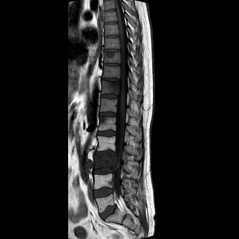
2 Anatomy of the spine MRI sagittal slice Vertebra Intervertebral Disc Vertebral Arch Lumbar Spine Vertebral Body
3 Vertebral compression fractures (VCFs) Partial collapse of vertebral bodies Traumatic VCFs raise no doubt about their etiology But a recent vertebral collapse without history of significant trauma creates difficulty in defining the cause of the VCF
4 Medical diagnosis Young patient with a VCF History of significant acute trauma Usually easy diagnosis
5 Medical diagnosis Elderly patient with VCF No history of significant acute trauma Diagnosis?
6 VCFs without history of significant trauma VCFs are the most common type of osteoporotic fractures The elderly have a high incidence of VCFs related to metastatic cancer affecting bone MRI is the most commonly used imaging method for spinal diseases and early detection of fractures

7 T1-Weighted MRI Osteoporotic VCF Metastatic VCF
8 Clinical classification of VCFs Osteoporotic VCFs classified as Benign VCFs Metastatic VCFs classified as Malignant VCFs
9 Benign VCFs in T1-weighted MRI Partial preservation of normal fatty bonemarrow signal in the vertebral body Degeneration of normally rectangular shapes of vertebrae into concave and rough shapes with indentations Rougher contours than malignant VCFs and normal vertebrae
10 Malignant VCFs in T1-weighted MRI Global reduction of signal intensity or nodular abnormality in the affected vertebral body Could result in a posterior convexity without substantial concavities May also cause the contours of vertebrae to be relatively smoothened due to convexity

11 Normal Benign VCFs Malignant VCFs
12 Benign vs Malignant VCFs Both tend to create concavities in the vertebral plateaus Could cause doubt in the diagnosis Correct classification is critical for planning treatment


13 Benign VCF Malignant VCF Which image has the malignant VCF and which one has the benign VCF?

14 Objectives Study the characteristics of VCFs in MRI Develop image processing techniques to extract features Classify VCFs Normal Benign Fractured Malignant
15 Study steps Selection of cases and images Manual segmentation of vertebral bodies Extraction of features of vertebral bodies Classification, validation, and statistical analysis
16 Database University Hospital of Ribeirão Preto Medical School University of São Paulo Cases and images collected from the Radiology Information System (RIS) Cases from September 2010 to March 2014 Philips 1.5T MRI System T1-weighted MRI
17 Database Lumbar vertebral bodies (L1 to L5) Median sagittal slice TIFF images with 8-bits/pixel 153 exams analyzed, 63 selected 38 women, 25 men Mean age: 62 years
18 Database 63 selected exams: At least one VCF per patient The nonfractured vertebral bodies of patients without malignant fractures are considered to be normal
19 Excluded cases Vertebral fractures secondary to trauma Infection and avascular necrosis Severe degenerative scoliosis Previous surgeries, radiotherapy, and chemotherapy
20 Database L5 L4 L3 L2 L1 Total Benign VCFs Malignant VCFs Normal Total
21 Examples of vertebral bodies Normal Benign VCFs Malignant VCFs


22 Manual segmentation MRI exam Vertebral body masks

23 Software flow chart

24 MRI exam and its mask

25 Detection of the coordinates of the vertebral bodies L1 L3 L2 L4 L5

26 Normalization of the MR images DiscsMean Extraction of blocks of intervertebral discs using the mask ROIs as reference. 5x5 disc block
27 Normalization of the MR images newimg i, j = imgoriginal(i, j) discsmean imgnorm i, j = 255 newimg i, j min newimg max newimg min newimg


28 MRI exam Mask Processing new image...

29 Detection of the ROIs

30 ROIs of the vertebral bodies Normal Benign VCFs Malignant VCFs
31 Computation of the features 3 Statistical gray-level features 14 Texture features 10 Shape features 27 Features

32 Statistical gray-level features Coefficient of variation Skewness Kurtosis
33 Statistical gray-level features Coefficient of variation (CV ) μ = i=1 x i CV = σ μ σ = i=1 x i μ 2
34 Statistical gray-level features Skewness skewness = (x i μ) σ 3 i=1
35 Statistical gray-level features Kurtosis kurtosis = (x i μ) σ 4 i=1
36 Differences in texture between normal and VCFs Malignant VCF Normal Malignant VCF Benign VCF Malignant VCF Malignant VCF
37 Texture features Gray-level cooccurrence matrix 14 texture features of Haralick et al.
38 Cooccurrence matrix Ex: Image 5x5 pixels, 3 gray levels Cooccurrence matrix p i, j for i = 0, j = Distance = 1 pixel Angle = ± Number of pixels of intensity 0 that are at ±45 degrees and distance 1 of pixels of intensity 2
39 14 texture features of Haralick et al. Angular second moment (Energy) f 1 = p(i, j) 2 Contrast i j Ng 1 Ng Ng f 2 = n 2 p i, j n=0 i=1 j=1 Ng: number of distinct gray levels in the quantized image
40 14 texture features of Haralick et al. Correlation f 3 = σ i σ j ij p i, j μ x μ y σ x σ y
41 14 texture features of Haralick et al. μ x, μ y means σ x, σ y standard deviations Ng p x i = p i, j j=1 Ng p y j = i=1 p i, j
42 14 texture features of Haralick et al. Sum of squares: Variance f 4 = i j i μ 2 p(i, j)
43 14 texture features of Haralick et al. Inverse difference moment f 5 = i j i j 2 p(i, j)
44 14 texture features of Haralick et al. Sum average 2Ng f 6 = i=2 i p x+y (i) Sum variance 2Ng f 7 = i=2 i f 8 2 p x+y (i)
45 14 texture features of Haralick et al. Ng Ng p x+y k = i=1 j=1 k = i + j p(i, j) k = 2,3,, 2Ng
46 14 texture features of Haralick et al. Sum entropy 2Ng f 8 = i=2 p x+y (i) log p x+y i Entropy f 9 = p i, j log p i, j i j
47 14 texture features of Haralick et al. Difference variance f 10 = variance of p x y Difference entropy Ng 1 f 11 = i=0 p x y (i) log p x y (i)
48 14 texture features of Haralick et al. Ng Ng p x y k = p(i, j) k = 0,1,, Ng 1 i=1 j=1 k = i j
49 14 texture features of Haralick et al. Information measures of correlation 1 f 12 = HXY HXY1 max HX, HY Information measures of correlation 2 f 13 = 1 exp 2 HXY2 HXY 1/2
50 14 texture features of Haralick et al. HXY = p i, j log i j p i, j HX and HY are entropy of p x and p y HXY1 = p i, j log i j p x i p y j HXY2 = p x i p y j log p x i p y j i j
51 14 texture features of Haralick et al. Maximal correlation coefficient f 14 = (second largest eigenvalue of Q) 1/2 where Q i, j = σ k p i,k p(j,k) p x i p y (k)
52 Shape features Compactness C o 4 A C o = 1 2 P, Perimeter P Vertebral area A
53 Shape features Fourier-descriptor-based feature FDF Z ( k) 1 N 1 = N n= 0 z( n)exp j 2 N nk k = -N/2+1,, -1, 0, 1, 2,, N/2 z(n) = x(n) + j y(n) n = 0, 1,..., N-1
54 Fourier-descriptor-based feature FDF Shape features + = = = + + = ) ( ) ( ) ( N N k N k k k k N k Z k Z k Z FDF

55 Shape features Convex deficiency CD CD = CH VA VA Convex hull CH Vertebral area VA
56 Shape features 7 Central invariant moments (Hu) M 1 = µ 20 + µ 02 M 2 = (µ 20 µ 02 ) µ 11 M 3 = (µ 30 3µ 12 ) 2 +(3µ 21 µ 03 ) 2 M 4 = (µ 30 + µ 12 ) 2 +(µ 21 + µ 03 ) 2
57 Shape features M 5 = µ 30 3µ 12 µ 30 + µ 12 [ µ 30 + µ µ 21 + µ 03 2 ] + (3µ 21 µ 03 )(µ 21 + µ 03 )[3(µ 30 + µ 12 ) 2 (µ 21 + µ 03 ) 2 ] M 6 = µ 20 µ 02 (µ 30 + µ 12 ) 2 (µ 21 + µ 03 ) 2 + 4µ 11 µ 30 + µ 12 µ 21 + µ 03 M 7 = (3µ 21 µ 03 )(µ 30 + µ 12 )[(µ 30 + µ 12 ) 2 3(µ 21 + µ 03 ) 2 ] (µ 30
58 Shape features µ 00 = m 00 = µ µ 10 = µ 01 = 0 µ 20 = m 20 µx² µ 11 = m 11 µxy µ 30 = m 30 3m 20 x + 2µx³ µ 21 = m 21 m 20 y 2m 11 x + 2µx²y µ 12 = m 12 m 02 x 2m 11 y + 2µx y² µ 03 = m 03 3m 02 y + 2µy³ µ 02 = m 02 µy²
59 Shape features m pq = i j i p j q img i, j, p, q = 0,1,2, x = m 10 m 00 y = m 01 m 00
60 Organization of the feature vector Coefficient of variation Skewness Kurtosis... M7

61 Files of features L1 L2 L3 txt files L4 L5

62 Inserting the reference classification Manual addition of the class Classification according to radiologist and biopsy Coefficient of variation Skewness Kurtosis... M7 Class VCF Normal Benign Malignant
63 Feature selection Software WEKA Wrapper method for feature selection knn with k = 1, 3,..., 13 Naïve Bayes RBF network Best first as search method Greedy search for the best subset of features
64 Classification Software WEKA Classifiers: k-nearest neighbor: k = 1, 3, 5, 7, 9, 11, 13 Naïve Bayes RBF network Stratified 10-fold cross-validation 9 folds for training, 1 fold for test
65 Clinical Classes VCF vs Normal Benign VCF vs Malignant VCF Malignant VCF, Benign VCF, and Normal
66 Validation Confusion Matrix Sensitivity Specificity AUROC % of correct classification
67 A z and p-values * for 0.01 p < 0.05 ** for p < 0.01 *** for p < p-values obtained using Wilcoxon rank-sum test NS indicates no significant difference NA indicates that A z could not be obtained
68 A z and p-values Benign VCF versus Malignant VCF All VCFs together versus Normal Feature Significance A z Significance A z CV NS *** Skew *** * Kurt *** NS H 1 *** NS H 2 *** * H 3 NS NS H 4 *** NS H 5 *** * H 6 *** *** H 7 *** NS H 8 *** ** H 9 *** *** H 10 *** ** 0.674
69 A z and p-values Benign VCF versus Malignant VCF All VCFs together versus Normal Feature Significance A z Significance A z H 11 *** ** H 12 *** NS H 13 *** *** H 14 NS NS C o *** *** FDF *** NS CD *** *** M 1 NS *** M 2 NS *** M 3 ** * M 4 * NS M 5 NS NS NA M 6 NS NS M 7 NS NS NA

70 A z and p-values


71 Mean and standard deviation of features
72 Mean and standard deviation of features Mean skewness of malignant VCFs is higher than that for benign VCFs T1 signals are distributed more on the lower side of the histogram for malignant VCFs H 6 and H 7 show large differences in their mean values for malignant VCFs versus benign VCFs

73 Mean and standard deviation of features
74 Feature selection
75 Feature selection k-nn did not select the gray-level features for benign vs malignant VCFs FDF, M 5, H 10, and H 13 were selected at least three times CV is statistically significant for all VCFs vs normal vertebral bodies and was selected for all classifiers
76 Feature selection Various texture features were selected for both types of classification Naïve Bayes selected the highest number of features for both types of classification
77 Classification Benign vs malignant VCFs Classifier ACC rate % AUROC k = k-nn k = k = k = Naïve Bayes RBF network All VCFs vs normal vertebral bodies Classifier ACC rate % AUROC k = k-nn k = k = Naïve Bayes RBF network k =
78 Classification RBF network classifier for benign vs malignant VCFs ACC rate was the lowest obtained AUROC is only better than that of 7-NN RBF network classifier for all VCFs vs normal vertebral bodies ACC rate is the highest obtained
79 Classification AUROC for classification of all VCFs together vs normal vertebral bodies is at least 0.92 AUROC of the naïve Bayes classifier is 0.97 for this purpose Better than the previous study using only shape features in which AUROC was This shows the importance of texture and gray-level features for this purpose
80 Classification AUROC for classification of benign vs malignant VCFs is 0.92 for naïve Bayes Better than the previous study in which the highest AUROC was 0.91 for 3-NN In a previous study using only shape features the highest AUROC was 0.78 This shows the importance of texture and graylevel features for this purpose
81 Benign VCFs, malignant VCFs, and normal vertebral bodies Predicted classification Malignant VCFs Benign VCFs Normal vertebral bodies True classification Malignant VCFs Benign VCFs Normal vertebral bodies Features selected: CV, Skew, H2, H3, H5, H6, H8, H9, H11, H12, H13,H14,Co, FDF, CD, M1, M3, and M7 Weighted average AUROC of 0.94 ACC rate of 82.7%
82 Limitations of the study Manual segmentation of the vertebral bodies Automatic segmentation methods could lead to the realization of a clinically useful CAD system Individual and separate analysis of the vertebral bodies ignores important information outside their regions
83 Limitations of the study The use of only the median sagittal slice Some lateral VCFs may be misclassified Extension of segmentation and feature extraction methods to 3D is desirable
84 Limitations of the study Analysis of only T1-weighted MRI Benign VCFs isointense vertebra in T2-weighted and T1-weighted MRI after gadolinium contrast Malignant VCFs heterogeneous or high signal in T2-weighted and in T1-weighted MRI after gadolinium contrast
85 Conclusion Most of the features presented are important for both types of VCF classification For benign vs malignant VCFs AZ values of texture and gray-level features are higher than those shape features For all VCFs vs normal vertebral bodies AZ values of shape features are higher than those of texture and gray-level features
86 Conclusion The features FDF and CV follow the opposite trend The naïve Bayes method was the best classifier in both types of classification The proposed methods are promising for CAD of VCFs
87 Conclusion Future works: Evaluate our methods with the inclusion of an automatic segmentation method Extend the methods to 3D analysis of vertebral bodies
88 Acknowledgment São Paulo Research Foundation (FAPESP) 2014/ and 2015/ National Council of Technological and Scientific Development (CNPq) Natural Sciences and Engineering Research Council of Canada Ph.D students Rafael de Menezes-Reis Faraz Oloumi
89 Feature k-nn k = 7 k = 9 k = 11 k = 13 Naïve Bayes RBF Network X X X X X X X X X Feature selection: benign vs malignant VCFs X X X X X X X X X X X X X X X X X X X X X X X X X
90 Feature k-nn k = 7 k = 9 k = 11 k = 13 Naïve Bayes RBF Network X X X X X X X X X X X X X Feature selection: all VCFs vs normal vertebral bodies X X X X X X X X X X X X X X X X X X X X X
Segmentation of vertebrae in lateral lumbar spinal X-ray images
 Segmentation of vertebrae in lateral lumbar spinal X-ray images Eduardo A. Ribeiro, Marcello H. Nogueira-Barbosa, Rangaraj M. Rangayyan, Paulo M. Azevedo-Marques School of Medicine of Ribeirão Preto, University
Improved Intelligent Classification Technique Based On Support Vector Machines
 Improved Intelligent Classification Technique Based On Support Vector Machines V.Vani Asst.Professor,Department of Computer Science,JJ College of Arts and Science,Pudukkottai. Abstract:An abnormal growth
COMPARATIVE STUDY ON FEATURE EXTRACTION METHOD FOR BREAST CANCER CLASSIFICATION
 COMPARATIVE STUDY ON FEATURE EXTRACTION METHOD FOR BREAST CANCER CLASSIFICATION 1 R.NITHYA, 2 B.SANTHI 1 Asstt Prof., School of Computing, SASTRA University, Thanjavur, Tamilnadu, India-613402 2 Prof.,
Yeast Cells Classification Machine Learning Approach to Discriminate Saccharomyces cerevisiae Yeast Cells Using Sophisticated Image Features.
 Yeast Cells Classification Machine Learning Approach to Discriminate Saccharomyces cerevisiae Yeast Cells Using Sophisticated Image Features. Mohamed Tleis Supervisor: Fons J. Verbeek Leiden University
Classification of mammogram masses using selected texture, shape and margin features with multilayer perceptron classifier.
 Biomedical Research 2016; Special Issue: S310-S313 ISSN 0970-938X www.biomedres.info Classification of mammogram masses using selected texture, shape and margin features with multilayer perceptron classifier.
COMPUTERIZED SYSTEM DESIGN FOR THE DETECTION AND DIAGNOSIS OF LUNG NODULES IN CT IMAGES 1
 ISSN 258-8739 3 st August 28, Volume 3, Issue 2, JSEIS, CAOMEI Copyright 26-28 COMPUTERIZED SYSTEM DESIGN FOR THE DETECTION AND DIAGNOSIS OF LUNG NODULES IN CT IMAGES ALI ABDRHMAN UKASHA, 2 EMHMED SAAID
Classification of cirrhotic liver in Gadolinium-enhanced MR images
 Classification of cirrhotic liver in Gadolinium-enhanced MR images Gobert Lee a, Yoshikazu Uchiyama a, Xuejun Zhang a, Masayuki Kanematsu b, Xiangrong Zhou a, Takeshi Hara a, Hiroki Kato b, Hiroshi Kondo
BREAST CANCER EARLY DETECTION USING X RAY IMAGES
 Volume 119 No. 15 2018, 399-405 ISSN: 1314-3395 (on-line version) url: http://www.acadpubl.eu/hub/ http://www.acadpubl.eu/hub/ BREAST CANCER EARLY DETECTION USING X RAY IMAGES Kalaichelvi.K 1,Aarthi.R
Comparison Classifier: Support Vector Machine (SVM) and K-Nearest Neighbor (K-NN) In Digital Mammogram Images
 and K-Nearest Neighbor (K-NN) In Digital Mammogram Images") JUISI, Vol. 02, No. 02, Agustus 2016 35 Comparison Classifier: Support Vector Machine (SVM) and K-Nearest Neighbor (K-NN) In Digital Mammogram Images Jeklin Harefa 1, Alexander 2, Mellisa Pratiwi 3 Abstract
JUISI, Vol. 02, No. 02, Agustus 2016 35 Comparison Classifier: Support Vector Machine (SVM) and K-Nearest Neighbor (K-NN) In Digital Mammogram Images Jeklin Harefa 1, Alexander 2, Mellisa Pratiwi 3 Abstract
Computer-aided diagnosis of subtle signs of breast cancer: Architectural distortion in prior mammograms
 Computer-aided diagnosis of subtle signs of breast cancer: Architectural distortion in prior mammograms Rangaraj M. Rangayyan Department of Electrical and Computer Engineering University of Calgary, Calgary,
Brain Tumour Detection of MR Image Using Naïve Beyer classifier and Support Vector Machine
 International Journal of Scientific Research in Computer Science, Engineering and Information Technology 2018 IJSRCSEIT Volume 3 Issue 3 ISSN : 2456-3307 Brain Tumour Detection of MR Image Using Naïve
Three-dimensional textural features of conventional MRI improve diagnostic classification of childhood brain tumours
 Research article Received: 5 December 2014, Revised: 3 June 2015, Accepted: 4 June 2015, Published online in Wiley Online Library: 9 August 2015 (wileyonlinelibrary.com) DOI: 10.1002/nbm.3353 Three-dimensional
Breast Cancer - FACTS: Mammography - TECHNIQUE: REPORTING: DILEMMA: Breast carcinoma leading cause of cancer death in womean Every 8-10
 Breast Cancer - FACTS: Breast carcinoma leading cause of cancer death in womean Every 8-10 th woman affected during lifetime About 4000 new cases/a in Austria Clustered mircrocalcifications one of early
Review of Image Processing Techniques for Automatic Detection of Tumor in Human Liver
 Available Online at www.ijcsmc.com International Journal of Computer Science and Mobile Computing A Monthly Journal of Computer Science and Information Technology IJCSMC, Vol. 3, Issue. 3, March 2014,
An automatic mammogram system: from screening to diagnosis. Inês Domingues
 An automatic mammogram system: from screening to diagnosis Inês Domingues Breast Cancer Workshop April 7th 2015 Outline Outline Outline Outline Outline Outline Outline Outline Outline Outline Outline Outline
A comparative study of machine learning methods for lung diseases diagnosis by computerized digital imaging'"
 A comparative study of machine learning methods for lung diseases diagnosis by computerized digital imaging'" Suk Ho Kang**. Youngjoo Lee*** Aostract I\.' New Work to be 1 Introduction Presented U Mater~al
MR Image classification using adaboost for brain tumor type
 2017 IEEE 7th International Advance Computing Conference (IACC) MR Image classification using adaboost for brain tumor type Astina Minz Department of CSE MATS College of Engineering & Technology Raipur
Cancer Cells Detection using OTSU Threshold Algorithm
 Cancer Cells Detection using OTSU Threshold Algorithm Nalluri Sunny 1 Velagapudi Ramakrishna Siddhartha Engineering College Mithinti Srikanth 2 Velagapudi Ramakrishna Siddhartha Engineering College Kodali
Classification of Breast Tissue as Normal or Abnormal Based on Texture Analysis of Digital Mammogram
 RESEARCH ARTICLE Copyright 2014 American Scientific Publishers All rights reserved Printed in the United States of America Journal of Medical Imaging and Health Informatics Vol. 4, 1 7, 2014 Classification
EXTRACT THE BREAST CANCER IN MAMMOGRAM IMAGES
 International Journal of Civil Engineering and Technology (IJCIET) Volume 10, Issue 02, February 2019, pp. 96-105, Article ID: IJCIET_10_02_012 Available online at http://www.iaeme.com/ijciet/issues.asp?jtype=ijciet&vtype=10&itype=02
PNN -RBF & Training Algorithm Based Brain Tumor Classifiction and Detection
 PNN -RBF & Training Algorithm Based Brain Tumor Classifiction and Detection Abstract - Probabilistic Neural Network (PNN) also termed to be a learning machine is preliminarily used with an extension of
Citation for published version (APA): Eijkelkamp, M. F. (2002). On the development of an artificial intervertebral disc s.n.
: Eijkelkamp, M. F. (2002). On the development of an artificial intervertebral disc s.n.") University of Groningen On the development of an artificial intervertebral disc Eijkelkamp, Marcus Franciscus IMPORTANT NOTE: You are advised to consult the publisher's version (publisher's PDF) if you
University of Groningen On the development of an artificial intervertebral disc Eijkelkamp, Marcus Franciscus IMPORTANT NOTE: You are advised to consult the publisher's version (publisher's PDF) if you
Texture analysis in Medical Imaging: Applications in Cancer
 Texture analysis in Medical Imaging: in Cancer Angel Alberich-Bayarri, PhD alberich_ang@gva.es angel@quibim.com 1 Biomedical Imaging Research Group (GIBI230) La Fe Polytechnics and University Hospital
A Multiple Classifier System for Classification of Breast Lesions Using Dynamic and Morphological Features in DCE-MRI
 A Multiple Classifier System for Classification of Breast Lesions Using Dynamic and Morphological Features in DCE-MRI Roberta Fusco 1, Mario Sansone 1, Antonella Petrillo 2, and Carlo Sansone 3 1 Department
Automatic Segmentation and Identification of Abnormal Breast Region in Mammogram Images Based on Statistical Features
 Automatic Segmentation and Identification of Abnormal Breast Region in Mammogram Images Based on Statistical Features Faleh H. Mahmood* 1, Alaa Ali Hussein 2 1 Remote Sensing Unit, College of Science,
Departement of Neurosurgery A.O.R.N A. Cardarelli- Naples.
 Percutaneous posterior pedicle screw fixation in the treatment of thoracic, lumbar and thoraco-lumbar junction (T12-L1) traumatic and pathological spine fractures. Report of 45 cases. G. Vitale, A. Punzo,
Basic Statistics 01. Describing Data. Special Program: Pre-training 1
 Basic Statistics 01 Describing Data Special Program: Pre-training 1 Describing Data 1. Numerical Measures Measures of Location Measures of Dispersion Correlation Analysis 2. Frequency Distributions (Relative)
Gray level cooccurrence histograms via learning vector quantization
 Gray level cooccurrence histograms via learning vector quantization Timo Ojala, Matti Pietikäinen and Juha Kyllönen Machine Vision and Media Processing Group, Infotech Oulu and Department of Electrical
Automatic Classification System for Lumbar Spine X-ray Images
 Automatic Classification System for Lumbar Spine X-ray Images Soontharee Koompairojn Kien A. Hua School of Electrical Engineering and Computer Science University of Central Florida Orlando, FL 32816 {soonthar,
Internal content classification of ultrasound thyroid nodules based on textural features
 COMMUNICATIONS IN SCIENCE AND TECHNOLOGY Homepage: cst.kipmi.or.id Communications in Science and Technology 1(2) (2016) 61 69 Internal content classification of ultrasound thyroid nodules based on textural
MRI Image Processing Operations for Brain Tumor Detection
 MRI Image Processing Operations for Brain Tumor Detection Prof. M.M. Bulhe 1, Shubhashini Pathak 2, Karan Parekh 3, Abhishek Jha 4 1Assistant Professor, Dept. of Electronics and Telecommunications Engineering,
Extraction of Texture Features using GLCM and Shape Features using Connected Regions
 Extraction of Texture Features using GLCM and Shape Features using Connected Regions Shijin Kumar P.S #1, Dharun V.S *2 # Research Scholar, Department of Electronics and Communication Engineering, Noorul
Ensemble Supervised Classification Method Using the Regions of Interest and Grey Level Co-Occurrence Matrices Features for Mammograms Data
 Iran J Radiol. 2015 April; 12(2): e11656. Published online 2015 April 22. WOMEN S IMAGING DOI: 10.5812/iranjradiol.11656 Research Article Ensemble Supervised Classification Method Using the Regions of
COMPUTER AIDED DIAGNOSTIC SYSTEM FOR BRAIN TUMOR DETECTION USING K-MEANS CLUSTERING
 COMPUTER AIDED DIAGNOSTIC SYSTEM FOR BRAIN TUMOR DETECTION USING K-MEANS CLUSTERING Urmila Ravindra Patil Tatyasaheb Kore Institute of Engineering and Technology, Warananagar Prof. R. T. Patil Tatyasaheb
A minimally invasive surgical approach reduces cranial adjacent segment degeneration in patients undergoing posterior lumbar interbody fusion
 A minimally invasive surgical approach reduces cranial adjacent segment degeneration in patients undergoing posterior lumbar interbody fusion T. Tsutsumimoto, M. Yui, S. Ikegami, M. Uehara, H. Kosaku,
Combined Radiology and Pathology Classification of Brain Tumors
 Combined Radiology and Pathology Classification of Brain Tumors Rozpoznanie guza mózgu na podstawie obrazu radiologicznego i patologicznego Piotr Giedziun Supervisor: dr hab. inż. Henryk Maciejewski 4
International Journal of Advance Engineering and Research Development EARLY DETECTION OF GLAUCOMA USING EMPIRICAL WAVELET TRANSFORM
 Scientific Journal of Impact Factor (SJIF): 4.72 International Journal of Advance Engineering and Research Development Volume 5, Issue 1, January -218 e-issn (O): 2348-447 p-issn (P): 2348-646 EARLY DETECTION
Signal intensity changes of the posterior elements of the lumbar spine in symptomatic adults
 ORIGINAL ARTICLE SPINE SURGERY AND RELATED RESEARCH Signal intensity changes of the posterior elements of the lumbar spine in symptomatic adults Kosuke Sugiura, Toshinori Sakai, Fumitake Tezuka, Kazuta
Repeatability and reproducibility of 18 F-NaF PET quantitative imaging biomarkers
 Repeatability and reproducibility of 18 F-NaF PET quantitative imaging biomarkers Christie Lin, Tyler Bradshaw, Timothy Perk, Stephanie Harmon, Glenn Liu, Robert Jeraj University of Wisconsin Madison,
Automated Approach for Qualitative Assessment of Breast Density and Lesion Feature Extraction for Early Detection of Breast Cancer
 Automated Approach for Qualitative Assessment of Breast Density and Lesion Feature Extraction for Early Detection of Breast Cancer 1 Spandana Paramkusham, 2 K. M. M. Rao, 3 B. V. V. S. N. Prabhakar Rao
LOCATING BRAIN TUMOUR AND EXTRACTING THE FEATURES FROM MRI IMAGES
 Research Article OPEN ACCESS at journalijcir.com LOCATING BRAIN TUMOUR AND EXTRACTING THE FEATURES FROM MRI IMAGES Abhishek Saxena and Suchetha.M Abstract The seriousness of brain tumour is very high among
Texture Analysis of Supraspinatus Ultrasound Image for Computer Aided Diagnostic System
 Original Article Healthc Inform Res. 2016 October;22(4):299-304. pissn 2093-3681 eissn 2093-369X Texture Analysis of Supraspinatus Ultrasound Image for Computer Aided Diagnostic System Byung Eun Park,
Objectives. Comprehension of the common spine disorder
 Objectives Comprehension of the common spine disorder Disc degeneration/hernia Spinal stenosis Common spinal deformity (Spondylolisthesis, Scoliosis) Osteoporotic fracture Destructive spinal lesions Anatomy
Brain Tumour Diagnostic Support Based on Medical Image Segmentation
 Brain Tumour Diagnostic Support Based on Medical Image Segmentation Z. Měřínský, E. Hošťálková, A. Procházka Institute of Chemical Technology, Prague Department of Computing and Control Engineering Abstract
THE data used in this project is provided. SEIZURE forecasting systems hold promise. Seizure Prediction from Intracranial EEG Recordings
 1 Seizure Prediction from Intracranial EEG Recordings Alex Fu, Spencer Gibbs, and Yuqi Liu 1 INTRODUCTION SEIZURE forecasting systems hold promise for improving the quality of life for patients with epilepsy.
A REVIEW ON CLASSIFICATION OF BREAST CANCER DETECTION USING COMBINATION OF THE FEATURE EXTRACTION MODELS. Aeronautical Engineering. Hyderabad. India.
 Volume 116 No. 21 2017, 203-208 ISSN: 1311-8080 (printed version); ISSN: 1314-3395 (on-line version) url: http://www.ijpam.eu A REVIEW ON CLASSIFICATION OF BREAST CANCER DETECTION USING COMBINATION OF
Computer-Aided Classification of Liver Lesions Using Contrasting Features Difference
 Computer-Aided Classification of Liver Lesions Using Contrasting Features Difference Hussein Alahmer, Amr Ahmed Abstract Liver cancer is one of the common diseases that cause the death. Early detection
102 Results RESULTS. Age Mean=S.D Range 42= years -84 years Number % <30 years years >50 years
 102 Results RESULTS A total of 50 cases were studied 39 males and 11females.Their age ranged between 16 years and 84 years (mean 42years). T1 and T2WI were acquired for all cases in sagittal and axial
EARLY STAGE DIAGNOSIS OF LUNG CANCER USING CT-SCAN IMAGES BASED ON CELLULAR LEARNING AUTOMATE
 EARLY STAGE DIAGNOSIS OF LUNG CANCER USING CT-SCAN IMAGES BASED ON CELLULAR LEARNING AUTOMATE SAKTHI NEELA.P.K Department of M.E (Medical electronics) Sengunthar College of engineering Namakkal, Tamilnadu,
Effect of Feedforward Back Propagation Neural Network for Breast Tumor Classification
 IJCST Vo l. 4, Is s u e 2, Ap r i l - Ju n e 2013 ISSN : 0976-8491 (Online) ISSN : 2229-4333 (Print) Effect of Feedforward Back Propagation Neural Network for Breast Tumor Classification 1 Rajeshwar Dass,
UNIVERSITY of PENNSYLVANIA CIS 520: Machine Learning Final, Fall 2014
 UNIVERSITY of PENNSYLVANIA CIS 520: Machine Learning Final, Fall 2014 Exam policy: This exam allows two one-page, two-sided cheat sheets (i.e. 4 sides); No other materials. Time: 2 hours. Be sure to write
CLASSIFICATION OF DIGITAL MAMMOGRAM BASED ON NEAREST- NEIGHBOR METHOD FOR BREAST CANCER DETECTION
 International Journal of Technology (2016) 1: 71-77 ISSN 2086-9614 IJTech 2016 CLASSIFICATION OF DIGITAL MAMMOGRAM BASED ON NEAREST- NEIGHBOR METHOD FOR BREAST CANCER DETECTION Anggrek Citra Nusantara
Posterior. Lumbar Fusion. Disclaimer. Integrated web marketing. Multimedia Health Education
 Posterior Lumbar Fusion Disclaimer This movie is an educational resource only and should not be used to make a decision on. All decisions about surgery must be made in conjunction with your surgeon or
Kinematic Analysis of Lumbar Spine Undergoing Extension and Dynamic Neural Foramina Cross Section Measurement
 Copyright c 2008 ICCES ICCES, vol.7, no.2, pp.57-62 Kinematic Analysis of Lumbar Spine Undergoing Extension and Dynamic Neural Foramina Cross Section Measurement Yongjie Zhang 1, Boyle C. Cheng 2, Changho
Brain Tumor Detection and Segmentation in MR images Using GLCM and. AdaBoost Classifier
 2015 IJSRSET Volume 1 Issue 3 Print ISSN : 2395-1990 Online ISSN : 2394-4099 Themed Section: Engineering and Technology Brain Tumor Detection and Segmentation in MR images Using GLCM and ABSTRACT AdaBoost
This supplement contains the following items:
 This supplement contains the following items: 1. Original protocol, final protocol, summary of changes 2. Original statistical analysis plan, final statistical analysis plan, summary of changes Original
Malignant Breast Cancer Detection Method - A Review. Patiala
 Malignant Breast Cancer Detection Method - A Review 1 Jaspreet Singh Cheema, 2 Amrita, 3 Sumandeep kaur 1,2 Student of M.tech Computer Science, Punjabi University, Patiala 3 Assistant professor, Department
MEM BASED BRAIN IMAGE SEGMENTATION AND CLASSIFICATION USING SVM
 MEM BASED BRAIN IMAGE SEGMENTATION AND CLASSIFICATION USING SVM T. Deepa 1, R. Muthalagu 1 and K. Chitra 2 1 Department of Electronics and Communication Engineering, Prathyusha Institute of Technology
AUTOMATIC BRAIN TUMOR DETECTION AND CLASSIFICATION USING SVM CLASSIFIER
 AUTOMATIC BRAIN TUMOR DETECTION AND CLASSIFICATION USING SVM CLASSIFIER 1 SONU SUHAG, 2 LALIT MOHAN SAINI 1,2 School of Biomedical Engineering, National Institute of Technology, Kurukshetra, Haryana -
Computer Assisted System for Features Determination of Lung Nodule from Chest X-ray Image
 Computer Assisted System for Features Determination of Lung Nodule from Chest X-ray Image Manoj R. Tarambale Marathwada Mitra Mandal s College of Engineering, Pune Dr. Nitin S. Lingayat BATU s Institute
A New Approach For an Improved Multiple Brain Lesion Segmentation
 A New Approach For an Improved Multiple Brain Lesion Segmentation Prof. Shanthi Mahesh 1, Karthik Bharadwaj N 2, Suhas A Bhyratae 3, Karthik Raju V 4, Karthik M N 5 Department of ISE, Atria Institute of
DESIGN OF ULTRAFAST IMAGING SYSTEM FOR THYROID NODULE DETECTION
 DESIGN OF ULTRAFAST IMAGING SYSTEM FOR THYROID NODULE DETECTION Aarthipoornima Elangovan 1, Jeyaseelan.T 2 1 PG Student, Department of Electronics and Communication Engineering Kings College of Engineering,
Classification of Thyroid Nodules in Ultrasound Images using knn and Decision Tree
 Classification of Thyroid Nodules in Ultrasound Images using knn and Decision Tree Gayana H B 1, Nanda S 2 1 IV Sem, M.Tech, Biomedical Signal processing & Instrumentation, SJCE, Mysuru, Karnataka, India
South Asian Journal of Engineering and Technology Vol.3, No.9 (2017) 17 22
 17 22") South Asian Journal of Engineering and Technology Vol.3, No.9 (07) 7 Detection of malignant and benign Tumors by ANN Classification Method K. Gandhimathi, Abirami.K, Nandhini.B Idhaya Engineering College
South Asian Journal of Engineering and Technology Vol.3, No.9 (07) 7 Detection of malignant and benign Tumors by ANN Classification Method K. Gandhimathi, Abirami.K, Nandhini.B Idhaya Engineering College
Analysis of the Retinal Nerve Fiber Layer Texture Related to the Thickness Measured by Optical Coherence Tomography
 Analysis of the Retinal Nerve Fiber Layer Texture Related to the Thickness Measured by Optical Coherence Tomography J. Odstrcilik, R. Kolar, R. P. Tornow, A. Budai, J. Jan, P. Mackova and M. Vodakova Abstract
SpineFAQs. Lumbar Spondylolisthesis
 SpineFAQs Lumbar Spondylolisthesis Normally, the bones of the spine (the vertebrae) stand neatly stacked on top of one another. The ligaments and joints support the spine. Spondylolisthesis alters the
UNIVERSITY of PENNSYLVANIA CIS 520: Machine Learning Midterm, 2016
 UNIVERSITY of PENNSYLVANIA CIS 520: Machine Learning Midterm, 2016 Exam policy: This exam allows one one-page, two-sided cheat sheet; No other materials. Time: 80 minutes. Be sure to write your name and
Module 1: Basic Comprehensive Course
 The Hellenic Spine Society organize 5 modules according to the following program, which is based on the Eurospine program Module 1: Basic Comprehensive Course SESSION1: SPINE THE BIGGER PICTURE Evidence
Introduction to Discrimination in Microarray Data Analysis
 Introduction to Discrimination in Microarray Data Analysis Jane Fridlyand CBMB University of California, San Francisco Genentech Hall Auditorium, Mission Bay, UCSF October 23, 2004 1 Case Study: Van t
Copyright 2007 IEEE. Reprinted from 4th IEEE International Symposium on Biomedical Imaging: From Nano to Macro, April 2007.
 Copyright 27 IEEE. Reprinted from 4th IEEE International Symposium on Biomedical Imaging: From Nano to Macro, April 27. This material is posted here with permission of the IEEE. Such permission of the
Efficient CAD system based on GLCM & derived feature for diagnosing Breast Cancer
 Efficient CAD system based on GLCM & derived feature for diagnosing Breast Cancer Rabi Narayan Panda #1, Mirza Ashad Baig *2, Dr. Bijay Ketan Panigrahi #3, Dr. Manas Ranjan Patro #4 #1 Associate Professor,
IMPLEMENTATION OF ULTRAFAST IMAGING SYSTEM FOR DETECTING THYROID NODULES
 Int. J. Engg. Res. & Sci. & Tech. 2016 Aarthipoornima Elangovan and Jeyaseelan, 2016 Research Paper IMPLEMENTATION OF ULTRAFAST IMAGING SYSTEM FOR DETECTING THYROID NODULES Aarthipoornima Elangovan 1 *
Quantitative Analysis of Vascular Canals in Vertebral Endplate
 Quantitative Analysis of Vascular Canals in Vertebral Endplate Kristine Tan 1, Won C. Bae, PhD 1, Tomonori Yamaguchi, MS 1,2, Kelli Xu, BS 1, Iris Shieh, BS 1, Jade He, BS 1, Robert L. Sah, MD, ScD 1,
IJREAS Volume 2, Issue 2 (February 2012) ISSN: LUNG CANCER DETECTION USING DIGITAL IMAGE PROCESSING ABSTRACT
 ISSN: LUNG CANCER DETECTION USING DIGITAL IMAGE PROCESSING ABSTRACT") LUNG CANCER DETECTION USING DIGITAL IMAGE PROCESSING Anita Chaudhary* Sonit Sukhraj Singh* ABSTRACT In recent years the image processing mechanisms are used widely in several medical areas for improving
LUNG CANCER DETECTION USING DIGITAL IMAGE PROCESSING Anita Chaudhary* Sonit Sukhraj Singh* ABSTRACT In recent years the image processing mechanisms are used widely in several medical areas for improving
CLASSIFICATION OF ABNORMALITY IN B -MASS BY ARCHITECTURAL DISTORTION
 CLASSIFICATION OF ABNORMALITY IN B -MASS BY ARCHITECTURAL DISTORTION #1 Venmathi.A.R., * 2 D.C.Jullie Josphine #1.Dept of ECE, Kings Engineering College * 2. Dept of CSE,Kings Engineering college Abstract-The
Estimation of Breast Density and Feature Extraction of Mammographic Images
 IJIRST International Journal for Innovative Research in Science & Technology Volume 2 Issue 11 April 2016 ISSN (online): 2349-6010 Estimation of Breast Density and Feature Extraction of Mammographic Images
Predictive Data Mining for Lung Nodule Interpretation
 Predictive Data Mining for Lung Nodule Interpretation William Horsthemke, Ekarin Varutbangkul, Daniela Raicu, Jacob Furst DePaul University, Chicago, IL USA {whorsthe,evarutba}@students.depaul.edu, {draicu,
DOWNLOAD OR READ : PREVALENCE OF MODIC DEGENERATIVE MARROW CHANGES IN THE CERVICAL SPINE PDF EBOOK EPUB MOBI
 DOWNLOAD OR READ : PREVALENCE OF MODIC DEGENERATIVE MARROW CHANGES IN THE CERVICAL SPINE PDF EBOOK EPUB MOBI Page 1 Page 2 prevalence of modic degenerative marrow changes in the cervical spine prevalence
Radiologists detect 'gist' of breast cancer before overt signs appear April 2018
 Radiologists detect 'gist' of breast cancer before overt signs appear April 2018 PC Brennan* Z Gandomkar, Epko E, K Tapia, S Lewis, Georgian-Smith D*, Wolfe J* MIOPeG & BREAST, University of Sydney; Department
GBM heterogeneity characterization by radiomic analysis of phenotype anatomical planes
 GBM heterogeneity characterization by radiomic analysis of phenotype anatomical planes Ahmad Chaddad *, Christian Desrosiers, Matthew Toews Laboratory for Imagery, Vision and Artificial Intelligence, Ecole
Investigating the performance of a CAD x scheme for mammography in specific BIRADS categories
 Investigating the performance of a CAD x scheme for mammography in specific BIRADS categories Andreadis I., Nikita K. Department of Electrical and Computer Engineering National Technical University of
National Imaging Associates, Inc. Clinical guidelines
 National Imaging Associates, Inc. Clinical guidelines Original Date: September 1997 THORACIC SPINE CT Page 1 of 5 CPT Codes: 72128, 72129, 72130 Last Review Date: May 2013 Guideline Number: NIA_CG_043
Classification of Mammograms using Gray-level Co-occurrence Matrix and Support Vector Machine Classifier
 Classification of Mammograms using Gray-level Co-occurrence Matrix and Support Vector Machine Classifier P.Samyuktha,Vasavi College of engineering,cse dept. D.Sriharsha, IDD, Comp. Sc. & Engg., IIT (BHU),
3 脊椎変形の相互関係とリスク分析の今後の方向
 Reprint request: DEGENERATIVE SPINE AND OSTEOPOROSIS IN ELDERLY PEOPLE Toshitaka NAKAMURA Department of Orthopedics, University of Occupational and Environmental Health School of Medicine Vertebral osteophytes,
MR imaging the post operative spine - What to expect!
 MR imaging the post operative spine - What to expect! Poster No.: C-2334 Congress: ECR 2012 Type: Educational Exhibit Authors: A. Jain, M. Paravasthu, M. Bhojak, K. Das ; Warrington/UK, 1 1 1 2 1 2 Liverpool/UK
Facet Joint Arthrosis Disc Degeneration and Lumbago. Dr.Ruchira Sethi Dr. Vishram Singh Department of Anatomy Santosh University, India
 Facet Joint Arthrosis Disc Degeneration and Lumbago Dr.Ruchira Sethi Dr. Vishram Singh Department of Anatomy Santosh University, India INTRODUCTION The initial division of spine into three columns for
Dr. P.V. Ramaraju 1, Satti Praveen 2 Department of Electronics and Communication SRKR Engineering College Andhra Pradesh, INDIA
 Classification of lung tumour Using Geometrical and Texture Features of Chest X-ray Images Dr. P.V. Ramaraju 1, Satti Praveen 2 Department of Electronics and Communication SRKR Engineering College Andhra
Validity of stiffness estimates of individual lumbar motion segments obtained in the intact spine
 2 Validity of stiffness estimates of individual lumbar motion segments obtained in the intact spine S.J.P.M. van Engelen A. Bisschop T.H. Smit M.H.M. Ellenbroek B.J. van Royen J.H. van Dieën Degeneration,
Detection and Classification of Brain Tumor using BPN and PNN Artificial Neural Network Algorithms
 Available Online at www.ijcsmc.com International Journal of Computer Science and Mobile Computing A Monthly Journal of Computer Science and Information Technology IJCSMC, Vol. 4, Issue. 4, April 2015,
Calculation of the Ejection Fraction (EF) from MR Cardio-Images
 from MR Cardio-Images") Calculation of the Ejection Fraction (EF) from MR Cardio-Images Michael Lynch, Ovidiu Ghita and Paul F. Whelan Vision Systems Laboratory School of Electronic Engineering Dublin City University Dublin 9,
Calculation of the Ejection Fraction (EF) from MR Cardio-Images Michael Lynch, Ovidiu Ghita and Paul F. Whelan Vision Systems Laboratory School of Electronic Engineering Dublin City University Dublin 9,
Research Article A Selective Ensemble Classification Method Combining Mammography Images with Ultrasound Images for Breast Cancer Diagnosis
 Hindawi Computational and Mathematical Methods in Medicine Volume 27, Article ID 4896386, 7 pages https://doi.org/5/27/4896386 Research Article A Selective Ensemble Classification Method Combining Mammography
Comparative Study of K-means, Gaussian Mixture Model, Fuzzy C-means algorithms for Brain Tumor Segmentation
 Comparative Study of K-means, Gaussian Mixture Model, Fuzzy C-means algorithms for Brain Tumor Segmentation U. Baid 1, S. Talbar 2 and S. Talbar 1 1 Department of E&TC Engineering, Shri Guru Gobind Singhji
Detection of occult vertebral fractures by quantitative assessment of bone marrow attenuation values at MDCT
 Detection of occult vertebral fractures by quantitative assessment of bone marrow attenuation values at MDCT Poster No.: C-1582 Congress: ECR 2014 Type: Scientific Exhibit Authors: F. O. Henes, M. Groth,
Research Article Abdominal Tumor Characterization and Recognition Using Superior-Order Cooccurrence Matrices, Based on Ultrasound Images
 Hindawi Publishing Corporation Computational and Mathematical Methods in Medicine Volume 2012, Article ID 348135, 17 pages doi:10.1155/2012/348135 Research Article Abdominal Tumor Characterization and
IJSRD - International Journal for Scientific Research & Development Vol. 2, Issue 06, 2014 ISSN (online):
:") IJSRD - International Journal for Scientific Research & Development Vol. 2, Issue 06, 2014 ISSN (online): 2321-0613 Detection and Classification of Brain cancer using BPNN and PNN Sahebgoud H Karaddi 1
IJSRD - International Journal for Scientific Research & Development Vol. 2, Issue 06, 2014 ISSN (online): 2321-0613 Detection and Classification of Brain cancer using BPNN and PNN Sahebgoud H Karaddi 1
FISH VERTEBRAE RADIOLOGIC VIGNETTE DONALD L. RESNICK
 ~ 1073 RADIOLOGIC VIGNETTE FISH VERTEBRAE DONALD L. RESNICK The term fish verfebru is applied to a vertebral body that has an abnormal shape characterized by biconcavity due to depression of its superior
Detection of architectural distortion using multilayer back propagation neural network
 Available online www.jocpr.com Journal of Chemical and Pharmaceutical Research, 2015, 7(2):292-297 Research Article ISSN : 0975-7384 CODEN(USA) : JCPRC5 Detection of architectural distortion using multilayer
Supervised Learning Approach for Predicting the Presence of Seizure in Human Brain
 Supervised Learning Approach for Predicting the Presence of Seizure in Human Brain Sivagami P,Sujitha V M.Phil Research Scholar PSGR Krishnammal College for Women Coimbatore, India sivagamithiru@gmail.com,vsujitha1987@gmail.com
Spinal canal stenosis Degenerative diseases F 06
 What is spinal canal stenosis? The condition known as spinal canal stenosis is a narrowing (stenosis) of the spinal canal that in most cases develops due to the degenerative (wear-induced) deformation
Classification of benign and malignant masses in breast mammograms
 Classification of benign and malignant masses in breast mammograms A. Šerifović-Trbalić*, A. Trbalić**, D. Demirović*, N. Prljača* and P.C. Cattin*** * Faculty of Electrical Engineering, University of