An automatic mammogram system: from screening to diagnosis. Inês Domingues
|
|
|
- Dale Cobb
- 5 years ago
- Views:
Transcription

1 An automatic mammogram system: from screening to diagnosis Inês Domingues Breast Cancer Workshop April 7th 2015

2 Outline
3 Outline
4 Outline
5 Outline
6 Outline
7 Outline
8 Outline
9 Outline
10 Outline
11 Outline
12 Outline
13 Outline


14 Pectoral muscle detection Polar coordinates and the shortest path (SPPC)
 Original image top half left half")
15 Pectoral muscle detection Shortest path with endpoints learnt by SVMs (SPLE) Original image top half left half thumbnail


16 Pectoral muscle detection Results Differences between SPPC and SPLE are not significant SPLE o o o if a robust estimation of the endpoints can be achieved the pectoral muscle boundary can be effectively predicted the prediction of the endpoints seems to be the main source of errors
17 Outline
18 Screening Portuguese Breast Cancer Screening Program


19 Screening Breast density almost entirely fatty scattered areas of fibroglandular density heterogeneously dense dense density has been associated with a higher risk of cancer masses and calcifications are harder to detect in dense breasts density decreases the sensitivity of automatic systems
20 Screening sensitivity and FNr better than reported for human specialists real clinical setting example replace one of the radiologists during the double-reading if a disagreement exists, the exam is sent for further investigation
21 Outline

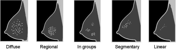
22 Detection of suspicious regions Some types of suspicious regions


23 Detection of suspicious regions Calcifications for each patch of the image o compute surprise if surprise > threshold calcification


24 Detection of suspicious regions Masses ACR density Sensitivity (%) False Positives I 52 3 II 30 3 III 26 6 IV 7 9 overall performance: Sensitivity = 38% with 5 false positives SVMs with RBF kernel features o original images o intensity value o Patch standard deviation o Patch 25 th percentile o Patch median value o Patch mean value o Patch 75th percentile o Patch maximum intensity o Iris filtered images o Patch 25th percentile o Patch median value o Patch maximum value SVMs with RBF kernel 9 shape features o area of the segmented region o area of the bounding box of the region o area of the region s convex hull o eccentricity o length of the major axis of the ellipse that has the same normalized 2nd-moments as the region o length of the minor axis of the ellipse that has the same normalized 2nd-moments as the region o diameter of a circle with the same area as the region, orientation o Perimeter 1 feature that uses both shape and intensity information o distance between the centroid and the weighted centroid
25 Outline

26 Characterization of suspicious regions
27 Characterization of suspicious regions Pearson correlation, distance correlation and Maximal Information Coefficient 7 calcification features: 1. Zernike moment of order 3 and repetition Zernike moment of order 4 and repetition 0 3. Zernike moment of order 4 and repetition Eccentricity extracted from the Spatial Density Function 5. Minimum of the mean intensities of the calcifications 6. Intensity std 7. Std of the mean intensities of the calcifications 9 mass features 1. Solidity 2. Compactness 3. Thinness ratio 4. Skeleton end points 5. Shape Index 6. Convexification 7. Extent 8. Contained lines 9. CC 2 = (Rmin / Rmax)
28 Outline
29 BI-RADS classification The scale BI-RADS Description 0 the exam is not conclusive 1 no findings 2 benign findings 3 probably benign findings 4 suspicious findings 5 high probability of malignancy 6 proved cancer
30 BI-RADS classification The scale BI-RADS Description 0 the exam is not conclusive 1 no findings 2 benign findings 3 probably benign findings 4 suspicious findings 5 high probability of malignancy 6 proved cancer
31 BI-RADS classification The scale BI-RADS Description 0 the exam is not conclusive 1 no findings 2 benign findings 3 probably benign findings 4 suspicious findings 5 high probability of malignancy 6 proved cancer


32 BI-RADS classification The scale BI-RADS Description 0 the exam is not conclusive 1 no findings 2 benign findings 3 probably benign findings 4 suspicious findings 5 high probability of malignancy 6 proved cancer


33 BI-RADS classification The scale


34 BI-RADS classification Motivation When more than one finding is present in the mammogram, the overall BIRADS in the medical report corresponds to the finding with the highest BI-RADS
35 BI-RADS classification Methods Max Ordinal Learning (MOL) o o MOL.LA MOL.CD Training set illustration White represents observed and gray not present features
36 BI-RADS classification non-mol Model A Model B
37 BI-RADS classification MOL.LA initialization Model A Model B
38 BI-RADS classification MOL.LA subsequent iterations Model A Model B
39 BI-RADS classification MOL.CD initialization Model A Model B

40 BI-RADS classification MOL.CD subsequent iterations Consider Model A fixed and update Model B ~ Model A = + And vice-versa Model B
41 BI-RADS classification Experiments Two kernels o Linear & Radial Basis Function Model parameterization selection o two-fold cross-validation Non-ordinal extension from binary to multi-class o one-against-one instantiated with SVMs Ordinal methods o Frank and Hall instantiated with SVMs o Data replication instantiated with SVMs o KDLOR instantiated LDA
42 BI-RADS classification Results Baseline techniques MOL.LA MOL.CD Standard Model Tri- Training Nonordinal Mass contorus Ground truth CaPTOR Frank&Hall 9 7 Data Replication 7 8 Frank&Hall 9 7 Data Replication 9 7 Automatic segmentation does not seem to negatively affect classification results Both the MOL.LA and MOL.CD techniques perform better than the standard methods o It is sufficient to test and compare MOL.LA and MOL.CD
43 Outline
44 Putting all together Component Ground truth Automatic AOM = 0.65 CM = 0.77 Pectoral muscle detection AD = 0.06 AMED = 0.07 HD = 0.17 TPr = 0.92 TPr = 0.82 Screening TNr = 0.18 TNr = 0.33 FNr = 0.08 FNr = 0.17 FPr = 0.82 FPr = 0.67 Calcification detection Sensitivity = 56.4 % Sensitivity = 63.8 % FP = 47 FP = 49 Mass detection Sensitivity = 47.6 % Sensitivity = 48.8 % FP = 4 FP = 4 BI-RADS classification MAE = 10 % MAE = 88 %
45 Thank you! Questions?
Classification of mammogram masses using selected texture, shape and margin features with multilayer perceptron classifier.
 Biomedical Research 2016; Special Issue: S310-S313 ISSN 0970-938X www.biomedres.info Classification of mammogram masses using selected texture, shape and margin features with multilayer perceptron classifier.
Breast Imaging! Ravi Adhikary, MD!
 Breast Imaging! Ravi Adhikary, MD! ACS Estimated Cancers Statistics 2014! Breast! New Cases in Women! 232,670 (+67,570 in situ)! Deaths in Women! 40,000! Colon! 48,380! 24,040! Cervical! 12,360! 4,020!
Colon cancer subtypes from gene expression data
 Colon cancer subtypes from gene expression data Nathan Cunningham Giuseppe Di Benedetto Sherman Ip Leon Law Module 6: Applied Statistics 26th February 2016 Aim Replicate findings of Felipe De Sousa et
S. Murgo, MD. Chr St-Joseph, Mons Erasme Hospital, Brussels
 S. Murgo, MD Chr St-Joseph, Mons Erasme Hospital, Brussels? Introduction Mammography reports are sometimes ambiguous and indecisive. ACR has developped the BIRADS. BIRADS consists of a lexicon in order
Classification of benign and malignant masses in breast mammograms
 Classification of benign and malignant masses in breast mammograms A. Šerifović-Trbalić*, A. Trbalić**, D. Demirović*, N. Prljača* and P.C. Cattin*** * Faculty of Electrical Engineering, University of
CLASSIFYING MAMMOGRAPHIC MASSES INTO BI-RADS SHAPE CATEGORIES USING VARIOUS GEOMETRIC SHAPE AND MARGIN FEATURES
 International Journal of Biomedical Signal Processing, (1), 011, pp. 43-47 CLASSIFYING MAMMOGRAPHIC MASSES INTO BI-RADS SHAPE CATEGORIES USING VARIOUS GEOMETRIC SHAPE AND MARGIN FEATURES B. Surendiran
arxiv: v2 [cs.cv] 8 Mar 2018
![arxiv: v2 [cs.cv] 8 Mar 2018](/thumbs/87/97094636.jpg "arxiv: v2 [cs.cv] 8 Mar 2018") Automated soft tissue lesion detection and segmentation in digital mammography using a u-net deep learning network Timothy de Moor a, Alejandro Rodriguez-Ruiz a, Albert Gubern Mérida a, Ritse Mann a, and
Automated soft tissue lesion detection and segmentation in digital mammography using a u-net deep learning network Timothy de Moor a, Alejandro Rodriguez-Ruiz a, Albert Gubern Mérida a, Ritse Mann a, and
BI-RADS Update. Martha B. Mainiero, MD, FACR, FSBI Brown University Rhode Island Hospital
 BI-RADS Update Martha B. Mainiero, MD, FACR, FSBI Brown University Rhode Island Hospital No Disclosures BI-RADS History 1980s Quality Issues ACR Accreditation BI-RADS 1994 2003 4 th Edition MRI, US January
Leonard M. Glassman MD
 BI-RADS The New BI-RADS Leonard M. Glassman MD FACR Former Chief of Breast Imaging American Institute for Radiologic Pathology Washington Radiology Associates, PC Breast Imaging Reporting and Data System
NMF-Density: NMF-Based Breast Density Classifier
 NMF-Density: NMF-Based Breast Density Classifier Lahouari Ghouti and Abdullah H. Owaidh King Fahd University of Petroleum and Minerals - Department of Information and Computer Science. KFUPM Box 1128.
Predictive Data Mining for Lung Nodule Interpretation
 Predictive Data Mining for Lung Nodule Interpretation William Horsthemke, Ekarin Varutbangkul, Daniela Raicu, Jacob Furst DePaul University, Chicago, IL USA {whorsthe,evarutba}@students.depaul.edu, {draicu,
Mammogram Analysis: Tumor Classification
 Mammogram Analysis: Tumor Classification Term Project Report Geethapriya Raghavan geeragh@mail.utexas.edu EE 381K - Multidimensional Digital Signal Processing Spring 2005 Abstract Breast cancer is the
Mammography limitations. Clinical performance of digital breast tomosynthesis compared to digital mammography: blinded multi-reader study
 Clinical performance of digital breast tomosynthesis compared to digital mammography: blinded multi-reader study G. Gennaro (1), A. Toledano (2), E. Baldan (1), E. Bezzon (1), C. di Maggio (1), M. La Grassa
Mammography is a most effective imaging modality in early breast cancer detection. The radiographs are searched for signs of abnormality by expert
 Abstract Methodologies for early detection of breast cancer still remain an open problem in the Research community. Breast cancer continues to be a significant problem in the contemporary world. Nearly
Compressive Re-Sampling for Speckle Reduction in Medical Ultrasound
 Compressive Re-Sampling for Speckle Reduction in Medical Ultrasound Professor Richard Mammone Rutgers University Email Phone Number Christine Podilchuk, Lev Barinov, Ajit Jairaj and William Hulbert ClearView
Investigation of multiorientation and multiresolution features for microcalcifications classification in mammograms
 Investigation of multiorientation and multiresolution features for microcalcifications classification in mammograms Aqilah Baseri Huddin, Brian W.-H. Ng, Derek Abbott 3 School of Electrical and Electronic
Mammographic Cancer Detection and Classification Using Bi Clustering and Supervised Classifier
 Mammographic Cancer Detection and Classification Using Bi Clustering and Supervised Classifier R.Pavitha 1, Ms T.Joyce Selva Hephzibah M.Tech. 2 PG Scholar, Department of ECE, Indus College of Engineering,
Mammographic Breast Density Classification by a Deep Learning Approach
 Mammographic Breast Density Classification by a Deep Learning Approach Aly Mohamed, PhD Robert Nishikawa, PhD Wendie A. Berg, MD, PhD David Gur, ScD Shandong Wu, PhD Department of Radiology, University
Ruud Pijnappel Professor of Radiology, UMC Utrecht. Chair Dutch Expert Centre for Screening Board EUSOBI
 Ruud Pijnappel Professor of Radiology, UMC Utrecht Best practice in Breast Imaging: what s new and what women need to know and Update on the Second Implementation Report of the 2003 Council Recommendation
The Radiology Aspects
 REQUIREMENTS FOR INTERNATIONAL ACCREDITATION OF BREAST CENTERS/UNITS The Radiology Aspects Miri Sklair-Levy, Israel RADIOLOGY GUIDELINES FOR QUALITY ASSURANCE IN BREAST CANCER SCREENING AND DIAGNOSIS Radiologists
Breast Density and Screening
 Breast Density and Screening Alberta Breast Cancer Screening Program Version: Jan 2019 What is this Booklet About? This is a guide to help you understand breast density and how it may affect you. Having
Imaging in breast cancer. Mammography and Ultrasound Donya Farrokh.MD Radiologist Mashhad University of Medical Since
 Imaging in breast cancer Mammography and Ultrasound Donya Farrokh.MD Radiologist Mashhad University of Medical Since A mammogram report is a key component of the breast cancer diagnostic process. A mammogram
Estimation of Breast Density and Feature Extraction of Mammographic Images
 IJIRST International Journal for Innovative Research in Science & Technology Volume 2 Issue 11 April 2016 ISSN (online): 2349-6010 Estimation of Breast Density and Feature Extraction of Mammographic Images
8/31/2016 HIDING IN PLAIN SITE, ARCHITECTURAL DISTORTIONS AND BREAST ASYMMETRIES ARCHITECTURAL DISTORTIONS ARCHITECTURAL DISTORTIONS
 HIDING IN PLAIN SITE, ARCHITECTURAL DISTORTIONS AND BREAST ASYMMETRIES DEBORAH THAMES R.T. (R)(M)(QM) ARCHITECTURAL DISTORTIONS Definition is disruption of the natural flow of breast pattern towards the
Amammography report is a key component of the breast
 Review Article Writing a Mammography Report Amammography report is a key component of the breast cancer diagnostic process. Although mammographic findings were not clearly differentiated between benign
Applying One-vs-One and One-vs-All Classifiers in k-nearest Neighbour Method and Support Vector Machines to an Otoneurological Multi-Class Problem
 Oral Presentation at MIE 2011 30th August 2011 Oslo Applying One-vs-One and One-vs-All Classifiers in k-nearest Neighbour Method and Support Vector Machines to an Otoneurological Multi-Class Problem Kirsi
Computerized image analysis: Estimation of breast density on mammograms
 Computerized image analysis: Estimation of breast density on mammograms Chuan Zhou, Heang-Ping Chan, a) Nicholas Petrick, Mark A. Helvie, Mitchell M. Goodsitt, Berkman Sahiner, and Lubomir M. Hadjiiski
LUNG CANCER continues to rank as the leading cause
 1138 IEEE TRANSACTIONS ON MEDICAL IMAGING, VOL. 24, NO. 9, SEPTEMBER 2005 Computer-Aided Diagnostic Scheme for Distinction Between Benign and Malignant Nodules in Thoracic Low-Dose CT by Use of Massive
Classification of Benign and Malignant Vertebral Compression Fractures in Magnetic Resonance Images
 Classification of Benign and Malignant Vertebral Compression Fractures in Magnetic Resonance Images Lucas Frighetto-Pereira, Guilherme Augusto Metzner, Paulo Mazzoncini de Azevedo-Marques, Rangaraj Mandayam
Breast Cancer Prevention and Early Detection using Different Processing Techniques
 e t International Journal on Emerging Technologies (Special Issue on ICRIET-2016) 7(2): 92-96(2016) ISSN No. (Print) : 0975-8364 ISSN No. (Online) : 2249-3255 Breast Cancer Prevention and Early Detection
Automated Approach for Qualitative Assessment of Breast Density and Lesion Feature Extraction for Early Detection of Breast Cancer
 Automated Approach for Qualitative Assessment of Breast Density and Lesion Feature Extraction for Early Detection of Breast Cancer 1 Spandana Paramkusham, 2 K. M. M. Rao, 3 B. V. V. S. N. Prabhakar Rao
Threshold Based Segmentation Technique for Mass Detection in Mammography
 Threshold Based Segmentation Technique for Mass Detection in Mammography Aziz Makandar *, Bhagirathi Halalli Department of Computer Science, Karnataka State Women s University, Vijayapura, Karnataka, India.
Mammogram Analysis: Tumor Classification
 Mammogram Analysis: Tumor Classification Literature Survey Report Geethapriya Raghavan geeragh@mail.utexas.edu EE 381K - Multidimensional Digital Signal Processing Spring 2005 Abstract Breast cancer is
Breast Imaging Lexicon
 9//201 200 BI RADS th Edition 201 BI RADS th Edition Breast Imaging Lexicon Mammographic Pathology and Assessment Categories Deborah Thames, R.T.(R)(M)(QM) The Advanced Health Education Center Nonmember:
Computer-Aided Diagnosis for Microcalcifications in Mammograms
 Computer-Aided Diagnosis for Microcalcifications in Mammograms Werapon Chiracharit Department of Electronic and Telecommunication Engineering King Mongkut s University of Technology Thonburi BIE 690, November
UW Radiology Review Course Breast Calcifications. BI-RADS 5 th Edition
 UW Radiology Review Course Breast Calcifications Grace Kalish, MD Vantage Radiology BI-RADS 5 th Edition Benign Skin Vascular Large rod like Coarse popcorn Suspicious Amorphous Coarse heterogenous Fine
BREAKING THE SOUND WAVES!
 BREAKING THE SOUND WAVES! SURVEY (HAND-HELD) VERSES AUTOMATIC BREAST ULTRASOUND WHAT S THE DIFFERENCE Patricia Gaspard (RT)(M)(R)(CRA)/Marie Barnett (RT)(M)(R) March 16, 2019 Hand Held vs ABUS (Automatic
Current Strategies in the Detection of Breast Cancer. Karla Kerlikowske, M.D. Professor of Medicine & Epidemiology and Biostatistics, UCSF
 Current Strategies in the Detection of Breast Cancer Karla Kerlikowske, M.D. Professor of Medicine & Epidemiology and Biostatistics, UCSF Outline ν Screening Film Mammography ν Film ν Digital ν Screening
Classification of Microcalcifications into BI-RADS Morphologic Categories Preliminary Results
 Biocybernetics and Biomedical Engineering 2009, Volume 29, Number 4, pp. 83 93 Classification of Microcalcifications into BI-RADS Morphologic Categories Preliminary Results TERESA PODSIADŁY-MARCZYKOWSKA
Leveraging Expert Knowledge to Improve Machine-Learned Decision Support Systems
 Leveraging Expert Knowledge to Improve Machine-Learned Decision Support Systems Finn Kuusisto, MS 1 ; Inês Dutra, PhD 2 ; Mai Elezaby, MD 1 ; Eneida Mendonça, MD, PhD 1 ; Jude Shavlik, PhD 1 ; Elizabeth
Malignant Breast Cancer Detection Method - A Review. Patiala
 Malignant Breast Cancer Detection Method - A Review 1 Jaspreet Singh Cheema, 2 Amrita, 3 Sumandeep kaur 1,2 Student of M.tech Computer Science, Punjabi University, Patiala 3 Assistant professor, Department
Improving Reading Time of Digital Breast Tomosynthesis with Concurrent Computer Aided Detection
 White Paper Improving Reading Time of Digital Breast Tomosynthesis with Concurrent Computer Aided Detection WHITE PAPER 2 3 Abstract PowerLook Tomo Detection, a concurrent computer-aided detection (CAD)
Artificial Intelligence in Breast Imaging
 Artificial Intelligence in Breast Imaging Manisha Bahl, MD, MPH Director of Breast Imaging Fellowship Program, Massachusetts General Hospital Assistant Professor of Radiology, Harvard Medical School Outline
CHAPTER 2 MAMMOGRAMS AND COMPUTER AIDED DETECTION
 9 CHAPTER 2 MAMMOGRAMS AND COMPUTER AIDED DETECTION 2.1 INTRODUCTION This chapter provides an introduction to mammogram and a description of the computer aided detection methods of mammography. This discussion
Automatic Classification of Breast Masses for Diagnosis of Breast Cancer in Digital Mammograms using Neural Network
 IJSTE - International Journal of Science Technology & Engineering Volume 1 Issue 11 May 2015 ISSN (online): 2349-784X Automatic Classification of Breast Masses for Diagnosis of Breast Cancer in Digital
Investigating the performance of a CAD x scheme for mammography in specific BIRADS categories
 Investigating the performance of a CAD x scheme for mammography in specific BIRADS categories Andreadis I., Nikita K. Department of Electrical and Computer Engineering National Technical University of
MIT International Journal of Electronics and Communication Engineering Vol. 3, No. 1, Jan. 2013, pp
 MIT International Journal of Electronics and Communication Engineering Vol. 3, No. 1, Jan. 013, pp. 43 47 43 A Novel Technique to Detect Abnormal Masses from Digital Mammogram Saurabh Verma Email: saurav.v84@gmail.com
Ana Sofia Preto 19/06/2013
 Ana Sofia Preto 19/06/2013 Understanding the underlying pathophysiologic processes leading to the various types of calcifications Description and illustration of the several types of calcifications, according
Lung cancer detection from chest CT images using spatial FCM with level set and neural network classifier.
 Research Article http://www.alliedacademies.org/archives-of-general-internal-medicine/ Lung cancer detection from chest CT images using spatial FCM with level set and neural network classifier. Manikandan
Computer aided detection of clusters of microcalcifications on full field digital mammograms
 Computer aided detection of clusters of microcalcifications on full field digital mammograms Jun Ge, a Berkman Sahiner, Lubomir M. Hadjiiski, Heang-Ping Chan, Jun Wei, Mark A. Helvie, and Chuan Zhou Department
Classification of Mammograms using Gray-level Co-occurrence Matrix and Support Vector Machine Classifier
 Classification of Mammograms using Gray-level Co-occurrence Matrix and Support Vector Machine Classifier P.Samyuktha,Vasavi College of engineering,cse dept. D.Sriharsha, IDD, Comp. Sc. & Engg., IIT (BHU),
AB MR Interpretation Overview
 AB MR Interpretation Overview Goal of AB MR interpretation is to maintain high sensitivity and specificity In order to minimize false positives and short term follow ups, it is fundamental to focus only
Localization of Breast Lesion Using Mammographic-Basic Views
 Research Article imedpub Journals www.imedpub.com Vol. 2 No. 1: 2 Localization of Breast Lesion Using Mammographic-Basic Views Nikki Mishra*, Meena Chakradhar, Ranjit Jha and Rauniyar RK Department of
Leonard M. Glassman MD Analysis of Breast Calcifications
 Importance of Calcification Leonard M. Glassman MD FACR American Institute for Radiologic Pathology Washington Radiology Associates, PC Washington DC 45% of all breast cancers present as calcification
A Novel Method For Automatic Screening Of Nonmass Lesions In Breast DCE-MRI
 Volume 3 Issue 2 October 2015 ISSN: 2347-1697 International Journal of Informative & Futuristic Research A Novel Method For Automatic Screening Of Paper ID IJIFR/ V3/ E2/ 046 Page No. 565-572 Subject Area
Comparison Classifier: Support Vector Machine (SVM) and K-Nearest Neighbor (K-NN) In Digital Mammogram Images
 and K-Nearest Neighbor (K-NN) In Digital Mammogram Images") JUISI, Vol. 02, No. 02, Agustus 2016 35 Comparison Classifier: Support Vector Machine (SVM) and K-Nearest Neighbor (K-NN) In Digital Mammogram Images Jeklin Harefa 1, Alexander 2, Mellisa Pratiwi 3 Abstract
JUISI, Vol. 02, No. 02, Agustus 2016 35 Comparison Classifier: Support Vector Machine (SVM) and K-Nearest Neighbor (K-NN) In Digital Mammogram Images Jeklin Harefa 1, Alexander 2, Mellisa Pratiwi 3 Abstract
FDA Executive Summary
 Meeting of the Radiological Devices Advisory Panel On October 24, 22, the panel will discuss, make recommendations, and vote on a premarket approval application supplement (P83/S) to expand the indications
The latest developments - Automated Breast Volume Scanning. Dr. med. M. Golatta
 The latest developments - Automated Breast Volume Scanning Dr. med. M. Golatta Automated Breast Volume US: Why? o Mammography is limited in dense breasts: high false negative rate o Many of these tumors
Computer Assisted System for Features Determination of Lung Nodule from Chest X-ray Image
 Computer Assisted System for Features Determination of Lung Nodule from Chest X-ray Image Manoj R. Tarambale Marathwada Mitra Mandal s College of Engineering, Pune Dr. Nitin S. Lingayat BATU s Institute
CLASSIFICATION OF ABNORMALITY IN B -MASS BY ARCHITECTURAL DISTORTION
 CLASSIFICATION OF ABNORMALITY IN B -MASS BY ARCHITECTURAL DISTORTION #1 Venmathi.A.R., * 2 D.C.Jullie Josphine #1.Dept of ECE, Kings Engineering College * 2. Dept of CSE,Kings Engineering college Abstract-The
Computer based delineation and follow-up multisite abdominal tumors in longitudinal CT studies
 Research plan submitted for approval as a PhD thesis Submitted by: Refael Vivanti Supervisor: Professor Leo Joskowicz School of Engineering and Computer Science, The Hebrew University of Jerusalem Computer
Automating Quality Assurance Metrics to Assess Adequate Breast Positioning in Mammography
 Automating Quality Assurance Metrics to Assess Adequate Breast Positioning in Mammography Gerald R. Kolb, JD, The Breast Group, Sunriver, OR; Kaier Wang, PhD, VolparaSolutions, Wellington, NZ; Ariane Chan,
High-Resolution Breast Cancer Screening with Multi-View Deep Convolutional Neural Networks
 High-Resolution Breast Cancer Screening with Multi-View Deep Convolutional Neural Networks Krzysztof J. Geras Joint work with Kyunghyun Cho, Linda Moy, Gene Kim, Stacey Wolfson and Artie Shen. GTC 2017
Quantification of Nodule Detection in Chest CT: A Clinical Investigation Based on the ELCAP Study
 Second International Workshop on -149- Quantification of Nodule Detection in Chest CT: A Clinical Investigation Based on the ELCAP Study Amal A. Farag, Shireen Y. Elhabian, Salwa A. Elshazly and Aly A.
Extraction and Identification of Tumor Regions from MRI using Zernike Moments and SVM
 I J C T A, 8(5), 2015, pp. 2327-2334 International Science Press Extraction and Identification of Tumor Regions from MRI using Zernike Moments and SVM Sreeja Mole S.S.*, Sree sankar J.** and Ashwin V.H.***
Breast Imaging & You
 Breast Imaging & You What s Inside: Breast Imaging... 2 Digital Breast Tomosynthesis (DBT) mammograms... 4 Breast cancer screening... 6 Dense breast tissue... 8 Automated breast ultrasound (ABUS)... 9
THE ONE STOP CLINIC FOR BREAST LESIONS. Philippe Vielh MD, PhD For the Breast Cancer Group Institut de cancérologie Gustave Roussy Villejuif, France
 THE ONE STOP CLINIC FOR BREAST LESIONS Philippe Vielh MD, PhD For the Breast Cancer Group Institut de cancérologie Gustave Roussy Villejuif, France CONTEXT IN 2004 French breast cancer screening program
Breast Imaging & You
 Breast Imaging & You What s Inside: Breast Imaging... 2 Digital Breast Tomosynthesis (DBT) mammograms... 4 Breast cancer screening... 6 Dense breast tissue... 8 Automated Breast Ultrasound (ABUS)... 9
Breast asymmetries in mammography: Management
 Breast asymmetries in mammography: Management Poster No.: C-1026 Congress: ECR 2015 Type: Educational Exhibit Authors: V. de Lara Bendahan 1, F. J. Hidalgo Ramos 2, J. L. Ortega Garcia 3, Keywords: DOI:
ACRIN 6666 IM Additional Evaluation: Additional Views/Targeted US
 Additional Evaluation: Additional Views/Targeted US For revised or corrected form check box and fax to 215-717-0936. Instructions: The form is completed based on recommendations (from ID form) for additional
Outlier Analysis. Lijun Zhang
 Outlier Analysis Lijun Zhang zlj@nju.edu.cn http://cs.nju.edu.cn/zlj Outline Introduction Extreme Value Analysis Probabilistic Models Clustering for Outlier Detection Distance-Based Outlier Detection Density-Based
Building an Ensemble System for Diagnosing Masses in Mammograms
 Building an Ensemble System for Diagnosing Masses in Mammograms Yu Zhang, Noriko Tomuro, Jacob Furst, Daniela Stan Raicu College of Computing and Digital Media DePaul University, Chicago, IL 60604, USA
BIRADS 3 and 4 lesions viewed by ultrasound and not seen in digital mammograms and tomosynthesis.
 Original article Anales de Radiología México 2016 Jul;15(3):205-213. BIRADS 3 and 4 lesions viewed by ultrasound and not seen in digital mammograms and tomosynthesis. García-Quintanilla JF 1, González-Coronado
Nature Neuroscience: doi: /nn Supplementary Figure 1. Behavioral training.
 Supplementary Figure 1 Behavioral training. a, Mazes used for behavioral training. Asterisks indicate reward location. Only some example mazes are shown (for example, right choice and not left choice maze
The Application of Image Processing Techniques for Detection and Classification of Cancerous Tissue in Digital Mammograms
 The Application of Image Processing Techniques for Detection and Classification of Cancerous Tissue in Digital Mammograms Angayarkanni.N 1, Kumar.D 2 and Arunachalam.G 3 1 Research Scholar Department of
Blood Vessel Detection from Fundus Image for Diabetic Retinopathy Patients using SVM
 Blood Vessel Detection from Fundus Image for Diabetic Retinopathy Patients using SVM Bibin. s 1, Manikandan. T 2 1 ME communication systems, Rajalakshmi Engineering College, TamilNadu, India 2 Associate
5/24/16. Current Issues in Breast Cancer Screening. Breast cancer screening guidelines. Outline
 Disclosure information: An Evidence based Approach to Breast Cancer Karla Kerlikowske, MDDis Current Issues in Breast Cancer Screening Grant/Research support from: National Cancer Institute - and - Karla
BI-RADS and Breast MRI. Kathy Borovicka, M.D. Thursday February 15, 2018
 BI-RADS and Breast MRI Kathy Borovicka, M.D. Thursday February 15, 2018 Learning Objectives Be familiar with the Breast Imaging Reporting and Data System (BI-RADS) Understand the components of a breast
Enhanced Detection of Lung Cancer using Hybrid Method of Image Segmentation
 Enhanced Detection of Lung Cancer using Hybrid Method of Image Segmentation L Uma Maheshwari Department of ECE, Stanley College of Engineering and Technology for Women, Hyderabad - 500001, India. Udayini
NAÏVE BAYES CLASSIFIER AND FUZZY LOGIC SYSTEM FOR COMPUTER AIDED DETECTION AND CLASSIFICATION OF MAMMAMOGRAPHIC ABNORMALITIES
 NAÏVE BAYES CLASSIFIER AND FUZZY LOGIC SYSTEM FOR COMPUTER AIDED DETECTION AND CLASSIFICATION OF MAMMAMOGRAPHIC ABNORMALITIES 1 MARJUN S. SEQUERA, 2 SHERWIN A. GUIRNALDO, 3 ISIDRO D. PERMITES JR. 1 Faculty,
Armed Forces Institute of Pathology.
 Armed Forces Institute of Pathology www.radpath.com Armed Forces Institute of Pathology Breast Disease www.radpath.org Armed Forces Institute of Pathology Evaluation of Breast Calcifications Leonard M.
Cancer Cells Detection using OTSU Threshold Algorithm
 Cancer Cells Detection using OTSU Threshold Algorithm Nalluri Sunny 1 Velagapudi Ramakrishna Siddhartha Engineering College Mithinti Srikanth 2 Velagapudi Ramakrishna Siddhartha Engineering College Kodali
Microcalcifications detected on mammography classified as BIRADS 4 and 5 and their correlations with histopatologic findigns
 Microcalcifications detected on mammography classified as BIRADS 4 and 5 and their correlations with histopatologic findigns Poster No.: C-0401 Congress: ECR 2010 Type: Educational Exhibit Topic: Breast
Statistical analysis to assess automated Level of Suspicion scoring methods in breast ultrasound
 Statistical analysis to assess automated Level of Suspicion scoring methods in breast ultrasound Michael Galperin a a Almen Laboratories, Inc., 2105 Miller Ave., Escondido, CA 92025 Abstract A well-defined
Detection of architectural distortion using multilayer back propagation neural network
 Available online www.jocpr.com Journal of Chemical and Pharmaceutical Research, 2015, 7(2):292-297 Research Article ISSN : 0975-7384 CODEN(USA) : JCPRC5 Detection of architectural distortion using multilayer
AMSER Case of the Month: November 2018
 AMSER Case of the Month: November 2018 52 year old female with an abnormal screening mammogram Areeg Rehman, MS 4 Nova Southeastern University Rebecca T. Sivarajah, MD Penn State University College of
Detecting Architectural Distortion in Mammograms Using a Gabor Filtered Probability Map Algorithm
 Detecting Architectural Distortion in Mammograms Using a Gabor Filtered Probability Map Algorithm O tega Ejofodomi, Michael Olawuyi, Don Onyishi, Godswill Ofualagba To cite this version: O tega Ejofodomi,
PLACE LABEL HERE. ACRIN 6657 MRI Form: Pre-Treatment (MRI-1)
") M3 ACRIN 6657 MRI Form: Pre-Treatment (MRI-1) If this is a revised or corrected form,indicate by checking box. ACRIN Study 6657 Case # Instructions: In accordance with the protocol, four MRI exams are
M3 ACRIN 6657 MRI Form: Pre-Treatment (MRI-1) If this is a revised or corrected form,indicate by checking box. ACRIN Study 6657 Case # Instructions: In accordance with the protocol, four MRI exams are
Available online at ScienceDirect. Procedia Computer Science 70 (2015 ) 76 84
 76 84") Available online at www.sciencedirect.com ScienceDirect Procedia Computer Science 70 (2015 ) 76 84 4 th International Conference on Eco-friendly Computing and Communication Systems Wavelet Packet Texture
Available online at www.sciencedirect.com ScienceDirect Procedia Computer Science 70 (2015 ) 76 84 4 th International Conference on Eco-friendly Computing and Communication Systems Wavelet Packet Texture
Accreditation Case Review: Mammography and Stereotactic Biopsy
 Accreditation Case Review: Mammography and Stereotactic Biopsy Brett T. Parkinson MD Breast Imaging Director Breast Care Services Intermountain Healthcare Chair, ACR Committee on Mammography Accreditation
Learning Objectives. 1. Identify which patients meet criteria for annual lung cancer screening
 Disclosure I, Taylor Rowlett, DO NOT have a financial interest /arrangement or affiliation with one or more organizations that could be perceived as a real or apparent conflict of interest in the context
COMPUTER AIDED DIAGNOSIS SYSTEM FOR DIGITAL MAMMOGRAPHY. Mohamed Eltahir Makki Elmanna
 COMPUTER AIDED DIAGNOSIS SYSTEM FOR DIGITAL MAMMOGRAPHY By Mohamed Eltahir Makki Elmanna A Thesis Submitted to the Faculty of Engineering at Cairo University in Partial Fulfillment of the Requirements
DESCRIPTION: Percentage of final reports for screening mammograms that are classified as probably benign
 Quality ID #146 (NQF 0508): Radiology: Inappropriate Use of Probably Benign Assessment Category in Screening Mammograms National Quality Strategy Domain: Efficiency and Cost Reduction 2018 OPTIONS F INDIVIDUAL
FALSE NEGATIVES IN MAMMOGRAPHY
 FALSE NEGATIVES IN MAMMOGRAPHY Abstract Dr. Gustavo Febles The concept of false negative in mammograms is defined and the factors which can define its occurrence are exposed. Mechanisms which can be used
MAESTRO TRIAL FINAL RESULTS. Gisela L.G. Menezes, MD, PhD
 MAESTRO TRIAL FINAL RESULTS Gisela L.G. Menezes, MD, PhD MAESTRO Angiogenesis Metastasis How does OA work? Malignant Benign How does OA work? D O How does OA work? Optoacoustic Imaging Fibroadenoma IDC
COMPUTERIZED SYSTEM DESIGN FOR THE DETECTION AND DIAGNOSIS OF LUNG NODULES IN CT IMAGES 1
 ISSN 258-8739 3 st August 28, Volume 3, Issue 2, JSEIS, CAOMEI Copyright 26-28 COMPUTERIZED SYSTEM DESIGN FOR THE DETECTION AND DIAGNOSIS OF LUNG NODULES IN CT IMAGES ALI ABDRHMAN UKASHA, 2 EMHMED SAAID
Lung Cancer Detection using CT Scan Images
 Available online at www.sciencedirect.com ScienceDirect Procedia Computer Science 125 (2018) 107 114 6th International Conference on Smart Computing and Communications, ICSCC 2017, 7-8 December 2017, Kurukshetra,
Tissue Breast Density
 Tissue Breast Density Reporting breast density within the letter to the patient is now mandated by VA law. Therefore, this website has been established by Peninsula Radiological Associates (PRA), the radiologists
Challenges to Delivery of High Quality Mammography
 Challenges to Delivery of High Quality Mammography Overview of Current Challenges Barbara Monsees, Washington University Geographic Access, Equity and Impact on Quality Tracy Onega, Dartmouth Medical School
Detection of Breast Masses in Digital Mammograms using SVM
 IJCTA, 8(3), 2015, pp. 899-906 International Science Press Detection of Breast Masses in Digital Mammograms using SVM Abstract: Breast Cancer stands to be the most deadly disease among women caused due