Quantitative Analysis of Vascular Canals in Vertebral Endplate
|
|
|
- Buddy Robertson
- 5 years ago
- Views:
Transcription
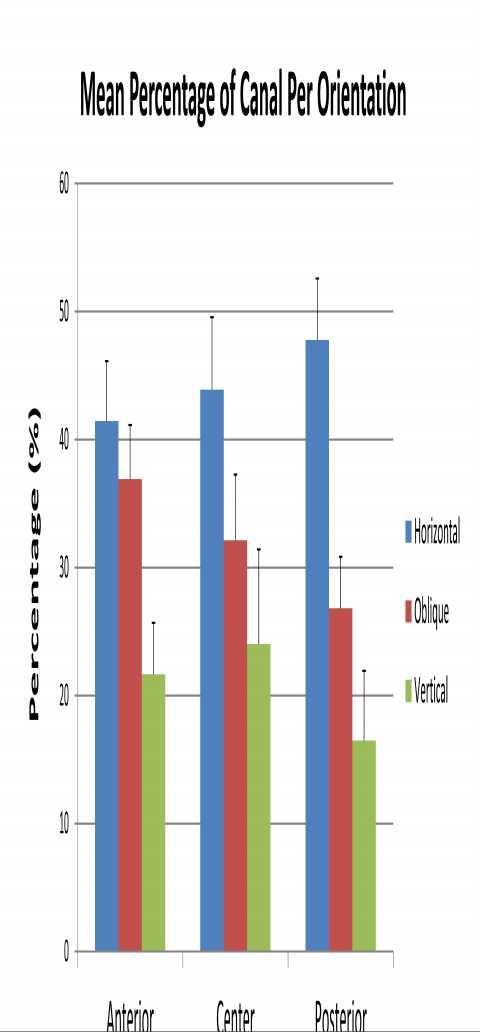
1 Quantitative Analysis of Vascular Canals in Vertebral Endplate Kristine Tan 1, Won C. Bae, PhD 1, Tomonori Yamaguchi, MS 1,2, Kelli Xu, BS 1, Iris Shieh, BS 1, Jade He, BS 1, Robert L. Sah, MD, ScD 1, Nozomu Inoue, MD, PhD 2, Koichi Masuda, MD 1. 1 University of California, San Diego, San Diego, CA, USA, 2 Doshisha University, Kyoto, Japan. Disclosures: K. Tan: None. W.C. Bae: None. T. Yamaguchi: None. K. Xu: None. I. Shieh: None. J. He: None. R.L. Sah: None. N. Inoue: None. K. Masuda: None. Introduction: Degenerative disc disease of the lumbar spine is associated with low back pain [1], and is an active target of therapeutics. Nutrition to intervertebral discs (IVD) is supplied via vascular canals in the bony endplate. Abnormal changes to vascular canals may lead to inhibition of nutrition and disc degeneration. Morphology of vascular canals has not been studied in detail. The purpose of this study is to describe techniques used to determine quantitative morphology of vascular canals in human vertebral endplates, and report preliminary results. Methods: Lumbar Spines: Lumbar spines (including L1 to L5) from cadavers (n=9, 59±11 yo, mean±sd; 2 females, 7 males) were obtained from a tissue bank within ~48 hours of death, and imaged using a 3-T magnetic resonance imaging (MRI) scanner to obtain T2-weighted images in mid-sagittal plane (FOV=16 to 20 cm, slice thickness=3 mm, TR=2000 ms, TE= 90 ms, image matrix= ). All intervertebral discs were graded using the Pfirrmann grading scheme [2]. Discs with grades 2 to 4 were used for this study. Endplate Samples: After MRI, disco-vertebral cores, 5 mm in diameter, were obtained from superior surfaces of L3 and L5, at anterior, central, and posterior regions (3 per level). A total of 54 cores were harvested. Micro-Computed Tomography (μct): The cores were imaged using μct (Shimadzu SMX 160CTS) at 2 µm resolution and 67 kv. Reconstruction was performed using Bone3D software. Analysis of Quantitative Morphology: Three-dimensional (3D) models representing the canals were obtained using Mimics software (Figure 1). Linear canal segments were isolated (a total of 595 canals) and their angle (relative to the endplate s surface normal vector), volume, and mean diameter were determined using a custom Matlab program. Based on the angle, the canals were categorized as horizontal (angle of 60 to 90 deg), oblique (30 to 60 deg) or vertical (0 to 30 deg). Statistics: Initial tests found no significant effect of level, so it was not considered further. ANOVA and posthoc Tukey test were used to determine the effects of region and canal orientation on canal volume and diameter. In addition, chi-square test was used to determine association of region and orientation, as well as MRI grade and orientation. Results: Mean canal diameter (μm) (Figure 2) varied significantly with region (p<0.001), but not orientation (p=0.19). The posterior region had canals with the smallest average diameter (~50 μm) relative to anterior (posthoc p<0.001) and central (posthoc p<0.001) regions which averaged ~60 μm. Average canal volume did not vary significantly with region or orientation (each p>0.5). Chi-square test revealed that the distribution of different canal orientations was significantly associated with region (p<0.01; Figure 3). The distribution of orientations by disc MRI grade was not significant (chi-square p=0.3). Discussion: A technique to determine quantitative morphology of vascular canals within vertebral endplate was described. Using a high resolution micro CT scanner, it was feasible to segment small vascular canals, demonstrating a complex 3D canal network consisting of vertical, oblique and horizontal canals. Vertical canals directly connect the bone marrow in the vertebral body to the IVD while horizontal canals interconnect the vertical canals and allow lateral distribution of nutrients across the endplate. This study showed that out of the three canal orientations, all three sample regions of the endplate had horizontal canals in the greatest number, suggesting an important contribution to nutrient transport within the entire endplate. In the center region, the smaller in number, but larger diameter, vertical canals indicate that the function of the vertical canals is to go through the endplate and connect the bone marrow to the IVD. This data is consistent with previous studies that found the center region of the endplate to be more porous than surrounding peripheral regions [3]. The mean diameter of the canals, ~50 to 60 μm, is also consistent with those seen on endplate surface on scanning electron micrographs [4]. In future studies, comparison of μct models with other reference measures, such as 3D histology [5] would be useful for further validation of our results. In addition, additional measures of interest, including porosity and density of canals will be analyzed. Significance: The determination of the quantitative structure of vascular canals in vertebral endplates is important for a better understanding of the etiology of disc degeneration. Acknowledgments: NIH NIAMS, General Electric, LifeSharing, Ministry of Education, Science, Sports and Culture, Grant-in-Aid for Scientific Research (C) , UCSD PRIME program, Julia Brown Research Scholarship

2 References: 1. Vanharanta+ Spine Pfirrmann+ Spine Nachemson+ Acta Orthop Scand Benneker+ Spine Jadin+ Biomaterials 2007.

3
4
5 ORS 2014 Annual Meeting Poster No: 0188
6
Disclosures: T. Yoshii: None. T. Yamada: None. T. Taniyama: None. S. Sotome: None. T. Kato: None. S. Kawabata: None. A. Okawa: None.
 Dynamic Changes in Spinal Cord Compression by Cervical Ossification of the Posterior Longitudinal Ligament Evaluated by Kinematic Computed Tomography Myelogram Toshitaka Yoshii, Tsuyoshi Yamada, Takashi
Disclosures: C.B. Raub: None. B.C. Hansen: None. T. Yamaguchi: None. M.M. Temple-Wong: None. K. Masuda: None. R.L. Sah: None.
 En Face Microscopy of Rabbit Knee Articular Cartilage Following Anterior Cruciate Ligament Transection Reveals Early Matrix Damage, Chondrocyte Loss and Cloning Christopher B. Raub, PhD, Bradley C. Hansen,
A Computational Model of Annulus Fiber Deformation in Cervical Discs During In Vivo Dynamic Flexion\Extension, Rotation and Lateral Bending
 A Computational Model of Annulus Fiber Deformation in Cervical Discs During In Vivo Dynamic Flexion\Extension, Rotation and Lateral Bending William Anderst, Mara Palmer, Joon Lee, William Donaldson, James
Citation for published version (APA): Eijkelkamp, M. F. (2002). On the development of an artificial intervertebral disc s.n.
: Eijkelkamp, M. F. (2002). On the development of an artificial intervertebral disc s.n.") University of Groningen On the development of an artificial intervertebral disc Eijkelkamp, Marcus Franciscus IMPORTANT NOTE: You are advised to consult the publisher's version (publisher's PDF) if you
University of Groningen On the development of an artificial intervertebral disc Eijkelkamp, Marcus Franciscus IMPORTANT NOTE: You are advised to consult the publisher's version (publisher's PDF) if you
Laminar Thickness: Correlation Against Symptoms of Spinal Stenosis and Changes with Age
 Laminar Thickness: Correlation Against Symptoms of Spinal Stenosis and Changes with Age Hamed Shalikar, MD 1, Sean L. Borkowski, MS 2, Juan Pablo Villablanca, MD 1, Sophia N. Sangiorgio, PhD 2, Edward
Hiroto Yamaguchi*, Hidetoshi Nojiri**, Kei Miyagawa*, Nozomu Inoue***
 Three-Dimensional High-Resolution Image Analysis of the Segmental Reduction Effect Obtained with Lateral Lumbar Interbody Fusion for Lumbar Intervertebral Deformity Hiroto Yamaguchi*, Hidetoshi Nojiri**,
Facet orientation in patients with lumbar degenerative spondylolisthesis
 35 J. Tokyo Med. Univ., 71 1 35 0 Facet orientation in patients with lumbar degenerative spondylolisthesis Wuqikun ALIMASI, Kenji ENDO, Hidekazu SUZUKI, Yasunobu SAWAJI, Hirosuke NISHIMURA, Hidetoshi TANAKA,
Depiction of Lateral Ligament Complex of the Ankle using 3D MRI in Healthy Subjects and Patients with Chronic Ankle Instability.
 Depiction of Lateral Ligament Complex of the Ankle using 3D MRI in Healthy Subjects and Patients with Chronic Ankle Instability. Satoshi Yamaguchi, M.D. 1, Hiroshi Matsumoto, R.T. 2, Atsuya Watanabe, M.D.
Signal intensity changes of the posterior elements of the lumbar spine in symptomatic adults
 ORIGINAL ARTICLE SPINE SURGERY AND RELATED RESEARCH Signal intensity changes of the posterior elements of the lumbar spine in symptomatic adults Kosuke Sugiura, Toshinori Sakai, Fumitake Tezuka, Kazuta
of the lumbar facet joints there
 Skeletal Radiol (1999) 28:215±219 International Skeletal Society 1999 A R T I C L E Dominik Weishaupt Marco Zanetti Norbert Boos Juerg Hodler MR imaging and CT in osteoarthritis of the lumbar facet joints
Mast Cell Tryptase Modifies The Contraction Of Human Joint Capsule Cells In Vitro
 Mast Cell Tryptase Modifies The Contraction Of Human Joint Capsule Cells In Vitro Kevin A. Hildebrand, MD 1, Lindsey M. Logan 1, Mei Zhang 1, David A. Hart, PhD 1, Paul T. Salo, MD 1, A. Dean Befus, PhD
Meniscus T2 Relaxation Time at Various Stages of Knee Joint Degeneration
 Meniscus T2 Relaxation Time at Various Stages of Knee Joint Degeneration Richard Kijowski, Michael Fazio, Benjamin Beduhn, and Fang Liu Department of Radiology University of Wisconsin School of Medicine
High Field MR of the Spine
 Department of Radiology University of California San Diego 3T for MR Applications Advantages High Field MR of the Spine Increased signal-to-noise Better fat suppression Increased enhancement with gadolinium
Spinal infection. Outline ANATOMY 6/2/2017. Anatomy Pathogen
 Outline Spinal infection Pramot Tanutit, M.D. Department of Radiology, Songklanagarind Hospital Faculty of Medicine, Prince of Songkla University Anatomy Pathogen Pyogenic spondylodiscitis Tuberculous
Facet Joint Arthrosis Disc Degeneration and Lumbago. Dr.Ruchira Sethi Dr. Vishram Singh Department of Anatomy Santosh University, India
 Facet Joint Arthrosis Disc Degeneration and Lumbago Dr.Ruchira Sethi Dr. Vishram Singh Department of Anatomy Santosh University, India INTRODUCTION The initial division of spine into three columns for
Morphological changes of the cervical spinal canal and cord due to aging on MR imaging
 Morphological changes of the cervical spinal canal and cord due to aging on MR imaging Shigeru Kobayashi, MD,PhD 1, Katsuhiko Hayakawa, MD, PhD 2, Takashi Nakane, MD, PhD 2, Riya Kosaka MD,PhD 3. 1 Department
DISCLOSURES. Goal of Fusion. Expandable Cages: Do they play a role in lumbar MIS surgery? CON 2/15/2017
 Expandable Cages: Do they play a role in lumbar MIS surgery? CON Jean-Jacques Abitbol, M.D., FRCSC San Diego, California DISCLOSURES SAB; K2M, Osprey, Nanovis, Vertera, St Theresa Royalties; Osprey, K2M,
A minimally invasive surgical approach reduces cranial adjacent segment degeneration in patients undergoing posterior lumbar interbody fusion
 A minimally invasive surgical approach reduces cranial adjacent segment degeneration in patients undergoing posterior lumbar interbody fusion T. Tsutsumimoto, M. Yui, S. Ikegami, M. Uehara, H. Kosaku,
Load Response of the Hindfoot Bones in patients with the Ankle Osteoarthritis : in vivo 3D study
 Load Response of the Hindfoot Bones in patients with the Ankle Osteoarthritis : in vivo D study Hara Y, Ikoma K, Kido M, Imai K, Maki M, Takatori R, Tokunaga D, Inoue N,, Kubo T Department of Orthopaedics,
An Advanced 3D Multi-Body System Model for the Human Lumbar Spine
 An Advanced 3D Multi-Body System Model for the Human Lumbar Spine Sousa, S. 1 and Claro, J. C. P. 2 1 University of Minho, Portugal, e-mail: sofia_sts@hotmail.com 2 University of Minho, Portugal, e-mail:
Analyzing Early Onset Scoliosis In 3d: How Does Growing Rod Surgery Affect The Three Planes Of Deformity?
 Analyzing Early Onset Scoliosis In 3d: How Does Growing Rod Surgery Affect The Three Planes Of Deformity? Diana A. Glaser, PhD 1, Burt Yaszay, MD 1, Gregory M. Mundis, MD 2, Behrooz A. Akbarnia, MD 2.
In Vivo Kinematic Analysis of the Hip during Activities of Daily Living.
 In Vivo Kinematic Analysis of the Hip during Activities of Daily Living. Daisuke Hara 1, Yasuharu Nakashima 1, Satoshi Hamai 1, Hidehiko Higaki 2, Takeshi Shimoto 3, Satoru Ikebe 2, Mio Akiyama 1, Masanobu
Degenerative spondylolisthesis is four times more
 Degenerative spondylolisthesis DEVELOPMENTAL OR ACQUIRED? T. W. Love, A. B. Fagan, R. D. Fraser From the Royal Adelaide Hospital, Adelaide, Australia Degenerative spondylolisthesis is four times more common
DOWNLOAD OR READ : PREVALENCE OF MODIC DEGENERATIVE MARROW CHANGES IN THE CERVICAL SPINE PDF EBOOK EPUB MOBI
 DOWNLOAD OR READ : PREVALENCE OF MODIC DEGENERATIVE MARROW CHANGES IN THE CERVICAL SPINE PDF EBOOK EPUB MOBI Page 1 Page 2 prevalence of modic degenerative marrow changes in the cervical spine prevalence
MEDICAL IMAGING OF THE VERTEBRAE
 MEDICAL IMAGING OF THE VERTEBRAE Vertebrae are your friends Matthew Harper MS-IV LECTURE OBJECTIVES INTRODUCE THE MOST COMMON MODALITIES OF MEDICAL IMAGING AND BASIC TECHNIQUES FOR READING THESE IMAGES
PREOPERATIVE RETROLISTHESIS IS A RISK FACTOR OF LUMBAR DISC HERNIATION AFTER FENESTRATION WITHOUT DISCECTOMY
 PREOPERATIVE RETROLISTHESIS IS A RISK FACTOR OF LUMBAR DISC HERNIATION AFTER FENESTRATION WITHOUT DISCECTOMY Shota Takenaka*, Noboru Hosono, Yoshihiro Mukai, Kosuke Tateishi, Takeshi Fuji Osaka Kosei-nenkin
Analysis of radiological factors promoting degeneration in adjacent disc levels after anterior cervical discectomy and fusion(acdf)
") Analysis of radiological factors promoting degeneration in adjacent disc levels after anterior cervical discectomy and fusion(acdf) Dept. of neurosurgery, Sapporo Azabu Neurosurgical Hospital*, Sapporo,
Analysis of radiological factors promoting degeneration in adjacent disc levels after anterior cervical discectomy and fusion(acdf) Dept. of neurosurgery, Sapporo Azabu Neurosurgical Hospital*, Sapporo,
Int J Clin Exp Med 2018;11(2): /ISSN: /IJCEM Yi Yang, Hao Liu, Yueming Song, Tao Li
: /ISSN: /IJCEM Yi Yang, Hao Liu, Yueming Song, Tao Li") Int J Clin Exp Med 2018;11(2):1278-1284 www.ijcem.com /ISSN:1940-5901/IJCEM0063093 Case Report Dislocation and screws pull-out after application of an Isobar TTL dynamic stabilisation system at L2/3 in
Int J Clin Exp Med 2018;11(2):1278-1284 www.ijcem.com /ISSN:1940-5901/IJCEM0063093 Case Report Dislocation and screws pull-out after application of an Isobar TTL dynamic stabilisation system at L2/3 in
Optimal Baseplate Rotational Alignment in Reverse Total Shoulder Arthroplasty: A Three-Dimensional Computer-Aided Design Study.
 Optimal Baseplate Rotational Alignment in Reverse Total Shoulder Arthroplasty: A Three-Dimensional Computer-Aided Design Study. Byron F. Stephens, MD 1, Casey T. Hebert 2, Thomas W. Throckmorton, MD 1,
Research Article Relationship of the Lumbar Lordosis Angle to the Level of Termination of the Conus Medullaris and Thecal Sac
 Anatomy, Article ID 1769, 4 pages http://dx.doi.org/1.1155/14/1769 Research Article Relationship of the Lumbar Lordosis Angle to the Level of Termination of the Conus Medullaris and Thecal Sac C. D. Moussallem,
Bilateral spondylolysis of inferior articular processes of the fourth lumbar vertebra: a case report
 Upsala Journal of Medical Sciences. 2012; 117: 72 77 CASE REPORT Bilateral spondylolysis of inferior articular processes of the fourth lumbar vertebra: a case report TOMOAKI KOAKUTSU 1,2, NAOKI MOROZUMI
The Quality Leader in 3D Cone Beam CT
 The Quality Leader in 3D Cone Beam CT The Complete 2-in-1 or 3-in-1 Multi-modality Solution PreXion, with over 15 years of innovation in the medical and dental fields, introduces the PreXion3D Eclipse.
Magnetic resonance imaging findings in patients with low backache
 Original Article Magnetic resonance imaging findings in patients with low backache Narayan Bikram Thapa 1, Suraj Bajracharya 2 1 Department of Radiology, KIST Medial College Teaching Hospital, Lalitpur,
Magnetic resonance of adolescent idiopathic scoliosis
 Australian Institute of Radiography The Radiographer 2005; 52 (2): 7 14 Magnetic resonance of adolescent idiopathic scoliosis University of Queensland, St Lucia Campus, Brisbane, Queensland, Australia
Supplementary Online Content
 Supplementary Online Content Schlaeger R, Papinutto N, Zhu AH, et al. Association between thoracic spinal cord gray matter atrophy and disability in multiple sclerosis. JAMA Neurol. Published online June
RECENT ADVANCES IN CLINICAL MR OF ARTICULAR CARTILAGE
 In Practice RECENT ADVANCES IN CLINICAL MR OF ARTICULAR CARTILAGE By Atsuya Watanabe, MD, PhD, Director, Advanced Diagnostic Imaging Center and Associate Professor, Department of Orthopedic Surgery, Teikyo
Revised Dec Spine MR Protocols
 Spine MR Protocols Sp 1: Cervical spine MRI without contrast Sp 2: Pre- and post-contrast cervical spine MRI Sp 3: Pre- and post-contrast cervical spine MRI (multiple sclerosis protocol) Sp 4: Thoracic
Exploring the Effect of Pore Size and Distribution on Shear Strength of Surface Porous Polyetheretherkeytone (PEEK)
") Exploring the Effect of Pore Size and Distribution on Shear Strength of Surface Porous Polyetheretherkeytone (PEEK) A Thesis Presented to The Academic Faculty by Rebecca Ellen Wyche In Partial Fulfillment
Exploring the Effect of Pore Size and Distribution on Shear Strength of Surface Porous Polyetheretherkeytone (PEEK) A Thesis Presented to The Academic Faculty by Rebecca Ellen Wyche In Partial Fulfillment
Fractures of the thoracic and lumbar spine and thoracolumbar transition
 Most spinal column injuries occur in the thoracolumbar transition, the area between the lower thoracic spine and the upper lumbar spine; over half of all vertebral fractures involve the 12 th thoracic
Metal Artefact Reduction in CT
 Metal Artefact Reduction in CT DANIEL MARRINER Metal Artefact Reduction in CT Metal Artefact Clinical Indications for MAR SEMAR and How It Works Technical Considerations Case Studies utilising SEMAR Metal
Added value of MR myelography using 3D COSMIC sequence in the diagnosis of lumbar canal stenosis: comparison with routine MR imaging
 Added value of MR myelography using 3D COSMIC sequence in the diagnosis of lumbar canal stenosis: comparison with routine MR imaging Poster No.: C-1099 Congress: ECR 2012 Type: Authors: Scientific Exhibit
Mineral Density Of Subchondral Bone May Be Quantitatively Evaluated Using A Clinical Cone Beam Computed Tomography Scanner
 Mineral Density Of Subchondral Bone May Be Quantitatively Evaluated Using A Clinical Cone Beam Computed Tomography Scanner Mikael J. Turunen, PhD 1, Juha Töyräs, PhD 1, Harri Kokkonen, PhD 2, Jukka S.
3D titanium interbody fusion cages sharx. White Paper
 3D titanium interbody fusion cages sharx (SLM selective laser melted) Goal of the study: Does the sharx intervertebral cage due to innovative material, new design, and lordotic shape solve some problems
Intra-Articular Tibiofemoral Injection of a Nonsteroidal Anti-Inflammatory Drug has no Detrimental Effects on Joint Mechanics in a Rat Model
 Intra-Articular Tibiofemoral Injection of a Nonsteroidal Anti-Inflammatory Drug has no Detrimental Effects on Joint Mechanics in a Rat Model Corinne N. Riggin, Jennica J. Tucker, Louis J. Soslowsky, PhD,
SpineFAQs. Lumbar Spondylolisthesis
 SpineFAQs Lumbar Spondylolisthesis Normally, the bones of the spine (the vertebrae) stand neatly stacked on top of one another. The ligaments and joints support the spine. Spondylolisthesis alters the
Validation of in vivo cancellous bone assessment using high resolution imaging and histologic examination
 Validation of in vivo cancellous bone assessment using high resolution imaging and histologic examination Luke Arentsen, PhD Medical Physics Resident University of Minnesota Cancellous bone has a higher
Ex. 1 :Language of Anatomy
 Collin College BIOL 2401 : Human Anatomy & Physiology Ex. 1 :Language of Anatomy The Anatomical Position Used as a reference point when referring to specific areas of the human body Body erect Head and
MRI and CT of the CNS
 MRI and CT of the CNS Dr.Maha ELBeltagy Assistant Professor of Anatomy Faculty of Medicine The University of Jordan 2018 Computed Tomography CT is used for the detection of intracranial lesions. CT relies
Case Report A Rare Case of Progressive Palsy of the Lower Leg Caused by a Huge Lumbar Posterior Endplate Lesion after Recurrent Disc Herniation
 Case Reports in Orthopedics Volume 2016, Article ID 5963924, 4 pages http://dx.doi.org/10.1155/2016/5963924 Case Report A Rare Case of Progressive Palsy of the Lower Leg Caused by a Huge Lumbar Posterior
Plasmapore XP. Stability that Starts at the Surface
 Plasmapore XP Stability that Starts at the Surface The Difference The Plasmapore XP Difference The unique structure, properties and processing of the Plasmapore XP coating results in early bone apposition
Morphologic Study of the Facet Joint in Spondylolysis and Isthmic Spondylolisthesis
 Abstract Morphologic Study of the Facet Joint in Spondylolysis and Isthmic Spondylolisthesis Chang Hoon Jeon, M.D., Woo Sig Kim, M.D., Jae Hyun Cho, M.D.*, Byoung-Suck Kim, M.D., Soo Ik Awe, M.D. and Shin
The Role of Two-Dimensional Ultrasonography in the Diagnosis of Protrusion of Cervical Intervertebral Discs in Adolescents
 American Journal of Clinical and Experimental Medicine 2017; 5(5): 176-180 http://www.sciencepublishinggroup.com/j/ajcem doi: 10.11648/j.ajcem.20170505.14 ISSN: 2330-8125 (Print); ISSN: 2330-8133 (Online)
JlntSocPlastination, Vol4:16-22,
 JlntSocPlastination, Vol4:16-22, 1990 16 SECTIONAL ANATOMY: STANDARDIZED METHODOLOGY Alexander Lane, Coordinator of Anatomy and Physiology, Triton College, Visiting Associate Professor, University of Illinois
WHAT IS SMART SPINAL FUSION?
 SMART SPINAL FUSION WHAT IS SMART SPINAL FUSION? FIRST-TIME-RIGHT SMART SPINAL FUSION = FIRST TIME RIGHT WHAT IS FIRST TIME RIGHT? Happy patients, pain free with excellent functionality with the intention
How to interpret computed tomography of the lumbar spine
 REVIEW Ann R Coll Surg Engl 2014; 96: 502 507 doi 10.1308/003588414X13946184902361 How to interpret computed tomography of the lumbar spine Z Ahmad 1, R Mobasheri 2,TDas 3, S Vaidya 4, S Mallik 5, M El-Hussainy
Artificial intervertebral disc
 The University of Toledo The University of Toledo Digital Repository Master s and Doctoral Projects Artificial intervertebral disc Vikas Ghai Medical University of Ohio Follow this and additional works
Positional Magnetic Resonance Imaging. Description
 Subject: Positional Magnetic Resonance Imaging Page: 1 of 6 Last Review Status/Date: June 2015 Positional Magnetic Resonance Imaging Description Positional magnetic resonance imaging (MRI) allows imaging
Range of motion of thoracic spine in sagittal plane
 DOI 10.1007/s00586-013-3088-7 ORIGINAL ARTICLE Range of motion of thoracic spine in sagittal plane Daigo Morita Yasutsugu Yukawa Hiroaki Nakashima Keigo Ito Go Yoshida Masaaki Machino Syunsuke Kanbara
UC San Diego UC San Diego Previously Published Works
 UC San Diego UC San Diego Previously Published Works Title Lumbar spine postures in marines during simulated operational positions. Permalink https://escholarship.org/uc/item/6d92t6wv Authors Berry, DB
Quality of Life. Quality of Motion.
 Quality of Life. Quality of Motion. Lateral Bend Vertical Translation Flexion Extension Lateral Translation Axial Rotation Anterior Posterior Translation Motion in all Directions Kinematics is the study
2/5/2019. Facet Joint Pain. Biomechanics
 Facet Arthropathy as a Pain Source Evaluation and Management Shelby Spine Jan 31 st Feb 2 nd, 2019 Kushagra Verma MD, MS Adult and Pediatric Scoliosis And Spine Deformity Beach Orthopaedics Specialty Institute
A morphometric study of the Pedicles of dry human typical lumbar vertebrae
 Original article: A morphometric of the Pedicles of dry human typical lumbar vertebrae Dhaval K. Patil 1 *, Pritha S. Bhuiyan 2 1Resident, Department of Anatomy, Seth G S Medical College, Parel, Mumbai-400012,
Subchondral Bone Density Distribution in Cam Type Femoroacetabular Impingement Hips
 Subchondral Bone Density Distribution in Cam Type Femoroacetabular Impingement Hips Nozomu Inoue MD, PhD Simon Lee MPH Shane J. Nho MD, MS Department of Orthopedic Surgery Rush University Medical Center
3 脊椎変形の相互関係とリスク分析の今後の方向
 Reprint request: DEGENERATIVE SPINE AND OSTEOPOROSIS IN ELDERLY PEOPLE Toshitaka NAKAMURA Department of Orthopedics, University of Occupational and Environmental Health School of Medicine Vertebral osteophytes,
University of Groningen. Thoracolumbar spinal fractures Leferink, Vincentius Johannes Maria
 University of Groningen Thoracolumbar spinal fractures Leferink, Vincentius Johannes Maria IMPORTANT NOTE: You are advised to consult the publisher's version (publisher's PDF) if you wish to cite from
Occult Lumbar Lateral Spinal Stenosis in Neural Foramina Subjected to Physiologic Loading
 Occult Lumbar Lateral Spinal Stenosis in Neural Foramina Subjected to Physiologic Loading Bruce H. Nowicki, Victor M. Haughton, Timothy A. Schmidt, Tae-Hong Lim, Howard S. An, Lee H. Riley III, Liyuan
1 Normal Anatomy and Variants
 1 Normal Anatomy and Variants 1.1 Normal Anatomy MR Technique. e standard MR protocol for a routine evaluation of the spine always comprises imaging in sagittal and axial planes, while coronal images are
ACR MRI Accreditation: Medical Physicist Role in the Application Process
 ACR MRI Accreditation: Medical Physicist Role in the Application Process Donna M. Reeve, MS, DABR, DABMP Department of Imaging Physics University of Texas M.D. Anderson Cancer Center Educational Objectives
Introduction of FIREFLY Technology
 Introduction of FIREFLY Technology FIREFLY Technology is a unique, patent-pending, pre-surgical planning and intra - operative navigation technology that is focused on spinal applications and is derived
It consist of two components: the outer, laminar fibrous container (or annulus), and the inner, semifluid mass (the nucleus pulposus).
, and the inner, semifluid mass (the nucleus pulposus).") Lumbar Spine The lumbar vertebrae are the last five vertebrae of the vertebral column. They are particularly large and heavy when compared with the vertebrae of the cervical or thoracicc spine. Their bodies
Lumbar Spine The lumbar vertebrae are the last five vertebrae of the vertebral column. They are particularly large and heavy when compared with the vertebrae of the cervical or thoracicc spine. Their bodies
Magnetic Resonance Classification of Lumbar Intervertebral Disc Degeneration
 SPINE Volume 26, Number 17, pp 1873 1878 2001, Lippincott Williams & Wilkins, Inc. Magnetic Resonance Classification of Lumbar Intervertebral Disc Degeneration Christian W. A. Pfirrmann, MD,* Alexander
Innovative Techniques in Minimally Invasive Cervical Spine Surgery. Bruce McCormack, MD San Francisco California
 Innovative Techniques in Minimally Invasive Cervical Spine Surgery Bruce McCormack, MD San Francisco California PCF Posterior Cervical Fusion PCF not currently an ambulatory care procedure Pearl diver
VERTEBRAL COLUMN VERTEBRAL COLUMN
 VERTEBRAL COLUMN FUNCTIONS: 1) Support weight - transmits weight to pelvis and lower limbs 2) Houses and protects spinal cord - spinal nerves leave cord between vertebrae 3) Permits movements - *clinical
Magnetic Resonance Imaging Findings in Degenerative Disc Disease of Cervical Spine in Symptomatic Patients
 Original Article J Nepal Health Res Counc 2015 Sep - Dec;13(31):196-200 Magnetic Resonance Imaging Findings in Degenerative Disc Disease of Cervical Spine in Symptomatic Patients Karki DB, 1 Gurung G,
PREPARED FOR. Marsha Eichhorn DATE OF INJURY : N/A DATE OF ANALYSIS : 12/14/2016 DATE OF IMAGES : 12/8/2016. REFERRING DOCTOR : Dr.
 Accent on Health Chiropractic 405 Firemans Ave PREPARED FOR Marsha Eichhorn DATE OF INJURY : N/A DATE OF ANALYSIS : 12/14/2016 DATE OF IMAGES : 12/8/2016 REFERRING DOCTOR : Dr. David Bohn This report contains
Study of Lumbar Vertebrae with respect to the dimensions of the pedicle in South Indian population
 Original article Study of Lumbar Vertebrae with respect to the dimensions of the pedicle in South Indian population 1 Dr.Sreevidya.J, 2 Dr.Dharani.V, 3 Dr.Savithri.K, 4 Dr.Sudha Seshayyan 1Senior Assistant
Christopher Ciarallo, MD Denver Health/Children s Hospital Colorado CRASH 2016
 Distribution of the vertebral level of the intercristal line by ultrasonography Christopher Ciarallo, MD Denver Health/Children s Hospital Colorado CRASH 2016 Korean J Anesthesiol. 2014 Sep;67(3):181 5
Injury Mechanisms of the Cervical Intervertebral Disc During Simulated Whiplash. Spine: Volume 29(11) June 1, 2004 pp
 June 1, 2004 pp") 1 Injury Mechanisms of the Cervical Intervertebral Disc During Simulated Whiplash Spine: Volume 29(11) June 1, 2004 pp 1217-1225 Panjabi, Manohar M. PhD; Ito, Shigeki MD; Pearson, Adam M. BA; Ivancic,
1 Injury Mechanisms of the Cervical Intervertebral Disc During Simulated Whiplash Spine: Volume 29(11) June 1, 2004 pp 1217-1225 Panjabi, Manohar M. PhD; Ito, Shigeki MD; Pearson, Adam M. BA; Ivancic,
Effects of Vertebral Axial Decompression On Intradiscal Pressure. Ramos G., MD, Martin W., MD, Journal of Neurosurgery 81: , 1994 ABSTRACT
 Effects of Vertebral Axial Decompression On Intradiscal Pressure. Ramos G., MD, Martin W., MD, Journal of Neurosurgery 81: 350353, 1994 ABSTRACT The object of this study was to examine the effect of vertebral
Study on Mechanical Characteristics of Lumbar Spine for Snatch Action in Weight Lifting Based on Finite Element Method
 ISSN 1750-9823 (print) International Journal of Sports Science and Engineering Vol. 04 (2010) No. 01, pp. 048-052 Study on Mechanical Characteristics of Lumbar Spine for Snatch Action in Weight Lifting
Automatic Classification System for Lumbar Spine X-ray Images
 Automatic Classification System for Lumbar Spine X-ray Images Soontharee Koompairojn Kien A. Hua School of Electrical Engineering and Computer Science University of Central Florida Orlando, FL 32816 {soonthar,
Case Report: CASE REPORT OF FACET ARTHROPATHY INDUCED NERVE ROOT COMPRESSION RESULTING IN MOTOR WEAKNESS AND PAIN
 Cox Technic Case Report #100 published at www.coxtechnic.com (sent October 2011 on 10/11/11 ) 1 Case Report: CASE REPORT OF FACET ARTHROPATHY INDUCED NERVE ROOT COMPRESSION RESULTING IN MOTOR WEAKNESS
ORIGINAL PAPER. Department of Orthopedic Surgery, Chubu Rosai Hospital, Nagoya, Japan ABSTRACT
 Nagoya J. Med. Sci. 77. 221 ~ 228, 2015 ORIGINAL PAPER RANGE OF MOTION DETERMINED BY MULTIDETECTOR-ROW COMPUTED TOMOGRAPHY IN PATIENTS WITH CERVICAL OSSIFICATION OF THE POSTERIOR LONGITUDINAL LIGAMENT
BIOMECHANICAL ANALYSIS OF THE DEADLIFT DURING THE 1999 SPECIAL OLYMPICS WORLD GAMES
 63 Biomechanics Symposia 2001 / University of San Francisco BIOMECHANICAL ANALYSIS OF THE DEADLIFT DURING THE 1999 SPECIAL OLYMPICS WORLD GAMES Rafael F. Escamilla, Tracy M. Lowry, Daryl C. Osbahr, and
OPERATIVE TECHNIQUE. CONSTRUX Mini PTC. Mini PTC Spacer System
 OPERATIVE TECHNIQUE CONSTRUX Mini PTC Mini PTC Spacer System TABLE OF CONTENTS Introduction 1 Operative Technique 2 Part Numbers 6 Indications For Use 7 INTRODUCTION 1 INTRODUCTION The CONSTRUX Mini PTC
Pott s kyphosis. University Affiliated Sixth People s Hospital, 600 Yishan Road, Shanghai , P.
 QJM Advance Access published November 17, 2014 Pott s kyphosis Author Names: Yi Zhang, Yong-Sheng Yu, Zheng-Hao Tang and Guo-Qing Zang Author Affiliations: Department of Infectious Diseases, Shanghai Jiao
AO CLASSIFICATIONS THORACO-LUMBAR SPINAL INJURIES
 AO CLASSIFICATIONS THORACO-LUMBAR SPINAL INJURIES T H E A O / A S I F ( A R B E I T S G E M E I N S C H A F T F Ü R O S T E O S Y N T H E S E F R A G E N / A S S O C I A T I O N F O R T H E S T U D Y O
Silvia Capuani*, Giulia Di Pietro*, Guglielmo Manenti, Umberto Tarantino^
 Silvia Capuani*, Giulia Di Pietro*, Guglielmo Manenti, Umberto Tarantino^ *CNR ISC, Physics Department Sapienza University, Rome, Italy Department of Diagnostic Imaging and Interventional Radiology, Molecular
Personalized Solutions. MRI Protocol for PSI and Signature Guides
 Personalized Solutions MRI Protocol for PSI and Signature Guides 2 Personalized Solutions MRI Protocol for PSI and Signature Guides Purpose and Summary This protocol is applicable for the Zimmer Biomet
EFFECTS OF VERTEBRAL AXIAL DECOMPRESSION (VAX-D) ON INTRADISCAL PRESSURE
 ON INTRADISCAL PRESSURE") EFFECTS OF VERTEBRAL AXIAL DECOMPRESSION (VAX-D) ON Gustavo Ramos, M.D., William Marin, M.D. Journal of Neursurgery 81:35-353 1994 Departments of Neurosurgery and Radiology, Rio Grande Regional Hospital,
EFFECTS OF VERTEBRAL AXIAL DECOMPRESSION (VAX-D) ON Gustavo Ramos, M.D., William Marin, M.D. Journal of Neursurgery 81:35-353 1994 Departments of Neurosurgery and Radiology, Rio Grande Regional Hospital,
Induction and Maintenance of Lordosis in MultiLevel ACDF Using Allograft. Saad Khairi, MD Jennifer Murphy Robert S. Pashman, MD
 Induction and Maintenance of Lordosis in MultiLevel ACDF Using Allograft Saad Khairi, MD Jennifer Murphy Robert S. Pashman, MD Purpose Is lordosis induced by multilevel cortical allograft ACDF placed on
The Temporomandibular joint: Anatomy, Mechanics, Pathology. Aditya Bahel, DO
 The Temporomandibular joint: Anatomy, Mechanics, Pathology Aditya Bahel, DO Outline Anatomy Mechanics and function Indications for TMJ imaging MR Protocols and pitfalls Pathology Treatment options Anatomy
Disclosures. Introduction. Purpose. Morphologic Variation in Lumbar Spinal Canal Dimensions by Gender, Race and Age
 Disclosures Morphologic Variation in Lumbar Spinal Canal Dimensions by Gender, Race and Age Jeremy D. Shaw, MD, MS; Daniel L. Shaw, BA; Daniel R. Cooperman, MD; Jason D. Eubanks, MD; Ling Li, MSPH; David
Radiographic Evaluation Of Dynamic Hip Instability In Lequesne s False Profile View
 Radiographic Evaluation Of Dynamic Hip Instability In Lequesne s False Profile View Ryo Mori 1, Yuji Yasunaga 2, Takuma Yamasaki 1, Michio Hamanishi 1, Takeshi Shoji 1, Sotaro Izumi 1, Susumu Hachisuka
Siglec-15 Is A Potential Therapeutic Target For Postmenopausal Osteoporosis
 Siglec-15 Is A Potential Therapeutic Target For Postmenopausal Osteoporosis Yusuke Kameda, Masahiko Takahata, Tomohiro Shimizu, Hiroki Hamano, Norimasa Iwasaki. Department of Orthopedic Surgery, Hokkaido
Functional Anatomy and Exam of the Lumbar Spine. Thomas Hunkele MPT, ATC, NASM-PES,CES Coordinator of Rehabilitation
 Functional Anatomy and Exam of the Lumbar Spine Thomas Hunkele MPT, ATC, NASM-PES,CES Coordinator of Rehabilitation Disclosure Anatomical Review Quick Review of Bony and Ligamentous structures Discal anatomy
FISH VERTEBRAE RADIOLOGIC VIGNETTE DONALD L. RESNICK
 ~ 1073 RADIOLOGIC VIGNETTE FISH VERTEBRAE DONALD L. RESNICK The term fish verfebru is applied to a vertebral body that has an abnormal shape characterized by biconcavity due to depression of its superior
Viviane Khoury, MD. Assistant Professor Department of Radiology University of Pennsylvania
 U Penn Diagnostic Imaging: On the Cape Chatham, MA July 11-15, 2016 Viviane Khoury, MD Assistant Professor Department of Radiology University of Pennsylvania Hip imaging has changed in recent years: new
Testing Micronized Amnion as a Therapeutic for OA in a Rat Model using Contrast Based Micro-CT Imaging
 Testing Micronized Amnion as a Therapeutic for OA in a Rat Model using Contrast Based Micro-CT Imaging Tanushree Thote, B.S 1, Sanjay Sridaran 2, Angela S.P. Lin 3, Nick J. Willett, Ph.D. 2, Robert E.
Fibular Malalignment in Subjects with Chronic Ankle Instability
 Fibular Malalignment in Subjects with Chronic Ankle Instability Takumi Kobayashi 1,2, Eiichi Suzuki 3, Naohito Yamazaki 3, Makoto Suzukawa 4, Atsushi Akaike 4, Kuniaki Shimizu 4, Kazuyoshi Gamada 1. 1