Advanced Radiation Therapies for Meningioma
|
|
|
- Miranda Marshall
- 6 years ago
- Views:
Transcription
1 Advanced Radiation Therapies for Meningioma by Jillian Doreen Maclean MBChB, FRCR Thesis submitted in partial fulfilment for the degree of MD(Res) University College London 1
2 I, Jillian Doreen Maclean confirm that the work presented in this thesis is my own. Where information has been derived from other sources, I confirm that this has been indicated in the thesis. 2
3 Acknowledgements At the risk of sounding cliched, the most important thing I learned undertaking this thesis was the value of teamwork. I was truly amazed by the extent that people will lend their expertise when it may lead to advances in therapy and patient benefit. The completion of this thesis is a testament to the enthusiasm and dedication of the staff of the Radiotherapy and Nuclear Medicine departments at University College London Hospital. In particular I must thank my supervisors Professor Susan Short and Dr Naomi Fersht for their guidance, encouragement and understanding. I must express my gratitude to Kevin Sullivan in radiotherapy for his persistence and good humour in viewing the numerous hurdles in the PET project as interesting challenges rather than insurmountable problems. I would like to thank Marcel Van Herk from the Netherlands Cancer Institute for providing essential assistance with PET MRI image registration. Likewise, without the hard work of Celia O Meara, John Dickson, Dr Jamshed Bomanji, Dr Irfan Kayani and Matthew Aldridge in UCLH Nuclear Medicine, many of the chapters of this thesis would have floundered. I would also like to express my gratitude to Chris Stacey in Radiotherapy Physics for meeting my endless requests for assistance with a smile and Paul Doolan for the huge amount he contributed to the proton chapter. Finally, I must sincerely thank all the patients who sacrificed their time and energy to participate in the studies contained within this thesis. Their willingness to participate in research, even in the absence of a direct personal benefit, was an inspiration. 3
4 Publications and Presentations Arising From Thesis Publications Maclean J, Fersht N, Bremner F, Stacey C, Sivabalasingham S, Short S. Meningioma causing visual impairment: outcomes and toxicity after intensity modulated radiation therapy. Int J Radiat Oncol Biol Phys. 2013;85 (4):e Maclean J, Fersht N, Short S. Controversies in radiotherapy for meningioma. Clin Oncol (R Coll Radiol). 2014;26(1):51-64 Maclean JD, Fersht N, Rosenberg I, D Souza D, Short S. Arc delivered intensity modulated radiotherapy achieves highly conformal radiotherapy for brain tumour patients with minimal hair loss. European J Clin and Med Oncology. 2011;3(4):10-16 Under review Maclean JD, Fersht N, Sullivan K, Kayani I, Bomanji J, Dickson J, O Meara C, Short S. Simultaneous 68 Ga DOTATATE PET/MRI in meningioma target contouring: feasibility and impact upon interobserver variability. Submitted to Clin Oncol (R Coll Radiol) Presentations at International Meetings Maclean JD, Fersht N, Sullivan K, Kayani I, O Meara C, Bomanji J, Short S. Interobserver variability in meningioma target volume contouring using simultaneous 68 Ga DOTATATE PET/ MRI. American Society for Radiation Oncology (ASTRO) 2014 Int J Rad Oncol, Bio, Phys. 2014; 90(1)S299 and National Cancer Research Institute (NCRI) 2014 ( Maclean JD, Fersht N, Short S. IMRT for Meningioma: outcomes of a prospective observational study, National Cancer Research Institute 4
5 (NCRI) 2013 ( Lalli N, Maclean JD, Short S, Fersht N. VMAT or IMRT: can we spare the hippocampus? UK Radiation Oncology (UKRO) October 2013 Maclean JD, Sullivan K, Fersht N, Short S. The introduction of simultaneous PET/MR to radiotherapy planning. European Society for Radiotherapy and Oncology (ESTRO) 2013 (short-listed for Young Scientist Award). Radiotherapy and Oncology 2013;106, S229. Maclean JD, Aldridge M, Haroon A, Bomanji J, Fersht N. Lutetium DOTATATE for advanced progressive meningioma: a pilot study. European Society for Radiotherapy and Oncology (ESTRO) 2013 (shortlisted for Young Scientist Award). Radiotherapy and Oncology 2013;106, S Aldridge M, Maclean JD, Haroon A, Fersht N Bomanji J. Lutetium DOTATATE for advanced progressive meningioma and pituitary tumours: dosimetric and clinical results. British Nuclear Medicine Society, April 2013 Maclean JD, Fersht N, Bremner F, Short S. Ophthalmological Outcomes and Toxicity Following IMRT for Meningioma. European Association of Neuro-ophthalmology April 2013 Maclean JD, Haroon A, Aldridge M, Bomanji J, Fersht N. 177 Lu-DOTA Therapy for Recurrent Meningioma: A Pilot Study. European Association of Nuclear Medicine, October 2012 Maclean JD, Fersht N, Bremner F, Short S. Ophthalmological Outcomes and Toxicity Following IMRT for Meningioma, European Society for Radiotherapy and Oncology (ESTRO) Radiotherapy and Oncology 2012;103, S
6 Abstract Radiotherapy has been used to treat meningiomas for decades, both in the primary setting when resection is not possible and as an adjunct to surgery in recurrent/ high grade disease. Newer radiotherapy planning and delivery techniques aim to optimise tumour control and minimise long-term toxicities. The purpose of this thesis was to explore the feasibility and potential for the use of advanced radiation planning and delivery techniques to treat meningiomas. In a prospective observational study of intensity modulated radiotherapy (IMRT) in fifty patients I demonstrated that IMRT is feasible and provided excellent dosimetric parameters. Medium term meningioma control rates were >90% in benign disease. Objective measures of toxicity were low. Visual symptoms improved in 38.5% of patients. In a pilot study of ten patients I showed that simultaneous 68 Ga DOTATATE PET/MRI can be utilised in meningioma radiotherapy planning. Baseline levels of interobserver variability in target volume definition between three Observers using CT/MRI alone were very high (mean target volume conformity levels of ). Levels of agreement improved only 4-5% with the addition of PET and there was negligible difference in contouring between standard PET(CT) and simultaneous PET(MRI). In a planning study of ten meningiomas I did not find a notable advantage for proton therapy (non-intensity modulated) over IMRT. The high quality of the IMRT plans left little room for improvement and range uncertainty restricted exploitation of proton dose deposition characteristics. In my review of the first six patients treated with the radionuclide 177 Lutetium DOTATATE for advanced progressive meningioma, tumour growth rates were found to slow, but there was generally disease progression during treatment. In conclusion, advanced radiation techniques for meningioma treatment are feasible and can confer clinical benefit. However, advances in technology do not necessarily translate into therapeutic gains. Careful prospective evaluation is required to ensure their optimal use. 6
7 Contents 1 Chapter 1: Introduction Background Epidemiology Pathology Anatomical Features and Grade Proliferation Markers Genetic Abnormalities Tumour Initiation Tumour Progression Prognosis Recurrence Survival Metastases Spinal and Primary Extradural Meningioma Cranial Meningioma Location and Symptoms Aetiology Population Statistics Genetic Environmental Radiation Hormones Head Injury Mobile Phone Use Diagnosis General
8 Computed Tomography Magnetic Resonance Imaging Imaging Mimics MR Spectroscopy Nuclear Imaging Clinical Evaluation Natural History of Untreated Meningiomas Treatment Options Overview Surveillance Surgery Radiation Therapy for Meningiomas Radiotherapy Planning and Delivery Techniques Dimensional Techniques Dimensional Conformal Techniques Intensity Modulated Radiotherapy Fractionated Stereotactic Radiotherapy Radiosurgery Radiation Toxicity Challenges in radiotherapy for meningioma What is the Target Volume? Hyperostosis Dural Tail Peri-tumoural oedema Post-operative Changes What Imaging Best Defines thetarget? What is the Optimal Radiotherapy Prescription Dose? Protons
9 Basics of Proton Therapy Protons in Meningioma Carbon Ion Therapy Systemic Therapy Targeted therapy Somatostatin Receptor Targeted Therapy Summary Aims of this Thesis Chapter 2: Outcomes and toxicity associated with Intensity Modulated Radiotherapy in the treatment of meningioma: a prospective observational study Introduction Background IMRT Study Aims Methods Patients Radiotherapy Procedure Radiotherapy Plan Evaluation Patient Evaluation Radiology Statistical analyses Results Radiotherapy Plan Evaluation Tumour control Symptoms Toxicity Quality of Life
10 2.4.6 MMSE Pituitary Discussion Treatment Plans Tumour Control Symptom Control Comparison to other studies Radiology Toxicities Quality of Life Neuropsychology Evaluation Endocrine Evaluation Conclusion Chapter 3: Simultaneous 68 Gallium DOTATATE PET/MRI in meningioma radiotherapy target volume delineation: a feasibility study with evaluation of the impact upon inter-observer variability in target volume delineation Introduction Challenges in Meningioma Target Volume Definition IMRT Confers a Greater Need for Precision in Target Volume Definition PET in Meningioma Target Volume Definition Interobserver Variability In Meningioma Target Definition Preliminary Work Simultaneous PET/ MRI Study Aims Materials and Methods Imaging Specifications Volunteer and Phantom work
11 3.3.3 Patient Imaging Protocol Image registration Contouring Protocol Differences in Target Volume Contours With and Without PET Results PET/MRI Technical Aspects Differences in Overall Volume Kouwenhoven Conformity Level Regions of difference Comparison of Different PET modalities Discussion Challenges with Target Volume Definition in Meningioma Baseline IOV Impact of PET on IOV PET/MRI versus PET/CT Technical implementation of PET/MRI planning Methods of determining IOV PET as a means of reducing IOV in radiotherapy planning for other tumour types Conclusion Chapter 4: Do protons improve plan parameters compared to photons for radiotherapy in meningioma? Introduction Aims Materials and Methods Photon Plans Proton plans Plan Analysis
12 4.4 Results Target Volume Characteristics TrueBeam (TRA) versus Clinac RapidArc (CRA) Proton versus RapidArc (Truebeam) Normal tissue sparing Discussion Plan parameters Difficulties exploiting the theoretical benefits of proton therapy Issues with planning studies comparing photons and protons Comparison to Other Planning Studies Clinical Use of Protons for Meningioma Conclusion Chapter 5: Preliminary evaluation of 177 Lutetium DOTATATE as a treatment for advanced progressive meningioma Introduction Background Evaluating disease status in meningioma Aims Materials and Methods Patients Imaging Therapy Imaging Assessment Dosimetry Results Patient features and clinical course Imaging Uptake Response
13 5.4.4 Outcomes Post PRRT Dosimetry Discussion Conclusion Chapter 6: Conclusions Introduction Important findings Implications of thesis Future Research Conclusion Appendices Appendix 1 EORTC Quality of Life Questionnaires Appendix 2 EEG Neuropsychology Pilot Substudy Protocol Analysis Appendix 3: Can doses to the hippocampus be reduced for patients treated with IMRT for meningioma? SGDMLC IMRT versus VMAT Appendix 4: Local Baseline Evaluation of Meningioma Recurrence Rate and Target Volume Definition Appendix 5: PET MRI of meningioma brain for radiotherapy planning work instruction
14 Table of Figures Figure 1-1 Proposed stepwise progression of meningioma [29] Figure 1-2 NCCN Guidelines for Treatment of Meningioma at Presentation Figure 1-3 NCCN Guidelines Follow-up and Treatment of Recurrent Disease. 44 Figure 1-4 Tumour location and surgical potential Figure field 3DCRT plan for a cavernous sinus meningioma Figure 1-6 Cavernous sinus meningioma IMRT treatment plans Figure 1-7 a) Abnormal bone associated with meningioma; b) Dural Tail Figure 1-8 Depth dose curves of photons and protons Figure 1-9 Location of pencil beam scanning proton spots in tumour Figure 2-1 Outcomes for patients with baseline visual deficits Figure 2-2 Non visual symptoms pre and post IMRT Figure 2-3 Acute Toxicities Figure 2-4 ECOG Performance Status pre and post IMRT Figure 2-5 Global quality of life for all patients Figure 2-6 Quality of life results for specific domains Figure 3-1 Comparison of Distribution of prescribed dose Figure 3-2 Radiotherapy Immobilisation and Imaging Equipment Figure 3-3 Imaging Protocol Figure 3-4 The Jaccard Coefficient Figure 3-5: Attenuation of PET signal Figure 3-6: Attenuation of PET uptake using Medibord prototype Figure 3-10 Examples of cases where PET could change target volume Figure 3-11 Regions of IOV per case Figure 4-1 Inclusion of the sinuses in the target Figure 4-2 Metal artefacts within the beam path Figure 4-3 Dose Volume Histogram mean brain-ptv Figure 4-4 Dose colour wash screen shots photons versus protons Figure 4-5 Dose colour wash screen shots photons versus protons
15 Figure 5-1 Regions of 177 Lu-DOTATATE therapy Figure 5-2 Growth rates pre and post PRRT Figure 5-3 SUV max after 4 cycles of treatment Figure 5-4 Cumulated activity within the meningioma for patient 4 (1 cycle) Figure 7-1 Mean doses to the bilateral hippocampus and whole brain-ptv Figure 7-2 Hippocampal dose distribution Figure 7-3 Bland Altman Plots of GTV and CTV between Observers Figure 7-4 GTV with and without PET
16 Table of Tables Table 1-1 WHO Classification of Meningioma Subtypes Table 1-2 Meningioma Site, Frequency and Symptoms Table 1-3 Simpson Grading of Extent of Meningioma Resection Table 1-4 Older Case Series of Radiotherapy Outcomes for Meningioma With Conventional Radiotherapy Table 1-5 Fractionated Radiotherapy Outcomes for Meningioma Table 1-6 Outcomes Following Radiotherapy for Optic Nerve Sheath Meningioma Table 1-7 Meningioma Studies Reporting Outcomes Following GTR, STR and GTR or STR plus RT Table 1-8 Current RTOG and EORTC Study Target Volume Specifications Table 1-9 Clinical Series of Proton Therapy for Meningioma Table 2-1 Neuropsychology tests added to protocol Table 2-2 Patient Characteristics Table 2-3 Clinical relevance of changes in scores Table 2-4 Scale for Scoring Visual Acuity Table 2-5 PRV: Dose Objectives Set and Actual Doses Accepted Table 2-6 Ophthalmology deficits at baseline and last follow-up Table 2-7 Univariate Analysis of Baseline Predictors of Any Response Table 2-8 Quality of Life Questionnaire returns Table 2-9 Quality of life scores for study population compared to general population and all cancers population Table 3-1 Previous studies of PET for meningioma radiotherapy planning Table 3-2 Patient Characteristics Table 3-3 Mean MRI distortion Table 3-4 KCLs for each case Table 4-1 Target features and beams used for each patient Table 4-2 Target Coverage for All Modalities Table 4-3 Brain-PTV Doses Per Patient
17 Table 4-4 OAR PRV Doses Table 5-1 Linear Criteria Used to Assess Disease Status on Imaging Table 5-2 Patient Demographics Table 5-3 Disease Status Post PRRT According to Linear Criteria Table 5-4 Disease Status Post PRRT: Volumetric and Growth Rate Analysis 207 Table 5-5 SUV max values pre and post PRRT Table 7-1 Characteristics of patients with PD following radical RT Table 7-2 Absolute volumes of contours Table 7-3 Conformity Level (CL) between Observers
18 List of Abbreviations ACTH AML ARSAC BS BTV CGE CI CNS CR CRA CRT CSF CT CTCAE CTV DVH EBRT ECOG EEG EORTC FSH FSRT G GH GTR GTV HI HU ICRU IMPT IMRT IOV KCL LG LH MLC MMSE MRI MTP NF NCCN NCI Adrenocorticotropic hormone Acute myeloid leukaemia Administration of Radioactive Substances Advisory Committee Brainstem Biological target volume Cobalt gray equivalent Conformity Index Central nervous system Complete response Clinac Rapidarc Conformal radiotherapy Cerebrospinal fluid Computed tomography Common Terminology Criteria for Adverse Events Clinical target volume Dose volume histogram External beam radiotherapy Eastern Cooperative Oncology Group Electroencephalogram European Organisation for Research and Treatment of Cancer Follicle stimulating hormone Fractionated stereotactic radiotherapy Grade Growth hormone Gross total resection Gross tumour volume Homogeneity index Hounsfield units International Commission on Radiation Units and Measurements Intensity modulated proton therapy Intensity modulated radiotherapy Interobserver variability Kouwenhoven conformity level Left globe Luteinizing hormone Multi-leaf collimator Mini mental state examination Magnetic resonance imaging Median time to progression Neurofibromatosis National Comprehensive Cancer Network (US) National Cancer Institute (US) 18
19 NCRI OAR ON ONSM OS PBS PBSTV PD PET PFS PR PRRT PRV PS PTV QoL QUANTEC RBE RCT RECIST RG RS RT RTOG SD SEER SFUD SG-DMLC SOBP SPECT SSTR STR SUV TFT TRA Tx ULN UTE VF VMAT WHO National Cancer Research Institute (UK) Organ at risk Optic Nerve Optic nerve sheath meningioma Overall survival Pencil beam scanning Pencil beam scanning target volume Progressive disease Positron emission tomography Progression free survival Partial response Peptide receptor radionuclide therapy Planning organ at risk volume Performance status Planning target volume Quality of life Quantitative analysis of normal tissue effects in the clinic Relative biological effectiveness Randomised controlled trial Response Evaluation Criteria in Solid Tumours Right globe Radiosurgery Radiotherapy Radiation Therapy Oncology Group Stable disease Surveillance, Epidemiology and End Results Program Single field uniform dose Static gantry dynamic multi-leaf collimator Spread out Bragg Peak Single-photon emission computed tomography Somatostatin receptor Subtotal resection Standardised uptake value Thyroid function tests Truebeam Rapidarc Treatment Upper limit of normal Ultrashort echo time Visual field Volumetric arc therapy World Health Organisation 19
20 1 Chapter 1: Introduction 1.1 Background Meningiomas are the most common non-glial brain tumour. Although the majority are benign, they can cause significant morbidity by pressure effects on critical structures and high grade disease limits life expectancy. Whilst surgery is the mainstay of therapy, radiotherapy is often used to treat the more challenging cases where resection is not possible or the tumour has recurred. Newer radiotherapy planning and delivery techniques aim to optimise tumour control whilst minimising long-term toxicities. However, the evidence base for radiotherapy use is largely based on retrospective case series. This thesis describes my research methodology and results obtained during my MD(Res) studies into advanced radiation techniques to treat meningioma. It begins with a comprehensive overview of meningiomas and describes the current role of radiation therapy in their management. In particular the many controversies surrounding radiotherapy due to the lack of prospective study are highlighted. The lack of available treatment options for progressive disease and targets for study are also discussed. 1.2 Epidemiology Meningiomas constitute approximately 25% of all histologically diagnosed primary intracranial neoplasms[1]. In a United States (USA) population-based study new symptomatic tumours were encountered annually in 2.0/ of the population and found incidentally on neuro-imaging in 5.7/100000, giving an overall incidence of 7.7/100000[2]. Prevalence of histologically-confirmed meningioma in the USA is 97.5/100000[3]. Screening and autopsy studies have reported that 1-3% of the adult population have a meningioma[4, 5] highlighting that most remain silent and asymptomatic. Incidence is increasing, but this may reflect increasing identification of hitherto silent disease with an increase in 20
21 the frequency of neuro-imaging for other reasons, advances in imaging quality and more robust reporting rather than a true increase in tumour frequency. 1.3 Pathology Anatomical Features and Grade Meningiomas arise from arachnoidal cap cells that are usually present in small clusters in the leptomeninges. They are firmly attached to the dura, usually in the skull base, convexity, and parasagittal regions or occasionally in intraventricular regions. Approximately 90% are intracranial and the other 10% arise in the spine[6]. Most commonly, meningiomas are superficially based globular masses that compress rather than invade adjacent brain tissue. Extension of the meningioma itself into dura and bone is not unusual a feature that is not necessarily indicative of higher grade tumours. Conversely, brain invasion does indicate non-benign disease. Meningiomas represent a diverse group of tumours with a range of subtypes and behaviours. Since the first World Health Organisation (WHO) classification of meningiomas in 1979, three grades (I-III) represent the spectrum of benign to increasingly malignant behaviour. Histological grade indicates the likelihood of disease recurrence and prognosis. As such, tumour grade has significant implications for patient management. Morphological subtype is one aspect of the grading system. Initially seven morphological subtypes were recognised, but further subtypes have been identified in the intervening period and the current 2007 classification recognises 15 distinct classes as shown in table
22 Table 1-1 WHO Classification of Meningioma Subtypes Grade 1 Grade 2 Grade 3 Meningothelial Clear cell Rhabdoid Fibrous (fibroblastic) Choroid Papillary Transitional (mixed) Atypical Anaplastic (malignant) Psammomatous Brain-invasive Angiomatous Microcystic Secretory Lymphoplasmocyte-rich Metaplastic A new feature in the 2007 grading system is that the presence of brain invasion in an otherwise benign meningioma automatically raises it to grade 2 classification. Additionally, any one or more of the following characteristics identify higher grade meningiomas: Atypical meningiomas (grade 2): 4 mitoses/10 high powered field (HPF) presence of at least 3 of the following characteristics: sheeting architecture hypercellularity macronuclei small cell formation spontaneous necrosis (not induced by embolisation or radiation) Anaplastic meningiomas (grade 3): 20 mitoses/ 10HPF Focal or diffuse loss of meningothelial differentiation at light microscopy resulting in sarcomatous, carcinomatous or melanoma-like appearance 22
23 The automatic upgrading of otherwise benign meningiomas to grade 2 disease if there is brain invasion has substantially increased the proportion of tumours that are classified as grade 2. Such meningiomas have the same rates of recurrence and mortality as other grade 2 meningiomas[7]. Using the 2007 classification approximately 75% of meningiomas are grade 1(previously approximately 90%), 18-22% are grade 2 and <5% grade 3[7] Proliferation Markers Although not currently a feature of the WHO grading system, markers of proliferation are often assessed on pathology specimens. MIB-1 is a monoclonal antibody directed at Ki-67, an important cellular marker of proliferation. Immunohistochemical staining for MIB-1 correlates with meningioma grade: 0.7%-2.2% for benign, 2.1%-9.3% for atypical and 11.0%- 16.3% for anaplastic meningiomas [8]. Several studies have advocated the use of such proliferation markers as predictive markers of tumour recurrence [9-12], whereas others have found limitations in using these mitotic markers to indicate likelihood of recurrence [13-15]. Among completely resected benign meningiomas, a MIB-1 index 3% was associated with a significantly shorter time to recurrence and the authors suggested that this could potentially identify a group who would benefit from post-operative radiotherapy [16]. 1.4 Genetic Abnormalities Tumour Initiation The most common genetic abnormality in sporadic meningioma involves the NF2 tumour suppressor gene on chromosome 22 [1]. This encodes the tumour suppressor protein merlin (also known as schwannomin) that is found predominately in Schwann cells and plays a role in controlling cell shape, movement and communication between cells. Loss of heterozygosity of the NF2 gene occurs in 40-70% of spontaneous meningiomas and almost all NF2 associated meningiomas. NF2 gene mutations in the form of small insertions, 23
24 deletions or nonsense mutations affecting splice sites are present in up to 60% of meningiomas [17-20]. Certain pathological subtypes are more likely to carry particular NF2 abnormalities, for example, bi-allelic inactivation of NF2 is less common in meningothelial meningiomas than transitional or fibroblastic types [19]. There is considerable variation in NF2 inactivation between subtypes within the same grade: NF2 abnormalities are present in approximately 80% of grade 1 fibroblastic and transitional meningiomas but <1% of grade 1 secretory meningiomas. Moreover, as the frequency of NF2 mutations across all grades of meningioma is generally equal, such mutations appear to be an important initiating event in meningioma tumorigenesis rather than acquired with disease progression [21]. DAL1, a gene of the same family as merlin, found on chromosome 18p has also been implicated in the early development of meningioma with reduced expression in approximately 60% of meningiomas regardless of grade [20]. There has only been one genome wide association study on meningioma and this identified a new susceptibility locus for meningioma at 10p12.31 [22] Tumour Progression Deletion of 1p is the second most common genetic mutation in meningiomas and appears to be acquired at disease progression rather than initiation as it is associated with higher grade tumours, disease recurrence and progression[23]. Loss of 1p is associated with a 30% recurrence rate compared to a 4.3% recurrence rate when 1p is intact [17]. The question of whether higher grade tumours develop from a lower grade precursor continues to generate debate. From a clinical point of view, most recurrent tumours retain the same grade as the original tumour - large series suggest that only fairly rarely are tumours upgraded [24, 25]. This suggests that most high-grade meningiomas occur de novo, at least when they are macroscopically detectable. However, at a cytogenetic level, malignant progression appears to be associated with a stepwise cumulative acquisition of chromosomal aberrations which creates a more aggressive subclone with a greater growth advantage [26, 27]. Grade 2 meningiomas generally maintain the genetic abnormalities found in grade 1 disease, whilst commonly showing 24
25 additional chromosomal losses (1p, 6q, 10, 14q, 18q) and gains (1q, 9q, 12q, 15q and 20). Similarly, the aforementioned abnormalities are usually present in anaplastic tumours with additional loss of 9p and 17q amplification [28, 29]. The overall number of chromosomal irregularities as detected by FISH correlates to invasive growth potential, tumour recurrence, and MIB-1 proliferation index [30]. Inactivation of various tissue inhibitors of matrix metalloproteinases, upregulation of several oncogenes including c-sis (22q) and STAT3 (17q), and signalling dysregulation of pathways such as the wingless (Wnt) pathway with alterations of E-cadherin and beta-catenin proteins, and the hedgehog pathway have been have all been found to play important, and perhaps complementary roles in meningioma development, progression, and recurrence [23, 20]. Figure 1.1 depicts the proposed genetic evolution of meningiomas. Considerable work is required to establish whether an individual s genetic tumour profile and proliferation indices should be incorporated into management decisions and whether targeted therapeutic strategies have a role in the treatment of progressive disease. 25
26 Figure 1-1 Proposed stepwise progression of meningioma [29] 26
27 1.5 Prognosis Recurrence Recurrence rates at 5 years after complete removal are approximately: 3-20% for grade 1, 38-50% for grade 2 and 33-78% for grade 3 meningiomas[31, 6, 32, 3]. These figures correspond to the pre-2007 grading system and therefore we may see a grade migration effect with lower recurrence rates in grade 1 tumours and higher recurrence rates for grade 2 disease Survival Despite being the most common type of brain tumour, registry data regarding survival in patients with meningioma is obscured because they are largely considered benign. As a result they are often poorly coded and excluded from registries. Data from the SEER database ( ) for 6737 patients with histologically confirmed benign meningiomas showed 3 year overall survival rates of 92.4% [33]. Most of these deaths are likely unrelated to the meningioma as a negligible death rate at 3 years in relation to a benign disease would be expected. Longer outcome data and disease-specific survival would provide more valuable information. For higher grade tumours, in the US National Cancer Database ( ), five year overall survival rates of 75% for atypical (grade 2) and 55% for malignant (grade 3) meningiomas were recorded [31]. In this analysis, disease recurrence was a clear adverse prognostic factor for morbidity and mortality [31]. Other reports are from institutional case series. Yang et al reported a mean overall survival of months and a mean progression-free survival of months for atypical meningiomas compared with a median overall survival of 39.8 months and a median progression-free survival of 32.2 months for malignant meningiomas. They also reported that 1.8% of 1098 patients in total experienced progression of tumour grade from their original tumours and that these recurrent higher grade tumours carried a worse prognosis than de novo higher grade disease [34]. Pasquier et al reported 5-year survival rates of 67.5% and 60% for grade 2 and 3 meningiomas respectively with corresponding 27
28 5-year progression-free survival rates of 62% and 48% [35]. Durand et al reported 5- and 10-year overall survival rates of 78.4% and 53.3%, respectively, for patients with grade 2 meningiomas and 44.0% and 14.2%, respectively, for patients with grade 3 meningiomas. Increased age is reproducibly demonstrated as an independent adverse prognostic feature in various studies [31, 36] Metastases Distant metastases from meningiomas are extremely rare, occurring in an estimated 0.1% of meningiomas and almost always in association with very large intracranial tumours [37]. The rarity of extracranial metastases in meningiomas may be due to the strong cohesiveness of meningioma cells, and extracranial organs may not supply a fertile soil for these tumours. However, the reported rate of metastatic spread may underestimate true prevalence as meningioma-related metastases are usually asymptomatic and systemic staging is not routinely performed. Discovery of metastasis often occurs after recurrence of the primary tumour, perhaps on a pre-operative chest x-ray, and the interval from first cranial surgery to discovery of metastasis ranges from 4 months to 15 years [38-41]. The most common secondary site is the lung (61%), followed by liver, lymph node, bone, pleura, and mediastinum [42-46]. Blood-borne passage of meningioma cells through venous channels is the most likely mechanism for distal spread of tumours as even benign tumours commonly invade the dural venous sinuses. Alternatively metastases could spread to the neuroaxis through the cerebrospinal fluid [47]. It has been postulated that surgical manipulation could release tumour from its normally cohesive state into the bloodstream or CSF. On the other hand, there is a very low incidence of metastasis associated with surgical management and metastases have been reported in non-operated cases [46, 48]. Traditional histological markers of malignancy do not appear to reflect metastatic potential in meningiomas and metastases often behave in a benign nature pursuing a very slow course [43, 45, 49, 50]. Surgical resection of metastasis can be curative [51]. 28
29 1.6 Spinal and Primary Extradural Meningioma Spinal meningiomas account for % of all meningiomas[6, 52]. The majority are located within the thoracic region[53]. In women, meningiomas are the most common type of spinal tumour, accounting for over 50%, while in men, gliomas and nerve sheath tumours are more common[54]. The tumour is located completely intradurally in 80-90% of cases, extradurally in 5-14% or both in approximately 5%[52, 55]. Unlike cranial tumours, the most common histological subtype of spinal meningioma is the psammomatous subtype[52, 56]. Pain is the most common initial symptom, usually proceeding neurological signs by several years and signs of myelopathy are present in most patients at diagnosis, with 64% having weakness and 32% nonambulatory [55]. To differentiate from metastatic disease, primary extradural meningioma/ ectopic meningioma refers to cases where there is no identifiable primary duralbased meningioma. However, the lack of consistency across case series in the precise definition of such cases obscures accurate incidence evaluation (variously quoted at 0.4-2% of meningiomas [57]). The vast majority occur in the orbit, paranasal sinuses, eyelids, parotid or facial bones [58]. Primary pulmonary meningioma has also been described in case reports where no central nervous system meningioma has been identified, although the exact origin such tumours is debated. Different theories have been advocated, such as intrathoracic differentiation of meningocytes/ arachnoid cells or ectopic proliferation of arachnoid cells [59-61, 41]. Detailed discussion of non-cranial meningiomas is outwith the scope of this thesis. In general, the pathology and natural history of these meningiomas is the same as for intracranial lesions with the vast majority being benign in nature. Management strategies follow the same principals as for intracranial meningioma. 29
30 1.7 Cranial Meningioma Location and Symptoms Meningioma related symptoms relate to tumour location. If close to critical structures, morphologically benign tumours can cause devastating disability. Table 1.2 details the intracranial sites and frequency within the brain along with potential symptoms[62]. In addition to site-specific symptoms, meningiomas can also cause seizures, headaches and other symptoms of raised intracranial pressure. Multifocality occurs in approximately 2.5% of cases (particularly in patients with neurofibromatosis) and meningiomas can grow en plaque in a diffuse flattened manner [58]. Table 1-2 Meningioma Site, Frequency and Symptoms Site Parasagittal/ falcine Anterior 1/3: (49%) Middle 1/3: (29%) Posterior 1/3: (22%) Approximate Frequency (%) [63] 25 Potential Symptoms Memory and behaviour changes Motor and sensory deficits Homonymous hemianopia Convexity 19 Motor and sensory deficits, skull defect Sphenoid ridge 17 Medial visual loss, cavernous sinus related cranial nerve deficits Lateral motor and sensory deficits Parasellar 9 Visual field defects (Bitemporal hemianopia), hormonal deficits, cavernous sinus cranial nerve deficits Posterior Fossa 8 Drowsiness, ataxia, ocular palsies, dizziness, hydrocephalus symptoms Olfactory Groove 8 Anosmia, change in mental status Meckel s cave 4 Trigeminal neuralgia Tentorium 3 Ataxia, visual loss, diplopia Peri-torcular 3 Homonymous hemianopia, cerebellar symptoms Lateral ventricle 1-2 Hydrocephalus symptoms Foramen Magnum 1-2 Nausea, ataxia, dysphagia, motor and sensory deficits Orbit/ Optic Nerve Sheath 1-2 Visual loss, proptosis 30
31 1.8 Aetiology Population Statistics Meningiomas show a female preponderance. The male-to-female ratio is approximately 1:2 overall, but this varies across age ranges with a maximum of 3.15:1 in the year age group[3, 64]. Female predominance is not evident with grade 3 tumours [63]. In a Los Angeles study there was a slightly higher incidence of meningioma in African Americans than in other races [65], but overall there does not appear to be significant differences in risk according to race [58]. The incidence of meningioma significantly increases with age. In the over seventies, meningioma comprises 50% of reported brain tumours - a 3.5 times higher incidence than in those below seventy [66]. Meningiomas can occur in children, but comprise only 2.2% of child/ adolescent CNS tumours and are associated with neurofibromatosis type 2 (NF2) in approximately 10% of cases [67]. In a recent large meta-analysis, there were more grade 3 tumours (10%) in the childhood population versus adults [67] Genetic Meningiomas occur in around 50% of patients with the syndrome of neurofibromatosis type 2 (not to be confused with the NF2 gene aberrations commonly found in all meningiomas). Approximately 30% of patients with NF2 have multiple meningiomas [68]. As such, it is important to consider the possibility of this syndrome in patients with multiple meningiomas. NF2 is an autosomal dominant syndrome caused by inactivating mutations of the NF2 tumour suppressor gene on chromosome 22q12 that predisposes to multiple benign tumours of the CNS. The prevalence of NF2 is approximately 1/56000[69]. In 50% of cases there is no family history and the condition is due to a de novo mutation [68]. The proportion of non-benign meningiomas is higher in patients with NF2 syndrome than general. However, there is in-built treatment bias in this statistic as only tumours displaying more aggressive features on 31
32 serial imaging will be removed; the majority of NF2 meningiomas are managed conservatively with a surveillance approach [70]. In contrast, the syndrome of neurofibromatosis type 1 (NF1) is not associated with an increased incidence of meningioma [71]. There are little data regarding family history of meningioma and risk outwith NF2 families. One study reported a standardised incidence ratio of 2.2 for the development of meningioma in first degree relatives of patients with meningioma [72]. However, there will be an effect of screening bias in families where one member has a meningioma. Nevertheless, spouses were unaffected and increased risk to first degree relatives was more evident in younger meningioma patients suggesting this may be a true finding. Another registry study demonstrated a five-fold increase in risk for persons with two affected first degree relatives [73]. No linkage or segregation analysis has yet been published for non-nf2 families. 1.9 Environmental Radiation The only clear environmental risk factor for the development of meningiomas is exposure to ionising radiation. An Israeli population-based study followed patients who had received radiotherapy for tinea capatis between 1948 and 1960 and found a relative risk of developing meningioma of almost 10[74]. Data from atomic bomb survivors showed an increased risk of meningioma but this was not found to be statistically significant. However, relative risk did increase in patients who had been closer to the site of the explosion [75]. In the Israeli study, a higher proportion of meningiomas had malignant characteristics, whereas the grade of tumour in the atomic bomb survivors reflected that found in the general population. The actuarial risk of developing a meningioma after radiation therapy in childhood has been reported as 0.53% at 5 years and 8.18% at 25 years [76]. The use of radiotherapy to treat head and neck and intracranial tumours has 32
33 been shown to increase future risk of meningioma [77, 74] with a median time from radiotherapy to meningioma diagnosis of 19 years [78]. Whether diagnostic level irradiation increases meningioma risk is less clear with studies (mainly focusing on dental x-rays) showing conflicting results [79-81] Hormones The female preponderance to meningioma, particularly within the reproductive years, suggests a potential hormonal influence. Receptors for oestrogen and progesterone are present on many meningiomas, although the functional activity of these receptors is not clear and there is no difference in their expression between sexes. There are several population studies addressing the relationship between meningioma risk and exogenous hormone exposure [82-86], but there is little data about hormonal drug composition or length of exposure. The data available does suggest a small increase in meningioma risk in those taking hormonal replacement therapy (HRT): three out of four studies that have addressed this question found a positive association with maximum odds ratio of 2.2. An association between hormonal contraception use and meningioma development was found in only one of three studies that reported on this. Conversely, one study found a protective association between hormone use and meningioma risk[86]. Conclusions cannot be made regarding whether there is an association between meningioma risk and pregnancy/ menstrual factors from available data. Again, several population and case-control studies have tackled the question [86, 83, 82, 85, 87] but outcomes are inconsistent and future research into hormonal associations should link associations with different hormonal receptor expression profiles to assess whether certain subtypes of tumour may be associated. There appears to be an association between breast cancer and meningioma risk. A review of the literature reported a relative risk of across studies [88]. The studies included were unable to control for potential hormonal risk factors and it is likely that, rather than a causal relationship, the association between the two illnesses is driven by shared hormonal risk factors. 33
34 1.9.3 Head Injury An association between head trauma and meningioma has been postulated. Studies are generally small and results conflicting positive associations may be a result of detection bias as patients who have experienced head trauma are more likely to undergo neuro-imaging. The largest cohort of over 200,000 patients followed up for an average of eight years after hospitalisation for head injury showed no statistically significant association after the first year[89] Mobile Phone Use Whether mobile phone use is associated with increased meningioma risk continues to be debated and multiple studies have investigated the question all with methodological deficits [90-92]. Currently, little evidence exists to support an association, but there are caveats to this conclusion: meningioma event numbers are quite low, follow-up time since mobile phone usage became widespread is still relatively short in view of the slow-growing nature of most meningiomas and measurement of the degree of phone usage is challenging Diagnosis General Patients may undergo imaging to investigate any of the symptoms detailed in Table 1.2 or the meningioma may be found incidentally whilst investigating unrelated symptoms. 85% of grade 1 meningiomas exhibit classical imaging characteristics [93] and, when combined with their select intracranial duraladherent locations, there is usually relative certainty in the diagnosis of meningioma without histology. The majority of patients undergo computed tomography (CT) initially followed by magnetic resonance imaging (MRI). Current imaging protocols cannot reliably predict the grade of tumour, which is only an issue if a non-surgical approach is to be followed. However, even a biopsy will not identify small regions of non-benign disease or brain invasion. 34
35 Computed Tomography Meningiomas are extra-axial and usually appear as sharply circumscribed lesions with a well-defined tumour/ brain interface. They are usually spherical or lobulated but can appear as en plaque lesions and can have a broad or narrow dural attachment appearing either sessile or pedunculated. They are isodense relative to the adjacent brain on nonenhanced images and dense, homogenous enhancement is typically seen following intravenous contrast [94]. Occasional heterogeneity can be due to the presence of blood products, necrosis or other tissue elements. Peritumoural oedema and calcifications have been reported in 25-60% of cases [94]. CT is more useful than MRI in identifying adjacent bone erosion or associated hyperostosis [95] Magnetic Resonance Imaging MRI is far better at showing soft tissue detail than CT. Meningiomas are typically iso- to slightly hypointense compared to the adjacent gray matter on non-enhanced T1 and T2-weighted imaging (T1W, T2W) and mildly hyperintense on fluid-attenuated inversion-recovery (FLAIR) sequences. Following gadolinium contrast, meningiomas typically exhibit homogenous enhancement. The dural tail sign is a term used to describe thickening of the dura adjacent to an intracranial neoplasm. It is best seen on T1W post-gadolinium sequences, but can also be identified on non-enhanced FLAIR sequences. It was originally described in relation to meningioma [96] and was initially thought to be pathognomic. However, subsequently it has been demonstrated in the context of numerous other pathologies, although is still most commonly associated with meningioma [97]. In addition to routine MRI sequences, MR perfusion images may be useful in differentiating between meningioma and schwannoma. Meningiomas are generally rapidly perfusing, hypervascular tumours (no blood-brain barrier), whereas schwannomas are hypovascular or avascular lesions [98]. Differentials in relative cerebral blood volume of a dural-based tumour may distinguish meningiomas from metastases [99]. 35
36 Preliminary work suggests that differential cerebral blood volume or diffusion weighted imaging may help to distinguish between atypical and typical meningiomas[100, 101], but this requires validation Imaging Mimics The list of other lesions that may resemble typical meningiomas on imaging is long: pituitary macroadenomas, craniopharyngiomas, lymphoma, plasmacytoma, ependymoma, primitive neuro-ectodermal tumours, glial tumours, granulomatous disease, schwannomas, glomus tumours and metastases [93]. However, tumour location and other imaging features usually allow differentiation. About 15% of grade 1 meningiomas and a higher percentage of non-benign meningiomas have uncharacteristic imaging appearances [93]. On CT these include intracranial tumour, osteolysis, and extracranial extension of the mass [102, 103]. MRI features that may be associated with higher grade meningiomas include markedly irregular tumour margins, irregular nodules, a mushrooming pattern and inhomogeneous enhancement [104]. Malignant meningeal tumours such as haemangiopericytomas, sarcomas and metastases are the most common differential diagnoses in these cases. In general, standard CT and MRI appearances will reliably diagnose a meningioma but, if doubt remains, several other imaging modalities can be used to distinguish meningiomas from other brain tumours MR Spectroscopy Magnetic resonance spectroscopy (MRS) complements MRI. While MRI uses the signal from hydrogen protons to form anatomic images, proton MRS uses this information to determine the concentration of brain metabolites such as N- acetyl aspartate (NAA), choline (Cho), creatine (Cr) and lactate in the tissue examined. Meningioma spectra lack N-acetyl aspartate, have high choline, alanine and glutamine levels and low creatine levels. [ ] Preliminary 36
37 results indicate that higher grade tumours may have different metabolic parameters compared to grade 1 tumours [108] Nuclear Imaging Somatostatin Receptors in Meningioma It has been known since the 1980s that meningioma cells strongly express somatostatin receptors (sstr)[109]. Somatostatin is a widely distributed neurotransmitter with a generally inhibitory and anti-proliferative role [110]. Sstr are present in many neuroendocrine tumours, but there has been little investigation into the function of these receptors in meningiomas. Many pathological studies have reported that 100% of meningiomas express sstr[111, 109, ], but analysis of only around 120 tumour samples have been reported in total and one larger study (n=42) reported only 88% positivity[115]. There are various methods of evaluating sstr positivity on tumour specimens with some degree of variation in test sensitivity. Of the five sstr subtypes, expression of subtype 2 (sstr2) is particularly strong in meningiomas[113]. Apart from high receptor positivity in the pituitary gland, sstr are not present to any degree in the normal brain, but they can be present on most other intracranial tumours, albeit to a far lesser extent (pituitary adenoma, high grade glioma, metastasis, lymphoma, sarcoma, abscess, chordoma)[116] Octreotide Scintigraphy/ SPECT Naturally occurring somatostatin is metabolically unstable and therefore synthetic somatostatin analogues have been developed. Octreotide is a selective, high affinity ligand for several sstr subtypes. Scintigraphy or single photon emission computed tomography (SPECT) using radiolabelled octreotide, usually DTPA chelate of 111 Indium or 111 In to [D-Phe 1 ]-octreotide, are nuclear medicine imaging modalities that have been used to identify meningiomas for the past two decades, with a reported sensitivity to correctly identify meningioma of %[ ]. To investigate whether negative imaging results were true or false negatives, Meewes et al, carried out immunohistochemical (ICH) evaluation of sstr in meningiomas that had been 37
38 negative for sstr on pre-operative scintigraphy (8/47 tumours)[112]. They found that all tumours were positive for sstr on ICH indicating that the negative scintigraphy results were false negative. Smaller tumours were more likely to be false negative. They concluded that although meningiomas are devoid of a typical blood brain barrier as they are durally based, a permeability barrier does appear to exist that possibly loses its integrity with tumour growth. In isolation, a positive sstr imaging result has a much lower specificity for meningioma as many other tumour types can display sstr. Specificity was as low as 27% in one study [122]. However, in the context of other radiological features typical of meningioma, SPECT or scintigraphy may aid diagnosis. Furthermore, such imaging may identify residual disease following surgery [123] and one study has suggested a possible relationship between a decrease in the concentration of sstrs at short-term scintigraphic follow-up after radiosurgery and early neurological improvement [124] Positron Emission Tomography (PET) A major drawback of SPECT is its difficulty in detecting meningiomas with a volume <10 ml[121]. PET imaging uses tracers that generate positron decay and captures projections on multiple directions. Compared to scintigraphy/ SPECT, PET has increased spatial resolution with a higher sensitivity to detect and record emitted events.[125] Furthermore, tracer biodistribution can be quantified with standardised uptake values (SUV). Although the experience with PET in meningioma is still limited, small scale studies indicate that it may be a promising molecular imaging modality and, when combined with CT, PET has potential to assist with radiotherapy target delineation (see section ). Several PET tracers may be of use in meningioma Gallium-DOTATOC/ DOTATATE In view of the sensitivity of octreotate scintigraphy to highlight tumours with sstr, somatostatin analogues for PET imaging and radionuclide therapy have been developed. Three compounds are in use, all of which utilise 68 Gallium DOTA conjugate peptides (DOTATOC, DOTATATE, DOTANOC). DOTA is the 38
39 chelating agent and TOC, TATE or NOC the somatostatin analogue. 68 Ga is produced from a generator and does not require a cyclotron. Most studies using 68 Ga DOTA imaging have been in the context of neuroendocrine tumours. There does not appear to be a clinically relevant difference between the various DOTA conjugate peptides, although DOTATATE has a 10 fold higher affinity for sstr2 than DOTATOC and DOTATOC may have higher SUV[126, 127]. 68 Ga-DOTATATE has been shown to be more sensitive than 111 In scintigraphy to identify neuroendocrine tumours[128]. In meningiomas 68 Ga-DOTATOC has been found to have higher sstr binding affinity than its SPECT counterpart and to identify much smaller tumours with a very high tumour-to-background ratio[129] F-FDG 18F-FDG is the most widely used PET tracer in oncology, exploiting the hypermetabolic state of most tumour cells. However, the high and regionally variable FDG uptake in normal brain often makes the delineation of brain tumours difficult and FDG-PET must be interpreted in conjunction with fused CT or MRI scans. There are increasing indications for 18-F FDG in malignant tumours, but the situation is quite different for meningiomas as the majority are benign. Some reports do show 18F-FDG uptake to be as high as in normal gray matter, but most demonstrate that meningiomas are hypometabolic resulting in low tumour-to-gray matter ratio[ ]. Glucose consumption appears to reflect aggressiveness of meningiomas and may predict probability of recurrence with only high grade tumours demonstrating higher than background FDG uptake[ ]. In this context it is unsurprising that a recent study suggests there is no role for 18F-FDG PET in tumour delineation or in monitoring response to radiosurgery, although it may be valuable for differentiating benign from malignant meningioma[130]. In contrast, another group found that the sensitivity of FDG PET to detect highgrade meningioma was low but suggested that it may predict recurrence and survival[134]. 39
40 Amino Acid and Membrane Component Tracers Several tracers are available that are markers of amino acid transport and protein synthesis. These accumulate significantly in meningioma. An advantage of using radio-labelled amino acids over FDG is the relatively low uptake of amino acids by normal brain tissue. 18-F Tyrosine 18F-tyrosine has a half-life of 110 minutes and meningioma-to-cortex activity ratio of approximately 2.5. One group evaluated the addition of 18F-Tyr PET to MRI in the follow-up of previously irradiated skull base meningioma (n = 11, GI = 8, unknown = 3). They found the PET positive region to be the same as the MR image in 54%, larger than the MR in 38% and smaller in 8%[135]. 11C Methionine 11C Methionine also shows high uptake in meningioma with a comparatively low uptake in normal brain and a half life of 20 minutes. It better identifies meningioma than 18F-FDG[136]. A heterogeneous 11C-MET uptake has been found to significantly correlate with tumour Ki-67 index[137]. Gudjonssen et al, evaluated 19 patients with this PET tracer pre and post proton therapy of whom 15 patients had a reduction in PET uptake at 36 months following treatment with a mean reduction of 20%[138]. 1-11C Acetate Acetate is readily taken up by normal cells and activated to acetyl-coa which is converted to carbon dioxide and water or amino acids. Tumour cells overexpress the enzyme that converts acetate into fatty acids and these are incorporated into intracellular membrane microdomains that are important for tumour growth[139]. A study using 1-11C acetate PET in meningioma (n = 22: GI = 8, GII = 2, GIII = 2, unknown grade = 10) showed that it was useful for detecting and evaluating the extent of meningioma and it could have a role in monitoring the response to radiosurgery[130]. However, 1-11C-acetate did not assist with radiological grading. The half life is also 20 minutes. 40
41 11C Choline 11C-Choline is a marker of phospholipid synthesis, which is increased in malignant tumours. Experience with meningiomas is very limited. A preliminary clinical study including 7 meningioma patients (GI = 5, GII = 2) showed that 11C-choline may better image meningioma compared to 18F-FDG[140]. Half life is 20 minutes Clinical Evaluation Amidst this myriad of complex imaging options the value of thorough clinical evaluation should not be diminished. Baseline documentation of neurological examination allows follow-up comparison and, depending on the tumours location, ophthalmological and auditory examination may be appropriate. If a patient is undergoing surveillance clinical follow-up is particularly important as symptoms and signs can develop in the absence of significant change on imaging. If a patient has undergone treatment, clinical evaluation is important to assess potential side-effects of treatments as well as monitor disease Natural History of Untreated Meningiomas An understanding of the natural history of meningiomas is essential to guide treatment decisions, particularly as increasing numbers of incidental tumours are being identified on imaging performed for other purposes. Most studies of surveillance (all retrospective) evaluated cases with mean follow-up periods of months. Results across the series are reasonably consistent with 22-37% of meningiomas showing disease progression on imaging within the study period, although patients usually remained asymptomatic and growth was usually in the order of 2.4-4mm/ year[ ]. In view of the complex shapes of meningiomas, volumetric analysis as opposed to linear measurement is a more sensitive measure of growth. The largest series of conservative management reported tumour growth rates within four years from diagnosis in 244 patients with meningioma followed-up for 1 year or 41
42 more (mean follow up 3.8 years) [146]. 44% of tumours demonstrated linear growth, 74% volumetric growth and 26.3% went on to have treatment in that time span. Although the proportion who developed symptoms is unclear, many patients did not require treatment despite some tumour growth. The significance of varying degrees of growth is largely dependent on tumour proximity to critical structures (even minor growth may be important if adjacent to a critical structure), therefore, the clinical relevance of volumetric assessment is not clear. Another 72 patients were followed-up for less than a year and of these, 22 (30.6%) required treatment in that period. They found the following factors to be associated with tumour growth: younger age, absence of tumour calcification on imaging, T2 hyperintensity on MRI and associated oedema. Initial tumour diameter >25mm, absence of calcification and younger age were associated with a shorter time to progression. The majority of studies evaluating conservative management have considerable limitations that hamper interpretation. Most have small patient numbers and limited follow-up periods. Furthermore, patients who undergo surveillance usually have their diagnosis of meningioma made on radiological grounds alone. As such the grade of the tumour is unknown and some tumours may not be meningiomas at all. There is also significant patient selection bias in that only patients who are asymptomatic or are deemed low-risk are selected for a surveillance policy Treatment Options Overview The aim of treatment for patients with meningiomas is to achieve local control with the least possible morbidity. Whilst improving overall survival is a considerable objective for higher grade meningiomas, grade 1 tumours in noneloquent areas often do not have a significant impact on survival. The optimum treatment strategy for each patient depends on tumour factors (location, size and grade) and patient factors (co-morbidity, age, performance status, 42
43 meningioma-related symptoms). First line management options for meningiomas include surveillance, surgical resection or radiation therapy. There are no randomised controlled trials (RCT) comparing treatment options for meningioma and published literature is largely based on retrospective single institution case series. Database studies, on the surface, suggest that surgery is associated with improved survival. McCarthy et al, evaluated outcomes for patients with meningioma pre-1992 in over 1000 US hospitals using the National Cancer Database (NCDB). They reported 5-year OS rates of 75% in those who underwent surgery versus 49.9% in non-operated patients [31]. A more recent analysis of the SEER database [33] evaluated outcomes for over patients classified as having non-malignant meningioma (only 55% had a histological diagnosis) and reported that 3 year survival rates were 93.4% for those who had undergone surgery versus 88.3% in those who had not. It is tempting in the absence of a RCT to draw conclusions about treatment efficacy from observational databases, but this is not possible as the lack of data held within databases, particularly regarding meningiomas, prevents reliable adjustment for confounding factors that may well account for the apparent survival differences. Low grade meningiomas are unlikely to impact upon survival to any degree at three years, therefore differences in short-term survival are unlikely to be associated with the treatment modality (unless there was significant treatment-associated mortality). Such differences are far more likely to be due to inherent patient factors driving treatment decisions; patients with short life expectancies due to severe comorbidity would not have been offered surgery. Such factors were not addressed in the multivariate analysis so these database studies do not reliably compare treatment modalities. The various treatment options are discussed below and the current National Comprehensive Cancer Network Guidelines for treatment out-with a clinical study are summarised in figures 1.2 and
44 Meningioma Asymptomatic Symptomatic Small (<3cm) Large (>3cm) Small (<3cm) Large (>3cm) Observe (preferred) or surgery if potential neurologic consequences and accessible, followed by RT if G3. Consider RT for subtotally resected G2 or RT if potential neurologic consequences Surgery if accessible, followed by RT if G3. Consider RT if incomplete resection and G1/2 or observe Surgery if accessible, followed by RT if G3 or RT Surgery if accessible, followed by RT if G3. Consider RT if incomplete resection in G1/2 or RT Figure 1-2 NCCN Guidelines for Treatment of Meningioma at Presentation Figure 1-3 NCCN Guidelines for Follow-up and Treatment of Recurrent Disease 44
45 Surveillance Most meningiomas are benign, slowly progressive tumours. In asymptomatic patients a surveillance approach appears safe with serial neuro-imaging until the tumour enlarges significantly, grows closer to critical structures or symptoms develop[145, 141]. Surveillance may be the treatment of choice for asymptomatic patients with incidental small tumours or in patient with significant co-morbidities whose symptoms are minimal and unlikely to progress within their lifetimes. Surveillance may also be appropriate in the minimally symptomatic patient when the tumour is inoperable and primary radiotherapy would be the active treatment option, with the aim of delaying the onset of potential radiotherapy-related toxicity (including increased second tumour risk). However, growth rates on an individual level are unpredictable and care has to be taken not to allow patients to develop significant irreversible symptoms prior to treatment or allow tumours to become inoperable. If treatment is likely to be required within the foreseeable future, it may be preferable to deliver treatment upfront depending on the patient s wishes. When complete surgical resection of a tumour appears possible at diagnosis, the extent of growth that would preclude complete excision must be taken into account when considering a surveillance policy: tumours close to critical structures may only have to increase in size a small amount to become inoperable. The NCCN meningioma guidelines suggest repeating MRI imaging at 3, 6 and 12 months in untreated patients, then 6-12 monthly for 5 years followed by 1-3 yearly indefinitely [147]. There is limited data regarding the percentage of patients diagnosed with meningioma who initially follow a surveillance program. This will be heavily dependent on the patient population and the interventional threshold of the department. One group report that approximately half of their patients undergo upfront surgery and the majority of the rest undergo primary surveillance (a small percentage have primary radiotherapy) [148]. 45
46 Surgery Complete surgical resection may be curative. Therefore surgery is the active treatment of choice where meaningful tumour resection can be achieved without significant predicted morbidity. The majority of patients undergoing treatment for meningioma undergo surgery. Surgical removal may permit reversal or improvement in neurological deficits caused by the meningioma, but this may not always be the case if permanent damage has already occurred. Surgical approach depends on the tumour location. Description of surgical techniques is covered in detail by Lee [149]. It should be noted that surgical techniques have progressed significantly in recent years including the development of intraoperative image guidance, microsurgery, endoscopic techniques and preoperative embolisation. In 1957, Simpson introduced a five-grade classification of surgical removal of meningiomas that correlated with tumour recurrence [150] (detailed in table 1.3). Most authors (including the RTOG and EORTC) classify a gross total resection (GTR) as Simpson grade 1-3 (abnormal bone may remain). The extent of surgical removal is the most important factor in predicting tumour recurrence after resection although Simpson s work predated statistical evaluation for confounding factors and formal histological grading of meningiomas (although there is considerable discussion of histology). Nevertheless, many surgical series in the decades following Simpson supported his findings, with some going on to recommend even more radical surgery [151, 152]. Clusters of meningioma cells have been observed within the arachnoid membranes in the vicinity of meningiomas and in the dura mater 3cm away, prompting some surgeons to favour a grade 0 surgery where at least a 4cm margin of dura mater is removed in all directions. Some groups noted a significant difference in recurrence rates with or without extensive arachnoid membrane removal [ ]. 46
47 Table 1-3 Simpson Grading of Extent of Meningioma Resection from 1957 Simpson Grade Description Recurrence/ Progression Rate I Macroscopically complete tumour removal 9% with excision of the dural attachment and any abnormal bone II Macroscopically complete tumour removal 16% with coagulation of its dural attachment III Macroscopically complete removal of the 29% intradural tumour without resection or coagulation of its dural attachment or extradural extensions IV Subtotal removal of the tumour 39% V Simple decompression of the tumour 89% However, whether Simpson s criteria remains relevant in the era of microscopic neurosurgery and embolisation has been questioned, particularly as sensitivity to identify subtotal resections at surgery and on subsequent imaging has improved. In Simpson s work and most reports published prior to the 1990s, recurrences were identified by clinical symptoms/ signs (or CT at the end of that period). Clearly current imaging techniques will more readily identify recurrences, although these may not necessarily be of clinical significance. Furthermore, base of skull tumours are now far more accessible than in the 1950s and the benefit of extensive resection is questionable as it is possible that tumour in bone may not behave in the same manner as tumours elsewhere. In fact the clinical effect of the advances in neurosurgical techniques over recent decades is underlined by the fact that overall recurrence/ progression rates have not increased despite imaging advances that more readily identify very small regions of tumour [156]. Sughrue et al published 5 year outcomes for 373 patients with grade 1 meningiomas undergoing primary surgery between 1991 and 2008 and found no significant differences in terms of recurrence or progression free survival between Simpson grades (I-IV) for tumours at all sites, and specifically for the base of skull group. However, they did not comment on the use of adjuvant therapies[157]. A recent study of 240 patients reported that those who underwent Simpson grade IV resections had higher recurrence rates than those with Simpson grade I-III resections, but that there was no difference in recurrence rates for those with Simpson grade I, II or 47
![III tumours. However, in patients who had Simpson grade II or III resections, those with a MIB-1index of >3% had shorter times to recurrence[16].](/docs-images/72/66553455/images/48-0.jpg "Clearly, the degree of resection possible is dependent on tumour location. Figure 1.4 depicts the difference in surgical accessibility between meningioma sites.")
48 III tumours. However, in patients who had Simpson grade II or III resections, those with a MIB-1index of >3% had shorter times to recurrence[16]. Clearly, the degree of resection possible is dependent on tumour location. Figure 1.4 depicts the difference in surgical accessibility between meningioma sites. Tumours of the convexity are more likely to be totally resected and have lower recurrence rates than parasellar or base of skull meningiomas. In one retrospective study of 225 patients, total resection rates were 96%, 58% and 28% for convexity, parasellar and sphenoid ridge tumours respectively which corresponded to 5 year progression rates of 3%, 19% and 34%[6]. The highest recurrence rates have been quoted in the skull base with 10-year recurrence rate post-surgery for tumours which invade the medial sphenoid wing and cavernous sinus of % [158]. Figure 1-4 Tumour location and surgical potential. a) large convexity meningioma, GTR possible; b) small cavernous sinus meningioma, no meaningful resection possible due to proximity of critical vessels/ nerves. A balance must be struck between the morbidity associated with attempted total resection and potential for meningioma-related morbidity associated with progressive disease. Even today few would argue with the conclusion of Simpson s 1957 paper: Surgery should be as radical as is safe [150]. 48
49 Several groups have performed retrospective studies to assess whether baseline imaging can indicate those grade 1 tumours likely to recur/ progress following surgery. Aspects analysed have been: tumour shape and size, relation to major sinuses, calcification, clarity of tumour/ brain interface, existence of a dural tail or oedema and residual tumour volume. Peritumoural oedema has been found to be an indicator of the likelihood of brain invasion for each centimetre of oedema, the probability of brain invasion increased by approximately 20%[159]. Furthermore, peritumoural oedema has been shown to relate to the aggressiveness of the tumour and correlate with a high meningioma MIB-1 index[9, 160] Radiation Therapy for Meningiomas External beam radiotherapy (EBRT) and radiosurgery (RS) are well-established treatments for meningioma. The different radiation modalities will be discussed in detail in section In general, for radical treatment EBRT involves a fractionated course of treatment with small doses (fractions) of radiation delivered on a daily basis (Monday to Friday) for approximately six weeks, whilst RS is usually delivered as a large single dose. Protons, heavy ions and radio-isotopes are beginning to be studied. Radiation therapy can be employed as a primary treatment, post-operatively or at recurrence. A consistent problem with the meningioma evidence base is that it is limited to retrospective case series, often within a single institution. The radiotherapy evidence base is further handicapped by the fact that outcomes are often analysed together regardless of treatment setting, technique or dose. Furthermore, many series have insufficient follow-up as progression/ recurrence can occur even after ten years. Local control rates 5-10 years following modern EBRT in benign tumours are generally >90% and recent RS series suggest similar results for local control [161]. Tables detail outcomes in published EBRT series. Where possible results are grouped for benign/ non-benign tumours and according to treatment timing, but should be interpreted with caution in view of the small number of progressions and non-benign tumours in most series. Only one study was prospective in nature. Several are from the same institution and therefore the 49
50 same patients may have been included in several reports. Many of the studies span decades and will incorporate varying levels of sophistication in radiotherapy planning techniques. Symptom control following radiation is not uniformly reported and analysis is clouded by high rates of previous surgery. However, some degree of clinical improvement is reported in % of patients following EBRT/ RS, with symptom stabilisation in most others with radiological stable disease [ ]. 50
51 Table 1-4 Older Case Series of Radiotherapy Outcomes for Meningioma With Conventional Radiotherapy Study Year Patients (n) Region Grade Primary Tx (n) Previous surgery % Planning Method Median Dose (Gy) Median F/up (months) Local Control (%) Barbaro 54 all N/A D at 10yr [168] Taylor 23 all D >70 >80 at 10yr [169] Glaholm 186 all All D at 15yr [170] Miralbell 36 all All D/ at 8yr [171] 3D+proton Goldsmith 117 all All D/ 3D at 10yr [172] Peele 42 SW N/A at 4.2yr PROS[173] Condra* 28 all D at 15yr [174] Nutting 82 SB D/ 3D at 10yr [175] Vendrely 156 all all D/ 3D at 5yr [176] Maguire 28 CS all D/ 3D at 8yr [177] Dufour 31 CS N/A D/ 3D at 10yr [178] Pourel 2001[179] 45 All (no ONS) all D/ 3D at 8yr 2.2 SW: sphenoid wing; SB: skull base; CS: cavernous sinus; ONS: optic nerve sheath; N/A: not applicable Late Toxicity (%) 51
52 Table 1-5 Fractionated Radiotherapy Outcomes for Meningioma (studies using solely 3DCRT or IMRT), continues on next page Study Pts (n) Region Grade (where known) Primary Tx (%) Previous surgery (%) Planning Method Median Dose (Gy) Median F/up (months) Local Control By Grade (%) Local Control By Timing (%) Debus* (2001) [180] 189 SB 1 and FSRT G1: 94 at 10yr NS 12 G2: 78 at 8yr Jalali (2002) [181] 41 All FSRT at 3yr NS 9.8 Uy (2002) [182] 40 All (no IMRT at 5yr NS 5 ONS) Pirzkall (2003) [183] 20 SB IMRT at 3yr NS 0 Torres** (2003)[184] 77 All all FSRT G1: 97.2 at mfu NS 5.2 G2: 60 at mfu Selch** (2004) [185] 45 CS FSRT at 3yr NS 0 Milker-Zabel* 317 All 1 and FSRT G1: 89 at 10yr 1ry: 4.7 at mfu [186] G2: 67 at 10yr Rec: 10 at mfu Sajja (2005) [187] 35 All 1 (2 pts IMRT at 3yr No Difference 5 G2) Henzel (2006) [188] 224 All all FSRT G1: 100 at 5yr NS 0 (plus11 RS) G2: 90 at 3yr G3: 83 at 3yr Milker-Zabel* 94 All all IMRT G1: 96.3 at 5yr NS [162] G2: 77.8 at 5yr Hamm (2008) [189] 181 SB all FSRT at 5yr NS 8.2 Litre (2009) [190] 100 CS NS FSRT at 3yr NS 0 Minniti (2011) [191] 52 SB FSRT at 5yr No Difference 5.5 Tanzler (2011) [192] 146 All FSRT (3D/ IMRT) Adeberg* (2012) [193] 85 All 2 and FSRT (3D), IMRT (+/- carbon) at 10yr 1ry:99 at 10yr Post-op: 93 at 10yr G2: 50% at 5yr No Difference 0-1 G3: 13% at 5yr Late Toxicity (%) 52
53 Study Pts (n) Region Grade (where known) Primary Tx (%) Previous surgery (%) Planning Method Median Dose (Gy) Median F/up (months) Local Control By Grade (%) Compter (2012) [163] 72 All all FSRT G1: 95 at 3yr G2: 40 at 3 yr Local Control By Timing (%) 1ry: 94 at 3yr Post-op: 71 at 3yr Rec: 58 at 3 yr Maclean (2013)[166] 30 All (vd) all IMRT at mfu No difference for symptom improvement Combs* (2013)[167] 507 SB all FSRT or IMRT *, **, : authors from the same institution and potentially the patient cross-over between series FSRT is 3DCRT with stereotactic set-up unless specified Overall: 88 at 10yr G1: 91 at 10yr G2/3: 53 at 10yr No Difference ONS: optic nerve sheath; FSRT: fractionated stereotactic radiotherapy (with 3DCRT); vd: visual deficits; NS: not specified; SB: skull base; mfu: at median follow up time; IMRT:intensity modulated radiotherapy; ND:no difference; Rec: treatment for recurrent disease; CS: cavernous sinus 1ry:primary treatment Late Toxicity (%) NS 53
54 Study Table 1-6 Outcomes Following Radiotherapy for Optic Nerve Sheath Meningioma Patients (n) F/up Months (mean/ median) RT Method Total Dose (Gy) Dose Per # (Gy) Vision Improved (%) Vision Stable (%) Vision Worse (%) Imaging stable/ improved (%) Late Toxicity (%) Liu 2002 [194] 5 36 FSRT Andrews FSRT [195] Becker FSRT [196] Narayan DCRT [197] Baumert FSRT [198] Richards FSRT [199] Landert (eyes) 57 FSRT [200] Sitathanee FSRT [201] Litre 2007 [202] 8 27 FSRT Arvold D or [203] proton Smee FSRT/ (some slight [204] 3D/ RS improvements) Milker Zabel FSRT [205] Saeed D/ FSRT N/A 35 (mild) [206] Lesser FSRT/3D/ [207] IMRT Metellus 2011 [208] FSRT Note: FSRT delivered with 3DCRT 54
55 Radiation as a Primary Treatment Radiotherapy is the treatment choice when meningiomas are deemed unresectable either due to tumour location (most commonly in close proximity to the optic apparatus or in the skull base) or when the patient requires treatment but is not suitable for surgery[147]. Durable PFS following radiotherapy is experienced by the majority of patients, although studies with longer follow-up periods tend to show lower progression free survival and most commonly symptoms stabilise but sometimes improvements are documented [170, 209, 210, 174, 179, 211]. One of the most recent large series presented separate outcomes for those treated with primary RT (88 patients) or following STR (57 patients) [192]. Local control rates at 5 and 10 years were as follows: definitive RT, 99% and 99%; postoperative RT, 96% and 93%; and overall, 97% and 96%, respectively. The 5- and 10-year cause-specific survival rates were: definitive RT 94% and 94%, postoperative RT, 100% and 96%; and overall 96% and 95% respectively. 5- and 10-year overall survival rates were: definitive RT, 81% and 75%; postoperative RT, 96% and 85%; and overall, 87% and 79% respectively. Severe RT complications occurred in 6.8% of RT patients; severe surgery-related complications occurred in 17% of patients treated surgically. Primary RS outcomes for meningioma are also impressive and data are available for larger patient cohorts than EBRT. Santacroce et al reported 10 year PFS rates of 92.7% following RS in nearly 3000 patients with imagingdefined meningiomas (implying no previous surgery) [164]. Pollock et al found no difference in 7 year PFS rates between RS and gross total resection (GTR) (>95% for both) [212]. Although some reduction in volume can occur, in general, meningiomas do not substantially shrink following radiotherapy and treatment should not be delayed until symptoms become severe. Some patients can have symptom improvement without significant change in tumour dimensions [208, 162, 166], probably because only a very small change may be required to relieve nerve compression in certain regions or perhaps reflecting vascular changes. If the tumour is large, even if GTR is not possible, surgery to relieve mass effect 55
56 followed by radiotherapy to the inoperable residual may be appropriate rather than radiotherapy alone Optic Nerve Sheath Meningiomas Optic nerve sheath meningiomas (ONSM) provide a considerable body of literature regarding outcomes following primary EBRT as this is usually the treatment of choice due to high rates of blindness associated with optic nerve infarction during surgery [213]. Overall, reported data suggest that ONSM remained stable or reduced in size following primary EBRT in % of cases ( months median follow-up). Clinical improvement figures vary as criteria differ, but 85% achieved stable disease in reported studies (table 1.6) Radiotherapy Following Subtotal resection Benign tumours Subtotal resection (STR) is associated with inferior PFS [214]. This is improved by post-operative radiotherapy. In case series PFS following STR plus EBRT appears comparable to GTR (table 1.7). RS rather than EBRT can treat the post-surgery remnant if size and location is appropriate. Although no studies directly compare the two, overall PFS rates following STR plus RS appear equivalent to GTR [161]. Unfortunately, a phase 3 EORTC study comparing observation with radiotherapy following STR in benign meningiomas closed due to low accrual (EORTC ). The ongoing RTOG 0539 study, designed to assess dose escalation in non-benign meningiomas, incorporates an observational arm for grade 1 meningiomas following GTR or STR, so should provide prospective outcome data for those who are not treated with radiotherapy. Most studies focus on PFS and do not address whether this impacts overall survival (OS). McCarthy et al analysed OS according to treatment in >8000 meningioma patients using the US National Cancer Database [31]. They reported equivalent OS in patients with GTR or STR and poorer outcomes in those treated with radiotherapy. However, considerable bias was unaccounted 56
57 for in the database and <5% of patients received radiotherapy. Soyeur et al reported no difference in OS amongst those with STR or STR plus up-front radiotherapy, suggesting it may be safe to delay radiotherapy until progression [215]. In practice, STR of meningioma is usually in locations where tumour growth would cause symptoms, hence upfront radiotherapy may still be favoured. Non-benign tumours OS does appear significantly shorter for patients with non-benign meningiomas who undergo STR as opposed to GTR [216, 217]. As such, post-operative radiotherapy is commonly recommended following STR of non-benign meningiomas. This is usually EBRT, although some groups support RS in this setting [218] [219]. Unfortunately the literature tends to group grade 2 and grade 3 meningiomas together. Table 1-7 Meningioma Studies Reporting Outcomes Following GTR, STR and GTR or STR plus RT Study Year Adegbite 1983 [151] Barbaro 1987 [168] Taylor 1988 [169] Miralbell 1992 [171] Peele 1996 [173] Condra 1997 [174] Soyuer 2004 [215] Patients (n) Histology Grade 5yr PFS after GTR (%) 5yr PFS after STR (%) 5yr PFS after STR + RT (%) 114 G1* No comment 132 G1* G1* N/A G1* N/A G1* G1*
58 Radiotherapy Following Gross Total Resection in Non-benign Meningiomas Five year PFS following GTR for G2 meningiomas is 40-50% [7, 24]. Such patients are therefore commonly offered adjuvant irradiation. However, study results are clouded by small patient numbers, combined outcomes for G2/G3 tumours, and significant variation in radiotherapy technique/ doses. Komotar et al reported recurrences in 22% of G2 meningiomas following GTR (median 44 months follow-up): 8% versus 41% for those with or without post-operative radiotherapy [220]. Likewise, Aghi et al described no recurrences in eight patients with atypical meningioma (out of 108) who had undergone GTR plus radiotherapy versus a 30% recurrence rate with GTR only (mean 3 year followup)[221]. However, modern imaging, highly sensitive to early recurrences, may permit a surveillance approach and in some locations repeat surgery in the event of re-growth may be preferable to radiotherapy. The largest study (n=114) found no benefit for post-operative radiotherapy in patients with G2 meningioma following GTR [219]. In practice, many factors are considered when deciding to offer adjuvant RT for grade 2 tumours including the fitness of the patient, the extent of the resection, the presence of brain invasion, markers of proliferation (mitotic index, MIB-1) and the likely morbidity of tumour re-growth or repeat surgery. It should also be noted that where there is no residual tumour on imaging defining the target volume is challenging. Database evaluation has not clarified the issue. Stessin et al evaluated postoperative radiotherapy for G2/G3 meningiomas using the SEER database [222] and concluded that adjuvant radiotherapy did not improve survival. However, they emphasised that the role of radiation remains uncertain due to selection bias, scant radiation and surgical detail and small patient numbers evaluable by the WHO 2007 criteria (n=82). There is a clearer case for post-operative radiotherapy for G3 tumours regardless of the extent of resection and a combined treatment strategy is generally accepted in the majority of the published literature [223]. Local recurrence is reported to be reduced [224] and GTR plus adjuvant radiation for 58
59 malignant meningiomas has been shown to independently predict improved disease-free and overall survival times - five-year disease-free survival improved from 15% without radiation to 80% with adjuvant radiation [225]. Coke et al reported local disease progression in 65% of patients after surgery alone, versus 18% after surgery plus radiation[226] Radiotherapy for Recurrent Meningioma Recurrent meningiomas pursue a more aggressive course than newly diagnosed meningiomas (they are a group selected by their propensity to recur). Older studies suggest improved salvage rates with surgery plus radiation or radiation alone versus surgery alone[171, 169, 227]. PFS with immediate or delayed post-operative radiotherapy is similar in some reports[169, 215], whilst others suggest that postponing radiation results in less effective tumour control[171, 179, 163]. No studies address OS. Again treatment decisions are individualised. Regarding timing of radiotherapy in relation to surgery, some publications show similar local control rates for patients treated with immediate or delayed postoperative radiotherapy[169, 215], whilst others suggest that postponing radiation results in less effective tumour control [179, 171]. Overall survival is not detailed in the studies and the absence of a randomised trial in this area means the question of whether radiotherapy can be safely delayed is unanswered; again decisions are taken on a case-by-case basis. Following surgery, particularly after a recurrence, patients generally undergo regular surveillance imaging and the increased sensitivity of modern imaging to identify very early recurrences may allow more leeway to delay radiotherapy. 59
60 1.13 Radiotherapy Planning and Delivery Techniques Dimensional Techniques There have been many advances in recent years in radiotherapy delivery techniques. Traditionally, EBRT was planned on orthogonal radiographs in a two-dimensional (2D) manner with dosimetric calculations made on the isocentre-axial slices of each field. Thus dosimetry was relatively crude and reflected dose deposition on a slice of tumour not the whole volume. Doses to organ at risk (OAR) were equally rudimentary. Only coplanar beam arrangements were possible. The majority of patients who received radiotherapy prior to the mid 1990s had treatment planned and delivered in a 2D fashion and much of the retrospective case series outcome data refers to these techniques. Some geographical misses and tumour underdosing were inevitable. Similarly, toxicity rates may have been higher with older techniques as doses to OAR were not accurately calculated and the ability to shape beams to avoid critical structures was very limited Dimensional Conformal Techniques 3D conformal radiotherapy (3DCRT) involves the delivery of radiation to a defined 3D target volume as opposed to a target area with 2D planning. The use of CT or MRI and advanced treatment planning software has enabled more accurate delineation of tumour/ OAR and allowed the high dose radiation region to be better shaped around the target while minimizing the dose to the adjacent OARs, although inevitably high dose will spill outside of the target (figure 1.5). Compared to 2D radiation delivery methods, 3DCRT has reduced radiationinduced toxicity and allowed safe escalation of radiation dose with resulting improved local tumour control for many tumour types. Goldsmith et al reported a 22% improvement in PFS for patients with meningiomas treated with immobilisation devices and CT or MRI based target definition in comparison to those treated without such techniques (p = 0.002)[172]. In older EBRT series that included 2-D planning, ten year PFS rates for benign meningioma were 60
![often <80%, significantly lower than modern series [228], although median follow-up is generally longer in older studies. Figure 1-5 A standard 3 field 3DCRT plan for a cavernous sinus meningioma.](/docs-images/72/66553455/images/61-0.jpg "The red line is the PTV, the red colour wash shows how the 100% isodose splashes outside of the PTV to include normal brain in the high dose region. The blue/green regions represent lower isodoses. 1.13.")
61 often <80%, significantly lower than modern series [228], although median follow-up is generally longer in older studies. Figure 1-5 A standard 3 field 3DCRT plan for a cavernous sinus meningioma. The red line is the PTV, the red colour wash shows how the 100% isodose splashes outside of the PTV to include normal brain in the high dose region. The blue/green regions represent lower isodoses Intensity Modulated Radiotherapy Intensity-modulated radiotherapy (IMRT) takes 3DCRT a step further in that the beam intensity can be varied whilst the beam is on. This allows the sculpting of complex dose distributions around irregular targets, reduces dose to normal tissues and permits delivery of the prescribed dose to regions of tumour adjacent to critical structures, potentially permitting dose escalation. Theoretically, as doses to OAR will be reduced, the risk of late normal tissue toxicity should also reduce, although there is some concern about the long-term effects of the low dose bath to normal brain and the additional radiation exposure associated with the daily image-guidance required to ensure accurate patient positioning with IMRT. In contrast to the iterative approach with older radiotherapy planning methods, in IMRT the prescribed dose to the target and maximum permissible doses to critical structures are entered into sophisticated planning software. This allows the planning physicists to determine the beam arrangement that best meets the dosimetry goals. Dose gradients between high and low dose are extremely 61
 technique was used where dose would be delivered in 5-9 set gantry positions.")
62 steep and dose delivered to the tumour and critical structures is much more accurately defined. When IMRT was first introduced a static gantry dynamic multi-leaf collimated (SG-DMLC) technique was used where dose would be delivered in 5-9 set gantry positions. More recently volumetric arc therapy (VMAT) has become available where IMRT dose is delivered continuously through a 360⁰ arc. A comparison of techniques is shown in figure 1.6. Multiple arcs can be used as required to optimise the plan. Planning studies have shown that both techniques are essentially equivalent in the brain in terms of dose to the target and avoidance of OAR, but VMAT has the potential advantage of shorter treatment times and uses less radiation compared to SG-DMLC [ ]. Figure 1-6 Cavernous sinus meningioma IMRT treatment plans: a) 5 field SG DMLC technique b) VMAT technique (1 arc of 270⁰). The red line is the PTV and the red colour wash the 100% isodose - conformality of the high dose is very high and similar for both plans. There are some differences in the regions of low dose distribution, but this can be altered as required. The few series available regarding outcomes following IMRT in patients with meningiomas are noted in table 1.5. All are retrospective in nature and have the associated methodological drawbacks. Pirzkall et al. compared conformal and IMRT plans used in nine patients and showed that the IMRT technique increased dose and target coverage while sparing OAR [183]. Uy et al. described treatment outcomes at five years following IMRT for 40 patients with 62
63 intracranial meningioma (40-56Gy, median 50.4Gy) and reported cumulative 5- year local control, PFS and overall survival as 93, 88 and 89%, respectively [182]. Two patients experienced tumour progression - one locally and one at a distance. OAR generally received a significantly lower dose than the target. Details on toxicity were limited, but the most commonly reported acute toxicity was mild headache. Milker-Zabel et al, reported their experience with IMRT in 94 patients with complex-shaped skull base meningioma. IMRT was used for recurrent disease in 54 patients, as a primary treatment in 26 patients and for postoperative residual disease in 14 patients. Overall local control was 93.6% at 4.4 years median follow up. Pre-existing neurological deficits improved in 39.8% and worsened in 4.3%. Two patients developed new clinical symptoms due to local tumour progression. [162] Of course, any improved outcomes seen in the IMRT era will also relate to improvements in imaging quality, treatment planning systems, patient immobilisation and set-up verification as well as improved dose distributions Fractionated Stereotactic Radiotherapy Stereotactic radiotherapy refers to treatment that is delivered with the patient immobilised using a stereotactic frame. Such frames position the patient in a more reproducible fashion than a standard radiotherapy shell and, as such, the margin added to the target volume to account for set-up errors can be reduced (usually to 2mm). This may reduce dose to critical structures surrounding the tumour. Stereotactic does not refer to the technique used to deliver radiation. Stereotactic treatment can be fractionated and delivered with 3DCRT or IMRT referred to collectively as fractionated stereotactic radiotherapy (FSRT). In the vast majority of case series FSRT in the main refers to treatment delivered by 3DCRT (certainly for patients treated prior to 2010). In most cases when IMRT has been delivered in a fractionated stereotactic manner the treatment has been classified as IMRT in publications. 63
64 Radiosurgery Stereotactic radiotherapy can also be given as a large single fraction, often described as radiosurgery (RS). As will be discussed in section , RS target volumes are generally smaller than EBRT volumes as margins for subclinical spread and set up error are usually not added (or are minimal). Various technologies exist to deliver RS. It can be delivered by a highly advanced linear accelerator or by the Gamma Knife. The therapeutic radiation in Gamma Knife therapy is gamma rays produced by radioisotope decay rather than photons, although the biological quality of gamma rays and photons is the same. For single-fraction treatments local control rates appear similar whether treatment was delivered with a linear-accelerator [ ] or by the Gamma Knife[ ]. Due to the sensitivity of normal tissue to dose per fraction, a meningioma size of >3.5cm mean diameter, optic nerve/ chiasm compression or ONSM are cited as contraindications to single fraction RS[239] and EBRT is often preferred to single-fraction RS when the tumour is close to sensitive critical structures, particularly the anterior visual pathway. To complicate the terminology, in recent years linear accelerator based technology has been developed to deliver RS with stereotactic accuracy without the need for a stereotactic frame due to precise and frequent onboard imaging (Cyberknife ). This has allowed hypofractionated radiosurgery where radical radiotherapy schedules are delivered in 5 fractions. Some groups now treat larger benign meningiomas or those close to critical structures in 5 fractions of radiotherapy rather than with EBRT. Published results so far appear equivalent to fully fractionated treatments, although again derive from small single institution case series with short follow-up [ ]. No randomised studies compare EBRT to RS. Overall, five and ten year PFS for G1 meningioma following either appear similar: % with RS [161] and 89-97% with modern EBRT techniques (table 1.5). Han et al reported equivalent outcomes in patients treated with either modality [246] and local control was the same for 28 cavernous sinus meningioma patients treated either with EBRT (50 Gy in 30 fractions) or with SRS (12 to 17 Gy at the 90% isodose) within the same centre [247]. 64
65 If patients are suitable for both EBRT and RS, RS is often favoured for patient convenience. In a retrospective multicentre analysis of 3768 patients with apparently benign meningiomas treated in 15 gamma-knife centres [164] 5 and 10 year PFS rates were 95.2% and 88.6% respectively with a permanent morbidity of 6.6% (4.8% moderate morbidity) which appears similar to EBRT. Symptomatic peritumoural oedema is rarely noted post-ebrt, but is reported in 6-35% of patients up to 18 months after RS (associated with larger tumours and parasagittal/ convexity locations where there is a greater parenchymal interface than in the skull base) [248, 161, 249]. As non-benign tumours are more likely to be infiltrative, subclinical disease may not be adequately treated by RS (no margin added to account for subclinical disease), and many institutions reserve RS for benign meningiomas. Furthermore, there is a theoretical radiobiological advantage of the higher total dose delivered in EBRT. However, some outcomes have been reported for higher grade meningiomas treated with RS [250, 242, 251]. The largest study (n=50) reported 40% 5 year PFS rates [242], but many patients received RS after progression following EBRT [242]. In general, failing EBRT is a negative predictor of PFS following RS and complications are more likely [242, 252], but treatment may be appropriate in the absence of other options. RS criteria is dependent on a centre s experience, one centre even reports use of single fraction RS to treat ONSM, although reported visual deterioration rates of 20% are higher than in most fractionated EBRT papers [253] Radiation Toxicity Historically radiotherapy was often avoided as it was felt to carry considerable toxicity. Concerns were raised regarding the rare circumstance of malignant degeneration as well as the more common relationship between irradiation and the development of meningiomas [223, 76]. The data available suggests that toxicity is substantially less than previously feared, particularly with modern techniques although limiting long-term toxicity remains of paramount importance as meningioma patients usually have long life expectancies. Data regarding 65
66 radiotherapy toxicity in the treatment of meningiomas mainly stems from the case series summarised in tables However, accuracy of these results is somewhat questionable in view of their retrospective nature. Overall permanent toxicity rates of 0-18% and % have been reported with modern EBRT and RS techniques [161]. However, the lack of prospective data necessitates caution in interpretation of these figures. For EBRT, optic neuropathy and retinopathy are rare with doses 54Gy and 45Gy respectively (<2Gy per fraction) [ , 172] and rates of severe dry-eye syndrome, retinopathy, and optic neuropathy increase steeply after doses of 40, 50, and 60Gy to related organs respectively [254, 255, 257]. Pituitary hormone insufficiency, seizures, hearing and other cranial nerve deficits and necrosis are occasionally reported [171, 191, 180, 183]. RS series rarely report doses to critical structures, although most centres largely follow the constraints suggested by Timmerman [258]. Bloch et al, cite increasing tumour size and supratentorial location to be associated with toxicity rather than prescribed dose. Likewise, Pollock et al reported increased tumour volume and a parasagittal/falx/convexity location as risk factors for permanent RS complications [259]. Formal evaluation of cognitive toxicity in meningioma is limited. Steinvorth et al reported a transient memory decline following the first fraction of FSRT. [260] However this subsequently improved, in association with improved mood, and no changes were later noted (only 14 patients had 1 year follow-up). Another group found that although meningioma patients exhibited long-term deficits in neurocognition, these seemed due to antiepileptic drugs and tumour location as there was no difference between the surgery only or surgery plus radiotherapy groups [261, 262]. There are no meningioma-specific second malignancy data. The relative risk of second malignancy following EBRT for pituitary adenoma compared to the normal population was reported as 10.5, with 10 and 20 year absolute risks of 2.0% and 2.4% [263]. Long-term follow-up of IMRT patients is required to assess whether this historic data remains comparable in view of the larger volumes of normal tissue receiving low-dose radiation with IMRT. There are 66
67 only a few reported cases of second malignancy following RS [264] and the largest study of almost 5000 patients showed no increase in second tumours compared to the general population, but no patient had 20 years follow-up (364 patients had >15 years follow-up) [265]. The lack of prospectively collected toxicity data, particularly with newer radiotherapy techniques, makes it difficult to counsel patients on the realistic likelihood of developing long-term toxicity following radiotherapy Challenges in radiotherapy for meningioma What is the Target Volume? The definition of target volumes correspond to ICRU 50/62 recommendations: gross tumour volume (GTV) outlines macroscopic tumour, clinical target volume (CTV) accounts for microscopic tumour spread and planning target volume (PTV) accounts for set-up errors [266]. However, optimal meningioma target definition has not been prospectively addressed and there is a lack of evidence to make recommendations. In EBRT reported PTVs include GTV plus 2cm [7], GTV plus 1cm [172], down to GTV plus 2mm (FSRT) [180]. In this latter report no margin failures were reported but median follow-up was too short to draw conclusions (35 months). As non-benign meningiomas are more likely to be infiltrative, many authors support an increased CTV margin for non-benign tumours. Adeberg et al recommend a CTV of GTV+1-2cm for grade 2 meningiomas and GTV+2-3cm for grade 3 [193]. The same group report using CTV margins of 1-3mm for benign skull base meningiomas [167]. The current RTOG and EORTC study specifications are detailed in table 1.8 (intermediate or high risk tumours). Most RS series do not specify target definition, but the general RS principle is to target enhancing disease alone (no CTV). Information about where recurrences occur in relation to target volume is scarce for meningioma. Askoxylakis et al, reported location of progression post-ebrt in 22 meningiomas [267]: marginal in 50% (most commonly in benign tumours) 67
68 and central in 50% (more common in non-benign tumours). This suggests improvement in target delineation and dose escalation may be more important for benign and non-benign meningiomas respectively. 68
69 Study Groups Margins Dose RTOG 0539 Low risk group: observational study G1 post surgery (GTR or STR) No RT No RT Non-low risk groups: Intermediate Risk: GTV = tumour bed, nodular dural enhancement, 54Gy in 30# phase 2 study G1 recurrent disease and G2 post hyperostotic/ directly invaded bone. Dural tail/ oedema NOT IMRT or protons GTR included CTV54= GTV + 1cm (reduce to 0.5cm at natural barriers) High Risk: G2 recurrent disease, G2 post STR GTV as for intermediate risk 60Gy in 30# and G3 any CTV54= GTV +2cm (reduce to 1cm at natural barriers) IMRT only CTV60= GTV + 1cm EORTC Group 1: observational study G2 and G3 post GTR GTV = post-op residual 60Gy in 30# CTV60= GTV + subclinical microscopic tumour (may include pre-operative tumour bed, peritumoral oedema, hyperostotic changes, pre-op dural enhancement/ thickening) + 1cm Group 2: phase 2 study G2 and G3 post STR GTV as Group 1 CTV60 as above CTV70 = GTV + 0.5cm Table 1-8 Current RTOG and EORTC Study Target Volume Specifications 70Gy in 35# 69
70 Hyperostosis Hyperostosis (figure 1.7a) is reported in association with meningioma in 25-75% of cases, most commonly in the convexities and sphenoid wing [268, 269, 95, 94]. Direct bony invasion from meningioma is well documented[95], but reactive bone expansion has also been demonstrated. The few pathological correlation studies available frequently demonstrate meningioma cells in bone when hyperostosis is present on imaging, but not all cases of bone invasion are identified on imaging. Goyal et al identified hyperostosis on pre-operative imaging in 75% of meningioma patients [269]. Tumour cells were present in bone in 23.3% of all patients, of whom 88% had hyperostosis on pre-operative imaging. Pieper et al, reported that of the 51 patients with CT hyperostosis, 26 had biopsy-proven bone invasion, but 10 more had bone invasion without CT hyperostosis [95]. The same group recently reported similar results: 13 of 14 patients with imaging-identified hyperostosis had meningioma in bone [270]. The majority of these tumours were grade1 with Ki67 <4%, indicating bone invasion does not itself indicate aggressive histology. Nakasu et al found that the association between bone invasion and recurrence disappeared when incomplete excision was considered[271]. However, one group reported an association between poorer prognosis and bone invasion in atypical meningiomas [272] Dural Tail Figure 1.7b depicts a dural tail, commonly seen in meningioma. The clinical significance of the dural tail remains unclear. The largest study of 179 patients with resected dural tails from convexity meningiomas found 88.3% contained tumour cells, of which 95% lay within 2.5cm of the tumour base [273] (no difference between benign/ non-benign tumours). This raises the question of whether margins required to pathologically clear disease differ from those required for radiotherapy as this extent of dural tail is often not included in radiotherapy target volumes and local control rates are excellent. In other series, approximately half of meningioma dural tails contained tumour and half were attributed to dural inflammation and vascular congestion (around 80 70
![patients total) [274-280].](/docs-images/72/66553455/images/71-0.jpg "The RTOG and EORTC studies specify GTV-inclusion of only nodular dural tails, although smooth dural tails appear as likely to contain meningioma cells (but are associated with benign disease) [273].")
71 patients total) [ ]. The RTOG and EORTC studies specify GTV-inclusion of only nodular dural tails, although smooth dural tails appear as likely to contain meningioma cells (but are associated with benign disease) [273]. DiBiase et al reported PFS rates of 96% versus 77.9% at 5 years for patients who did or did not have the dural tail included in the RS prescription isodose respectively [281]. However, this association did not remain statistically significant on multivariate analysis and some argue that recurrences are no more likely in the dural tail than any other portion of dura next to the main tumour mass and that improved control with dural tail inclusion simply reflects larger target volumes[282 ]. Practice varies between centres and there is a need for prospective evaluation with quality assurance for contouring and dosimetry. Figure 1-7 a) Abnormal bone associated with meningioma; b) Dural Tail Peri-tumoural oedema Peritumoural oedema has been found to be an indicator of the likelihood of brain invasion (for each centimetre of oedema, the probability of brain invasion increased by 20%) [159] and has been shown to relate to tumour aggressiveness, correlating with a high meningioma MIB-1 index[9, 160]. Most 71
72 authors do not specifically include oedema within the target volume, although the current EORTC study states that it may be included in CTV (non-benign disease) Post-operative Changes In general outlining the tumour volume on the pre-operative MRI assists in distinguishing between tumour and post-operative changes. However, many patients have had several operations by the time they come to radiotherapy planning or there may have been further growth since their operation. Conversely, there may be no visible tumour on imaging where higher grade tumours are treated with adjuvant radiotherapy. As such, defining target volumes in the post-operative setting often remains a challenge What Imaging Best Defines thetarget? Standard Imaging The success of radiotherapy depends on the accurate determination of target volumes and OAR. Target volume delineation can be challenging in many meningiomas and targeting certainty is increasingly important with growing utilisation of highly conformal treatment techniques such as IMRT/ RS, where dose fall-off is very sharp out-with the defined target. Contrast-enhanced MRI co-registered to planning CT is the current standard imaging: meningioma out-with bone is clearest on post-contrast T1-weighted MRI [283] and bone is clearest on CT. Better soft tissue definition also permits more accurate delineation of many OAR on MRI. Only one study has specifically evaluated the need for coregistered MRI and CT in meningiomas. Khoo et al described a successful method of MRI-CT co-registration and reported that, in 7 patients with meningiomas, target volumes defined on MRI were typically larger than those defined on CT, but that the MRI-defined region did not necessarily include all of the CT-defined region and both volumes could be markedly different[284]. Many other studies evaluating the combination of both imaging modalities for radiotherapy planning of other CNS tumours have 72
73 shown that MRI information considerably alters target volume definition.[ ] [288] PET To try and better delineate target volumes in challenging regions, several groups have evaluated whether PET/CT is useful in addition to standard MRI and planning CT [ ]. 68 Gallium DOTATOC which binds to somatostatin receptors and the amino acid tracer 11C Methionine have been evaluated. In general, PET information results in alterations in the target volume in the majority of patients. In all studies the PET information resulted in bi-directional changes in target volumes as the PET positive regions were both smaller and larger than the CT/MRI volume in different patients. Most differences were apparent in the bone What is the Optimal Radiotherapy Prescription Dose? Standard treatment doses for EBRT are 50-60Gy in 2Gy per fraction. Data regarding whether a dose-response relationship exists is scarce and study quality poor (retrospective case series, small numbers of heterogeneous patients, varying treatment schedules and combined results for tumour grades). Katz et al found no apparent improvement in local control for non-benign meningiomas following hyperfractionated treatment plus RS boost (approx 60Gy in twice daily fractions plus Gy boost) [295]. Others report improved local control with doses >52-53Gy in both benign and higher grade tumours [172], lower recurrence rates in patients who received >50Gy (combined tumour grades) [176] and improved PFS and OS with 60Gy in nonbenign meningiomas [223].The EORTC and RTOG are currently running nonrandomised studies investigating dose escalation in non-benign meningioma (table 1.8). Optimal RS dose is also debated. The median marginal dose in studies published since 2000 has been 11-18Gy. Several groups propose a minimal marginal dose of 12-16Gy [296, 239, 297], but others prefer 14-15Gy [241]. Pollock et al [241] highlighted that a 12Gy single fraction only equates to 42Gy of EBRT in 2Gy fractions (alpha/beta ratio 2). However, they found no 73
74 improvement in local control with increasing dose. The few reports of RS for non-benign meningiomas generally used higher median marginal doses (approximately 18Gy) [242]. Attia et al reported improved PFS for atypical meningiomas treated with >14Gy [251] and some recommend doses >20Gy in this setting [298]. Interestingly, non-benign meningiomas are more commonly reported in the convexity or parasagittal regions where there may be more scope to increase dose compared to other regions Protons Basics of Proton Therapy All photon radiotherapy techniques are limited by the physical properties of photon travel and energy deposition characteristics. After a short build-up region, photon dose decreases relatively slowly with increasing tissue depth. Treatment plans generally aim to encompass the target within the 95% isodose. This means that the surrounding tissues inevitably receive a percentage of the overall dose. The ideal therapeutic radiation would deliver a defined dose distribution within the target volume and none outside it. Charged particles have been considered for this purpose since the 1940s based on their theoretically favourable dose deposition characteristics compared to photons, although the vast cost and complexity involved in developing specialist facilities has limited their widespread use. Protons are the most commonly used charged particle in the therapeutic setting. The typical dose deposition curve differences between photons and protons are shown in figure 1.8. When a charged particle enters tissue the energy it deposits is approximately inversely proportional to the square of its velocity: as the particle slows, ionisation events increase [299]. Decreasing velocity at the end of the particle s path results in a rapid accumulation of ionisation events and causes a dose deposition peak - the Bragg peak. The particles have very little energy beyond their Bragg peak and deposit minimal energy past that 74
75 point. This physical characteristic of protons gives them a theoretical advantage over photons because the region of maximum energy deposition can be positioned within the target for each beam. However, to cover a tumour adequately Bragg peaks have to be spread out (SOBP). Figure 1-8 Depth dose curves of photons and protons highlighting the rapid dose fall-off with protons compared to photons. Note that the entrance dose with both is similar for clinical use as protons require a SOBP The original and still widely used method to create a SOBP involves the use of sequentially penetrating absorbers of variable thickness, known as passive scattering. This requires a different compensator and collimator for each field for each patient, making it logistically difficult and it cannot account for the proximal contour of the target. Furthermore, neutrons are produced from proton interactions with the scattering foil which can be damaging to surrounding normal tissue. Newer facilities tend to exploit the fact that protons can be deflected magnetically with scanning techniques. There are a variety of subtly different scanning approaches, but the core principle is that narrow monoenergetic "pencil" beams of different energies are sequentially scanned magnetically across the target volume layer by layer to cover the full depth of tumour (the highest energy beam treats the deepest layer and the lowest 75

76 energy treats the most superficial), as shown in figure 1.9. No compensators are required and neutrons are not produced. Figure 1-9 Representation of the location of pencil beam scanning proton spots in tumour The biggest potential advantage of scanning proton techniques may be that it is possible to intensity-modulate the beam and provide intensity-modulated proton therapy (IMPT) in a manner akin to IMRT. As with IMRT, IMPT theoretically produces better dose distributions than standard therapy, but there is an even higher risk that IMPT could be too accurate and risk geographical target misses or high dose deposition in critical structures adjacent to target due to the very small region of dose deposition in proton therapy. There are many other unresolved questions around proton therapy. From a physics standpoint there are numerous uncertainties meaning that in proton plans what you see is not what you get. Fundamentally, although we know protons deposit their energy at the Bragg peak, we do not know exactly where the Bragg peak is for each beam. Furthermore, lateral penumbras are uncertain (probably no better than photon penumbras), CT Hounsfield units cannot be used directly to calculate absorbed dose as in photon therapy, the consequences of nuclear interactions are unclear (neutrons) and the effect of organ motion and immobilisation devices may have significant impact on where 76
77 the dose is delivered. From a biological point of view, whether the target prescription and OAR tolerance doses from photon therapy can be directly transferred to proton therapy is unknown as is the true relative biological effectiveness (RBE) of protons compared to photons. Proton RBE is widely simplified to a generic value of 1.1, but there is clear experimental evidence in vitro and in vivo that the proton RBE does not have a single value, with variations in the clinically relevant dose range of 10%-15% Protons in Meningioma Several meningioma planning studies suggest that protons may provide better dose distributions than photon plans, mainly in terms of reduced integral dose which may result in fewer long term toxicities. A small planning study indicated that proton therapy may half the risk of second malignancy and reduce doses to neurocognitive and visual/ auditory structures [300]. However, there are few clinical study reports. As we have seen for other data regarding outcomes following treatment for meningioma, most reports are retrospective and relatively small. Protons have been used as a monotherapy fractionated course, as single fraction RS or combined with photon therapy as a boost. Published clinical outcomes are summarised in table 1.9. Overall the results appear comparable to modern photon therapy, although in some circumstances toxicity appears greater with protons. That said, as with the transition from 2D to 3DCRT to IMRT in photon therapy, there have been continuing advancements in proton therapy that are not necessarily reflected in these reports. An increase in understanding of the biology and physics of proton therapy is required to fully exploit their theoretical benefits. Whether there is a clinical benefit with protons over photon therapy in meningioma remains to be established, particularly as toxicity rates with IMRT are expected to be low, meaning that any absolute benefits of protons may be very small. 77
78 Table 1-9 Clinical Series of Proton Therapy for Meningioma Series Institute Year Wenkel MGH 2000*[301] Vernimmen Tygerberg 2001[302] Noel Orsay 2005[303] Boskos Orsay 2009[304] Halasz MGH* 2011[305] Weber PSI 2011[306] Slater Loma Linda 2012[307] Combs Heidelberg 2013 [308] Pts Grade F/up months (mean or median) Photons plus protons CGE (protons: 8-34 #; photons: 0-23#) 23 NS 40 Protons only Group 1:18 pts 20.3CGE in 3# Group 2: 5 pts CGE in # 51 1** 25.4 Photons plus protons Photons: median 30.6Gy Protons: 30CGE (1.8-2CGE per #) 24 II, III 32.2 Photons plus protons Photons: median 30.96CGE Protons: median 34.05CGE (1.8-2CGE per #) 50 1** 32 Protons only 13CGE single # 90% isodose (range CGE) 39 All 62 Protons only Grade 1: CGE Grade II/III: /-5.3CGE (1.8-2CGE per #) 72 1(47) UK (21) II (4) 74 Protons only G1: CGE G2: CGE (1.8CGE per #) 71 1 NS Protons only 57.6CGE Treatment OS PFS Late Toxicity (% G3) 93% 5yr 77% 10yr 100% 5yr 88% 10yr 20% NS 88% 5yr (group 1) 100% 5yr (group 2) 13% 100% 4yr 98% 4yr 4% 42.6% 8yr 46.7% 8yr 4% NS 94% 3yr 5.9% 81.8% 5yr All Grades: 84.8% 5yr Benign: 100% 5yr 13% NS 96% 5yr 5.5% (but tumours encasing optics) NS 100% 0% MGH: Massachusetts General Hospital; PSI: Paul Scherrer Institute; NS: not stated; UK: unknown; *Wenkel patients from ; Halasz patients from ); **where pathology available; paper details a wide variety of tumours, f/up for benign meningiomas unclear (high grade meningiomas treated with photons and carbon ions) 78
79 Carbon Ion Therapy Carbon ions confer favourable dose deposition characteristics in a similar manner to protons and also have increased relative biological effectiveness. A phase II single-centre study is currently evaluating progression-free survival, overall survival, safety and toxicity of a carbon ion boost applied to the macroscopic tumour in conjunction with photon radiotherapy in patients with atypical meningiomas after incomplete resection or biopsy [309]. The same group have reported that re-irradiation with carbon therapy following progression after previous photon radiotherapy can be safely achieved for meningiomas and may offer palliation (3 patients) [310] Systemic Therapy There is very little role for systemic therapies in routine management of meningiomas. Benign meningiomas have excellent local control and survival rates and as such, even the most potentially effective adjuvant therapies would offer little absolute benefit. Systemic agents have been evaluated in the palliative setting as a treatment for the small numbers of refractory or inoperable high grade meningiomas. Studies have been small and nonrandomised. To date chemotherapy and hormonal therapies have shown little benefit. Hydroxyurea has been most studied [ ] as it has been shown to be a potent inhibitor of cultured meningioma cells in vitro [317]. However, in humans, although well tolerated and convenient, it has shown little efficacy the largest reports showed a median PFS of 2 months and 4 months for high grade and recurrent grade 1 disease respectively [316, 318]. Temozolomide and irinotecan have also appeared inactive in this disease in small studies [319, 320] Targeted therapy Approximately 60% of meningiomas have prolactin receptors and 30% oestrogen receptors. Consequently, a variety of hormonal therapies have been evaluated in the treatment of recurrent meningiomas. Results for megace, mifepristone and tamoxifen have all been disappointing and these agents have 79
80 not warranted further study [321, 322]. Like hydroxyurea, the immunomodulatory agent interferon alpha is reported to have some cytostatic activity in meningiomas in vitro, but results of study in humans have been underwhelming [323]. Molecular-based targeted therapies may hold more promise than conventional chemotherapy, but a greater understanding of the molecular genetics of meningioma is required. Meningiomas are vascular tumours and VEGF expression is increased in atypical and anaplastic histologies, compared with benign meningiomas [324]. Two retrospective case series have reported outcomes following the use of bevacizumab (an anti-vegf monoclonal antibody) for heavily pre-treated progressive meningiomas (15 and 14 patients each, across different institutions). Six month PFS rates of 43-86% are reported and further prospective study may be warranted [325, 326]. The majority of reports of other targeted molecular therapies have produced results probably not dissimilar to what would be expected without treatment. Preliminary evaluation of imatinib as a PDGF inhibitor has not been promising 6 month PFS was 29% [327]. Likewise erlotinib/ gefitinib, sunitinib and vatalanib all report 6 month PFS of 25-40% [ ] Somatostatin Receptor Targeted Therapy As discussed in section , somatostatin receptors, especially the sstr2a subtype, are present on most meningiomas [111, 115]. In vitro, the addition of somatostatin usually inhibits meningioma growth, but there are some studies where growth is stimulated. Three small phase 2 studies/ case series have reported the use of long acting somatostatin analogues in patients with meningiomas with very varied results. One study reported a median time to progression (MTP) of 17 weeks (11 patients) [331] in patients with recurrent progressive disease whilst another found the MTP to be 115 months (13 patients) [332]. It should be noted that this later report was a retrospective case series in patients with benign residual disease post-operatively who had not undergone radiotherapy so it represents a very different population from patients with advanced meningioma usually treated with systemic therapy 80
81 (indeed, the outcomes may have simply reflected the natural history of the disease rather than any treatment effect). The largest formal phase 2 study of patients with progressive disease (16 patients) reported a partial radiographic response in 31% and a 44% PFS at 6 months [333]. The other reports did not find any radiographic response. A newer somatostatin analogue with higher affinity and a wider sstr spectrum (including subtypes 1, 2, 3 and 5) than the sustained release somatostatin described above is being examined in a phase II trial for patients with recurrent or progressive meningiomas [334] Radioisotopes The fact that meningiomas possess a high density of sstr could potentially be exploited for therapeutic gain with peptide receptor radionuclide therapy (PPRT) in a manner similar to sstr-positive neuroendocrine tumours. PPRT involves systemic administration of synthetic somatostatin analogues, radiolabelled with a suitable beta-emitting radionuclide. The radiopeptides generally used for PRRT for neuroendocrine tumours are 90 Y-DOTATOC ([ 90 Y-DOTA0,Tyr3]- octreotide) or more recently 177 Lu-DOTATATE ([177Lu-DOTA0,Tyr3]- octreotate). For meningiomas, PRRT remains experimental and experience is limited mainly to the palliative setting. The largest case series reported results following 90 Y DOTATOC therapy for 29 patients with meningiomas that had progressed following standard therapy [335]. All tumours were scintigraphically positive for sstr2. Patients received intravenous 90 Y-DOTATOC for 2 6 cycles for a cumulative dose in the range of 5 15 GBq. The treatment was well tolerated in all patients. 66% had stable disease on MRI three months after treatment completion and 34% PD. Better results were obtained in patients with grade I meningioma than in those with grade II III, with median time to progression (from beginning PRRT) of 61 months in the low-grade group and 13 months in the high-grade group. Sabet et al reported a case of progressive metastatic meningioma with severe associated symptoms where stable disease was achieved with significant symptomatic improvement following therapy with 177 Lu-DOTATATE [336]. Van 81
82 Essen et al included 5 meningiomas in their case series reporting the use of 177 Lu [337]. Patients received 2-4 cycles at an interval of 6-10 weeks. At the end of treatment 2/5 patients had SD (one patient had SD prior to treatment). Patients with high grade bulky disease did not appear to respond to therapy. Kreissl et al targeted patients earlier in the disease process in a pilot study assessing the feasibility and tolerability of a combination of standard EBRT (median dose 53Gy) with a 7.2Gy PRRT boost ( 177 Lu) [338]. They found that this treatment regime was tolerated well and there was a minor reduction in tumour size overall, but longer follow up is required to comment on outcomes. The biological rationale for PRRT in meningiomas is compelling and further study is warranted Summary Meningiomas are common, although many remain clinically silent and do not require treatment. When intervention is required, surgical excision is usually the treatment of choice, but tumour location may prevent complete resection. Radiotherapy is often used for the more challenging cases where the meningioma cannot be excised because of its close proximity to critical structures, when it has recurred after previous resection or in non-benign disease. Overall control rates following radiotherapy appear impressive for benign tumours, but the evidence base regarding outcomes and toxicity is poor with a lack of prospective studies. As patients treated with radiotherapy for meningioma can generally be expected to live for many years, minimising longterm toxicity is of paramount importance. Newer techniques of planning and delivering radiation such as IMRT have the potential to spare normal tissue and reduce side-effects. However, tumour control with these techniques is heavily reliant on precise target volume definition which can be challenging and there has been little work into methods for improving target contouring in meningiomas. Proton radiation therapy has been reported to reduce integral dose to normal tissue, an attractive prospect in the brain, but reports of proton 82
83 therapy are prone to hyperbole and further study is required before this becomes a standard treatment for meningiomas. Finally, there is no effective treatment option for meningiomas that have progressed following radiotherapy and the increased understanding of tumour surface receptor expression opens the door for exploration of receptor-targeted therapies Aims of this Thesis The remainder of this thesis is structured into four parts each of which explores a particular aspect of advanced radiation therapies for meningiomas. The first section describes outcomes in a prospective observational study in patients with meningioma treated at University College London Hospital with IMRT. In the second section I report my findings regarding the development and evaluation of the use of simultaneous PET/MRI using 68 Gallium DOTATATE in meningioma radiotherapy planning. The technical hurdles that had to be overcome to allow PET/MRI to be integrated into meningioma target volume definition are described and the effect of PET/MRI use on interobserver variability analysed. Part three is a planning study analysing dosimetry to tumour and normal tissue using protons versus optimal intensity modulated radiotherapy. My institution is currently commissioning one of two UK proton centres. Although meningiomas are not currently one of the indications for proton therapy for UK patients being sent abroad for treatment, the list of indications for proton therapy is likely to increase when there are two UK facilities. In view of the possible long-term neurocognitive effects from the exposure of normal brain to radiation and the associated potential for inducing second malignancies and the fact that several proton centres treat meningiomas, I evaluated whether the proton planning techniques we plan to use improve treatment plans versus our current VMAT IMRT plans. 83
84 The final section details my preliminary evaluation of the use of the radionuclide 177 Lutetium DOTATATE to treat patients with advanced meningioma whose disease had progressed following radiotherapy. This was carried out to establish whether a larger formalised study was warranted and to explore methods of evaluating response to therapy in meningioma studies. 84
85 2 Chapter 2: Outcomes and toxicity associated with Intensity Modulated Radiotherapy in the treatment of meningioma: a prospective observational study 2.1 Introduction Background As discussed in Chapter 1, the dose of therapeutic radiation that can be delivered to intracranial tumours is often limited by the proximity of tumour to radiosensitive critical structures. Furthermore, our understanding of tolerance levels of many regions of the brain important for higher mental functions is still in its infancy. Intensity modulated radiotherapy (IMRT) has dosimetric advantages over 3D conformal radiotherapy (3DCRT) as described in section IMRT provides better conformity of high dose around target, improved target coverage and better sparing of critical structures. This is particularly relevant in meningiomas where patients usually have long life expectancies. Similar advantages can be provided with single fraction radiosurgery, but IMRT is applicable to a wider group of patients as it is not subject to the contraindications of radiosurgery [239]. Although the theoretical benefits of IMRT are clear in terms of plan parameters, there are drawbacks associated with IMRT compared to 3DCRT. From an economic standpoint, the creation, quality assurance and delivery of IMRT plans is significantly more time consuming than 3D CRT, experienced planners are required to produce optimal plans and expensive hardware/ software is necessary (although usually already available due to IMRT use in other tumour 85
86 sites). Due to the steep dose gradients associated with IMRT there is potentially more risk of overdose to critical structures, concomitant radiation dose is higher due to the required daily image-guided set-up and concerns remain regarding the low-dose bath region IMRT Study The potential clinical advantages of IMRT are clear, but there remains a paucity of patient data evaluating the use of IMRT in the brain. There has been concern regarding the effects of a possible increase in integral dose to the normal brain with IMRT, although emerging evidence indicates that this is not necessarily the case with careful IMRT planning [339]. Virtually all published series regarding outcomes following radiotherapy for meningioma are retrospective, lack objective measures and combine outcomes for many radiation techniques. Therefore, it was deemed important to introduce IMRT for meningiomas to my institution within a prospective observational study. This was designed to evaluate the potential of IMRT to reduce the dose to neurological dose limiting structures and to collect dosimetric, toxicity and outcome data. A single arm observational design was followed rather than a randomised approach (between 3DCRT and IMRT) for several reasons. Firstly, documenting IMRT outcome data as a single arm approach would provide greatest patient numbers and the most robust data. Despite the paucity of published data, it is intuitive that toxicity from meningioma radiotherapy is largely dependent on tumour location and a vast number of patients over many centres would be required to show any difference between two randomised treatment arms which would not have been feasible. Furthermore, it was anticipated that IMRT would become standard therapy within a few years of commencing the study and so continued randomisation to an inferior 3DCRT would have been unethical. Indeed, one of the benefits of introducing IMRT to a department within a study is the clear protocol and review process, which is enhanced by having larger patient numbers. The specified primary study outcome measure was the proportion of patients suffering grade 2 late neurotoxicity at 1 year, assessed in the standard manner by the National Cancer Institute Common Toxicity Criteria for Adverse 86
87 Events (NCI CTCAE) scale v.3.0. Secondary outcome measures were to determine the feasibility of delivering IMRT to patients with meningiomas, to assess progression free and overall survival and other acute and late toxicity. Important and appropriate aspects of toxicity with objective measures were included: ophthalmology, quality of life and neuropsychology. At the inception of the study the plan had been to include a comprehensive battery of formal neuropsychology testing administered by our neuropsychology department. However, there was no funding available to support this. Therefore, for practical purposes the Folstein mini mental state examination (MMSE) was used as the only objective measure of higher mental functioning for the majority of patients in this study. In the intervening years it has become clearer that this is not a sensitive tool to identify changes in neurocognition for patients with CNS tumours in clinical trials [343]. In 2011, expansion of my institution s neuropsychology department permitted more advanced neuropsychology testing within the study and, following a substantial amendment the tests listed in table 2.1 are now carried out in patients who chose to participate in this aspect of the study. However, these data are not presented here as further patient recruitment and follow-up is required to draw conclusions. The study began recruitment in I was not involved in the original study design, but collected patient follow-up data and recruited new patients from I was solely responsible for data analysis and carried this out when 50 patients had been recruited with a minimum follow-up of 1 year. I made several substantial amendments to the protocol in 2011 to allow: inclusion of patients with a firm radiological diagnosis of meningioma. Initially a histological diagnosis of meningioma (any grade) was required, but many meningioma patients are diagnosed on radiological grounds without histology due to the risks of biopsy. a volumetric arc technique (VMAT) to deliver IMRT. The original protocol specified a static gantry dynamic MLC radiotherapy technique (SG DMLC) with 4-9 non-opposing coplanar fields. However, as discussed in section , volumetric arc techniques (VMAT) have since been 87
88 proven to produce equivalent plans that can be delivered in a shorter time with less monitor units. the use of PET information to assist in target volume definition (chapter 3) more extensive formal neuropsychology testing (table 2.2) with evaluations at baseline, 3 months, 1 year and 3 years. the inclusion of an EEG substudy (appendix 2). Table 2-1 Neuropsychology tests added to protocol Test Name Weschler Adult Intelligence Scale/ National Adult Reading Test Recognition Memory Test for Words and for Faces Adult Memory and Information Processing Battery: Story and Figure recall/ List and design learning Graded Difficulty Naming Test Incomplete Letters (vosp) Stroop Colour Word Test Trail Making Test (parts A and B) Controlled Oral Word Association Test Symbol Digit Modalities Test Hospital Anxiety and Depression Scale What Test Measures Intelligence estimate Visual and verbal memory, distinguishes between right (visual) and left (verbal) hemisphere damage Speed and accuracy of information processing Word-finding difficulties Visual object and space perception Selective attention and executive function Visual motor speed and executive function Verbal fluency General cerebral dysfunction Mental states that may interfere with other test results 2.2 Aims To prospectively assess the following: The feasibility of using IMRT to treat meningiomas Symptom response and toxicity following IMRT for meningioma The rate of local control of meningiomas following IMRT 50.4Gy in 28 fractions Patient reported quality of life following IMRT for meningioma 88
89 2.3 Methods Patients Patients due to receive radiotherapy for meningioma were recruited between November 2006 and November All patients would receive IMRT. Ethical approval for the conduct of this study was obtained by the Regional Ethics Committee (reference 06/Q0502/81). Inclusion criteria were: age over 18 and ECOG performance status 0-2. Patients were excluded if they had previous radiotherapy to the region or other illness that interfered with the protocol treatment plan. Patients could undergo IMRT as a primary treatment or following previous surgery. Fifty patients were evaluated. Patients were largely recruited from the local neurooncology practice, although some patients were referred from centres where IMRT was not available. For the purposes of data collection, these patients continued their follow-up at my institution. All patients were deemed appropriate for IMRT following discussion in the neurooncology multidisciplinary meeting where their clinical scenario, radiology and pathology were reviewed. Patients provided written informed consent to undergo IMRT. Median follow-up was 36 months (range months). Table 2.2 details baseline patient demographics. The patients with visual symptoms are detailed in a separate column to assist in interpretation of the visual outcome data which was the most robust measure. 89
90 Table 2-2 Patient Characteristics Characteristic Total Patients n (%) Patients with visual symptoms n (%) Total (78% of total) Sex Male Female 20 (40) 30 (60) 14(36) 25 (64) Age (years) Median Range Location Sphenoid Wing Parasagittal Cavernous Sinus Suprasellar Optic Nerve/ Apex Frontal Cerebellopontine Angle Occiput Middle Ear Number of Previous Operations* None Biopsy only (28) 7 (14) 11 (22) 5 (10) 4 ( 8) 3 ( 8) 3 ( 6) 2 ( 4) 1 ( 2) 5 (10) 5 (10) 28 (56) 9 (18) 3 ( 6) 14 (36) 2 ( 5) 11 (28) 5 (13) 4 (10) 1 ( 3) 0 2 ( 5) 0 5 (13) 4 (10) 20 (51) 7 (18) 3 ( 8) Timing of Radiotherapy Primary Immediate post STR Immediate post GTR Progression post surgery >1 year previously 7 (14) 20 (40) 6 (12) 17 (34) 6 (15) 18 (46) 0 15 (39) Time to PD between last operations (months) Median Range Grade Not possible (14) 27 (54) 15 (30) 1 ( 2) (18) 24 (62) 8 (21) 0 90
91 2.3.2 Radiotherapy Procedure Patients were immobilised with a thermoplastic shell and CT scanned in treatment position (2.5mm slices). Planning CT scans were fused with T1 plus gadolinium MRI sequences (pre and post-operative, slice thickness varied 3-6mm). From July 2012, select patients also had 68 Gallium PET imaging coregistered (chapter 3). Target volumes were delineated using Oncentra Masterplan. GTV encompassed the visible tumour. A 1cm margin was applied in the plane of dural enhancement, bone or brain invasion to form the CTV. A 5mm margin was applied to create PTV. PTV margin was reduced to 3mm from 2012 as per institution guidelines. A 3mm margin was added to organs at risk (OAR) to create a planning organ at risk volume (PRV). Treatments were planned on Eclipse version 8.9, Varian Medical Systems, Palo Alto, using the Anisotropic Analytical Algorithm (AAA) and a 2.5mm calculation grid. Each plan was optimised for a 6MV beam on a Varian 2100 Series Clinac or Truebeam STx and normalised to the mean target dose prescribed. The IMRT method initially used was a 5-9 field SG DMLC technique; this was replaced by VMAT (Varian RapidArc ) in March The VMAT technique consisted of either a single arc or two arcs of ⁰ (coplanar or non coplanar) to meet plan constraints. Prescribed dose was 50.4Gy to mean target dose in 28 daily fractions. Plan optimisation was performed to reflect the following PTV and PRV constraints: 99% PTV receives >90% dose; 95% PTV receives > 95% dose; 50% PTV receives 100% dose; a maximum of 5% PTV receives >105% dose; a maximum of 2% PTV receives >107% dose; brainstem receives < 55Gy (not an issue for prescribed dose of 50.4Gy); each lens receive <6Gy; each optic nerve receives < 50Gy; optic chiasm receives < 50Gy. Patients were moved on-set to correct for systematic errors with daily online Kv imaging and weekly cone beam CT (frequency of CT increased if difficulties with set-up) Radiotherapy Plan Evaluation Radiation plans were accepted if they met the above constraints. However, to provide more detail in accordance with the International Commission of 91
92 Radiation Units Report 83, I evaluated further data that was not part of the initial plan analysis: the D98% and D2% to the PTV. These represent the near minimum and near maximum doses respectively. The mean values plus standard deviations (SD) are presented when data distribution was Gaussian and median plus interquartile range (IQR) when data was skewed. PTV homogeneity index = (D2%-D98%) D50% Conformity index (CI) = VPTV95% V95% This CI, described by Wagner et al [231], reflects the fact that 100% of the target is not necessarily covered by the 95% isodose and thus can be interpreted in a meaningful way (e.g. a CI of 0.8 means that 80% of the 95% isodose lies within the PTV and 20% outwith) Patient Evaluation General Formal evaluation was scheduled pre-imrt, at one month, 3-6 months and 12 months post-imrt and annually thereafter. I carried out these evaluation as the clinical research fellow from Prior to this, evaluations were performed by the previous research fellow or the consultant in charge of the patient s care. Evaluations were recorded and stored in hard copy (no database). Toxicities were recorded fortnightly during treatment. ECOG Performance Status (PS), medications, medical problems and clinical examination features were recorded each visit. Symptoms and early and late treatment toxicity was recorded using the CTCAE version 3. Alopecia was assessed in relation to hair loss within the radiotherapy fields. Neurocognition was assessed by the MMSE at each visit. I considered a changes in an 92
93 individual s score 4 points significant as per other published literature and the EORTC meningioma study [342] Quality of life It was chosen to assess quality of life with the validated and widely used EORTC questionnaires QLQ-C30 and QLQ-BN20 (Appendix 1). Patients were asked to complete EORTC questionnaires QLQ-C30 and QLQ-BN20 at each visit. QLQ-C30 evaluates global health status, functional status (physical, role, emotional, cognitive, social) and symptoms (fatigue, nausea/vomiting, pain, dyspnoea, insomnia, appetite, bowel upset, financial difficulties). EORTC QLQ- BN20 is a brain specific module evaluating future uncertainty, visual loss, motor dysfunction, communication deficit, headaches, seizures, drowsiness, itchy skin, alopecia, weak legs and bladder dysfunction. The standard EORTC scoring procedure requires a raw score for each scale (15 scales for QLQ-C30 and 11 scales for QLQ-BN20). This is the mean value of all responses for each scale. Raw scores are linearly transformed to a scale as shown in Box 2.1. Box 2.1 Linear transformation of QLQ scores Functional Scales: Score = 1 - (RS-1) x 100 Range Symptom Scales: Score = (RS-1)} x 100 Range Global Quality of Life: Score = (RS-1)} x 100 Range RS: raw score; Range: difference between maximum and minimum possible raw scores (i.e. for a scale of 1-4 the range is 3). Interpreting the clinical relevance of changes from baseline to 3 years for each scale in EORTC QLQ-C30 varies between papers. I chose the method proposed by Cocks et al [340] as these specify clinically relevant small and 93
94 medium sized changes for each subscale (different for improvement and deterioration). Table 2.3 summarises the criteria. Such detailed guidelines are not available for interpretation of QLQ-BN20 and I therefore set a minimal clinically important difference as a standard change of 10 points for both improvement and deterioration in each subscale as this has been used in other brain tumour studies [341]. Table 2-3 Clinical relevance of changes in scores as proposed by Cocks et al [340] Subscale Improvement Small Medium Large Deterioration Small Medium Large Physical 2-7 >7 NE >17 Role 6-12 >12 NE >22 Function Cognitive 3-7 >7 NE 2-10 >10 NE Emotional 6-9 >9 NE 3-12 >12 NE Social 3-8 >8 NE 3-12 >12 NE Global Health Status 5-8 >8 NE >16 Symptom Fatigue >9 4-9 NE >17 Nausea 3-9 >9 NE >16 Finance >3 NE NE 2-10 >10 NE Pain > >20 Constipation 4-10 >10 NE 5-15 >15 NE Diarrhoea 3-11 >11 NE 5-15 >15 NE Dyspnoea 2-9 >9 NE 5-11 >11 NE Insomnia 5-9 >9 NE >17 Appetite 7-13 >13 NE >26 Individual patient scores for global health status were also analysed. There is no guidance as to what would be deemed a significant change for individual 94
95 patients but changes of 3 points were recorded. A pituitary blood screen (random time) was performed pre-imrt and at follow-up: FSH, LH, testosterone/ oestradiol, GH, IGF-1, TFTs, cortisol, and prolactin. Routine stimulation tests were not performed Ophthalmic Evaluation Patients with baseline visual symptoms had ophthalmic evaluation performed by a neuro-ophthalmologist. Both eyes were evaluated. Visual acuity was assessed with a Snellen chart (+/- pinhole) and scored as per table 2.4. A defect was defined as 6/12 vision or worse; an increase of 2 points was an improvement. A defect in colour vision, as assessed by Ishihara plates, was defined as 3/17 plates not read; an increase in 3 plates read constituted an improvement. The test plate had to be read correctly to indicate sufficient acuity. Visual fields were assessed by Humphrey automated perimetry when vision allowed (expressed as the mean deviation (MD) in decibels (db)) or Goldmann kinetic perimetry (expressed as mean radial degrees (MRD) on the 14e isopter). In Humphrey perimetry an improvement was a decrease of 3dB; in Goldman perimetry improvement was an increase of 10 MRD. Improvements had to be sustained during subsequent testing. Deterioration criteria were the converse of improvement criteria. Pupil examination, fundoscopy, ocular motility, ocular pressures, retinopathy, cataract, keratitis, ptosis, proptosis and dry/ watery eye were documented according to the CTCAE version 3.0. Toxicities were deemed short-term or persistent depending on whether they lasted for less or more than 6 months following IMRT. 95
96 Table 2-4 Scale for Scoring Visual Acuity Snellen Chart Reading Score 6/5 12 6/6 11 6/9 10 6/12 9 6/18 8 6/24 7 6/36 6 6/60 5 Finger Counting 4 Hand movements 3 Light Perception 2 No light perception Radiology Patients underwent MRI plus gadolinium pre-imrt ( 8 weeks prior), at 3 months, 1 year then annually. Tumour status was assessed according to the modified RECIST criteria detailed in the RTOG study (Box 2.2). Standard multiplanar sequences performed were T1, T1 plus gadolinium, T2, FLAIR Statistical analyses Influence of prognostic factors on outcome was assessed using logistic regression in a univariate model and the association between dose to lacrimal gland and the development of a dry eye was assessed by logistic regression with dose as a continuous variable. SPSS version 21 was used for all statistical analysis with a value of p<0.05 deemed statistically significant. 96
97 Box 2.2 Modified RECIST Criteria Used Continued No Evidence of Disease (CNED): no measurable residual meningioma pre or post IMRT Complete Response (CR): disappearance of any residual, measurable meningioma. Partial Response (PR): measurable meningioma decreases by 20% in any diameter, but does not meet the criteria for CR. Minor Response (MR): measurable meningioma decreases in any diameter by <20%. Stable Disease (SD): measurable meningioma remains unchanged, or increases in any diameter by <20%. Progressive Disease (PD): measurable meningioma increases in any diameter by >20%, or new nodular enhancement occurs in patients with no measurable meningioma pre-imrt Neurologic Progression (NP): new or progressive neurologic deficit attributed to meningioma, with or without measurable meningioma growth. 2.4 Results Radiotherapy Plan Evaluation 39 patients had SG DMLC plans with 4-9 fields (mean 5) and 11 had VMAT plans (1-2 arcs). Mean monitor units to deliver the plan was 618 for IMRT (SD 166.8) and 368 for VMAT (SD 55.6). The median PTV for all cases was 89.5cm 3 (IQR cm 3 ) with a median 95% isodose coverage of 96% of PTV 97
98 (IQR %). Mean PTV D98% was 94.4% prescribed dose (SD 1.7%) and mean PTV D2% was 104% (SD 1.4%). Mean homogeneity index was 0.1 (SD 0.03): there was an average 10% difference of dose across the PTV. Mean conformity index was 0.83 (SD 0.08): an average of 83% of the 95% isodose lay within the PTV. Mean brain-ptv dose was 10.4Gy (SD 3.9Gy). Doses to PRVs are recorded in table 2.5. In select cases doses to PRV above those specified were accepted, e.g. higher ipsilateral eye PRV doses were accepted in the case of an ipsilateral blind eye when the aim of treatment was to spare contralateral vision. In general, PTV coverage was not compromised to reduce dose to lenses. All patients completed the prescribed treatment course Tumour control 88% of patients had measurable meningioma on imaging prior to IMRT. Four patients had a minor radiological response and 42 patients SD. No patients had a CR or PR. Of those with a minor response, the tumour reduction had occurred on the one year scan in two and the two year scan in two. Four patients exhibited radiological PD (3 were G2, 1 was G1). One progression occurred at 24 months, two at 30 months (new symptoms prompted imaging) and one at 64 months (G1). Radiological local control rates at a median follow-up of 36 months were 96.3% for known G1 tumours, 81.3% for known higher grade tumours and 100% for tumours diagnosed radiologically (most likely G1). Of the four patients with PD, one had further surgery plus experimental radioisotope therapy (chapter 5), one had experimental radioisotope therapy alone and two have had no further treatment (one asymptomatic and one unfit for surgery). One further patient with a cavernous sinus tumour developed increased ptosis presumably related to tumour progression but without clear change on MRI (neurological progression). Two patients in the cohort died, one due to progressive meningioma and one of unrelated causes. 98
99 Table 2-5 PRV: Dose Objectives Set and Actual Doses Accepted OAR dose Maximum Dose Objective Set (Gy)* Accepted Dose Range (Gy) Accepted Dose Median (Gy) All Pts (n=50) Pts with visual symptoms (n=39) Number of Patients with accepted dose > objective dose ** All Pts (n=50) Pts with visual symptoms (n=39) Right lens Left lens Right ON PRV Left ON PRV Chiasm PRV Right globe PRV Left globe PRV Brainstem PRV Right lacrimal gland None (mean) n/a 12 n/a n/a Left lacrimal gland None (mean) n/a 8 n/a n/a *Dose objective data refers to 2Gy per fraction, in our patients 1.8Gy per fraction was being delivered. Dose equivalents were not calculated due to associated uncertainties. **Doses to PRV > objective were accepted for example if risk of damage from meningioma > than risk of damage from radiation or in already blind eyes. 99
100 2.4.3 Symptoms 95% of patients had meningioma-related symptoms pre-imrt. All symptoms had been present for 6 months or more (median 24 months, IQR months) Visual Symptoms Baseline Visual symptoms were the most common, present in 39/50 (78%) patients. 35/39 (89.7%) had tumours affecting the anterior visual pathways including three patients with ONSM. At baseline 25/39 patients had a defect in visual acuity and/or colour vision, six of whom were blind in the ipsilateral eye (no light perception). 18 had a defect in visual field and 16 a defect in extraocular movements. Thirty-two had more than one ophthalmological symptom/ sign, often related to the same nerve/ pathway. Eight had ptosis and eight had proptosis. Improvements The original visual deficits improved in 15/39 patients (38.5%) and were stable in 24 (61.5%). In patients with more than one visual deficit, improvements were often congruous across symptoms/ signs related to the same nerve. Outcome data are stated in relation to the number of patients that had each defect at baseline. There were improvements in visual acuity in 1/20 patients, colour vision in 4/10 and visual field in 5/18. Rarely both eyes were affected: one patient had colour vision abnormalities in both eyes that improved in both eyes and three patients had field defects in both eyes that improved in both eyes in one patient. The majority of patients had some changes noted in measurable variables too small to meet the pre-defined criteria that indicated test/ re-test reliability. Indeed, even clinically relevant test improvements were not necessarily such that the patient noted an improvement. Figure 2.1 displays changes noted in patients with defects in acuity, colour vision and field. Table 2.6 details overall baseline symptoms and outcomes. 100
101 Extra-ocular movements improved in 5/16 patients. 3/8 patients with ptosis noted significant improvement and proptosis improved in 2/8 patients. Improvement in extra-ocular movement or proptosis corresponded with reduced patient-reported diplopia. Three patients had convergence defects prior to IMRT that remained stable and were treated with prismatic correction. Median time to improvement following completion of IMRT was 6 months (range 1 month- 30 months). One patient with ptosis had a significant improvement at 3 months that returned to baseline by 1 year then remained stable. On univariate analysis, none of the following were found to be predictive of a clinical response in vision following IMRT: age, sex, grade, type of baseline clinical abnormality, blind eye, previous surgery, time since surgery, location of tumour, time since symptom onset or PTV size (table 2.7). There was a trend towards a baseline deficit in visual acuity being a negative predictive factor for any form of visual clinical response (OR=0.27, CI= , p=0.066). One of the six patients with a blind eye had an improvement, but this was in the contralateral visual field. Three patients with baseline optic disc pallor had clinical visual improvements associated with improved optic nerve function without change in disc appearance. 101
102 Table 2-6 Ophthalmology deficits at baseline and last follow-up in patients with baseline deficit Deficit Number of patients with baseline defect (eye number if different) Outcome at last follow-up Number of patients (eye number if different) Improvement Stable Deterioration Acuity 20 1 (1) 38 0 Colour Vision 10 (11)* 4 (5) 35 0 Visual Fields 18 (22) 5 (7) 31 1 Pupil Defect Disc Swelling Ophthalmoplegia Ptosis Proptosis Retinopathy (unrelated) *10 further patients could not see the test plate due to poor acuity; 2 patients not included in outcome analysis had field changes unrelated to IMRT (glaucoma surgery and retinal detachment) 102
103 Figure 2-1 Outcomes for patients with baseline visual acuity, colour or field deficits 103
104 Table 2-7 Univariate Analysis of Baseline Predictors of Any Response Baseline Characteristic Odds Ratio 95% CI P value Age (continuous) Female Impaired Acuity Impaired Colour Vision Disc Abnormality Pupil Abnormality Visual Field Restriction Proptosis Ptosis Ophthalmoplegia Blind Prior Surgery Size of PTV (continuous) Region of Tumour* Time Since Symptom Onset *data categorised into ONSM, cavernous sinus, sphenoid wing, parasellar, posterior pathway; all categories were non-significant so detail not displayed data categorised into < 12 months, 1-2 years, 2-5 years and >5 years; all categories were nonsignificant so detail not displayed Other Symptoms Forty patients completed evaluation of non-visual symptoms and ECOG performance status. Headache was the most common with ten patients reporting moderate or severe headache prior to treatment. The severity of headache had reduced to at most mild (not requiring regular analgesia) in 7/10 patients by one year post-imrt, although four remained on the neuropathic agent they had been taking prior to IMRT. Two patients had an increase in headaches following radiotherapy: scar pain increased from moderate to severe at three months following radiotherapy in one patient but returned to moderate following lidocaine injection at one year and the other patient with baseline mild headache developed severe headache one year following radiotherapy which returned to mild headache following treatment for anxiety and depression. 104
105 Five patients experienced seizures before IMRT (four had simple partial seizures, one had grand mal seizures). The frequency of seizures reduced in two patients in the two years following IMRT, but they also had alterations in anti-epileptic medications around the time of IMRT. One patient had resolution of seizures and stopped anti-epileptic medication. One patient developed new seizures as a late toxicity. Other symptoms were very varied and largely dependent upon tumour location. Figure 2.2 details the grade of non-visual symptoms at baseline and most recent review. Overall, 20 patients reported G2 (moderate) non-visual symptoms at baseline (34 moderate symptoms in total as some patients had more than one moderate symptom). On their most recent visit ten patients reported moderate non-visual symptoms (15 moderate symptoms in total) equating to a 50% decline in patients reporting moderate symptoms Toxicity Acute Fatigue, alopecia, dermatitis, nausea, ocular surface irritation (watery or inflamed eye) and a sensation of ear fullness causing some irritation and conductive hearing reduction were reported (figure 2.3). Acute side-effects peaked at around ten weeks from commencing treatment (1 month posttreatment) and were almost at baseline by 6 months post treatment. 87.5% experienced at least moderate fatigue, but >30% of patients had reported this level of fatigue at baseline, many of these patients had recent surgery, were taking strong analgesia/ anti-epileptic medication or had other health problems that could have accounted for the fatigue. Fatigue levels were at baseline by one year. 77.5% reported moderate alopecia at ten weeks following treatment although 15% had baseline alopecia due to recent surgery (unrelated alopecia not included). Ocular surface irritation refers to erythema and watering of the eye. Two patients reported new G1 diplopia during treatment that had resolved by week

106 Figure 2-2 Non visual symptoms pre and post IMRT 106
107 Figure 2-3 Acute Toxicities Performance Status A temporary deterioration in ECOG performance status, largely related to fatigue, was documented in 16 patients (figure 2.4): 12 patients increased from PS0 to PS1, three patients from PS1 to PS2 and one patient from PS2 to PS3. These all returned to baseline by 6 months. PS improved between baseline and six months in three patients: one improved from PS2 to PS0 and two from PS1 to PS0, although the baseline poor PS in two of these patients could be attributed to recent surgery rather than tumour. 107

108 Figure 2-4 ECOG Performance Status pre and post IMRT Persistent Toxicity One patient developed grade 2 keratitis and dry eye at week ten which remained at 28 months and was managed with lubricants and anti-inflammatory eye drops. Doses to globe PRV in this case were: mean 25Gy, maximum 41Gy, D2% 40.7Gy, lacrimal gland mean 30Gy. The same patient developed grand mal seizures two years after treatment with an apparent new region of meningioma beside the original tumour that was found to be necrotic tissue without tumour at surgery. The patient has had no further seizures. One patient with a suprasellar meningioma developed asymptomatic persistent patchy contralateral temporal hemi-field loss first detected at 6 months (pre-imrt MD - 1.1dB; 6 months MD -4.1). There was no radiological progression which suggests the cause was chiasm-related radiotherapy toxicity (chiasm PRV max 49.5Gy, D2% 49.12Gy). Three patients developed ipsilateral G1 cataract not requiring intervention at 10, 15 and 18 months - lens doses (organ, not PRV): 5Gy, 12Gy and 16Gy. One patient developed posterior vitreous detachment in week 6 and contralateral retinal detachment 18 months later (globe doses 39 and 41.5Gy). These were thought to be probably unrelated to treatment. No 108
109 patient with baseline uniocular symptoms developed toxicity in the contralateral eye. Five patients reported mild dry eye (requiring intermittent use of over the counter drops) and two patients had persistent mild watery eye after 1 year. Lacrimal gland doses in the patients who developed G1 dry eye were Gy (median 29Gy). Eight other patients with lacrimal gland doses >23Gy did not report dry eye. Increasing dose (in Gy) to the lacrimal gland was associated with the development of dry eye (OR=1.18 for each 1Gy increase in dose to the lacrimal gland, CI= , p=0.003). The doses quoted to the lacrimal gland were delivered to the organ itself; no PRV was created as dose constraints were not specified. One patient with a meningioma in the region of the auditory canal reported increased tinnitus three months following radiotherapy that was still present at 18 months. One patient developed in-field ophthalmic herpes zoster (HZ) at 10 months. The skin rash resolved following standard HZ therapy, but neuropathic pain persisted at 30 months Quality of Life Baseline QLQs were available for 40 patients. Time points were analysed when >50% of patients initially surveyed completed follow-up QLQs: baseline, 3-6 months, 12 months and 2-3 years post-imrt (table 2.8). Table 2-8 Quality of Life Questionnaire returns Time point Expected Received % Return % of Initial Baseline month post RT months post RT 12 months post RT months post RT 48 months post RT
110 Table 2.9 compares the baseline and 1 year QoL scores for this cohort of patients with EORTC scores from the general population and all cancer patients [344]. Despite the fact that most meningioma patients had histologically benign disease, their baseline mean global health status was almost identical to the all cancer patients. Functional scores in our patients were lower than all cancer patients particularly fatigue, insomnia and financial difficulties. At one year post-imrt mean global health status in our patients had risen to almost equate the general population mean, although functional and symptom scores generally remained similar to those reported by all cancer patients. Using the criteria by Cocks et al [340] there was a medium improvement in mean global QoL by 1 year (increase of 8.4 points over baseline) that persisted but was classified as small at 2-3 years (increase of 5.9 points over baseline). Small improvements were also documented in pain by 3 months (reduction in 5.5 points from baseline), appetite by 1 year (reduction in loss of appetite of 8.1 points) and finances by 2-3 years (reduction in financial symptoms of 7.3 points). A medium reduction in cognitive functioning was identified at 3 months (reduction in cognitive function of 17.2 points), but this returned to baseline level by 1 year and remained the same at 2-3 years. For QLQ-BN20 the only change classified as significant ( 10 point change) was an increase in daytime drowsiness at 2-3 years (11.2 point change). This had been stable at one year. 110
111 Table 2-9 Quality of life scores for study population compared to general population and all cancers population Scale General Population (Mean) All Cancers (Mean) Meningioma Patients Pre- IMRT (Mean) Meningioma Patients 1 year post- IMRT (Mean) Global Health Status Physical Functioning Role Functioning Emotional Functioning Cognitive Functioning Social Functioning Fatigue Nausea Pain Dyspnoea Insomnia Appetite Loss Constipation Diarrhoea Financial Difficulties n.b. For Global Health Status and Functional subscales the closer the score is to 100 the better; for Symptoms subscales the closer the score is to 0 the better As only 57% of those with baseline scores had 2-3 year scores, I re-analysed changes from baseline to 2-3 years in only those with 2-3 year scores. The improvements detailed above were maintained, but small improvements were also noted at 2-3 years in role functioning and insomnia, and the improvement in appetite was medium rather than small. Furthermore, the significant increase in daytime drowsiness no longer remained significant (6.2 point increase as opposed to 11.2). For individual patients, eight patients had significant improvements in global health status ( 3 points) between baseline and their most recent visit. In all 111
112 cases these improvements had occurred by 1 year (three patients had an initial decline at 3 months). Three patients had a decline in global health status between baseline and their most recent visit. Unrelated issues accounted for this decline in two of these three patients. Figures 2.5 and 2.6 depict trends in quality of life measures in relation to IMRT. Figure 2-5 Global quality of life for all patients 112
113 Figure 2-6 Quality of life results for specific domains 113
114 2.4.6 MMSE Forty patients had baseline and subsequent MMSE scores. Three patients had baseline MMSE 25/30 (classified abnormal). The median baseline MMSE was 30/30. One patient s score declined by 4 points at 2 years (from 29 to 25), but this was attributable to disease progression rather than radiotherapy. All other patients remained within 3 points of their baseline score at their last follow-up, i.e. no significant change Pituitary Thirty-one patients had full pituitary blood screens done at every visit (median 32 months follow-up), 2 others had thyroid function only checked. Three patients had baseline panhypopituitarism (two long-standing diagnosis, one new diagnosis following the study bloods). Two other patients had baseline high prolactin levels (>2x ULN). Three other patients developed prolactin levels up to 3 x upper limit of normal one year following radiotherapy. All patients with high prolactin levels were female and all had disease in the pituitary/ hypothalamus region, except one whose hyperprolactinaemia was felt to be medication-related. Prolactin levels remained elevated in the patients with high baseline values, but reduced to <1 x ULN by two years in the patients who developed raised levels after radiotherapy. 2.5 Discussion Treatment Plans IMRT provided excellent target coverage and organ at risk avoidance. However, the prescribed dose of 50.4Gy was within tolerance of most OARs except the retina. IMRT would be preferable to 3DCRT if higher doses were prescribed particularly around visual pathways. The overall impression was that dosimetry for patients treated with SG-DMLC IMRT or VMAT was comparable, although duel planning would be required to confirm this. As previously discussed, VMAT 114
115 became the preferred mode of IMRT delivery due to the reduced treatment time and monitor units. Conformity and homogeneity indices are not routinely evaluated in plan approval and it may be useful to include such indices when standard dose constraints are fairly easily met. The significance of a mean brain-ptv dose of 10.4Gy is unclear. In recent years, the OAR sparing capabilities of IMRT has naturally stimulated research into whether constraints should be set for other regions of the brain related to higher mental function. The hippocampus is one region which has an important role in memory function and appears to be extremely radiosensitive. Gondi et al, found that equivalent dose in 2Gy per fraction of >7.3Gy to 40% of the bilateral hippocampi was associated with some degree of memory impairment at 18 months [345]. These results should be considered preliminary due to the very small sample size, but they provide a rationale for minimising dose to the region. To ensure that VMAT techniques would not be inferior to SG-DMLC for hippocampal sparing I carried out a small planning study detailed in Appendix 3 to establish whether the hippocampal dose constraint suggested by Gondi s work could be achieved for meningiomas using both techniques. In summary, dose to the hippocampi could be markedly reduced by specifically setting hippocampal constraints in the optimiser, but it was difficult to meet the D40% 7.3Gy (in 2 Gy per fraction) constraint for tumours close to the hippocampi. VMAT was generally superior to SG-DMLC Tumour Control Tumour control rates in this study compare well to published series where the majority of patients were treated with 3DCRT (stereotactic or standard): 92% overall radiological stable disease (96% for G1 and 81% for G2). However, larger patient numbers and longer follow-up is required as late PD can occur. Progression-free survival curves have not been presented here as they are misleading in view of the recurrence at 64 months in the benign group in relation to the small number of patients followed up to that point. 115
116 2.5.3 Symptom Control Vision In the patients with baseline visual symptoms and detailed neuroophthalmological assessment, 38.5% had objective sustained improvement following IMRT. Symptoms remained stable in the others. Symptoms associated with optic nerve impairment appeared as likely to respond as those related to cavernous sinus cranial nerves, whilst improvements in proptosis or ptosis were also recorded. The likelihood of overall clinical response was assessed but no clear predictors were found although, there was a trend towards a deficit in acuity being a negative predictor (p=0.06). Rush et al reported that visual improvement occurred predominantly in patients with limited baseline visual deficits who were treated with primary radiotherapy for pituitary macroadenomas [346]. Adeberg et al reviewed outcomes in 40 patients with ONSM treated with FSRT (mostly 3DCRT) of whom 29 had baseline visual impairment. Long-term visual outcomes were better in patients without previous surgery and larger tumour volumes [165]. None of the three ONSM patients in our study had a response, but two patients were blind prior to treatment. Patient numbers in both these studies and our own are too small to draw firm conclusions regarding predictors of response Other symptoms 50% of patients with G2 (moderate/ severe) non-visual symptoms reported a reduction in the symptom to G1 (nil/mild). Improvements were often noted in more than one symptom. However, symptomatic improvements are likely to be multi-factorial and the role of IMRT is hard to define. Headache was the most common non-visual symptom and 7/10 patients no longer required regular analgesics one year post-imrt, but the addition of regular neuropathic agents in some patients confound the issue. Similarly, the reduction in seizure frequency documented may also be largely related to medication changes. Measurement of non-visual symptoms is relatively subjective, particularly when patients were assessed by different clinicians. Notwithstanding, the symptom 116
117 scales used are frequently applied in EORTC-badged studies and are the best available tools Comparison to other studies Several retrospective studies have reported outcomes following radiotherapy (a variety of techniques and dose schedules) in larger groups of patients. Combs et al, published a retrospective evaluation of outcomes in 508 meningioma patients following radiotherapy [167]. They report visual improvements in 29% and patient reported other symptom improvements in 26%, but criteria for improvement are undefined. Milker-Zabel et al, reported improvements in preexisting neurological deficits in 47.9% following all types of radiotherapy in all meningioma locations (317 patients)[186], an improvement in 39.8% of patients receiving IMRT for base of skull meningiomas (94 patients)[162] and a clinical response in 19% of patients with cavernous sinus meningioma (57 patients) [347]. Most improvements concerned diplopia, exophthalamus and trigeminal hypo/ dysesthaesia, but there is no detail regarding response criteria. Stiebel- Kalish et al found that 38% and 50% of patients with anterior visual pathway meningiomas treated with FSRT had an improvement or stabilisation in vision respectively (16 patients) [348]. There are many retrospective surgical series categorised by tumour location and surgical technique. Jacob et al, found an improvement in vision in the ipsilateral eye in 23%, stable vision in 27% and deterioration in 50% of patients who underwent surgery for cavernous sinus meningioma[349]. Improvements in visual symptoms following surgery for suprasellar meningioma are reported in 42-78% with deterioration in 13-28%[350]. Post-operative improvements in vision has been reported in 30% of cases of spheno-orbital ridge meningiomas, with a reduction in proptosis in 85%, but new cranial nerve deficits in 21%[351]. There are no direct randomised trials comparing surgery with radiation, although a few studies have compared outcomes in patients treated with radiotherapy against a historical surgical cohort. Andrew s et al, reported 150% higher probability of visual improvement in the radiotherapy group [195] and Turbin et al, found that patients who received radiation alone showed the 117
118 highest rate of vision preservation [352]. Whether radiotherapy was the primary treatment or followed previous surgery did not predict clinical response in our cohort, but patient numbers were too low and confounding factors too high to make any recommendations regarding the merits of either approach. No patients in our study had rapid visual deterioration so it is not possible to advise upon whether radiotherapy would be an alternative to surgery in this situation. An improvement in symptoms was reported in 40% of patients with meningioma-related visual field disturbances following radiosurgery with no deteriorations [353]. In view of the higher theoretical potential for toxicity with large single fractions, radiosurgery dose reductions to areas of meningioma adjacent to visual pathways are often necessary. Longer term results and further study is required. It must be noted that single fraction radiosurgery would not have been suitable for the majority of patients in this study in view of relative contraindications. However, short course fractionated radiosurgery, e.g. with Cyberknife, may be a suitable and convenient alternative to IMRT Radiology Despite the number of patients with a notable improvement in symptoms, few had radiological improvement and no patient had a response sufficient to be regarded as a PR by RECIST criteria. Improvement in symptoms without radiological tumour shrinkage has been reported previously [208, 353, 162]. This may be due to very minor changes in normal tissue and tumour geometry, a reduction in peritumoural oedema or be related to changes in tumour vasculature. MRI based measures such as apparent diffusion co-efficient or perfusion values may give additional information about response to therapy. Although our radiology protocol included such MRI sequences, unfortunately issues in radiology protocoling resulted in inadequate numbers undergoing baseline diffusion/ perfusion sequences prior to 2011 and there are currently insufficient patient data to evaluate. Clearly, the current linear-based criteria used for response evaluation in meningiomas (RECIST or WHO) are insufficient to predict clinical response and even the modified criteria used here appear insensitive. Volumetric analysis may be a more sensitive alternative (discussed further in section 5.5). 118
119 2.5.6 Toxicities Acute toxicities peaked one month post-imrt and generally resolved by six months. Fatigue and alopecia were experienced by the majority, although alopecia was confined to small areas and appeared to be less frequent in those treated with VMAT rather than SG-DMLC IMRT. I have previously found that scalp surface radiation dose was approximately 10Gy less for VMAT plans compared to the same brain tumour volumes planned with SG-DMLC IMRT [354]. No unexpected acute toxicities occurred. Persistent toxicity was low, the main symptom being low grade dry eye. An increase in dose to the lacrimal gland was associated with an increase in reported dry eye, although, the odds ratio of 1.18 per 1Gy increase in dose to the lacrimal gland may represent a mean as there is likely to be a threshold dose where risk increases exponentially. Lacrimal gland tolerance levels are not well defined and larger numbers would be required to perform modelling. There are no specified lacrimal gland dose constraints in meningioma study protocols or the QUANTEC initiative. Bhandare et al developed a normal tissue complication probability model predicting a total dose of 34Gy and 38Gy to the major lacrimal gland would correspond to a 5% and 10% incidence of severe dry eye syndrome (DES) following radiotherapy for head and neck cancer [355]. They reported a decrease in latency of DES onset with an increase in total dose and dose per fraction to the lacrimal gland. It is unclear whether total dose corresponded to mean or maximum dose (plans from 1965 onwards). Mean dose may be more relevant for lacrimal gland function (similar to parotid). However, the sensation of dry eye is likely to be influenced by dose to other ocular glandular structures and interventions such as previous surgery. Moreover, dry eye is a relatively common complaint in the general population with a reported prevalence of 14.4% [356]. Of note, the patient in our study with persistent grade 2 dry eye had experienced acute keratitis. She also went on to develop a small region of radiation necrosis with associated oedema that presented as a grand mal seizure. These symptoms developed despite all structures being within standard tolerance levels, suggesting a degree of individual radiation sensitivity. Another patient developed visual field deterioration consistent with chiasm 119
120 damage. Chiasm PRV was within tolerance levels and she was not diabetic. These cases highlight the fact that there is still some risk to normal structures at doses considered acceptable and fraction sizes of <2Gy. It has been reported that a dose per fraction <1.9Gy minimises the risk of radiation-related optic neuropathy or retinopathy[255, 254] and that long term morbidity increases with a total dose >54Gy[172]. Within my study, dose constraints to ocular PRV were conservative and readily achievable in view of the 50.4Gy prescription. Nevertheless, little is known about dose constraints for structures such as the extraocular muscles/ nerves and posterior visual pathways and it is reassuring that I documented no toxicity associated with these regions Quality of Life Quality of life assessments are most commonly associated with studies in the palliative setting and QoL evaluation is lacking for meningiomas. However, it is very important for meningioma patients as survival is generally not the issue and there are various treatment options to consider that may have varying impact on QoL. It is interesting to note that although only one meningioma was malignant, the overall QoL scores closely resembled the scores for patients with all cancers rather than the general population. This highlights the considerable impact of meningiomas upon QoL. Clinically significant improvements were documented in global health status, pain, appetite and finances following IMRT (most improvements by 1 year). The decline in self-reported cognitive function at 3-6 months post-imrt is likely related to fatigue as both returned to near baseline at 1 year. Daytime drowsiness appeared to increase at 2-3 years following IMRT, but this finding disappears when the baseline results of those who had not completed 2-3 year questionnaires are excluded. The drop-out rate of patients completing QoL forms over time (due to death, lack of follow-up or non-compliance) is a major problem with interpretation of QoL data. This study followed standard reporting for QoL in brain tumours by presenting findings when 50% of those completing 120
121 baseline forms had completed forms at a certain time-point, but with a small cohort this will cause interpretation difficulties. In recent years, several groups have attempted to define what constitutes a meaningful clinical difference in mean QoL values. However, it remains difficult to interpret what a mean change of 10 points represents in real-terms. It can be more intuitive to report changes in the percentage of patients experiencing a symptom not at all or very much but this is not possible with many aspects of the EORTC questionnaires where the score averages responses to several questions. Furthermore, with personal involvement in assessing the patients in this study it was clear to me that the QoL responses were often largely influenced by other health/ personal problems unrelated to the meningioma or radiotherapy. For example, one patient correctly reported pain very much but this related to a recent skiing accident and another patient reported insomnia and worrying very much but this related to marital difficulties. Furthermore, an individual s personality appears to have a large impact on responses - several of the most symptomatic patients actually reported the best global health status and vice versa. The reporting of mean scores should balance out such discrepancies when patient numbers are large. Two accounts of QoL following radiotherapy for meningioma are published. Henzel et al, reported a prospective study of 44 patients with two years followup after 3DCRT [357] using a well-validated but generic QoL assessment tool not specifically designed for cancers (SF36). They found that patients with meningiomas had impaired QoL compared to the German national population and reported an initial decline in QoL following radiotherapy that recovered towards pre-treatment values by two years without significant improvements. They used a higher dose (59.4Gy for non-benign tumours) and had generally smaller target volumes than this study. Combs et al, included a retrospective estimation of QoL in their study of 507 patients[167]. They had a 56% response rate to a bespoke questionnaire (283 patients) sent out months after treatment (no baseline comparator). Assessment appears to have been based on the question how do you feel after radiotherapy? They reported QoL to be unchanged, improved or worse following radiotherapy in 47.7%, 37.5% and 11% respectively. Whilst this is a pragmatic attempt to generate some estimation of QoL following radiotherapy, a more formal prospective evaluation 121
122 is required. Unfortunately no QoL assessment is included in the protocols for the current RTOG or EORTC meningioma dose escalation studies. Jakola et al, recently published QoL data prospectively collected on 54 patients following surgery for meningioma using the EQ-5D questionnaire (complete data on 46 patients). Like the SF36, this is a generic questionnaire which does not evaluate aspects such as cognitive function. However, the group had previously demonstrated a good correlation between the EQ-5D and performance status in patients with gliomas and shown it to be responsive to new neurological deficits [358]. They reported clinically significant improvements in 25 patients (49 %) following surgery and significant deteriorations in 10 patients (20 %). Improvements were mostly related to reduced pain/discomfort or anxiety/depression and improved capability of performing usual activities Neuropsychology Evaluation No patient had a significant reduction in MMSE score attributable to radiotherapy (decline of 4 points), although one patient did in relation to progressive disease. As only three patients had baseline scores 26/30, the sensitivity of the MMSE to detect any improvements was limited. The MMSE has been widely used to assess neurocognition in brain radiotherapy studies [342], [359] and it is the sole method of cognitive assessment in the current EORTC meningioma study. However, cognitive functions affected by radiation are likely to include impairments of learning and memory, processing speed, executive function, and fine motor control related to frontal-subcortical white matter dysfunction. Such aspects of cognition are not assessed by the MMSE which instead concentrates assessments on markers of dementia including aphasia, apraxia, orientation, and attention, unlikely to be significantly influenced by radiation (although brain tumours themselves could obviously affect such functions). As detailed in section 2.12, a more comprehensive battery of neuropsychology assessments is now carried out, but it will be several more years before this can be analysed. The new neuropsychology protocol was developed specifically to assess many different aspects of cognitive function as effects may differ depending on the location of the 122
123 meningioma. It is carried out by a trained neuropsychologist and takes around 1 hour. For larger multicentre studies this may be impractical and Meyers et al have suggested an abbreviated battery to be carried out by oncologists/ cancer nurses (after a short period of training) that take approximately 20 minutes [343]. Whilst radiation-related cognitive decline is a concern in treating patients with meningiomas, little formal study data has been published. Steinvorth et al [260] reported a transient decline in memory following the first fraction of FSRT. However memory and attention subsequently improved, associated with improved mood, and no later changes were noted (only 14 patients had 1 year follow-up). Another group found that long term neurocognitive deficits in meningioma patients were largely due to antiepileptic drugs and tumour location and there was no difference between the surgery only or surgery plus radiotherapy groups [261, 262]. A greater understanding of the effect of radiation on neurocognition and better assessment of changes in higher mental function following radiotherapy is of increasing relevance now that IMRT techniques would permit sparing of important neurocognitive structures. Whilst clinical neuropsychology evaluation is a relevant method to evaluate effect of radiation on a patient s functioning it only shows the outcome rather than offering detail on the cause of any changes Endocrine Evaluation IMRT has so far not had significant effects on pituitary function in our patients. Two patients had baseline tumour-related panhypopituitarism. 16% of patients (5/31) with full endocrine evaluation had high prolactin levels, 3 of which developed after IMRT (one probably unrelated). In all patients the pituitary received >40Gy. A recent review reports mild to modest elevation in prolactin level in 20 50% of adult females with pituitary doses >40Gy although it can be impossible to separate tumour and radiation effects when the tumour is in the pituitary region [360]. Tumour-related hyperprolactinaemia is thought to relate to compression of the pituitary stalk reducing the delivery of the inhibitory neurotransmitter dopamine to the anterior pituitary where it reduces prolactin 123
124 secretion. Radiation-induced hyperprolactinemia is also thought to be largely due to reduced dopamine and is usually subclinical. Prolactin levels can gradually decline to normal, possibly reflecting late radiation-induced damage to the pituitary lactotroph. Some degree of growth hormone deficiency (GHD) following radiotherapy is reported in % of patients at pituitary doses of 30-50Gy for non-pituitary adenomas [360]. However this mostly relates to childhood irradiation and other factors influenced diagnosis of GH dysfunction: radiation schedule (larger fraction size causing more problems) length of post-irradiation follow up and the diagnostic thresholds for deficiency. Bloods taken in our study were at random times and GH itself is a poor measure of true GH function due to its diurnal variation. IGF-1 was also measured as it is a more stable GH-dependent marker, but a stimulatory insulin tolerance test (ITT) is gold standard. However, this is clearly not a practical screening tool and testing in adults is only indicated in patients who manifest signs and symptoms suggestive of severe GH deficiency. Compensated deficiency is described (impaired GH response to stimulation but normal spontaneous GH function), but no treatment is required. Following 30-50Gy to the pituitary, gonadotrophin deficiency is reported in 20 50% (usually related to younger age at irradiation), TSH deficiency in 3 9% long-term and ACTH deficiency in 3% long-term [360], but this rarely required hydrocortisone replacement. As such routine testing in the absence of clinical symptoms is probably not indicated, but the potential for pituitary dysfunction should be borne in mind: in this study panhypopituitarism was discovered on study bloods in one patient, but they were symptomatic. 2.6 Conclusion IMRT undoubtedly produces more conformal treatment plans than 3D CRT. However, prospective evidence is required to show that this translates into better patient outcomes in terms of toxicity and perhaps tumour control for meningioma. This study reports encouraging tumour control rates with low toxicity rates, but longer follow-up and a larger patient cohort is required to confirm these findings. Objective improvements in visual symptoms were documented in a significant proportion of patients. A substantial number of 124
125 patients also reported improvements in non-visual symptoms, but these are harder to quantify and medication changes can confound interpretation of results. Clinical and QoL improvements can occur without significant MRI change. QoL is an important aspect of treatment outcomes largely neglected in meningioma studies. It was clear from this cohort that, whilst meningiomas are largely pathologically benign and often do not impact upon life expectancy, baseline QoL scores were similar to patients with malignancies indicating that there is significant morbidity associated with meningiomas. However, this study also highlights the considerable challenges to interpreting QoL information. Further work is required to investigate QoL outcomes particularly as there are often several treatment options available (surgery, fractionated EBRT or radiosurgery). Likewise, evaluation of the impact of treatment on higher mental function requires more sophisticated prospective study particularly as IMRT techniques would permit sparing of regions of the brain associated with cognition. 125
126 3 Chapter 3: Simultaneous 68 Gallium DOTATATE PET/MRI in meningioma radiotherapy target volume delineation: a feasibility study with evaluation of the impact upon inter-observer variability in target volume delineation 3.1 Introduction Challenges in Meningioma Target Volume Definition Target volume definition for meningiomas treated with radiotherapy can be challenging. As discussed in section 1.15, target definition protocols are often vaguely reported in case series and differ widely between centres and even within the same centre over time. Although some groups have attempted to recommend appropriate margins, there is a lack of prospective evaluation. Furthermore, even for a specified protocol, the exact tumour borders of meningiomas treated with radiotherapy can be hard to define due to bone thickening, enhancing dural tails and post-operative changes. Uncertainty in target volume definition has two potential consequences: the target is larger than required and excess normal tissue falls within the high dose region increasing the risk of treatment-related toxicity, or the target is too small and regions of meningioma are undertreated which in turn increases the likelihood of disease progression. 126

127 Knowledge about where post-radiotherapy failures occur in relation to the defined target is required to evaluate optimal target delineation. Progression within the high dose region implies inherent tumour radioresistance; progression at the margins points to a geographical miss of tumour cells. There is a paucity of information regarding the location of progressions following radiotherapy in meningiomas, presumably reflecting the fact that there are few multi-centre studies and, as control rates following radiotherapy are high, the number of cases of post-radiotherapy progression recorded in a single institution is low. Furthermore, the range of planning techniques and dose scheduling practices, even within the same institution, make it difficult to draw conclusions. One group reported a 50:50 ratio for central versus marginal progressions following EBRT (n= 22) with non-benign tumours more likely to progress centrally [267] IMRT Confers a Greater Need for Precision in Target Volume Definition Although local control rates following radiotherapy for meningioma are very good, this does not negate the need for accurate target definition. Furthermore, older radiotherapy techniques may have compensated for undercontouring as extra normal tissue was inevitably covered by the 95% isodose (figure 3.1). Figure 3-1 Comparison of Distribution of prescribed dose: IMRT (left) versus 3D CRT (right) showing that extra normal tissue was included in the high dose region with 3D CRT (only the 95% isodose and above is shown in colour wash) 127
128 IMRT techniques produce steep dose gradients permitting sculpting of the high dose region closely around the target. Increasing use of IMRT therefore confers a greater need for accurate target definition to ensure tumour coverage. Conversely, target volumes in EBRT may have traditionally been too generous, particularly in view of the comparable outcomes following radiosurgery where no margin for subclinical disease is usually added. Therefore improved target contouring would better exploit the normal tissue sparing capabilities of IMRT PET in Meningioma Target Volume Definition The current gold standard imaging for meningioma target volume definition is contrast-enhanced MRI co-registered to planning CT. Meningioma out-with bone is best visualised on post-contrast T1-weighted MRI [283], but bone itself is better visualised on CT [361]. Several groups, detailed in table 3.1, have published data regarding the integration of functional information from PET/CT. PET tracers used are either somatostatin analogues or are amino-acid based. PET positive regions can be smaller or larger than the CT/MRI volume in different patients. Whether the addition of PET information improves the accuracy of target volume definition is not clear. 128
129 Table 3-1 Previous studies of PET for meningioma radiotherapy planning Tracer Study Design Findings 68 Ga Milker-Zabel DOTATOC 2005 [289] 26 patients All regions FSRT PTV alterations in 73% PTV 35% ; 38% Main benefit base of skull Pituitary/ tumour border unclear Gehler 2009[290] 26 patients All regions IMRT CTV alterations in 65% GTV 38%; 50% Most changes in base of skull or after surgery Nyuki 2009[291] 42 patients All regions SRT (no detail) 2 pts -ve PET GTV alteration in 72% GTV 23%, GTV 49% All bidirectional changes Mean GTV 9cm 3 Additional lesions seen on PET Main changes in bone Graf 2012[363] 16 patients Infracranial extension Infracranial extension detected by PET > MRI/ CT 11C- Methionine Astner 2008[293] 32 patients Skull-base FSRT GTV alterations 91% PTV 16%, 75% Mean GTV increase 9.4% Grosu 2006 (same study as Astner)[294] IOV: intraobserver variability 10 patients FSRT Evaluation of IOV with and without PET IOV with addition of PET 129
130 3.1.4 Interobserver Variability In Meningioma Target Definition Analysis of interobserver variability (IOV) in target definition between appropriately trained observers is commonly used to assess optimal imaging modalities or protocols for radiotherapy planning [364]. A reduction in IOV acts as a surrogate for improved delineation. There has been little formal evaluation of IOV in meningioma target definition. Khoo et al, reported a reduction in IOV when combined MRI and CT was used to define target volumes compared to CT alone in 7 patients, with 70-75% overlap between two observers in small volume tumours <20cc [284]. Grosu et al evaluated target volumes drawn by two observers with and without 11C-methionine PET in ten meningiomas treated with primary radiotherapy [294]. They reported a considerable baseline IOV in GTV definition with a mean agreement of 69% using CT/MRI and agreement of 80% between contoured volumes in only 1/10 patients. The addition of PET information resulted in a 10% increase in the median volume of intersection between observers. From the limited information available, it appears there is substantial IOV in target volume definition in meningiomas, even when using the same protocol. This indicates differences in image interpretation. However, data are limited and it is possible that the addition of a further imaging modality may actually compound differences in interpretation and not necessarily reduce IOV in target definition [286]. Further work is therefore required to evaluate methods of standardising meningioma target volume definition Preliminary Work I carried out preliminary work at my institution to evaluate the location of meningioma disease progression in relation to the radiotherapy fields, the extent of baseline IOV in target definition and whether the use of 68 Ga-DOTA PET changed target definition for patients treated with radiotherapy for meningioma. This work is detailed in Appendix 4. In summary, of the 90 patients with meningioma who underwent radiotherapy when combined CT/MRI was used to define target volumes, there were 11 progressions (8/11in non-benign disease). Five progressions were clearly central, three clearly marginal and it was unclear in the other three. The mean level of agreement in defined GTV 130
131 and CTV was 67% between two observers in ten meningiomas, similar to that reported by the groups detailed above. The addition of 68 Ga-DOTATATE PET/CT information substantially altered target definition by a single observer in two of the three cases assessed. These findings supported the need for further evaluation into the use of PET in target volume definition at my institution Simultaneous PET/ MRI PET is generally obtained on a combined PET/CT scanner, in which each imaging modality is sequential to the other. However, there are limitations with PET information derived from PET/CT: spatial resolution is relatively poor at 5-7mm, there can be considerable partial volume effects and noisy reconstruction algorithms and patient motion between the PET and CT acquisitions can cause poor image co-registration. Recently combined PET/MRI imaging has become commercially available in which imaging modalities are obtained simultaneously, but clinical applications of this technology in oncology are yet to be defined. Theoretically PET/MRI has better spatial resolution and it appears that PET/MR can identify separate small meningiomas not seen on PET/CT [365]. The first case report of PET/MR for radiotherapy treatment planning showed that PET/MR identified meningioma infiltration along the falx that had not been included in the treatment plan created for the PET/CT plus co-registered MRI volume [366]. The same group also reported identification of additional small regions of meningioma with PET/MRI, although the clinical relevance of identifying such disease is questionable [367]. It should be noted that these reports used sequential rather than simultaneous imaging. The rigid anatomy of the intracranial tumours makes them an ideal region to initially explore the use of PET/MRI although it is likely that there may be more advantage of the simultaneous nature of PET/MRI outside the brain where organ movement during imaging may be a significant issue (e.g. in the neck or pelvis). 131
132 3.2 Study Aims 1. To assess the feasibility of using simultaneous 68 Gallium-DOTATATE PET/MRI for target volume definition in meningiomas 2. To evaluate whether PET information changed individual target volume definition and altered interobserver variability in meningioma contouring 3. To establish whether there were differences in target volumes defined using PET/CT or PET/MRI. 3.3 Materials and Methods Imaging Specifications ARSAC and regional ethical approval was obtained to study 68 Ga DOTATATE PET/MRI in radiotherapy planning. A Siemens Biograph Molecular 3 Tesla MRI scanner (PET/MR) was used for radiotherapy planning at my institution. The PET detector assembly has 8 rings of 56 detector blocks with 8 x 8 lutetium oxyorthosilicate crystals (4x4x20mm) per block, coupled to an array of 3x3 water-cooled avalanche photodiodes installed between the gradient and radiofrequency coils to prevent interference and inhomogeneities between PET and MRI modalities. PET/MR pixel size was 1.4 x 1.4mm with a 2-dimensional 2mm slice thickness ( mm possible). The full width half maximum (FWHM) axial resolution was 4.21mm at the centre of the field of view (FOV) (6.62mm 10cm from the centre). PET reconstruction was performed using the Poisson ordered subset expectation maximisation (OSEM) resolution modelling algorithm with 6 iterations, 16 subsets and 3D scatter correction. Attenuation correction was performed according to a 2-point Dixon MR sequence, which segments the Dixon images into 4 compartments: air, lung, fat, and soft tissue. PET/CT imaging (sequential) was performed using a Siemens Biograph HiRez 16 unit scanner, equipped with 4 x 4 x16 mm 3 lutetium oxyorthosilicate (LSO) scintillation crystals in combination with a 16-slice CT. Pixel size was 1.95 x 1.95mm with a slice thickness of 3.27mm and FWHM axial resolution of 5.1mm at the centre FOV. 132
.")
 acrylic baseboard, (b) body coil, (c) thermoplastic bridge over shell, (d) shell, bridge and coil To evaluate the extent of")

133 3.3.2 Volunteer and Phantom work The feasibility of imaging patients in the radiotherapy treatment position with thermoplastic (TP) shell was assessed. An MRI-compatible acrylic flat couchtop designed and manufactured in-house was used and the MRI body coils placed upon a TP bridge over the shell (figure 3.2). A healthy volunteer was scanned to assess the tolerability of scanning with the shell in the 60cm bore scanner. a b c d Figure 3-2 Radiotherapy Immobilisation and Imaging Equipment: (a) acrylic baseboard, (b) body coil, (c) thermoplastic bridge over shell, (d) shell, bridge and coil To evaluate the extent of PET attenuation by the radiotherapy equipment (i.e. couch and shell), a striatal phantom (seven regions of interest filled with FDG) was imaged with and without the radiotherapy equipment. The percentage attenuation of PET SUV from a prototype couchtop designed by Medibord specifically to reduce PET attenuation was assessed by means of a germanium phantom. Imaging was performed on PET/CT as a surrogate for PET/MR as standard PET/MR attenuation correction (AC) sequences do not depict the radiotherapy equipment and hence will not account for any attenuation. As there may have been potential for the PET detectors to interfere with the MRI images, we assessed MRI image distortion using a bespoke water-based phantom and co-registration of PET/MR to CT using a Lucy phantom. 133
134 3.3.3 Patient Imaging Protocol A departmental work instruction was written to ensure consistency of imaging and contouring (Appendix 5). Ten patients with meningioma underwent PET/MR imaging followed immediately by PET/CT. Table 3.2 details their clinical features. Patients were selected for PET imaging when it was felt that their meningiomas were likely to be difficult to define using standard imaging. The imaging protocols used are detailed in figure 3.3. A separate planning CT was not required as the PET/CT was performed in the radiotherapy treatment position. Table 3-2 Patient Characteristics Case Tumour Location Grade Clinical Situation 1 Parasellar/ Sphenoid 2 Resection 18 months previously with small residual Post-operative RT delayed due to co-morbidity Recent growth of residual on MRI 2 Occiput 1 Surgery 20 years previously Significant growth on MRI without symptoms Surgery not repeated as CR impossible due to sagittal sinus invasion and previous post-operative complications 3 Frontal/ Falx 2 Continued MRI progression after multiple Previous operations and radiosurgery 4 Foramen Magnum 1 Previous debulking surgery with residual disease 5 Cerebellopontine Angle 1 Surgery 5 years previously with major post-operative complications, residual disease in critical location 6 Sphenoid UK Primary Treatment (CR not possible due to proximity to visual apparatus) 7 Occipital UK Primary Treatment (CR not possible due to proximity to sagittal sinus) 8 Frontal 2 Multiple previous operations, residual disease 9 Occipital 2 En-plaque disease, multiple previous operations 10 Sphenoid 1 Debulking surgery (for severe proptosis) six months previously with gross residual disease UK: unknown (no histology) 134
135 Figure 3-3 Imaging Protocol Time (mins) 0 100MBq iv 68 Gallium DOTATATE 5-30 PET/MRI (no head shell) T2 and DW sequences PET/MRI (head shell)t1 (-/+ gadolinium) Simultaneous PET Attenuation Correction PET/CT (head shell) No iodine contrast Laser alignment Static emission scans in 2 overlapping bed positions in tumour region. 135
136 3.3.4 Image registration The PET/CT and MRI data were co-registered with Eclipse version 10 or 11 radiotherapy planning software for contouring using a standard rigid coregistration technique (automated with manual adjustment as required). Evaluation of an appropriate method of registering the PET/MR data to radiotherapy planning software was carried out. The degree of rotation (x, y, z) required for co-registration to the planning CT of the T2 (without shell) and the T1 (with shell) sequences was evaluated to establish the accuracy of the immobilisation equipment used in the MRI. It was not anticipated that the use of the shell would significantly improve coregistration accuracy in the brain but an overall manual check was made. The intention had been to formally assess the differences in co-registration at certain landmarks in the planning CT to the two MRIs, especially where the tumour extended below the base of skull, but this proved impossible to perform accurately in view of differences in appearance in the two MRI sequences and the lack of CT contrast. In regions of rigid anatomy, such as the brain, other groups have used the distance between the skin on each side of the head to assess alignment, but as the shell compresses the skin this was not a reliable method of registration comparison between imaging with the shell on or off Contouring Protocol Three radiation oncologists with experience in neuro-oncology provided the contours. Observers 1 and 2 were those whose contours were evaluated in appendix 4. All observers were affiliated with the same institution and used the same contouring protocol in their standard practice. Margins were the same for grade 1 and 2 disease: GTV1 = visible tumour on MRI and CT GTV2 = visible tumour on MRI, CT and PET(from PET/CT) GTV3 = visible tumour on MRI, CT and PET(from PET/MR CTV1 = GTV1 + 1cm in the plane of dural enhancement, bone or brain invasion CTV2 = GTV2 + 1cm in the plane of dural enhancement, bone or brain invasion CTV3 = GTV3 + 1cm in the plane of dural enhancement, bone or brain invasion 136
137 For reference, a guide PET positive region for PET(CT) and PET(MRI) (the biological target volume BTV) was defined by a nuclear medicine consultant without an SUV threshold limit, but the inclusion of this in the GTV was at the discretion of the individual observer. To reduce intra-observer variability in CT/MRI interpretation, each observer copied the GTV1 and CTV1 using the automatic software function and altered as they wished in view of the PET information Differences in Target Volume Contours With and Without PET Absolute target volumes defined with MRI/CT alone and MRI/CT plus either PET modality were compared and a qualitative evaluation made of regions of difference. Qualitative differences in PET images produced by either modality were also noted. The Jaccard coefficient (figure 3.4) is widely used in the radiotherapy literature to reflect IOV between two observers. It represents the ratio of the intersection volume divided by the composite volume, but cannot be used to directly compare more than two observers. Therefore, the Kouwenhoven conformity level (KCL) was calculated [368]. The KCL is a mathematically based generalisation of the Jaccard coefficient unbiased to the number of delineations and specifically designed for evaluation of IOV in radiotherapy studies. It takes an average value from a conformity histogram formed by distributing the total of delineated volumes according to the number of times the particular volume appears. 100% is full concordance and 0% is no concordance. KCL was evaluated using Surrey Heuristic Engine for Radiotherapy, Radiobiology and Imaging (Sherri) software version This software is endorsed by the National Cancer Research Institute (NCRI) Radiotherapy Trials Quality Assurance Group. Statistical evaluation of differences in KCL was performed on SPSS version 21 by one way repeated measures analysis of variance (ANOVA) for the overall group and Tukey s multiple comparison test to compare between all pairs of results. A statistical significance level of 0.05 was set. 137
. Coregistration of the MRI to planning CT as measured on the Lucy phantom resulted in <1mm uncertainties throughout.")
138 Jaccard Coefficient = b a+b+c Figure 3-4 The Jaccard Coefficient represents the similarity of two volumes: the intersection volume divided by the total volume 3.4 Results PET/MRI Technical Aspects Mean MRI distortion was less than 1mm at the isocentre (table 3.3). Coregistration of the MRI to planning CT as measured on the Lucy phantom resulted in <1mm uncertainties throughout. Attenuation of PET SUV by the radiotherapy equipment (shell, headrest and couchtop) and from the Medibord is detailed in figures 3.5 and 3.6. Although an underestimation of SUV of 1.8-5% was still noted with the Medibord, this is within the pre-determined test-retest probability and is substantially less than the other couchtop. Table 3-3 Mean MRI distortion Position Mean Distortion X axis (mm) Mean Distortion Y axis (mm) Axial isocentre Saggital isocentre Coronal isocentre Axial Top slice Axial Bottom slice
139 Direct registration of the PET/MR data to Eclipse version 10 was not possible as the systems did not recognise the PET data despite it being in DICOM format. Therefore a two-step process was required for the first five patients: rigid coregistration of PET/MR (T1 plus gadolinium sequences) to planning CT (plus PET) based on a co-ordinates system was carried out on bespoke software provided by the Netherlands Cancer Institute and subsequently this precoregistered data could be transferred to Eclipse version 10 for contouring. An upgrade to Eclipse version 11 allowed direct co-registration of all modalities from patient six onwards. The PET images could be viewed and windowed in greyscale only (for contouring), but this was satisfactory as the European Association of Nuclear Medicine recommends the use of grey-scale rather than colour for outlining PET target volumes [369]. All patients completed the planned imaging protocols and reported that imaging with the shell on was tolerable but they universally preferred the PET/CT to the PET/MR. Four patients reported mild to moderate anxiety in the PET/MR with the shell, of whom two reported similar anxiety in the CT scanner with the shell and in the MRI scanner without the shell. The bespoke couchtop and shell fixation system provided effective immobilisation as mean rotations required to co-register images without the shell were: x 5.33⁰, y 3.21⁰ and z 6.12⁰ and with the shell were: x 0.56⁰, y 0.41⁰ and z 0.46⁰. However, the shell was not necessarily required for meningiomas as manual inspection of the coregistered images showed no clear differences with or without shell. 139


140 Figure 3-5: Attenuation of PET signal using standard radiotherapy equipment Figure 3-6: Attenuation of PET uptake using Medibord prototype 140
141 3.4.2 Differences in Overall Volume Absolute volumes contoured are detailed in figures 3.7a and 3.7b. Using standard CT/MRI only Observer 1 consistently contoured the largest volumes and Observer 3 the smallest (up to 91% smaller). With the addition of PET information, Observer 1 still tended to contour the largest volumes. There was a reduction in the difference between the largest and smallest GTV of >10% when PET was added in six cases and an increase in three cases (for both PET modalities). There was less alteration in CTV differences with a >10% decrease in the difference between the largest and smallest CTV in two cases (for both PET modalities). The observers approach to altering volumes based on PET images varied greatly. Observer 1 rarely changed GTV in relation to PET information: no changes or very minor change in 8/10 cases, with a change of >10% in only one case. Conversely Observers 2 and 3 made changes of >10% in GTV in relation to PET information (either modality) in eight and ten cases respectively, corresponding to a respective CTV change of >10% in five and nine cases. Observer 2 increased GTV in relation to PET in six cases with a decrease in two, and increased CTV in four cases with a decrease in one. Observer 3 only increased volumes in relation to PET information. There were several cases where change in absolute volume with PET information differed >10% between PET/MR and PET/CT. On occasion observers included equivocal PET positive regions in CTV rather than GTV. The largest change in volume with the addition of PET imaging was seen for case 4 by Observers 2 and 3. This was a very unusual case where the meningioma had tracked down through the skull base into the soft tissue of the facial muscles. The small tumour above the skull base was clearly seen on MRI, but the inferior portion of tumour was less clear. It was obvious that the tumour extended into the soft tissue on both PET modalities (figure 3.8). 141
142 Figure 3-7a GTVs per Observer for each case 142
143 Figure 3-7b CTVs per Observer per case 143
.")
144 Figure 3-8: Example case where PET highlighted tumour not identified on MRI, but in retrospect visible on MRI. All observers outlined the upper part of the tumour on MRI (a). This region was confirmed as PET positive on b. Only Observer 1 included the lower tumour using MRI/ CT (c and d). Observer 2 and 3 included the lower region when it was highlighted with PET imaging (e and f) Kouwenhoven Conformity Level The KCL between contours with each group of imaging modalities were generally low reflecting significant IOV in target volume delineation (table 3.4 and figure 3.9). Overall, the addition of PET information resulted in only a very small improvement in mean KCL. GTV1 mean CL was 0.34 (range ), versus 0.38 (range ) for GTV2 versus 0.39 (range ) for GTV 3 (p=0.06 for difference between all three groups, p>0.05 for difference between individual pairs). CTV1 mean CL was 0.31 (range ) versus 0.35 (range ) for CTV2 versus 0.35 (range ) for CTV3 (p=0.04 for 144
145 difference between all three groups, p>0.05 for difference between individual pairs). The small overall improvement with PET (both PET/CT and PET/MRI) was largely influence by case 4 (figure 3.8). This was the only case where there was an improvement in KCL of 10% in both GTV and CTV (for both PET modalities). Therefore, although the absolute volumes often became more consistent with the addition of PET information, this did not necessarily translate into the volumes being more similar overall. Table 3-4 KCLs for each case GTV/CTV1 (CT/MRI only); GTV/CTV2 (CT/MRI plus PET(CT); GTV/CTV3 (CT/MRI plus PET(MRI) Case Conformity Level GTV1 GTV2 GTV3 CTV1 CTV2 CTV Mean SD

146 Figure 3.9 GTV and CTV KCL per case 146
147 3.4.4 Regions of difference The most common region for differences in contours between observers was bone (five cases), followed by dural tail (three cases) and post-operative changes (three cases). There were also two occipital tumours where there were clear differences in inclusion or exclusion of the confluence of the sinuses and the transverse sinuses. There were only minor differences between observers in the inclusion of the contents of the pituitary fossa. Most differentiation between hot or cold regions on PET was in bone (figure 3.10). However although observers consistently increased their volumes to include hot bone if they had not already done so, they were not always willing to reduce volumes in relation to PET information. On occasion, differences in the superior or inferior extent of GTV were irrelevant to the treated volume because all the bone in the region was included in CTV. For the majority of cases, a correct volume could not be defined. However, Observers 2 and 3 clearly missed soft tissue extension of tumour without the PET information in case 4 (figure 3.8). Figure 3.11 depicts the regions of difference for each case. 147

, but appears negative on PET")
148 A1 B1 C1 A2 B2 C2 Figure 3-70 Examples of cases where PET could potentially decrease or increase target volume. (A1 and A2) bone suspicious on CT; the same region is ill-defined on MRI (B1 and B2), but appears negative on PET (C1 and C2). 148

 2 Sinus inclusion Extent of dural tail 3")
 4 Soft tissue extension Bone inclusion See")
149 Figure 3-8 Regions of IOV per case (table continues to page 147) Case Regions of IOV Example of GTV1 contours in regions of IOV 1 Sphenoid bone inclusion (the two smaller contours almost overlap on this slice, the other is very different) 2 Sinus inclusion Extent of dural tail 3 Sphenoid bone inclusion (as for Case 1 two contours are very similar and one very different ) 4 Soft tissue extension Bone inclusion See figure


150 Case Regions of IOV Example of GTV1 contours in regions of IOV 5 Extent of tumour bulk/ dural tail 6 Bone inclusion 7 Sinus inclusion 150


 10 Bone")

 giving a more defined edge to the PET")
 guide BTV was smaller than the PET")
151 Case Regions of IOV Example of GTV1 contours in regions of IOV 8 Secondary region of tumour: on the left slice only 2/3 Observers contoured tumour at that point Differentiation of tumour from post-op changes Extent of dural tail 9 Extent of tumour bed Extent of dural tail (on the left slice there is considerable IOV but volumes are very consistent on the right) 10 Bone Differentiation of tumour from post-op changes Dural Tail Comparison of Different PET modalities In general there was a reduced halo effect around the positive region with PET(MR) images compared to PET(CT) giving a more defined edge to the PET positive region (figure 3.12). As a result, the PET (MRI) guide BTV was smaller than the PET (CT) guide BTV in 9/10 cases but this rarely impacted upon the 151


152 GTV and CTV delineated by the radiation oncologists (figure 3.13). The PET positive region appeared clearer when using an SUV colour-scale (figure 3.14), but as there is no defined SUV threshold for meningiomas, this may be misleading. Greyscale was used in the study and indeed the colour scale could not be used for contouring on ARIA version 11. Figure 3-12 Four cases with tumours in different regions: corresponding MRI, PET(CT) and PET(MRI) showing the sharper image with PET(MRI). This led to smaller guide BTVs with PET(MRI) 152
 BTV was smaller than PET (CT), but this did not impact upon volumes defined by radiation")

153 Figure 3-13 Three nuclear medicine physicians defined BTV on PET(CT) (left) and PET(MRI) (right). PET(MRI) BTV was smaller than PET (CT), but this did not impact upon volumes defined by radiation oncologists using co-registered MRI, CT and PET information. Figure 3-14 SUV colour scale clearly demarcates regions of different SUV, but currently no SUV threshold for meningioma is known so this could not be used to define tumour. 153
154 3.5 Discussion Challenges with Target Volume Definition in Meningioma There is no consensus regarding the protocol for defining radiotherapy target volumes in meningiomas. Paradoxically, prospective evaluation of margins is hampered by the excellent radiotherapy control rates for benign tumours and the fact that disease progression can occur many years after treatment. Multicentre studies would be required to generate enough patients with progressive disease to permit assessment of regions of recurrence as a surrogate for margin suitability in meningioma and there are considerable differences in radiotherapy treatment techniques and approaches to contouring between centres and even within the same centre over time. Theoretically, defining the GTV should be less contentious than CTV. However, defining the extent of bone, dural tail and even enhancing residual tumour after multiple operations is subject to interpretation of complex imaging data. In view of the excellent long-term control rates in benign disease, one could question the need to improve consistency in meningioma contouring. However, these control rates are based on 2D or 3D treatment planning methods where some normal tissue outside of the target inevitably received high radiation doses. Furthermore, the default position in contouring is often to include equivocal regions in the target and if dose escalation proves effective in the ongoing multi-centre studies in non-benign meningiomas, tighter target volumes would be preferable. A raft of procedures aiming to reduce uncertainties and errors has accompanied the introduction of IMRT, e.g. daily image guidance and improvements in co-registration software including deformable packages. However, as this study highlights, the target volume contours drawn by the radiation oncologist are the biggest source of uncertainty. Fundamentally, to derive maximum benefit from IMRT it is therefore important to understand where the differences lie between contours drawn by different observers and to study measures that may improve contouring. 154
155 3.5.2 Baseline IOV This study confirms anecdotal evidence and the findings of Khoo and Grosu et al, [284, 294] that target volume definition varies greatly in meningiomas. It also refutes the suggestion that such variation is merely attributable to the use of different protocols between centres. The mean conformity levels are very low for both GTV and CTV. Even the removal of case 4, where two observers certainly missed disease, only slightly improved mean KCL. However, the very high level of IOV in this study may be an overestimate because patients in this study were specifically selected to undergo PET because it was anticipated that defining target volume may be challenging. Interestingly, Mukesh et al [370], used the KCL to evaluate IOV in CTV for parotid tumours and organs at risk (spinal cord, brainstem, contralateral parotid gland). They reported an overall mean KCL of 30% for CTV, 23% for brainstem and 25% for the spinal cord (axial conformity for brainstem and spinal cord 44.8% and 60.3% respectively). Theoretically, contouring organs at risk should be less contentious than meningiomas. This suggests that our low KCL results are not exceptional. The involvement of more observers would increase confidence that our results are a true reflection of IOV in meningioma contouring, although an outlier will always skew results. However, the fact that this was a single institution study limited the number of appropriately trained observers. There were several reasons to keep the study in-house. Firstly as observers were used to following the protocol this increased the likelihood that IOV was due to interpretation of the imaging rather than interpretation of the protocol. Furthermore, the software requirements involved restricted contouring to the study institution. Other studies comparing IOV have included staff of less experience or radiologists, however, this would have introduced further variables to cause IOV. A separate study to analyse GTVs delineated by radiologists compared to radiation oncologists would be interesting and could help to define the gold standard that is missing from meningioma literature. There seemed to be two factors driving the high baseline IOV in this study: image interpretation and clinical judgement. The biggest differences in image interpretation occurred in regions of bone. Specifying the window level for 155
156 evaluating bone may have reduced interpretation issues and window levels should ideally be pre-specified in the protocol of IOV studies. It also seemed clear that observers tended to go off-protocol and include equivocal bone in the CTV rather than the GTV. As the observers themselves had written the protocol this underlines the challenges in achieving protocol compliance in radiotherapy studies. However, the fact that some observers included bone in GTV and others CTV cannot explain the high IOV as conformity levels were very similar for both. As expected, other common regions of discrepancy were the dural tail and post-operative bed. There was also considerable variation in relation to inclusion of the dural sinuses. Meningiomas are known to invade the venous sinuses and sinus invasion is often a reason preventing complete surgical excision. Clearly if there is documentation of sinus invasion at surgery then the region should be included in the target volume, but invasion is often not clear cut on imaging and what extent of sinus to include is not known. Differentiation between tumour and pituitary is ill-defined with 68 Ga DOTA as there is high uptake in the normal pituitary, but there was only minor baseline variation within this region, probably as a result of deliberate case selection. In the parotid contouring study by Mukesh et al, discussed above, the CTV KCL improved to 54% with the use of a standardised COSTAR study segmentation protocol. Unfortunately, a segmentation protocol would not be possible for meningiomas as there is such variability in tumour location and appearance. However, publication of MRI/ CT references by an expert body regarding what constitutes involved and uninvolved bone, dural tail and sinuses may be a useful reference tool. On a local level it may also be possible to reduce IOV due to image interpretation by involving neuroradiologists in planning sessions and by discussing operative findings with the treating surgeon in patients treated post-operatively. Clinical judgement as a cause of IOV is perhaps harder to define and is far harder to influence than image interpretation. However, it was clear that the observers followed a general pattern in their approach to contouring: Observer 1 nearly always contoured the largest volumes, followed by Observer 2 then Observer 3. This reflects each individual s approach to risk benefit analysis of undertreating tumour versus overtreating normal tissue. Likewise, Observer 1 156
157 minimally altered contours in relation to the PET information, whereas Observers 2 and 3 were more willing to make changes (particularly in terms of reducing volumes). Many centres use universal 3D margins to grow GTV to CTV, which should reduce CTV IOV. The more complicated CTV margin in this protocol added further scope for clinical judgement, highlighted by the fact that there was slightly greater IOV in CTV definition Impact of PET on IOV The 4-5% improvement in KCL with the addition of PET data was disappointing and is probably not of major clinical significance as the baseline level was so low. Although observers did alter contours with PET information, this did not necessarily improve consistency. From these results I could not recommend inclusion of PET into standard imaging protocols to improve target volume contouring in meningiomas, but PET is likely to add useful information in particularly challenging cases. Notwithstanding, one of the main reasons that IOV did not improve substantially was that there were differences between observers in their willingness to allow PET information to influence the target contoured, particularly with regard to reducing GTV in relation to PET. Again, this reflects differences in clinical judgement between observers and confidence regarding the ill-defined sensitivity of PET. This was particularly noted for Observer 1 who consistently contoured the largest volumes and did not reduce volumes. However, the approach of this observer was no doubt influenced by their status as the treating physician. This is likely to have introduced some bias to the results. Ideally contouring studies should involve test cases where no physicians are the treating physician and all have exactly the same information regarding the patient, but this would be challenging in a single institution study. The impact of this bias was particularly apparent in case 4 where Observer 2 and 3 clearly missed disease on standard imaging. Although the disease was much clearer on PET, it was apparent on the standard MRI with careful analysis. This highlights the importance of full evaluation of the standard imaging available, rather than the need to necessarily add in another imaging modality. 157
158 The target volumes in this study were generally larger than in other meningioma case series and as observers rarely reduced volumes with regard to PET information, the potential for PET to improve conformity may have been limited. With the exception of case 4, PET rarely highlighted disease that had not been included on the CT/MRI contours. As the sensitivity of PET for small volume disease is not known, it seems reasonable that the observers exercised caution in reducing volumes. Further work, is necessary to clarify PET sensitivity and confirm that the PET halo is simply an artefact rather than subclinical disease. This could be approached in several ways. The gold standard would be histological evaluation. Ethically and practically it is unlikely that such a study could be carried out in patients undergoing primary radiotherapy, but preoperative PET could potentially be performed in patients undergoing primary surgery with sampling from equivocal regions of the bone/ dural tail. Alternatively discordant regions could be further evaluated with more complex MRI sequences, such as diffusion weighted sequences, or more than one PET tracer could be analysed. More simply, it is possible that using 1mm MRI and CT slices for contouring, as is often the case for radiosurgery, would clarify the target to some extent by reducing the partial voluming effect of 3mm cuts. Controversially, counter to the general first principles of radiotherapy, it may be that not all peripheral meningioma cells need necessarily be included in the target volume to achieve disease control in benign disease. In the largest pathological study of meningioma dural tails, 95% lay within 2.5cm of the tumour base [273], but this extent of dural tail is rarely included in radiotherapy target volumes and local control rates remain excellent and margins are generally not added in radiosurgery. Margins required to obtain pathological clearance may differ from those required to achieve control with radiotherapy PET/MRI versus PET/CT The reduced ionising radiation exposure incurred with PET/MR compared to PET/CT and the potential to reduce the number of scans required in situations 158
159 where MRI and PET negate the need for CT are clear practical advantages of PET/MR in the diagnostic setting. However, the CT scan remains the primary data set for radiotherapy planning and the decision to treat a patient is usually based on a recent standard MRI meaning that a further MRI is not required. Therefore, the number of scans and radiation exposure for radiotherapy patients may not be reduced with PET/MRI if the PET/CT can be carried out in the planning position. Although PET/MRI images are acquired simultaneously, these still require coregistration and simultaneous image acquisition may not substantially improve anatomical contouring, particularly within the brain where coregistration is usually straightforward. Study of PET/ MRI contouring in other regions that rely on MRI soft tissue definition but are subject to more movement, such as the pelvis or the neck would be warranted. Whilst the planning CT remains the primary data set, the argument could be made that it would be better to have simultaneous PET/CT than PET/MRI. However, simultaneous PET/CT would require the development of new detectors that can simultaneously detect and discriminate gamma rays and CT x-rays. Of note, iodine-based CT contrast probably attenuates PET SUVs more than gadolinium-based MRI contrast [371] which may have to be factored in if a threshold SUV was set to define tumour. There was no significant difference between the small improvements in IOV with PET whether the PET images were generated on the PET/CT or PET/MRI. Theoretically the PET(MRI) images may have technical advantages over PET(CT) in terms of better spatial resolution and smaller voxel size. Indeed, the PET (MR) images did have a reduced halo around the tumour compared to PET(CT) images. However, the smaller slice thickness will also have reduced the partial voluming effect. Nonetheless, in practice, the apparent improvement in sharpness of PET images did not appreciably alter the contours drawn when CT/MRI and either PET were co-registered. The reduced halo with PET MRI does however weaken the argument that this equivocal region around the clear PET positive area is subclinical disease and make it more likely to be imaging artefact. 159
160 It should be noted that to minimise systemic radiation exposure and streamline the process, only one radioisotope injection was given. As such, there will have been a small amount of isotope decay between PET(MR) and PET(CT), but this was kept to a minimum and did not appear to have had any impact on the results Technical implementation of PET/MRI planning Several technical questions regarding attenuation and co-registration were addressed as they arose which limits the strength of the data. Overall, it was feasible for patients to undergo PET/MRI in the radiotherapy planning position with the shell on, but a pre-defined patient questionnaire may have been helpful to better assess patient comfort. At the analysis stage I found that it was not possible to effectively compare co-registration with and without shell due to the differences between T1 and T2 weighted images and the fact that we had not used CT contrast. Qualitatively the shell appeared to make little difference to co-registration of cranial tumours and therefore would not necessarily be required for future brain cases. However, it did prove that it will be possible to undertake studies of regions where reproducible immobilisation is required for coregistration purposes. Likewise, the phantom work carried out in this study was not formally preplanned. Accordingly, due to phantom and isotope availability, different phantoms were used to compare the standard radiotherapy equipment attenuation and the different couchtops. Although, this does not invalidate the overall conclusion that the Medibord was substantially less attenuating than standard equipment, it does preclude a robust comparison. In the diagnostic PET/MRI setting, Mantlik et al observed an underestimation of PET activity of up to 22% without accounting for the vacuum mattress (also sometimes used for radiotherapy positioning), but relatively insignificant attenuation by the foam pads used in the lower limbs [372]. An attenuation factor could be calculated on separate CT images for a dedicated set of positioning devices and matched to the estimated extent of these devices on MR images but, this would be timeconsuming and error-prone. A more attractive proposition is likely to be ultrashort echo (UTE) MRI sequences that are currently under evaluation. 160
161 These may allow delineation of positioning aids as well as bone on MRI. This is a particularly attractive concept for intracranial tumours where bone is important. PET attenuation by the positioning aids was probably not of significance in this study, but if an SUV threshold was used to determine PET-positivity or if treatment response were to be monitored according to SUV values, attenuation by positioning aids would become more important. There is no data regarding whether a change in 68 Ga DOTATATE PET SUVs for meningiomas occurs following radiotherapy and whether such a response would be prognostic. Although this is an interesting potential research question, the clinical relevance is not clear as it would be unlikely to alter management and would expose patients to more ionising radiation during follow-up (although PET/MRI would be preferable to PET/CT). A major challenge that became apparent after the first study patient had been scanned was that the planning software available did not have the capacity to display the PET images from the PET/MRI. To prevent treatment delays, patients were treated using volumes delineated using the PET/CT plus coregistered MRI until bespoke software became available that allowed a workaround, although this was extremely time-consuming and impractical for standard practice. Fortunately an upgrade of the planning software solved the problem. This underlines the fact that advances in diagnostic imaging often require corresponding software advances in the therapeutic setting Methods of determining IOV Despite analysis of IOV being a common research topic, there is no widely accepted method of contour comparison. The majority of studies report absolute volumes, but as demonstrated in this study, volume alone is misleading. In several cases the absolute volumes in this study were very similar, but overall conformity levels were rarely above 50%. Many studies therefore assess deviation in contours from a gold standard. However, a gold standard volume could not be defined in this study in view of the controversy around meningioma target volume contouring. 161
162 In this situation, the conformity level (or index) is used to describe how similar volumes are. The Jaccard coefficient (figure 3.4) cannot be directly applied to more than two observers - the result would be dependent on the number of delineated volumes which would prevent comparison between studies with different numbers of observers (i.e. the more observers, the smaller the coefficient of intersection to composite volume). The two acceptable methods of determining conformity level are either to determine an average of the Jaccard coefficient for all possible sets of pairs or to use the KCL which was specifically devised for radiotherapy. Both methods give very similar results, but the KCL was chosen for this study as it is more sensitive in situations when one volume is quite different from the other two. The mathematics of this is explained in detail by Kouwenhoven [368], but the basic principle is that if two volumes are identical the average of the Jaccard coefficient for pairs can never fall below 0.33 regardless of the size of the third contour. The KCL may therefore be more sensitive at lower conformities. For studies using greater numbers of observers the KCL is more practical. IOV is increasingly studied in the radiotherapy literature and standardisation of the mode of reporting IOV would allow easier inter-study comparisons PET as a means of reducing IOV in radiotherapy planning for other tumour types The previous studies evaluating the use of PET for meningioma radiotherapy planning have shown that the addition of PET alters target volumes, but whether this reduces IOV is not well studied. Grosu et al, reported a 10% median increase in the region of agreement in meningioma GTV defined by two observers with the addition of 11C methionine PET/CT. The same group reported similar results for glomus tumours [373]. There has been more work regarding the effect of the addition of PET information on IOV in target volume delineation in other tumour types, mainly using 18F FDG. This tracer is not of use in meningiomas due to the very high background uptake of FDG in the normal brain and meningiomas are usually hypointense for FDG. In most studies target volumes are generally altered in relation to PET information, but there are varied results regarding whether this 162
163 reduces IOV. Several studies show that PET does improve IOV in defining rectal GTV, although this may not necessarily impact on CTV which includes prophylactic treatment of the pelvic nodes (although some centres boost the rectal GTV) [374, 375]. The results in lung cancer largely indicate that the use of PET reduces IOV and phase two studies regarding boosting the PET-positive region are ongoing [376, 377]. However, one study showed that PET did not improve concordance between trained radiation oncologists, although it did result in trainees contours being more similar to a reference contour [378]. Head and neck studies are less consistent [379] and an initial study in cervix cancer did not show an improvement in IOV with PET [380], whereas a study evaluating the use of pre-chemotherapy PET in lymphoma target volume delineation did show reduced IOV. It is important to note that several of the studies that showed a reduction in IOV with PET involved auto-contouring of the PET volume based on a set SUV level which is intuitively likely to increase consistency amongst observers. SUV thresholding is not currently possible for meningiomas. 3.6 Conclusion This study showed very high baseline levels of IOV in meningioma target volume definition using standard coregistered CT/MRI. It is feasible to use 68 Ga DOTATATE PET/MRI, but this only slightly improved consistency in contours. Some observers did increase target volumes on occasion to include regions of tumour identified on PET that they had missed on standard imaging, but these were usually identifiable on CT/ MRI following re-evaluation of the standard imaging. If PET is used, its coregistration to MRI and CT appears more important than whether the PET is acquired on PET(CT) or PET(MRI), although, in isolation, the PET/MRI images do appear slightly sharper. Overall there remained considerable variation in contouring between observers even with the addition of PET and further work is required to clarify whether volumes can be safely reduced in PET negative regions. In theory, IOV in the target defined should be accounted for in the CTV to PTV margin. However, typical PTV margins in the brain of 3mm will not achieve this and contouring variability is likely to remain the biggest uncertainty in radiation 163
164 planning and delivery for meningiomas. With the increasing use of highly conformal radiotherapy techniques, further work to reduce IOV is required. 164
165 4 Chapter 4: Do protons improve plan parameters compared to photons for radiotherapy in meningioma? 4.1 Introduction In section , I detailed the excellent control rates following EBRT for benign meningiomas (approximately 90% local control at 10 years) and described the improved target coverage and reduced critical structure dose associated with IMRT compared to older photon delivery techniques. In Chapter 2 the clinical outcomes of patients undergoing IMRT for meningioma in a prospective study were evaluated. Documented toxicity rates were low, although longer follow-up is required. The OAR sparing capabilities of IMRT has stimulated research into whether dose escalation can improve control rates for non-benign meningiomas and whether constraints should be set for other regions of the brain related to higher mental function. The hippocampus is one region which has an important role in memory function and appears to be extremely radiosensitive. Gondi et al, found that equivalent dose in 2Gy per fraction of >7.3Gy to 40% of the bilateral hippocampi was associated with some degree of memory impairment at 18 months [345]. These results should be considered preliminary due to the very small sample size, but they provide a rationale for minimising dose to the region. As detailed in appendix 3, advances in photon technology allow for marked dose reduction to the hippocampus when specific dose constraints are set for this region, but it remains difficult to meet the D40% 7.3Gy (in 2 Gy per fraction) constraint for tumours close to the hippocampi. However, many regions other than the hippocampus are involved in higher mental functions. Several groups have reported that moderate dose to the temporal lobes is associated with impairments in memory, IQ and other 165
166 cognitive functions [381, 382] and working memory is independent of temporal lobe structures and can be impaired following cranial irradiation [345, 383, 384]. Therefore, any radiation delivery method that can minimise dose outside of the target is of significant interest. Furthermore, all IMRT techniques are associated with an increased low dose bath to normal tissue. What effect, if any, this will have in the long term is unknown as more twenty years follow-up in a large number of patients would be required to evaluate second cancer risk. Treatment-related second malignancies are of particular concern for patients with meningiomas because these are usually benign tumours that often do not reduce life expectancy. As discussed in section 1.16, all photon radiotherapy techniques, regardless of their complexities, are limited by the physical principles of photon travel and energy deposition characteristics. Compared to photons, proton therapy offers the theoretical advantage of more localized deposition of energy due to the dose deposition characteristics of the Bragg peak (figure 1.8). Several planning studies have indicated that there may be a role for protons in treating meningiomas. Most contained a variety of tumour types. Baumert et al, initially compared photon 3DCRT to proton beams (spot scanned) in a brain tumour planning study (including one meningioma) and reported that, although protons offered no advantage for simple geometries or superficial lesions, the conformity index with protons was better than for photons for complex or concave lesions or when the PTV was adjacent to critical structures [385]. The same group later compared treatment plans using various radiotherapy modalities for six patients with base of skull tumours (including four meningiomas): static gantry IMRT (photons), spot-scanned protons and intensity modulated protons (IMPT). They concluded that dose conformity was generally equivalent and that OAR sparing was best for IMRT and IMPT (better than standard protons). They noted a reduced integral dose with protons, particularly a reduced volume of normal brain receiving <30% of the prescribed dose [386]. Bolsi et al compared three photon techniques (3DCRT, VMAT and IMRT) with spot scanned protons and passive scattered proton plans for benign brain tumours including five meningiomas. They reported that proton techniques were superior to all photon approaches in terms of target homogeneity, conformity and OAR sparing [387]. The same group later concluded that spot- 166
167 scanning was the best proton method as it produced the lowest maximum significant dose to healthy brain and the best conformity index [388]. In the UK, there are currently no high energy proton centres and a very limited list of indications for the funding of proton therapy abroad for adults: base of skull and spinal chordoma, base of skull chondrosarcoma and spinal/ paraspinal sarcomas (non Ewings). More paediatric tumours types are funded but these are rare. Two UK proton centres are now being developed (including my institution). Therefore, there may be capacity to expand the list of protonapproved conditions in the UK. I therefore performed a planning study comparing dosimetric parameters in meningioma cases between optimal IMRT and protons using the non-intensity modulated spot-scanning proton technique my institution plans to take forward. This was to establish the practical aspects of using proton therapy within the brain and to investigate whether there was support for the use of protons over optimal photon therapy for meningiomas. 4.2 Aims Ten patients had three radiotherapy plans created: two volumetric arc therapy (VMAT) IMRT photon plans (Clinac RapidArc and Truebeam RapidArc) and a single field uniform dose (SFUD) proton plan to: establish the optimal VMAT radiotherapy plan parameters for each case compare the optimal VMAT plan to the proton plan for each case and evaluate whether protons appear advantageous identify practical issues with the SFUD proton planning method and photon/ proton planning studies 167
168 4.3 Materials and Methods Photon Plans As discussed in chapter 2, VMAT (Varian RapidArc) is now the preferred method of IMRT delivery in my institution where there are two linear accelerator models with RapidArc capability: the Clinac 2100Ex (CRA) and the Truebeam Stx (TRA). TRA can deliver radiosurgical treatments and has theoretical advantages over CRA for fully fractionated radiotherapy: the multileaf collimaters (MLCs) are smaller, the field is almost entirely defined by the MLCs rather than the jaws and tight jaw tracking reduces leaf transmission. However, it is not clear whether plan parameters are improved with TRA. Duel VMAT plans (CRA and TRA) were created for ten unselected consecutive cases of meningioma to establish our best VMAT plan. The CRA is equipped with Millenium multileaf collimator (MLC) (120 leaves with 5mm resolution over the central field) and the TRA with HD 120MLC (120 leaves with 2.5mm resolution over central field). Patients were immobilised with a thermoplastic shell and CT scanned in the treatment position with slice spacing of 2.5mm. Scans were fused with the appropriate MRI sequences (section 2.3.2) and target volumes were delineated on Oncentra Masterplan. Photon treatments were planned on Eclipse version 10, Varian Medical Systems, Palo Alto, using the Anisotropic Analytical Algorithm (AAA) and a 2.5mm calculation grid. A 3mm margin was added to the CTV to produce a PTV and to OAR to create planning organ at risk volume (PRV). One or two arcs were used as required to meet constraints. The prescription was 50.4Gy mean target dose in 28 daily fractions of 1.8Gy and plans were normalised to the mean target dose. ICRU83 recommends prescribing to the median target dose. Our institution is moving towards this, but we have noted previously that the mean and median are within 0.2% of each other for brain tumours (RA). Plan optimisation was performed to reflect the following PTV and PRV constraints: 99% PTV receives >90% dose; 95% PTV receives > 95% dose; 50% PTV receives 100% dose; a maximum of 5% PTV receives >105% dose; a maximum of 2% PTV receives >107% dose; brainstem receives < 55Gy (not an issue for prescribed dose of 50.4Gy); each 168
169 lens receives <6Gy; each optic nerve receives < 50Gy; optic chiasm receives < 50Gy. Patients were treated using the VMAT plan that provided the best target coverage and OAR avoidance. Patients underwent weekly cone beam CT, daily Kv imaging and were repositioned as required before treatment to exclude setup error Proton plans Proton plans were created for the same ten cases. Plans were produced under direct supervision of personnel experienced in proton planning at the Roberts Proton Centre in the University of Pennsylvania (UPenn). The proton beam data refers to therapy delivered by pencil beam scanning (PBS) protons with a horizontal fixed beam line and 27 clinical energies between MeV using Ion Beam Applications (IBA) Proteus hardware. The PBS technique employed was non-intensity modulated single field uniform dose (SFUD). This method of PBS delivery was selected as it is the most robust of the scanning techniques and as such it will be used at our centre initially rather than intensity modulated protons (IMPT). Treatment plans were created on Eclipse version 10 using a 2.5mm grid and a proton convolution superposition dose calculation algorithm with a simultaneous spot optimisation algorithm. Unlike photon therapy, absorbed dose in protons cannot be directly derived from CT Hounsfield units (HU). Therefore a HU to stopping power calibration curve was determined using stoichiometric calibration. Patient 5 had metal clips inside the target and the HU had to be overridden in contouring because of artefact. An appropriate stopping power was assigned to the clips at the higher extent of the established range taking account of the fact that the clips were in the target where the proton energy is lower and the stopping power higher. GTV and CTV are the same for photon and proton plans as they are independent of treatment delivery technique. In photon therapy, treatment plans are optimised to the PTV that takes account of set-up error, machine tolerances and intra-treatment variations. A 3mm universal margin is added to the CTV to create PTV. The concept of PTV is more complicated for proton therapy due to additional uncertainties in proton plans, in particular the range uncertainty, i.e. 169
170 uncertainty where the protons stop. In accordance with the UPenn guidelines, a uniform pencil beam scanning target volume (PBSTV) of 5mm was added to the CTV to account for the range uncertainty of protons. The photon and proton plans produced were therefore optimised to different targets: PTV for photons (CTV + 3mm) and PBSTV for protons (CTV + 5mm). This introduces a significant issue in comparing the OAR sparing-capabilities of the modalities directly, but reflects the reality of proton use in clinical practice. The OAR PRV dose constraints were the same for photons and protons. Proton doses were corrected to a relative biologic effectiveness (RBE) value of 1.1 and expressed as cobalt gray equivalent (CGE). Due to range uncertainty, beam angle was chosen to meet the general rule that no more than 2 out of 3 beams should be in direct alignment with a critical structure (or 1 of 2 beams). A range shifter was applied to each field where the proximal edge of the targets was less than the range of the lowest energy (100 MeV, has a range of 7.4 cm). To ensure sufficient coverage and to reduce the possibility of high-weighted spots close to the edge of the target (which could result in high OAR doses if the patient moved), at least one spot was positioned laterally outside the target. Prioritising of target coverage or organ sparing was the same for both photon and proton plans (dependent on individual circumstances, e.g. if already unilaterally blind, the ipsilateral optic nerve would be compromised rather than target) Plan Analysis DVHs were compared between photon and proton plans. Target doses for PBSTV with protons and PTV for photons are detailed as the respective plans were optimised for these volumes. However doses to CTV for both modalities are also quoted as this volume was the same for both photon and proton plans. As per ICRU 83 recommendations, the D2% (near maximum) and D98% (near minimum) doses are quoted. The D95% to target is also quoted as this has historically been important and a D95% of 95% remained one of the target constraints. 170
171 Dose conformity around target is represented by the conformity index (CI) described by Wagner et al [231]: For photons: CI = VPTV 95% V95% For protons: CI = VPBSTV 95% V95% As detailed in Chapter 2, this CI reflects the fact that 100% of the target is not necessarily covered by the 95% isodose. A CI of 1 is perfect conformity, a CI of 0.8 means that 20% of the 95% isodose lies outside of the PTV. Homogeneity of dose across the target is represented by the homogeneity index: HI = D2% - D98% D50% A HI of 0.1 indicates a 10% spread of dose between the near maximum and near minimum in the target. The integral dose to the brain is represented by mean brain dose and the V50%, V30% and V10% of Brain-PTV with a mean brain-ptv dose volume histogram (DVH). For OAR, the D2%, the mean dose and the number of cases where the D2% exceeded the dose constraint is quoted to the optic nerves, globe, optic chiasm, lens, brainstem PRV. Standard deviations were obtained using GraphPad Prism v4. Statistical testing between plan parameters was not carried out as the role of chance in creating different plan parameters is far outweighed by factors such as beam choice and treatment planning system used. 171
172 4.4 Results Target Volume Characteristics Table 4.1 summarises target volume characteristics and treatment plans created. The mean PTV was 90.24cc (SD 55.87) and proton PBSTV cc (SD 65.25). Eight tumours were located in the skull base, one was frontal and one parafalcine TrueBeam (TRA) versus Clinac RapidArc (CRA) All plans met the PTV and PRV constraints (lens dose exceeded for five lenses). There was no discernible difference in target coverage or OAR sparing between modalities (table 4.2). Likewise the dose conformity was essentially the same (TRA mean CI: 0.87, SD 0.05; CRA mean CI: 0.86, SD 0.07) as was dose homogeneity across the target (TRA mean HI: 0.07 SD 0.02; CRA mean HI: 0.08, SD 0.02). The D2% to the majority of OAR PRV was less with TRA than CRA, up to 3.5Gy in some instances, but the mean difference on OAR PRV D2% was negligible (TRA mean 34Gy, SD 9.1Gy, CRA mean 34.5Gy, SD 8.9Gy). The most noticeable difference between the two RA modalities was a reduced dose 0.5cm superior and inferior to the PTV with TRA than CRA (mean dose difference between TRA and CRA superior 16.9%, inferior 14.3%). However, the mean brain dose was only a little less with TRA (mean 0.6Gy less, SD 0.4Gy). Similarly the volume receiving 50% was a little less in all patients with TRA rather than CRA (mean 8.9% less, SD 4.5%). There was no clinically relevant difference in number of monitor units used between the two modalities (TRA mean 362, SD 32; CRA mean 351, SD 30). On visual inspection, the TRA plans were felt on balance to be slightly superior to the CRA plans. Therefore all patients were treated on the TRA and the TRA plans were compared to the proton plans. 172
173 Table 4-1 Target features and beams used for each patient Patient Tumour Target Size Grade Previous Tx Arcs Proton Beams Proton Comments Location PTV PBSTV 1 Skull Base N/A Nil 1 R lateral and RSO No issues 2 Parafalcine Surgery 2 Single vertex PBSTV overlaps globe To get good coverage had to increase dose outside target laterally because of spot size 3 Frontal x surgery 2 2 x oblique vertex PBSTV overlaps globe and optic nerve Difficult to get full coverage in nasal cavities Dose shooting through nasal cavities and frontal sinus 4 Skull Base Surgery 2 L lateral and LSO PBSTV overlaps globe Dose shooting into nasal cavities 5 Skull Base Surgery 2 L lateral and LSO Some shadowing behind clips Dose shooting into nasal cavities 6 Skull Base Surgery 1 L lateral and LSO PBSTV overlaps globe and chiasm Difficult to get coverage anteriorly due to air cavities 7 Skull Base N/A Nil 2 L lateral and LSO Difficult to get coverage because of overlapping OAR and spot size Dose shooting through sinus 8 Skull Base N/A Nil 2 L lateral and LSO PBSTV overlaps globe Dose shooting into nasal cavities 9 Skull Base Surgery 2 R lateral and RSO PBSTV and globe overlap Spot size increased dose to left globe 10 Skull Base N/A Nil 2 L lateral and LSO PBSTV overlaps optic PRVs Anterior PTV coverage limited by sinuses L = left; R = right; SO = superior oblique 173
174 4.4.3 Proton versus RapidArc (Truebeam) Target Coverage Due to the increased margin on CTV required for protons (5mm as opposed to 3mm with photons), the PBSTV was larger than the corresponding photon PTV in all cases (mean 37.9% larger, range 24-61%). The proton D98% (near minimum dose) to PBSTV was less than the photon D98% (to PTV) in nine of ten cases (proton PBSTV mean D98% 5.1% less, SD 4.6%). The 95% isodose did not cover 95% of the PBSTV in four cases, but did so in all TRA plans. There was no notable difference in the CTV D98% or 95% coverage for either modality. The D2% (near maximum) was marginally higher for the proton plans (mean D2% for protons 103.9%, SD 1.5; photons mean 102.5%, SD 0.6). Accordingly, dose homogeneity was better with photons than protons in nine cases (one the same): mean proton HI 0.14 (SD 0.07), mean photon HI 0.07 (SD 0.02). The HI was 0.1, representing a 10% range of dose to PBSTV in nine cases with protons and one case with photons. CI was better with photons in six of ten patients, better with protons in two patients and there was no difference in the other two: mean CI 95 with protons 0.8 (SD 0.1) and photons 0.87 (SD 0.1), i.e. an average of 7% more of the 95% isodose was outside of the target with protons (PBSTV) versus photons (PTV). Figure 4.1 demonstrates the difficulty conforming dose around the air spaces of the sinuses with protons and the larger PBSTV often includes more of the sinuses. Metal artefacts within the beam path also cause more problems with protons (figure 4.2). 174
175 Patient Modality PTV cc (PBSTV)* d2% PTV (PBSTV)* d98% PTV (PBSTV)* d95% PTV (PBSTV)* CI 95% HI CTV d98% CTV d95% 1 Pro TRA CRA Pro TRA CRA Pro TRA CRA Pro TRA CRA Pro TRA CRA Pro TRA CRA Pro TRA CRA Pro TRA CRA Pro TRA CRA Pro TRA CRA Table 4-2 Target Coverage for All Modalities *For proton plans the PBSTV figure is quoted rather than PTV, but CTVs are the same 175
.")
 This can result in underdosing of the target (patients 3, 4 and 7) VMAT (RapidArc) PHOTONS")
176 Figure 4-1 Inclusion of the sinuses in the target causes problems with protons (especially as the PBSTV often includes more of the sinuses than PTV). Photon PTV (red line), proton PBSTV (green line). See table 4.1 for proton beam detail. A) This can result in underdosing of the target (patients 3, 4 and 7) VMAT (RapidArc) PHOTONS PROTONS Patient 3 Patient 4 Patient 7 176
 Note that patient")

177 B. Alternatively, this can result in overdosing outside the target (patient 4 and 5) Note that patient 4 appears in both underdosing and overdosing circumstances. VMAT (RapidArc) PHOTONS PROTONS Patient 4 Patient 5 177

178 Figure 4-2 Metal artefacts within the beam path cause more problems with proton than photon dosimetry. See table 4.1 for proton beam detail. PHOTONS PROTONS Patient 5 178
179 4.4.4 Normal tissue sparing Brain-PTV The mean total dose and dose per fraction to brain-ptv was less with protons versus photons in 7/10 cases, but the clinical relevance of this is small as the mean total dose was only 0.65Gy less with protons (mean total dose 7.69Gy with protons, 8.51Gy with photons). The V50% to brain-ptv was higher with protons in all cases. However, the V30% and V10% to brain-ptv were considerably lower with protons than photons in virtually all cases (table 4.3 and figure 4.3). Table 4-3 Brain-PTV Doses Per Patient Pt Tx Mean Dose V50% V30% V10% (Gy) 1 Pro TRA Pro TRA Pro TRA Pro TRA Pro TRA Pro TRA Pro TRA Pro TRA Pro TRA Pro TRA
180 Figure 4-3 Dose Volume Histogram showing mean brain-ptv dose for protons and photons (all patients) Figure 4.4 shows that whilst the RA photon plans appear to spread the low dose across a larger volume of normal tissues on the axial slices, the proton plans do the same on the sagittal view. This results in the minimal differences in overall mean dose to brain-ptv. Again, the brain-ptv doses will be influenced by the larger PBSTV for protons. 180


181 VMAT (RapidArc) PHOTONS PROTONS Patient 3 Patient 8 Figure 4-4 Dose colour wash screen shots photons versus protons 1. See table 4.1 for proton beam detail. Axial images show far less dose to normal brain with protons, but coronal and sagittal slices show higher entrance dose for protons outside the target. 181
182 Specific OAR PRV Doses As detailed in table 4.4, the planning constraints for the OAR PRVs (except the lenses) were met in all cases for both photons and protons. The maximum dose to lenses was above 6Gy in 9 of 19 lenses assessed for protons and 4 of 19 lenses for photons. However, a different angle could have been chosen for the proton beams to reduce lens dose if sparing lenses was deemed critical. Dose to individual OAR PRV obviously fluctuated depending on tumour position and the proton beam angle chosen, but the near maximum for OAR PRV was generally similar for both modalities: PRV median D2% with photons was 41.6Gy (IQR Gy) versus 43Gy (IQR Gy) with protons. Although the larger PBSTV sometimes increased dose to OAR PRV, the beam angle chosen for protons could exploit their rapid distal dose fall off to spare specific regions of the brain if desired, e.g. temporal lobe (figure 4.5). 182
183 Pt Tx BS D2% Gy BS mean Gy RG D2% Gy RG mean Gy LG D2% Gy LG mean Gy RON D2% Gy RON mean Gy LON D2% Gy LON mean Gy Chiasm D2% Gy 1 Pro TRA CRA Chiasm mean Gy 2 Pro TRA CRA Pro TRA CRA Pro TRA CRA Pro TRA CRA Pro TRA CRA Pro TRA CRA Pro TRA CRA Pro n/a n/a TRA n/a n/a CRA n/a n/a Pro TRA CRA Table 4-4 OAR PRV Doses 183


184 PHOTONS PROTONS Patient 8 Patient 10 Patient 1 Figure 4-5 Dose colour wash screen shots photons versus protons 2. See table 4.1 for proton beam detail. The PBSTV for protons was larger than the PTV for photons. This could increase dose to OAR such as the globe. Conversely, the proton field angle and distal dose fall-off can completely spare specific regions of the brain if this is desired, e.g. temporal lobes or the globe. However, as shown in figure 4.4, the proton dose is largely redistributed elsewhere. 184
185 4.5 Discussion Plan parameters There was little difference in plan parameters between the two different modes of delivering VMAT (TRA versus CRA). TRA does have other favourable features that would not be identified in a planning study and may result in more accurate treatment delivery: tighter mechanical tolerances, a proactive rather than reactive control system and a couch with 6 degrees of freedom allowing correction of rotational set-up errors. Overall, both TRA and CRA plans were very acceptable and met planning constraints (apart from the lens doses in a few cases). The TRA plans were chosen as the comparator optimal photon arm as the penumbra was slightly less. In some cases this did reduce the near maximum dose to the PRVs by several Gy, but the average difference was <0.5Gy. Improved superior and inferior dose fall off was identified in all TRA plans with lower dose at 0.5cm above and below PTV, although the effect of this on brain-ptv dose was marginal. The theoretical dosimetric advantages of proton therapy were not clearly evident in this study mainly due to two factors. Firstly, the photon plans were extremely good and met the dose constraints, meaning there was little room for improvement with protons. The prescription dose of 50.4Gy used for benign meningiomas is within tolerance of most standard OARs in the brain. Secondly, there are more uncertainties associated with proton therapy than photons, in particular the range uncertainty. Centres have different approaches to this. We chose to follow the UPenn model of adding an additional margin in all dimensions. This meant that the proton PBSTV was considerably larger than the photon target (PTV), hence the target was often closer to OAR. Although the margin added to CTV was only 2mm larger for protons than photons, this increased the target volume by a substantial mean of 35.7%. Air spaces are difficult to cover in photon therapy and even harder with protons. Often the larger PBSTV required for protons included more sinus than the PTV leading to poorer PBSTV coverage. Arguably, dose through air spaces may not be of clinical significance, but it could result in target underdosing. Of note, the CTV coverage was comparable for the photon and proton plans. 185
186 The main postulated benefit of protons and the study hypothesis was that protons can reduce dose to normal brain and OAR. The brain-ptv dose was only marginally less with protons and the dose to particular OAR PRV was slightly more with protons (although still generally within planning constraints). However, again the larger PBSTV will have had a significant influence upon this. Most proton literature publishes axial slices showing the distal dose fall-off which clearly favours protons, but this study highlights that it is very important to display the proton entry dose in the sagittal and coronal images. Although the reduction in overall brain-ptv doses were unimpressive with protons, the V30% and V10% were generally better. As shown in the mean DVH, the brain-ptv DVH lines for photons and protons crossed to favour protons at around 15Gy (30%). It is low dose of this order (as low as 2.5Gy) to normal brain that has been implicated in second cancer risk and impairment of higher mental function. Moreover, the reduction in integral dose may be more significant if higher prescription doses were applied. Further work is required to identify appropriate dose constraints for radiosensitive regions of the brain involved in higher mental function, such as the hippocampus, that could be also become OAR avoidance structures, as radiation delivery technology has now evolved to potentially spare such regions without compromising target dose. I compared my institution s optimal method of IMRT planning (TRA) to SFUD proton plans. Arguably, this is not comparing like with like. Intensity modulated proton therapy (IMPT) plans are likely to provide better plans than SFUD. However, the practical purpose of this study was to explore whether it would be beneficial for patients with meningiomas to be considered for proton therapy in the UK when this becomes available. I therefore felt it was most useful to evaluate the proton technique that will be used initially in the UK. It would have been interesting to add an IMPT comparator arm to this study, but the available planning expertise and software did not permit this and IMPT is likely to have progressed further by the time it becomes available in the UK. Clearly bias was introduced in favour of the photon plans by the disparity in experience between the photon and proton planners. However, this was minimised as much as possible by the fact that the proton planning was heavily supervised and reviewed by highly experienced proton planners at UPenn, who 186
187 deemed all proton plans in this study acceptable, although it is possible that they could have been improved further Difficulties exploiting the theoretical benefits of proton therapy In general the lateral penumbra of PBS protons is relatively similar to photons as a spot is always positioned outside of the target. This is to prevent spots near the edge of the target having a very high weighting which would potentially overdose OARs close to target in the event of set-up error. In PBS, the spot size is larger than the maximum range of motion, hence for brain cases the minimum spot size is 3mm. This can limit dose conformity. Furthermore, as shown in this study, the entrance dose of protons is not necessarily better than photons. As more than one proton field is usually required, the entrance dose can cover a significant volume of normal tissue. The theoretical benefit of protons lies in the rapid dose fall off distal to the Bragg peak. However, the major uncertainties associated with proton biology currently prevent full exploitation of the Bragg Peak. Uncertainties in proton therapy relate to inaccuracies arising from CT reconstruction, conversion of CT HU to proton stopping power and inaccuracies arising from the dose algorithm. HU uncertainties contribute approximately +/- 3% uncertainty in range even after site-specific CT scanner calibrations have been carried out [389]. As such, the exact point of the Bragg Peak for each beam is not known. This range uncertainty means that distal fall off cannot be safely relied upon to spare OAR adjacent to the target from all beams. In some cases in this study, slightly better OAR sparing may have been achievable with the addition of a third beam angled directly at the OAR, however, the trade off would have been more spread of dose to the normal brain. This approach was not favoured as the dose constraints were generally met by the plans produced. Several centres treat patients with a combination of photons and a proton boost, using the Bragg Peak to boost only the GTV without a margin (hence reducing the risk of overdosing an adjacent critical 187
188 structure). Indeed many full proton treatment plans involve boosts to smaller targets. Again, this was not required with the relatively modest dose used in this study although, if dose escalation for non-benign meningiomas proves to be beneficial, such a protocol may have to be revisited. In brain photon therapy a uniform 3D CTV to PTV margin is standard to account for penumbra uncertainties, set up, motion and physician inaccuracies / interobserver variability (although this last concept is rarely accounted for in practice). Cho et al, found that uniform CTV expansions were adequate for the majority of clinical cases [390]. Margins for proton therapy are more complicated and are a trade-off between ensuring target coverage, exploiting the advantages of protons and minimising risk of errors. Whilst the same PTV factors have to be considered for protons as photons, range uncertainty is the biggest issue and can itself vary when a patient s anatomy along the beam path is changed by variations in set-up. However, if a margin for range uncertainty was added to set-up certainty, the resulting margins would be unacceptably large. Adding larger margins for protons than photons reduces the plan conformity and, as shown in this study, even the addition of an extra 2mm seems to have largely negated most of the potential benefits of proton therapy. In practice, it is range uncertainty that largely dictates the margin applied to CTV for proton therapy. ICRU 78 discusses the addition of margins to CTV to create a proton planning target volume similar in concept to the PTV margin in photons [391]. It proposes that the proton PTV be defined relative to the CTV on the basis of inaccuracies of the lateral beam alone and that adjustments are made in the beam-design algorithm to account for range uncertainties. However, different institutions approach range uncertainty and margins in different ways and there can be variation between tumour sites. The universal margin approach is unsatisfactory when there are significant changes in density in the proximal beam path as this affects the range. This is rarely an issue for brain lesions as the beams are generally positioned to avoid entry through the sinuses. For the purposes of this study, I followed the UPenn approach, where 5mm universal margins are added for intracranial tumours. This is largely driven by range uncertainty and effectively disregards set-up uncertainties distally as errors are 188
189 added in quadrature: e.g. if distal range uncertainty was taken as 5mm and standard set-up uncertainties as 3mm, combined these uncertainties are (5mm 2 + 3mm 2 ) = 5.8mm. Although range uncertainty differs depending on depth of each individual s tumour, an isotropic margin is a pragmatic solution in the brain as the ranges to distal beam edge are all quite similar. It also negates the need to add a separate margin to each beam as this could lead to significant errors in a new centre. Starting with larger margins in a new centre is the safest approach and also highlights the importance of choice of beam angle, an important factor in planning training. However, some studies report that universal geometric expansion of CTV is inadequate for proton planning [392, 393]. Park et al, recently published phantom work detailing the use of beam specific PTVs in proton therapy to account for range uncertainty and setup errors for the specific beam angle (in the prostate) [394]. They found that beam specific PTVs ensured better target coverage. The PTV concept may not be the best solution to ensure target coverage when dealing with range uncertainty in IMPT as this is vastly more complex than SFUD protons [395, 396]. It requires more sophisticated solutions than the simple addition of margins and directly incorporating uncertainties into the optimisation algorithm is proposed [397] Issues with planning studies comparing photons and protons The fact that the target volume may be different between photons and protons is a major hurdle in planning studies. In fact, the majority of planning studies comparing photons and protons optimise to the same PTV with no accounting for uncertainty within the optimisation algorithm. A fundamental issue making comparisons between proton and photon plans difficult is that what you see is not what you get with protons. Comparing plans for the two modalities as you would with photons is somewhat artificial and may systematically over or underestimate the benefits of proton therapy. As per UPenn guidelines, the proton plans for the intracranial lesions in this study were optimised to a PBSTV larger than the PTV. However, I also evaluated CTV coverage for both. Nevertheless, the larger PBSTV undoubtedly increased integral dose and dose to adjacent OAR with protons. I did not produce two plans for protons optimising for PTV coverage as this is not the approach my institution will take. A reduction in the range uncertainty associated with protons would undoubtedly improve 189
190 photon versus proton plan comparisons. Proton imaging is currently being developed that would remove the uncertainty in CT HU to proton stopping power conversion, although it is likely to be sometime before this is commercially available. A second considerable uncertainty surrounds the relative biological effectiveness (RBE) of protons. Protons have a higher RBE than photons: a lower dose of protons than photons is required to achieve an equivalent biological effect. A generalised 10% difference in biological effect is assumed when prescribing clinical treatments and proton doses are expressed as their photon-equivalent Gy (RBE)/ Cobalt Gy Equivalent (CGE), i.e. the proton dose is multiplied by 1.1 [398]. This calculation was already factored into the proton planning system used in this study as is the usually the case. Whilst the generic value of 1.1 is practical and facilitates comparison between when proton and photon studies, it is based on historic in vitro cell survival experiments with some in vivo animal experiments [399]. Undoubtedly, it is a gross simplification and the true RBE value is likely to fluctuate depending on many factors e.g. beam energy, depth of penetration in tissue, specific tissue features, biological endpoint, dose per fraction, position in the beam path and initial beam properties. It is suggested by animal and cell work that the true proton RBE may vary 10%-15% in the clinically relevant dose range [398]. However, this would be impossible to reliably incorporate into a treatment planning system and the uncertainty is too great to propose RBE values specific to tissue, dose/fraction, and proton energy. Therefore, proton DVHs have to be viewed as an approximation, where the same DVH dose for photons and protons may not be of exact biological equivalence. This is true for tumour and normal tissues. As such, further study is required to establish whether the normal tissue dose constraints are the same for photons and protons. The broader relevance of proton versus photon planning studies is also challenging as each proton facility is unique, to a greater extent than photon facilities, in terms of hardware characteristics (proton equipment, range shifter use etc), how uncertainties are addressed and planning techniques. Experience of planners varies considerably. 190
191 4.5.4 Comparison to Other Planning Studies There are a few other planning studies comparing photons to protons in meningiomas [ , 300]. The majority favour protons mainly due to reduced dose outside of the target, although this reduction was not always present or significant. There are notable differences between this study and previous reports. Meningiomas are a unique tumour type as target volumes are often very irregular. Most of the previous planning studies had only a few meningioma cases within a mixture of other brain tumours and target volumes were generally very small (<30cc versus mean of cc in this study). Bolsi et al, concluded that proton therapy was more effective at reducing integral dose in superficial lesions [387]. The majority of tumours in my study were deeper base of skull lesions with a resulting substantial proton entry dose. Of significance is the fact that previous studies, with one exception [300], did not obviously take account of proton uncertainties. The most fundamental issue is that the majority of previous planning studies were published in the early 2000s and evaluated less advanced photon techniques 3DCRT, static gantry IMRT or old arc techniques. Modern photon planning techniques, such as those used in this study, may leave little room for clinically significant improvement with protons. In a recent planning study Arvold et al, concluded that protons approximately half second tumour risk, but they evaluated static gantry IMRT photon technique which is associated with higher integral dose than VMAT [300]. Their study was entirely theoretical and the predicted absolute decrease in risk was small (1.3 per versus 2.8 per ). Furthermore, many assumptions were made, including the fact that proton and photons inherently carry the same propensity to cause second tumours, and doses to the whole brain were significantly higher than in this study for both protons and IMRT (mean 19Gy and mean 22.8Gy in Arvold s study versus mean 7.96Gy and 8.51Gy in this study for protons and photons respectively) Clinical Use of Protons for Meningioma There are limited published clinical outcome data following proton treatment for meningiomas, despite the fact that many proton centres regularly treat this 191
192 tumour type. The published case series were detailed in table 1.9 and detail 376 patient outcomes. A wide variety of protocols were used, with some centres combining photon and proton therapy, some using large single fraction proton therapy and others using fractionated courses of proton therapy alone. In general, the data available suggest that proton therapy is likely to be a reasonable treatment option for meningiomas, but so far do not support a clinical advantage for proton therapy over modern photon techniques. Outcomes appear relatively comparable to photon case series in terms of tumour control and documented toxicity can actually be worse, although most proton series used higher doses than is standard with photons for benign meningiomas and many altered their protocols. The lack of obvious clinical advantage calls into question the merits of further investigation into the use of protons for meningioma therapy. However, these reports include only a small number of patients and have all the methodical problems associated with retrospective case series previously discussed in this thesis. Secondly, the studies were not performed with modern image guidance and proton delivery technology has improved since most patients in the studies were treated. Furthermore, aspects associated with subtler toxicity, such as cognitive effects, were not evaluated and far longer follow-up is required to evaluate second tumour risk. 4.6 Conclusion Despite the theoretical dose distribution advantages of proton therapy over photons associated with the Bragg Peak, in this meningioma planning study the photon plans were generally favourable to the proton plans. This can likely be attributed to several factors. Firstly, the modern photon intensity modulated arc technique used as the photon comparator produced excellent plan dosimetry with little room for improvement. Indeed, the SFUD proton technique compared was arguably more rudimentary and IMPT may have provided better proton plans. The prescribed dose of 50.4Gy was also within tolerance of most structures. Most fundamentally, the uncertainties associated with protons necessitated optimising to a larger proton target volume and this largely negated the potential advantages of protons. As a greater understanding of protons is developed, it may be possible to better exploit the Bragg Peak. However, the practical purpose of this study was to evaluate whether 192
193 meningiomas would be an appropriate indication for SFUD proton therapy in my institution. On the basis of these results, I would not favour the routine use of protons over optimal IMRT photons for meningiomas. Additionally, the published retrospective case series of clinical outcomes do not indicate a significant advantage for protons over photon therapy, although both delivery techniques have improved since these analyses and neurocognition was not assessed. Nevertheless, I did find that the normal brain received slightly lower doses with protons, particularly below V30%. This may be more significant if dose escalation proves to be beneficial for higher grade meningiomas. If further clinical evaluation of protons for meningioma therapy is carried out this should be within carefully planned and executed prospective studies. 193
194 5 Chapter 5: Preliminary evaluation of 177 Lutetium DOTATATE as a treatment for advanced progressive meningioma 5.1 Introduction Background The previous chapters have explored techniques of optimising EBRT to maximise local control rates and limit toxicity in the treatment of meningioma. As demonstrated in Chapter 2, local control rates following IMRT for grade 1 meningiomas are excellent and modern case series with longer follow-up consistently report 10 year local control rates of >90% (table 1.5). However, as benign meningiomas are relatively common, disease progression is nevertheless encountered in clinical practice. Furthermore, although outcome data for higher grade disease treated with radiotherapy is more limited, local control rates are undoubtedly poorer: in the order of 50-70% at 10 years for grade 2 disease [167, 186] and as low as 13% at 5 years for grade 3 disease [193]. Treatment options for meningiomas that progress following radiotherapy are limited. Surgical excision is preferred, but often tumour location limits the extent of excision possible. Even with optimal surgery, meningiomas that have recurred have a propensity to do so again and some tumours relentlessly progress despite multiple operations. As discussed in Chapter 1 studies of systemic chemotherapy agents and targeted therapies have been limited to case series or small phase 2 designs, but these have proven disappointing and none have so far warranted larger study. Radiation remains the only non-surgical treatment with proven efficacy in meningioma and limited reports of re-irradiation in the setting of progressive disease (radiosurgery or EBRT) have been published. Wojcieszynski et al, 194
195 reported outcomes for the largest series of 19 patients with meningioma following re-irradiation [252]. The median time from original radiation was 40 months and the Kaplan-Meier estimate for PFS at one year following reirradiation was 66% (considerably worse for G2/3 than G1 disease). No significant toxicities were reported, but dose was prescribed to cover the GTV alone to minimise toxicity (no CTV or PTV margin), which may limit the effectiveness of re-irradiation in more infiltrative higher grade tumours. Targeted radioisotopes have the potential to circumvent many of the problems of re-irradiation as radiation dose outside of the tumour should be minimal. Somatostatin receptors (sstr) on meningiomas are a key potential target due to their abundant expression, although their functional role remains unclear [111, 115]. Despite the fact that somatostatin analogues have been reported to stimulate proliferation of meningioma cells in vitro, the use of long-acting somatostatin analogues in patients with progressive meningiomas has been associated with progression free survival rates of 44% at 6 months with median time to tumour progression of 5 months (n=16) [333] or a median time to progression of 17 weeks (n= 11)[331]. In chapter 3, I showed how sstrs can be manipulated with 68 Ga DOTATATE PET imaging to aid radiological diagnosis or potentially assist in radiotherapy planning. Somatostatin analogues such as DOTATATE (or other DOTA compounds), can also be bound to the radionuclides 90 Yttrium ( 90 Y) or 177 Lutetium ( 177 Lu) rather than 68 Ga for therapeutic purposes. 177 Lu is an ideal isotope for peptide receptor radionuclide therapy (PRRT) due to its relatively long half life (6.65 days) and therapeutic beta emissions (E max 0.5 MeV). Unlike 90 Y, it also decays with a low abundance of gamma emissions that can be directly measured (113keV; 11% abundance and 208 kev; 13% abundance), offering the potential for quantitative evaluation of uptake. 177 Lu DOTATATE therapy is well established at my institution (UCLH) for the treatment of adult neuroendocrine tumours (NETS) and a phase 2 study to assess its therapeutic role in children with relapsed or refractory high-risk neuroblastoma is currently underway. This was undertaken after outcome analysis of six children with poor prognosis neuroblastoma treated on 195
196 compassionate grounds indicated that 177 Lu DOTATATE appeared to be safe, tolerable and potentially beneficial [400]. Evaluation of the therapeutic potential of 177 Lu DOTATATE in patients with progressive meningioma in a similar manner was pursued as the widespread expression of sstr on meningiomas provided a rationale for potential benefit in this patient group who have no other proven treatment options. Furthermore, a small body of published work has described activity of sstr-targeted PRRT against meningiomas [335, 337, 336, 338]. The largest study by Bartolomei et al, reported outcomes following 90 Y DOTA therapy (2-4 cycles totalling 5-15GBq) in 29 patients with meningiomas that had progressed following standard therapy [335]. 66% had stable disease (SD) on MRI 3 months after treatment completion and 34% progressive disease (PD). Van Essen et al, included 5 meningiomas in a 177 Lu octreotate case series [337]. Patients received 2-4 cycles at an interval of 6-10 weeks. At the end of treatment 2 patients had SD (one of whom had SD prior to treatment). Sabet et al, also reported a case of progressive metastatic anaplastic meningioma with severe associated symptoms where SD was achieved with significant symptomatic improvement following 177 Lu DOTATATE [336]. Six patients with advanced progressive meningioma were treated with 177 Lu DOTATATE on compassionate grounds at my institution between September 2010 and January I undertook evaluation of outcomes in these patients to establish if a formal study was warranted and reviewed various methods of categorising disease status in meningiomas to assess the most appropriate measures to evaluate if a larger study was undertaken Evaluating disease status in meningioma Evaluating disease status is particularly challenging in meningiomas because the aim of treatments is usually SD on imaging and control of symptoms. They do not tend to significantly regress following radiation and symptoms can improve/ deteriorate without significant imaging changes. Even the categorisation of stable or progressive disease is more challenging in brain tumours, particularly meningiomas, than most solid tumours. Various methods of imaging analysis exist. Linear criteria are the best established and remain the 196
197 most common method of categorising results in the majority of brain tumour studies. They apply to diameters measured on a single axial section where the tumour is largest and are detailed in table 5.1. Cystic or necrotic tumour and leptomeningeal lesions are nonmeasurable according to RECIST criteria clearly an issue for meningiomas that arise from the dura. This is not specified in the WHO/ MacDonald Criteria. Modified RECIST criteria form the basis of radiological evaluation in the current RTOG 0539 meningioma dose escalation study and were used in the IMRT study in Chapter 2. These allow changes in any diameter to be measured rather than specifically the largest diameter. This is likely to be a more appropriate measure in meningiomas as they often grow irregularly. Several other relevant categories are included in these modified RECIST criteria. Table 5-1 Linear Criteria Used to Assess Disease Status on Imaging Disease Criteria Status RECIST (1D) Modified RECIST (2D) WHO/ MacDonald (2D) Max Diameter* Any Diameter Product of Max Diameter on slice with largest tumour area* PD 20% 20% or new nodule 25% NP N/A New/ progressive neuro N/A symptoms due to meningioma but no measurable growth SD All other No growth or growth All other <20% MR N/A size but <20% N/A PR 30% 20% 50% CR Resolution of all visible tumour Resolution of all visible tumour Resolution of all visible tumour CNED N/A No N/A *or sum of maximum diameters if 2 lesions PD: progressive disease; NP: neurologic progression; SD: stable disease; MR: minor response; PR: partial response; CR: complete response; CNED: continued no evidence of disease Evaluation of changes in tumour volume is now more feasible due to advances in radiology software. In malignant gliomas, only the volumetric measurement of tumour size was found to be predictive of survival and linear dimensions were 197
198 not comparable with direct volumetric measurement [401]. Likewise, in schwannomas linear measurements were found to underestimate tumour growth rate compared to volumetric measurements [402]. However, there are no accepted tumour response criteria for volumetric analysis in brain tumours. Furthermore, as demonstrated in Chapter 3, defining meningioma tumour volume is difficult with significant interobserver variability due to bony hyperostosis and dural tails that may represent disease or simply a benign reaction to adjacent tumour and post-operative changes. Analysis of changes in tumour growth rate has been proposed as a sensitive indicator of therapeutic potential in early phase studies. Pre-treatment growth rates are compared to those during the treatment period. It has been postulated that this could substantially improve the assessment of treatment efficacy in drug development as linear response is dependent on the natural history of tumours [403]. Finally, a novel potential component of a larger study of 177 Lu DOTATATE would be evaluation of the absorbed dose of the radioisotope into the meningioma. This information could be used to formulate individualised treatment protocols based upon a desired absorbed dose. I therefore also evaluated the feasibility of performing meningioma dosimetry in a larger study. 5.2 Aims 1. To evaluate outcomes in the first cohort of patients treated on compassionate grounds with 177 Lu DOTATATE for advanced progressive meningioma at my institution in terms of toxicity and response (symptom and imaging). 2. To evaluate the potential to calculate absorbed dose of 177 Lu DOTATATE for an individual patient. 3. To establish whether a formal phase 2 study is warranted based on experience from this first cohort and what response parameters would be most appropriate should further investigation be pursued. 198
199 5.3 Materials and Methods Patients Six patients with recurrent progressive meningioma were assessed for 177 Lu- DOTATATE therapy between September 2010 and January Four patients came from the local neuro-oncology practice and two were referred from elsewhere due to a lack of other treatment options. Patients underwent 68 Ga DOTATATE PET/CT to establish tumour sstr status. Compassionate treatment with 177 Lu-DOTATATE in patients with sstr-positive disease and no other proven treatment options had been approved by the local ethics committee. The cases of all patients were discussed at the neuro-oncology and nuclear medicine multi-disciplinary meetings at my institution. All patients had previously undergone surgery and/ or radiation therapy. Patients had no other proven therapeutic options and the risks of further surgery or re-irradiation were deemed too high to pursue at the point of evaluation. Each patient gave written informed consent for treatment. For radiation protection purposes, patients were excluded from receiving PRRT if they were unable to self-care and they were required to have adequate haematologic, renal and hepatic function Imaging Patients were required to have progressive disease (PD) at baseline on a postcontrast T1 weighted MRI scan and sstr positivity on 68 Ga-DOTATATE PET/CT. Nuclear imaging was carried out according to the local protocol. Patients were imaged on a Discovery STE PET/CT system (GE Healthcare) minutes post intravenous injection of 68 Ga DOTATATE (139MBq +/- 35MBq). A lowdose scout projection (120kVp; 10 ma; pitch 1.75) was used to localise the region required for imaging. PET acquisitions incorporated 2 bed positions to cover the head and neck regions. PET was performed in 3-dimensional mode with 5 min per bed position. Whole-body acquisitions were not routinely performed. Iterative reconstruction with 21 subsets was performed with attenuation correction derived from the CT. Patients were considered eligible for 199
200 PRRT if their meningioma showed avid uptake of 68 Ga, although no specific SUV max criteria was set Therapy Patients were admitted into a dedicated in-patient radioisotope treatment room. 177 Lu DOTATATE was obtained commercially (IDB, Holland) and re-constituted in the in-house radio-pharmacy. The planned treatment was four cycles of 7400 MBq of 177 Lu DOTATATE, with 8 to 10 weeks between cycles. An intravenous infusion of amino acids (2.5% L-Lysine HCl and 2.5% L-arginine in water for injection, 1L over 4 hours) was commenced 30 minutes before 177 Lu DOTATATE to saturate renal tubular uptake and reduce radiation to the kidneys. Ondansetron and a short course of oral dexamethasone were prescribed to counteract nausea and prevent acute oedema. 177 Lu DOTATATE was administered intravenously via a second pump over 30 minutes ( ml/hour). Suitability for discharge was based on external dose-rate measured at 1m from the patient, with the appropriate restrictions advised regarding radiation protection. Prior to each cycle, patients were assessed to evaluate symptoms, ECOG performance status (PS) and treatment toxicity according to the Common Toxicity Criteria of the National Cancer Institute (CTCAE) version 3.0. Full blood count (FBC) was monitored weekly for 6 weeks after each administration and biochemistry was assessed prior to each cycle Imaging Assessment All patients underwent post-therapy imaging on Day 2 of each cycle to assess uptake of 177 Lu DOTATATE. This comprised whole-body imaging followed by a single SPECT/CT (on GE discovery 670), bed position (40cm) covering the head and neck. All acquisitions were performed using a medium energy general purpose collimator. SPECT data was acquired using 2 emission windows (113 kev and 208 kev) and 3 scatter windows located around the emission photopeaks. Projection time was 30 seconds with 120 views. CT was performed with 140 kv and modulated ma. Slice thickness was 1.25mm. 200
201 Patients did not undergo imaging between cycles provided that they were clinically stable. Follow-up 68 Ga DOTATATE PET/CT and MRI scans were performed approximately 3 months following administration of cycle 4. The standard imaging reports issued during treatment consisted of assessment of metabolic activity on 68 Ga DOTATATE PET/CT (pre treatment and after 4 cycles) by SUV max and a general evaluation of the tumour mass on MRI by a consultant in nuclear medicine/ neuro-radiology as appropriate. More detailed evaluation was carried out retrospectively by a single neuro-radiology consultant to explore methods of evaluating meningioma response on MRI according to the linear criteria specified in table 1 and tumour volume. T1- weighted MRI post with gadolinium. Tumour volumes were delineated as a region of interest using OsiriX Medical Image Software ( Only the soft tissue disease was measured as response in bone is difficult to assess. Two pre-treatment MRI scans were available for all patients to establish pretreatment growth rates and two methods of expressing growth rate were explored: a standard percentage tumour volume increase per month and a logarithmic transformation to account for exponential growth kinetics [403]: GR= log10 (Vt/V0)/dt Where Vt and V0 are the tumour volume at time t and time 0, and dt is the time in months elapsed between time 0 and time t. Statistical evaluation was not carried out due to the small patient numbers Dosimetry Only Patient 4 had undergone the necessary imaging to allow evaluation of tumour dosimetry (cycle 2 only). This consisted of several post-therapy scans to evaluate dose to tumour over time rather than just a single scan to assess whether there was tumour uptake. With the assistance of a nuclear physicist, I evaluated this according to the standard Medical Internal Radiation Dose (MIRD) schema [404]: 201
202 D T A ~ S h h r T r S DT is mean absorbed dose (Gy). A ~ h is total cumulated activity (both uptake and retention Bq per sec). S is the mean absorbed dose per unit cumulated activity (Gy/Bq per second). A ~ h was established by measuring the total counts in the meningioma on SPECT/CT at several time points post-treatment (2.2, 4.8, 21.4 and 92.6 hrs) and converting these to activity using the SPECT sensitivity factor (counts per second/ MBq) specific for our scanner (determined by 177 Lu- DOTATATE phantom imaging). A time-activity curve was plotted and a MATLAB program used to fit a biexponential to the curve to derive cumulated activity ( A ~ h ).S includes consideration of the types and energies of the radiations emitted, geometrical aspects such as the size and shape of the source and target regions and the distance between them. OLINDA/EXM software was used to derive the relevant S factors for the mass of the meningioma region of uptake [405]. 5.4 Results Patient features and clinical course All six patients had sufficient 68 Ga DOTATATE uptake to warrant PRRT. Table 5.2 details patient demographics and previous treatment history. All had grade 2 or 3 meningiomas. Due to issues pertaining to production and labelling, the mean administered activity of 177 Lu DOTATATE was 7.32MBq (range MBq). The mean interval between treatments was ten weeks (range 8-14 weeks). The length of in-patient stay required for radiation protection purposes was one night, although for logistical reasons patients stayed two nights. Four patients completed the full course of four cycles. Two patients continued to deteriorate neurologically in relation to their tumour prior to cycle 2 causing significant radiation protection issues and did not undergo further cycles. 202
203 During therapy two patients demonstrated objective symptomatic benefit. One had significant reduction in seizure frequency (without change in medication) and the other had an increase in power of previously weak limb, which improved their mobility. There were no significant acute toxicities. Two patients reported grade 1 fatigue and one patient had grade 1 thrombocytopenia (platelet nadir 103 x10 9 /L 6 weeks post 4 th administration). However, Patient 3 developed acute myeloid leukaemia (AML) 15 months post-therapy. The treating haematology team felt that the complex karyotype that was found in the bone marrow biopsy was typical of a therapy-related AML and PRRT was implicated as he had not previously had chemotherapy, although the latency period was very short. 203
204 Table 5-2 Patient Demographics Patient Age (yr) Sex 1 67 M 2 53 M 3 60 M 4 67 M 5 67 F 6 45 M PS (ECOG) Main Site/ Symptoms 1 Sphenoid Proptosis 1 Parieto-occipital Seizures 1 Sphenoid Blind L eye 1 Parafalcine Weak leg 3 Cav sinus CN III, IV, V, V1 unsteadiness 3 Temporal Blind and deaf (R) Type Grade Chordoid 2 Atypical 2 Meningothelial 2 Chordoid 2 Atypical 2 Anaplastic 3 Time since initial tx (months) Previous surgeries Previous RT 34 1 EBRT: 50.4Gy 28# (IMRT) 29 months prior EBRT: 55Gy in 30# (2D) 192 months prior ɣ-knife RS: 14Gy to 45% isodose 36 months prior 95 2 EBRT: nil ɣ-knife RS: 16Gy to 50% isodose 58 months prior 53 2 EBRT: 50.4Gy 28# (IMRT) 29 months prior 32 0 EBRT: 50Gy in 25# 54 2 EBRT: 45Gy in 25# (3D) 30 months prior ɣ-knife: 16Gy to 50% isodose 9 months prior Other tx and notes Had PRRT immediately post debulking surgery rather than at further PD Tamoxifen since 4 months prior Had TBI as a child 204


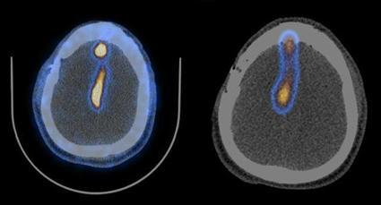
205 5.4.2 Imaging Uptake Regions of 177 Lu-DOTATATE therapy uptake on SPECT scan mirrored regions of tracer avidity on pre-therapy 68 Ga-DOTATATE scans (figure 5.1). 68 Ga-DOTATATE 177 Lu-DOTATATE Pt Figure 5-1 Regions of 177 Lu-DOTATATE therapy uptake on SPECT scan mirrored regions of tracer avidity on pre-therapy 68 Ga-DOTATATE scans 205
206 5.4.3 Response Tables 5.3 and 5.4 detail the disease status and growth rates pre and post treatment for each patient. There was at least 12 months between the pretreatment and post-treatment imaging due to cycle length and time to posttreatment MRI, hence disease status post-treatment reflects a long time period. It was not possible to calculate time to progression as patients had significant intervals between imaging. Patients 1-4 completed all 4 cycles. On MRI performed 3 months post-therapy, three of these four patients had SD by RECIST criteria, two by WHO criteria and one by modified RECIST criteria. By volumetric analysis, all had an increase in tumour volume across the treatment period, but the extent of this increase varied widely between 2-460%. The only patient with SD by modified RECIST criteria had minimal volume increase over the treatment period (2%). Tumour growth rate was markedly slower in the posttreatment period in three patients. Percentage increase in tumour volume was 7-255% per month pre-treatment versus % per month through treatment. Assuming exponential growth, this equates to a growth rate of versus Figure 5.2 depicts changes in growth rate. Patient 2 had also developed several new intracranial meningioma deposits in the four months prior to the baseline scan and no further new deposits occurred in the 12 months between the baseline and post-treatment scans. Patient 5, who terminated treatment after one cycle, actually had a reduction in tumour growth rate on MRI performed post cycle 1 compared to pre-treatment imaging (14% per month pre-treatment versus 2.5% per month during treatment), although the patient had continued to deteriorate clinically and was unfit for cycle 2. Both patients who did not complete therapy had a poorer baseline performance status compared to those who completed 4 cycles (PS 3 versus PS 1). As detailed in table 5.6, there was no clear association between SUV max and changes in growth rate: patient 3 had the most striking reduction in both growth rate and SUV max (figure 5.2), but in other patients SUV max response was variable. 206
207 Table 5-3 Disease Status Post PRRT According to Linear Criteria Pt Linear assessment Post-PRRT RECIST Modified RECIST WHO 1 PD PD PD 2 SD PD SD 3 SD SD SD 4 SD PD PD SD: stable disease; PD: progressive disease Pt Table 5-4 Disease Status Post PRRT: Volumetric and Growth Rate Analysis Vol pre- PRRT 1 (ccm) Vol pre- PRRT 2 (ccm) Vol post- PRRT (ccm) Vol between pre and post-tx MRI (%) Time between scans Pre 1 and 2; Pre 2 and Post (months) Growth pre- PRRT (% per month)* Growth Over PRRT (% per month)* Growth Rate pre-prrt** ; ; ; (but had surgery after so 8.2 pre- PRRT) ; N/A 6; N/A N/A 1.5; NA 60 N/A N/A Performance status deteriorated so patients received only 1 cycle Growth Rate over PRRT** 207
208 Figure 5-2 Growth rates pre and post PRRT 208

209 Table 5-5 SUV max values pre and post PRRT Patient Pre-PRRT SUV max Post-PRRT SUV max Figure 5-3 Patient 3 was the only patient with a considerable reduction in SUV max after 4 cycles of treatment. 209
210 5.4.4 Outcomes Post PRRT Disease progression continued in all patients following completion of PRRT. Patient 1 died 18 months after completion of therapy following continued gradual disease progression. Further surgery was not pursued as only limited debulking would have been possible and was associated with significant risks. Patient 2 underwent further debulking surgery 10 months after cycle 4 due to PD causing impaired mobility. 18 months following completion of 177 Lu (8 months post-surgery) gradual disease progression had continued but further surgery is being reserved until symptoms progress. Patient 3 had the best reduction in growth rate and SUV max during treatment, but underwent re-irradiation with EBRT for disease progression 15 months following completion to try and prevent loss of vision (accepting the risk of reirradiation). As discussed, he was diagnosed with AML shortly afterwards and received palliative treatment. Disease progression in patient 4 has continued at the same rate in the 12 months follow-up post PRRT, although only in one specific region of the tumour (frontal). Surgery is being reserved for when symptoms develop. Rapid disease progression continued in the two patients who terminated treatment after one cycle and both died within five months of commencing PRRT Dosimetry The estimated mean absorbed dose of radiation within the meningioma in the single cycle evaluated in patient 4 was 33.9Gy. The cumulated activity graph is shown in figure
211 Figure 5-4 Cumulated activity within the meningioma for patient 4 (1 cycle) 5.5 Discussion The purpose of reviewing outcomes in the first patients with progressive meningioma treated with 177 Lu DOTATATE PRRT at my institution was to establish if a larger formal study was warranted, to assess treatment regime tolerability and to explore methods of evaluating response and dosimetry in meningiomas. The patient cohort was necessarily small and results are therefore descriptive. The definition of stable or progressive disease in brain tumours, particularly meningiomas, is challenging. In this analysis, measuring response by three different linear criteria produced three different results. Of the four patients who completed therapy, the disease was classified stable at 1 year in three patients using RECIST criteria and two patients by WHO criteria. Neither measure adequately reflected the clinical picture as they did not take account of the fact that particular regions of meningioma were growing in most patients, 211
212 but were not the largest dimensions. Modified RECIST criteria did reflect this and seemed the most appropriate linear measure, as the only patient with SD according to these specifications also had very little change in volume (Patient 3). Volumetric measurements offer more information, but no cut-off values exist to define stable or progressive disease. Henson et al, extrapolated the RECIST and WHO linear criteria to equivalent volume criteria assuming a spherical lesion using the formula V= 4/3πr 3 [406]. They reported that disease would be classed as progressive when the tumour volume increased by 73% or 40% for RECIST and WHO respectively. This large discrepancy indicates that further work would be required before volumetric criteria could become standard in studies. Volumetric analysis may also not reflect important changes in meningiomas where the exact region of disease progression can be more relevant than the extent of progression per se, as even minimal growth in eloquent areas can result in major symptoms. Conversely, for similar reasons objective symptomatic improvements can occur following radiation with very little change in imaging (as shown in chapter 2). Another issue previously discussed is that meningioma volumes can be difficult to define due to dural tails, post-operative changes and bony hyperostosis, although it is likely to be the extent of soft tissue disease that dictates prognosis. To try and standardise MRI evaluation in this report, only regions of bone positive on 68 Ga DOTATATE PET were defined as disease and all measurements were performed by one consultant radiologist to avoid inter-observer variability. Evaluation of growth rate was explored in these patients. Meningioma growth rates slowed considerably in three of four patients who completed 4 cycles. However, tumour growth continued in all patients and debate is required regarding the extent of growth rate reduction that could be considered clinically relevant and would justify the cost of therapy. The two methods of expressing growth rate (% per month and logarithmic scale) appeared relatively equivalent when plotted, although meningiomas may not grow in the same exponential manner as most malignant tumours. Changes in tumour metabolic activity on PET imaging may have a role in assessing response to treatment, although there was no obvious correlation 212
213 between reduction in growth rate and SUV max in these patients. A larger cohort would be required to formally evaluate this. Patient 3 had a notable reduction in SUV max and growth rate during treatment, but developed progressive disease post-therapy at a similar rate to the other patients who completed four cycles. Indeed, whether meningiomas reduce sstr expression in response to radiation is not known as sstr do not necessarily reflect metabolic or mitotic activity and such receptors may not down-regulate following successful treatment. One group actually reported an increase in SUV max values on 68 Ga DOTA PET in the majority of patients following one cycle of 177 Lu DOTA for meningioma [338]. The 177 Lu DOTATATE protocol used in this cohort of patients was well tolerated acutely. It was the same regime as that used in neuroendocrine tumours where patients have usually been heavily pre-treated with other marrow-depleting therapies. As this is not the case for patients with meningioma, there could be scope to increase the dose or frequency/ number of cycles of treatment, but this would obviously incur greater cost and potential side effects. There were considerable limitations with this analysis, largely due to the fact that a pre-defined pilot study protocol was not used. Although a defined treatment protocol was used, a formal study protocol would have improved consistency of imaging and symptom analysis and strengthened outcome assessment. Timing between imaging differed considerably (particularly pretreatment imaging). This made it impossible to accurately define time to disease progression. As such growth rate results are an approximation as they would not necessarily have been consistent over the time period between imaging. Ideally, in a formal study, two pre-treatment MRIs at a specified interval would be required for growth rate evaluation and more frequent MRIs during therapy would allow time to progression/ progression free survival rates to be established at set time points. This would allow comparison to studies of systemic agents. Accepting these limitations, overall this outcome analysis in the six initial patients with advanced progressive meningioma treated with this PRRT suggested limited potential clinical benefit. Recently, the cost of 177 Lu DOTATATE therapy has risen significantly due to patenting that restricts in- 213
214 house re-constitution. As such, we have decided not to pursue a more formal study. Furthermore, although treatment was well tolerated in the acute setting, a causative relationship between PRRT and the development of AML 18 months following treatment in Patient 3 must be considered. It cannot be definitively concluded that this was secondary to PRRT, particularly in view of the short latency period, but the complex karyotype was indicative of a secondary leukaemia. The potential for ionising radiation to induce leukaemias is well established, most notably in survivors of the atomic bombs [407]. Although rare, an increase in secondary leukaemias has also been reported in patients receiving external beam radiotherapy for a wide range of conditions [408, 409] and cytotoxic chemotherapy [410]. Overall, therapy-related myeloid neoplasms are thought to account for 10-20% of all cases of AML and myelodysplastic syndrome [411]. Most reports of leukaemia following PRRT concern the use of 131 I for thyroid cancer and a recent meta-analysis reported a 2.5 fold increased relative risk for the development of leukaemia in patients treated with radioiodine for thyroid cancer [412]. Case reports exist regarding AML following the use of other radionuclides in cancer therapy [413, 414]. Most relevant to this case, are reports of acute leukaemia following treatment with 177 Lu or 90 Y DOTA in patients with neuroendocrine tumours, but causation cannot be assumed from case reports [415, 416]. Kossman et al, reported two incidences of AML 17 and 26 months following 89 Strontium for metastatic prostate cancer [413], although a causative relationship with the radionuclide was questioned in view of the fact that the patients had previously received other cancer therapies and the relatively short latency period [417]. The potential to induce second malignancies carries more importance if PRRT were to be used earlier in the course of meningiomas, particularly as radiotherapy/ radiosurgery are generally very effective. Minutoli et al, recently reported their experience of using 111 Pentetreotide in eight patients with meningioma [418]. The treatment schedules varied between patients and sometimes combined 90 Y DOTA cycles. They reported PR in two patients, stable disease in five and progression in one after 2-4 cycles of therapy. However, four patients (50%) had stable disease prior to therapy and as the 214
215 majority had grade 1 disease, PD would not necessarily have been expected in that timescale in the absence of any treatment. Furthermore, only one patient had previously received radiotherapy (six had surgery) and this certainly remains the recommended treatment when effective surgery is not possible. Kriessl et al, also evaluated the use of PRRT earlier in the course of meningiomas [338]. They carried out a pilot study in 10 patients with meningioma (majority grade 1) to establish the feasibility and tolerability of a combination of standard EBRT (median 53Gy) with a 7.2Gy PRRT boost ( 177 Lu). Treatment was well tolerated and there was a minor reduction in tumour size overall. However, whether the PRRT altered outcomes compared to EBRT alone cannot be evaluated and it remains unproven whether dose escalation in general is beneficial in meningioma, particularly for grade 1 disease. All patients in my cohort had G2 or 3 meningiomas with relatively rapid rates of progression. Patients 5 and 6, with poor performance status continued to rapidly progress and were not able to receive a second cycle of PRRT. Poor outcomes in less fit patients are typical in studies of systemic agents, and an ECOG PS of 2 is usually required. Most previous reports of the use of PRRT in meningiomas suggested that those with less aggressive disease may gain more benefit from PRRT. In Bartolomei s study of 29 patients treated with 90 Y DOTA, unsurprisingly patients with G1 disease had a much longer time to progression than those with higher grade disease (61 v 13 months) [335]. Likewise, there was no response in high grade bulky meningiomas treated with 177 Lu octreotate in the report by Van Essen et al [337]. However, Sabet et al, reported a dramatic improvement in a single patient with very poor PS following PRRT for meningioma [336]. In general, large tumour size is reported to limit the efficacy of PRRT[419]. It may be that the patients in my cohort had disease that was too advanced to achieve substantial benefit from 177 Lu DOTATATE therapy or that their disease had already become largely resistant to radiation therapy. An understanding of absorbed radiation dose following PRRT would help evaluate the relative merits of PRRT in meningioma. The ability to perform individual dosimetry could theoretically permit personalised dosing of radioisotope to achieve a desired uptake. Preliminary evaluation of meningioma uptake of 177 Lu was carried out in this study to assess whether this would be a feasible component of a larger study. The calculated absorbed dose of 33.9Gy 215
216 with one cycle was greater than expected, although not impossible as reported absorbed doses for neuroendocrine tumours vary between 0.9 to 42Gy/MBq following a single administration of 90 Yttrium DOTATOC therapy [420, 421]. However, there are substantial inherent inaccuracies with dosimetric evaluation of PRRT uptake. Most significantly, there is a rapid uptake or wash-in phase of 177 Lu into the tumour and a longer clearance or wash-out phase (half-life 6.7 days). Therefore the accuracy of the cumulative time activity curve is dependent on the number of time points used to create the curve. Imaging was performed at four time points to create the curve used but there was a large gap between point 3 and 4. More imaging time points would have increased certainty regarding the shape of the curve. A small degree of error may also have been introduced by the fact that the CT component of the SPECT/CT was used to calculate the tumour mass when MRI is the optimal imaging modality to define meningiomas, although the patient s tumour was clearly visible on SPECT/CT. Accurate mass is required to determine the appropriate S factor. Furthermore, the pre-calculated standard S factors provided by OLINDA/EXM software assume that tumours are isolated unit-density spheres with a uniform activity distribution and do not take into account individual patient/ tumour morphology or cross dose between tumour and normal tissue. However, the effect of this for 177 Lu in the brain will be minimal as there is little uptake outside the pituitary. Finally, when data are acquired shortly after isotope administration the gamma camera may be unable to accurately process the high activity, although a deadtime correction can be applied. The only other published work evaluating PRRT uptake in meningiomas comes from the previously mentioned pilot study of combined EBRT/ PRRT by Kriessl et al, and a subsequent paper by the same group correlating pre-treatment 68 Ga DOTA SUV max and PRRT uptake in the same patients [338, 422]. Patients remained in hospital for 4-5 days after PRRT and underwent daily nuclear imaging. They reported absorbed doses of Gy (median 7.2Gy) and a strong correlation between pre-treatment 68 Ga DOTA SUV max and 177 Lu DOTA retention in the voxels with the highest uptake. However, the limitations in accuracy of tumour dosimetry must be considered when interpreting results and further study would be required before conclusions could be drawn. 216
217 Unfortunately, more accurate assessment of meningioma dosimetry in a larger study would be extremely costly and onerous for patients due to the number of scans required. In view of the apparent limited efficacy of PRRT in patients with advanced progressive disease, such evaluations may not be feasible. 5.6 Conclusion There are no effective treatment options for patients with advanced, progressive meningiomas previously treated with radiotherapy. The abundant expression of sstr on meningiomas provided scientific rationale to explore the use of PRRT with 177 Lu-DOTATATE in such patients. Assessment of treatment response is challenging in meningiomas and standard linear criteria do not appear to accurately reflect disease status. Modified RECIST criteria appeared the most useful and simple to assess of the linear criteria. Volumetric analysis is appealing, but reproducibility within large studies may be difficult and impair response evaluation. In the first six patients with meningiomas treated with 177 Lu DOTATATE on compassionate grounds at my centre, growth rates did appear to slow. However, tumours continued to grow during treatment and only one patient had obviously stable disease over the treatment period. In view of the recent increase in cost of 177 Lu-DOTATATE, we did not feel that it was feasible to pursue further study in this patient population. 217
218 6 Chapter 6: Conclusions 6.1 Introduction The purpose of this thesis was to explore the feasibility and potential of advanced radiation planning and delivery techniques in the treatment of meningioma. Meningiomas are the most common non-glial brain tumour and radiotherapy is a well established treatment, with case series indicating ten year local control rates following radiotherapy of approximately 90% for benign disease (Table 1.5). In view of these excellent control rates, minimising treatment toxicity is of significant importance. Advanced radiation planning/ delivery techniques such as IMRT (including VMAT) and protons have the potential to reduce toxicity as they theoretically better spare normal tissue compared to 3DCRT. These techniques may also permit dose escalation for non-benign meningiomas where long-term reported tumour control rates postradiation are generally 50% (Table 1.5). Although the theoretical potential of such techniques to improve outcomes is clear, they are expensive to implement, require extreme precision of delivery and robust quality assurance protocols. Furthermore, questions remain regarding the low-dose radiation bath associated with IMRT. As highlighted in Chapter 1, the majority of reports detailing outcomes following radiotherapy for meningioma are retrospective case series which limit the estimation of local control, symptom improvement and toxicity rates. Careful clinical evaluation within prospective studies is required. Newer radiation techniques carry a greater potential for error than 3DCRT due to the conformity of the high dose region around the target. Translating their theoretical advantages into true clinical benefit will be highly dependent on accurate target delineation. Paradoxically, the use of advanced radiation techniques could be detrimental to outcomes if the target is poorly contoured. The definition of the appropriate target volume in meningiomas has not been established. Even the delineation of GTV is contentious as differentiating postoperative changes from viable meningioma can be challenging and the 218
219 appropriate extent of bone, dural tail and venous sinuses to include can be unclear on CT/MRI. Therefore, research into techniques that could clarify GTV is attractive. In Table 3.1 I detailed several reports that described alterations in the meningioma target volume defined in relation to the addition of 68 Ga DOTATATE PET/CT imaging to CT/MRI. This is appealing as 68 Ga DOTATATE is relatively specific to meningioma within the skull (outwith the pituitary). However, although there are reports of clinicians altering target volumes in relation to PET information, whether this improves the accuracy of contouring and whether PET/MRI offers any benefits over and above PET/CT has not previously been evaluated. Whilst more accurate target definition will maximise the benefits of all newer radiation planning and delivery techniques, the unique characteristics of the proton Bragg Peak has led many to consider whether protons could provide additional benefit in treating meningiomas over even the most conformal of photon techniques. As described in Chapter 4, several older planning studies indicated that protons improve normal tissue sparing in the treatment of meningiomas and many proton centres in other countries routinely treat meningiomas. This is of particular relevance as the first UK proton centres are being commissioned and appropriate treatment indications need to be defined. However, the recent advances in photon technology confer a need to evaluate whether the proton techniques to be used initially in the UK are likely to improve radiation treatment planning parameters against the current gold standard photon therapies for meningiomas. Unfortunately, despite impressive local control rates following radiotherapy, meningioma progression is encountered in clinical practice due to their relatively high prevalence. Currently, there are no established treatment options for patients with meningiomas that have progressed following radiotherapy. They appear unresponsive to cytotoxic chemotherapy. As for other tumour types, advances in treatments for meningiomas that recur following surgery and radiotherapy are likely to lie with receptor-targeted agents. The same somatostatin receptors exploited diagnostically with 68 Gallium DOTATATE PET offer an appealing target for peptide receptor radionuclide therapy (PRRT) with 177 Lutetium DOTATATE. Such therapy had been introduced at my institution on 219
220 a compassionate basis and evaluation of patients treated was required to establish whether a larger study was warranted. IMRT/ VMAT, PET-based planning, proton therapy and PRRT all carry the potential to improve outcomes for patients with meningioma. However, the cost implications associated with their use must be justified and it is essential to identify where they may offer most significant clinical benefit. Large scale studies themselves require considerable resources and should be supported by feasibility study evidence. Therefore, the specific aims of this thesis were: 1. To evaluate IMRT as a treatment for meningiomas within a prospective observational study in terms of toxicity, clinical and imaging response, quality of life and local control rates. 2. To assess the feasibility of integrating simultaneous 68 Gallium DOTATATE PET/MRI into meningioma radiotherapy target volume definition and to establish whether this impacted upon interobserver variability in contouring in relation to CT/MRI alone and PET/CT plus MRI. 3. To compare dosimetric parameters from optimal VMAT photon radiotherapy plans to SFUD proton plans for the treatment of meningioma and identify practical issues with SFUD proton planning. 4. To evaluate whether a formal phase II study of 177 Lutetium DOTATATE therapy for advanced progressive meningioma is warranted based upon outcomes in the first cohort of patients. 6.2 Important findings In Chapter 2 I demonstrated that IMRT is feasible for meningiomas and provided excellent target coverage/ OAR avoidance. This resulted in medium term tumour control rates of >90% in benign disease and provided more robust toxicity data than previous studies. Objective measures of toxicity were low. Improvements in visual symptoms were documented in a significant proportion 220
221 of patients and usually occurred without significant change in tumour dimensions on MRI. Analysis of non-visual symptoms and quality of life were more subjective and subject to confounding, but a substantial number of patients did report improvements. Longer follow-up and a larger patient cohort are required to strengthen these findings. As such, I would advocate IMRT/ VMAT as the radiation method of choice to treat meningiomas. However, in Chapter 3 I identified very high rates of interobserver variability in meningioma target volume definition. This requires further evaluation if more highly conformal radiotherapy techniques are to be widely adopted. Significant levels of IOV indicate over/ undertreatment of the true target depending upon the contouring physician. I found that analysing the Kouwenhoven Conformity Level highlighted the extent of IOV far more than the absolute volumes and would suggest that analysis of conformity with a method such as the KCL be analysed in all future studies of IOV. It would be useful if this were standardised to allow interstudy comparisons. Although the incorporation of simultaneous PET/MRI into treatment planning was feasible, it resulted in only a very small reduction in IOV compared to the standard CT/MRI and there was no clinical advantage to PET/MRI over PET/CT when all three modalities were coregistered. There was one case (of ten) where the PET information clearly assisted two of the observers in identifying meningioma that had unexpectedly extended into the soft tissue below the base of skull. However, on reflection, this could be identified on MRI with careful evaluation. Therefore, although there may be specific situations where PET could add valuable information, I would not advocate the routine use of PET (from MRI or CT) in meningioma contouring without more evidence to indicate a significant benefit. Thus, the theoretical potential of 68 Ga DOTATATE PET to improve meningioma contouring did not appear to translate into a substantial benefit in real clinical cases. In a similar manner, despite the favourable dose deposition characteristics of the proton Bragg Peak, I did not find a notable advantage for SFUD protons compared to an advanced photon IMRT technique (Truebeam VMAT) in the planning study in Chapter 4. This was largely due to the high quality of the photon plans that left little room for improvement and the significant uncertainties associated with protons that necessitated optimisation to a larger proton target volume. I did identify a slightly lower V10-30% for 221
222 normal brain with protons which could become more significant if advances in knowledge of proton dose deposition characteristics reduce the need for the addition of extra margins to guarantee target coverage. Overall, my findings in Chapter 4 do not support the clinical introduction of SFUD protons for meningioma treatment currently. In my analysis of 177 Lu DOTATATE therapy for advanced meningiomas presented in Chapter 5, I again found that the theoretical promise of advanced radiation techniques did not necessarily translate into substantial clinical benefit. Although meningioma growth rates appeared to slow during 177 Lu DOTATATE therapy, the tumours did generally progress during treatment. The cost of embarking upon a larger study would be substantial and the magnitude of benefit and relevant patient population relatively small. Therefore, although 177 Lu DOTATATE appeared to confer some anti-meningioma activity, further study was not feasible. This chapter also highlighted the challenges associated with defining imaging criteria that reflects the response of meningiomas to treatments. It was clear in Chapter 2 that many patients experienced objective symptomatic improvement following IMRT despite no obvious change on MRI and the converse is no doubt true. The modified RECIST criteria appeared the most useful and practical of the linear criteria as it includes a neurological progression category to reflect this. Growth rate/ volumetric analysis seemed more sensitive to identify meningioma growth and may be useful for future studies evaluating treatments particularly in advanced meningioma, but clinically relevant changes in growth rate would have to be debated and would vary depending upon tumour location. Furthermore, the IOV in contouring meningiomas noted in Chapter 3 would also be likely to affect volume analysis by reporting radiologists. 6.3 Implications of thesis Chapter 2 provided preliminary clinical evidence to support the more widespread use of IMRT rather than 3D CRT in the treatment of meningiomas. In addition, it demonstrated that introducing an advanced radiation technique to 222
223 clinical practice within an observational/ phase 2 study can be an effective tool to increase patient access to advanced treatments whilst ensuring quality assurance and allowing prospective data collection. I would argue that randomised studies are unnecessary and indeed unethical to evaluate advanced radiation techniques in situations where the dose distributions achievable are significantly superior to the standard technique, particularly for meningiomas where it would take many years to evaluate outcomes. Randomised trials of radiation techniques for meningiomas have previously failed due to poor accrual. These are particularly unlikely to succeed in benign meningioma as extended follow-up would be required to establish tumour control rates and clinical outcomes, which are themselves highly dependent on tumour location, baseline features and previous interventions. Even for nonbenign meningiomas, treatment decisions are highly influenced by individual tumour features such as the tumour location and clinician/ patient preferences hamper randomisation. Currently, a phase 3 multi-centre RCT (ROAM Study) is in set-up in the UK with the aim of evaluating the impact of adjuvant radiotherapy on recurrence rates following complete resection of atypical meningiomas. This is a relatively clean question and a randomised study could succeed, although the feasibility/ acceptability of the study to eligible patients and clinicians will be initially analysed to determine whether the full study can proceed. Whilst in specific circumstances RCTs may therefore remain appropriate to study radiotherapy for meningiomas, more novel trial design must be considered. The IMRT study in Chapter 2 will provide robust long-term outcome information that is far more reliable than retrospective case series in the largest patient population possible within a single centre. However, such an approach could potentially be extended to include multiple centres and the robust radiotherapy quality assurance required would in itself improve the quality of radiotherapy delivered. As was apparent throughout this thesis, the support of professionals outside of the radiotherapy department is essential to implement advances in technology and to accurately monitor clinical outcomes (such as ophthalmology) and a trial setting appeared to facilitate interdepartmental support. In Chapters 3-5 I found that the theoretical advantages of advances in radiation planning/ delivery techniques do not necessarily translate into likely clinical 223
224 benefit. Clearly a balance must be struck between early implementation of new technologies for the benefit of current patients and a need to delay widespread clinical use whilst supporting evidence is obtained. Nevertheless I would advocate the need to obtain evidence in support of a technology from feasibility/ planning studies prior to embarking upon expensive large studies or incorporating the technique in routine clinical practice. Contouring protocols have been changed in some centres on the basis of reports that the integration of a new imaging modality alters target volume definition, but, as demonstrated in Chapter 3, this is not necessarily evidence that the new imaging improves contouring. Although evaluation of IOV is only a surrogate for improved target definition, it is more meaningful than change alone. Methods of evaluating IOV vary widely throughout the literature and it would be helpful if a standardised approach could be endorsed by bodies such as the EORTC/ RTOG to allow comparison between different studies in the future. In the UK, direction from the NCRI CTRad group can help standardise studies and provide appropriate software, such as that used to analyse IOV in Chapter 3. Although the number of patients who received PRRT for advanced meningiomas in Chapter 5 was very small and there were considerable issues with data analysis, the apparent minimal activity of the radioisotope therapy suggested radioresistance in meningiomas that progress following previous radiation treatment (EBRT, RS or both). Future research efforts for this patient group should concentrate upon systemic targeted non-radiation therapies. The recent publication of promising phase 2 data reporting outcomes following the use of sunitinib, a tyrosine kinase inhibitor targeting vascular endothelial growth factor receptor (VEGFR) and platelet derived growth factor receptor (PDGFR), to treat surgery and radiation refractory grade 2 and 3 meningioma offers an avenue for future randomised study [423]. Thirty-six patients were enrolled and six month PFS rates were 42% with a median PFS of 5.2 months and overall survival of 24.6 months. However, the rate of progression pre-treatment was not stated and radiological outcome measures were limited by the use of the MacDonald (2D) response criteria as previously discussed. Expression of VEGFR2 appeared predictive of PFS, although this could just be a marker of more favourable tumour biology. Considerable toxicity was also noted with 60% experiencing grade 3 side-effects. 224
225 6.4 Future Research The ability of IMRT to sculpt radiation dose demonstrated in Chapters 2 and 4 raise the question of whether other regions of the brain, particularly those known to be associated with higher mental function, should become organs at risk. The hippocampus has an important role in short term memory and has been shown to be extremely radiosensitive. As shown in appendix 3, doses to the hippocampus can be significantly reduced with IMRT by specifying this as an avoidance structure. However, many other regions of the brain responsible for functions such as attention and information processing are likely to be just as important as the hippocampus and clinical study with formal neuropsychology testing is required to establish whether reducing dose to the hippocampus reduces toxicity and if so what the appropriate dose constraint should be. Indeed, neuropsychological impairment following brain radiotherapy is very poorly characterised and will be significantly influenced by other factors such as tumour location, previous surgery, anti-seizure medications and comorbidities such as depression. The true incidence and nature of neuropsychological impairment following brain radiotherapy have been very poorly studied and this should be a priority for future clinical study. Strategies to reduce the high baseline level of IOV in meningioma contouring identified in Chapter 3 require evaluation. Clinical judgement regarding areas at risk appeared to be a significant factor determining IOV and simple clarification of the location of meningioma recurrences/ progressions may help to standardise physicians views on what constitutes the target. Due to the efficacy of radiotherapy, multicentre observational/ non-randomised phase 2 studies would be required to generate this information. Although I did not find that PET information significantly reduced IOV, further work aiming to clarify the sensitivity of PET tracers to identify small volume meningioma may improve confidence to reduce volumes in relation to PET information. This may make it more likely to assist in standardising contours. The most appropriate way of studying this would be pathological examination of tissue in patients undergoing surgery for meningioma from regions that appear suspicious on pre-operative CT/MRI but are PET negative. It would be impractical to attempt biopsy in patients undergoing primary radiotherapy. 225
226 Although I did not find a notable improvement in meningioma plan dosimetry with SFUD protons, range uncertainty necessitated optimisation to larger target volumes for protons than photons which compromised their potential to improve radiotherapy plans. However, in view of the excellent results achievable with modern photon techniques, I do not feel that further research into the use of protons to treat meningioma is a priority, but more general research into proton dose deposition characteristics and biology would allow better exploitation of their potential clinical benefit. Development of proton-based imaging is an important area of research that is likely to advance our understanding of absorbed dose and reduce uncertainties. It is likely that additional avoidance structures in the brain will be proposed in the coming years, particularly for aspects of higher mental function and it is possible that protons may become a useful treatment option if more certainty regarding the location of the Bragg Peak can be achieved. 6.5 Conclusion The likely clinical benefits associated with some advanced radiation techniques in meningioma are clear, such as IMRT versus 3DCRT. However, throughout this thesis I have found that the theoretical benefits of some technologies do not necessarily translate into dosimetric/ clinical improvements in real cases. Planning studies and early feasibility assessments are essential to focus radiation research in the most appropriate areas and to identify technical challenges prior to embarking upon expensive large scale studies or routinely introducing complex techniques into clinical practice. The RCT is not necessarily the most appropriate form of study for research into advanced radiotherapy techniques, particularly for slow-growing tumours like meningiomas. Technology develops far faster than results of studies would take to mature. However, other carefully planned and executed prospective approaches, such as non-randomised observational/ phase II studies, can provide strong evidence for the dosimetric advantage, safety and clinical effectiveness of new techniques. Radiotherapy confers excellent long term 226
227 control rates for meningioma and therefore research focused on minimising long-term toxicity rates is important. Such studies require a co-ordinated multidisciplinary approach both in design and execution. A raft of quality assurance procedures to ensure optimum treatment planning and delivery has accompanied the implementation of modern highly conformal radiation techniques. However, the largest variable influencing outcomes is likely to be delineation of the target by the treating clinician. This appears to be a particular issue for meningiomas, although it is undoubtedly a feature in many other tumour types. Research to establish variables in target definition between clinicians and strategies to improve consistency should be seen as a priority to allow the theoretical promise of advanced radiation therapies to be translated into real clinical benefit. 227
228 7 Appendices 228
229 7.1 Appendix 1 EORTC Quality of Life Questionnaires EORTC QLQ - BN20 Copyright 1994 EORTC Quality of Life Group Patients sometimes report that they have the following symptoms. Please indicate the extent to which you have experienced these symptoms or problems during the past week. During the past week: Not A Quite a Very at all Little bit much 31 Did you feel uncertain about the future? Did you feel you had setbacks in your condition? Were you concerned about disruption of family life? Did you have headaches? Did your outlook on the future worsen? Did you have double vision? Was your vision blurred? Did you have difficulty reading because of your vision? 39 Did you have seizures? Did you have weakness on one side of your body? Did you have trouble finding the right words to express yourself? 42 Did you have difficulty speaking? Did you have trouble communicating your thoughts? Did you feel drowsy during the daytime? Did you have trouble with your coordination? Did hair loss bother you? Did itching of your skin bother you? Did you have weakness of both legs? Did you feel unsteady on your feet? Did you have trouble controlling your bladder?
230 EORTC QLQ-C30 (version 3) We are interested in some things about you and your health. Please answer all of the questions yourself by circling the number that best applies to you. There are no "right" or "wrong" answers. The information that you provide will remain strictly confidential 1 Do you have any trouble doing strenuous activities, like carrying a heavy shopping bag or a suitcase? Not at all A little Quite a Very bit much Do you have any trouble taking a long walk? Do you have any trouble taking a short walk outside of the house? 4 Do you need to stay in bed or a chair during the day? 5 Do you need help with eating, dressing, washing yourself or using the toilet? During the past week: 6 Were you limited in doing either your work or other daily activities? 7 Were you limited in pursuing your hobbies or other leisure time activities? 8 Were you short of breath? Have you had pain? Did you need to rest? Have you had trouble sleeping? Have you felt weak? Have you lacked appetite? Have you felt nauseated? Have you vomited? Have you been constipated? During the past week: 17 Have you had diarrhoea? Were you tired? Did pain interfere with your daily activities? Have you had difficulty in concentrating on things, like reading a newspaper or watching television? 21 Did you feel tense? Did you worry? Did you feel irritable? Did you feel depressed? Have you had difficulty remembering things? Has your physical condition or medical treatment interfered with your family life? 27 Has your physical condition or medical treatment interfered with your social activities? 28 Has your physical condition or medical treatment caused you financial difficulties?
231 For the following questions please circle the number between 1 and 7 that best applies to you 29. How would you rate your overall health during the past week? Very poor Excellent 30. How would you rate your overall quality of life during the past week? Very poor Excellent Copyright 1995 EORTC Quality of Life Group. All rights reserved. 231
232 7.2 Appendix 2 EEG Neuropsychology Pilot Substudy Protocol Background In the standard IMRT Study protocol we now use clinical neuropsychology testing to evaluate whether there are any changes in higher mental functions such as memory, attention etc. following radiotherapy for meningioma. Measuring event related potentials (ERP) with electroencephalogram (EEG) recording during neuropsychology testing is a more complex assessment protocol for neuropsychology assessment used in research into dementia and other cognitive disorders. Many groups have reported that such testing has allowed a better understanding of why patients develop deficits and in some situations can identify abnormalities before they clinically manifest. More information about electrical changes in the brain following radiotherapy would permit a better understanding of the cause of any cognitive changes and the sensitivity of various brain regions to radiotherapy. Currently there are no reports of using such technology to study patients following radiotherapy. Objectives We will carry out a pilot study to establish whether EEG monitored neuropsychology testing is practical for patients undergoing radiotherapy, whether patients find the protocol acceptable and how it would best be incorporated into the main IMRT study. We will assess what changes are seen on the EEG in patients who have neuropsychology test scores that have reduced following radiotherapy. 232
233 Patient Selection Criteria Inclusion Patients should already have undergone neuropsychology testing in the main IMRT study with scores in any domain below the 50% percentile on the most recent test or with a drop of 25% from their baseline. Exclusion Patients deemed unlikely to be able to participate in the full 2 hour assessment. Patient Numbers Five patients will undergo EEG testing to assess the feasibility of testing and potential value of including it for other patients. Withdrawal Patients will be free to stop the session and withdraw from the study at any point. Trial Design This is a pilot study to evaluate acceptability of EEG neuropsychology testing to patients, practical issues regarding the feasibility of carrying out such testing and is hypothesis generating in terms of what EEG features may be seen in patients following radiotherapy. If this pilot substudy indicates that a larger study of EEG testing is warranted then before and after treatment testing may be incorporated into the main IMRT study (following study amendment). 233
234 Interventions Patients will have one session of neuropsychology assessment with EEG recording. The EEG will be measured using 24 electrodes, which are applied to the subject s scalp by means of an electrode cap. Reference electrodes will be placed on the left and right mastoids, and a ground electrode will be placed on the sternum. After application of the electrodes, a water soluble electrode paste is applied to the electrodes. The application of the electrodes, will take about 30 minutes in total. A variety of neuropsychology tasks relating to different cognitive processes will be carried out with EEG recording. These are used frequently in EEG studies. The tasks are presented in the visual modality on a computer screen which will display varying commands. Patients will respond by pressing different computer keys as instructed with either left or right hand or with different fingers of the same hand. The following tests will be undertaken: 1. Information processing: S1-S2 Test (stimulus/ response) 2. Memory processes: Relational and Item Specific Encoding Task 3. Response related information processing: Eriksen Flanker Task A mental fatigue and patient opinion questionnaire will be completed at the end of the session. Total testing time will be around two hours (including short rest periods). Criteria of Evaluation Behaviour analysis During the experiment, computer key presses of the subject will be recorded. Average response accuracy and reaction times will be used as a measure of performance during each task. EEG analysis EEG analyses will be performed, using Vision Analyzer (Brain Products GmbH). 234
235 Statistical Considerations Behavioural data will be analysed using repeated measures ANOVA. Analyses will be performed using SPSS. The univariate approach for repeated measures will be used. Mean amplitudes will be entered as dependent variables into SPSS. In the ERP analyses, erroneous responses will be rejected from the analyses. Ethical Issues The substudy will be conducted in accordance with the declaration of Helsinki. A patient information sheet specific to the substudy will be given to the patient. The patients are already enrolled in the main IMRT study so that information sheet will not be given again. Any questions arising from the patient information sheet will be discussed before the patient gives written informed consent to be entered into the substudy. GPs of patients that have consented should be informed in writing Analysis Five patients took part in the substudy. Feasibility data was obtained. All patients completed the tasks, but found them onerous and tiring. They would not have been willing to undertake such assessment on a regular basis. The results could not be further analysed due to technical issues. 235
236 7.3 Appendix 3: Can doses to the hippocampus be reduced for patients treated with IMRT for meningioma? SGDMLC IMRT versus VMAT Background Preclinical evidence suggests that radiation dose to neurogenic stem cells in the hippocampus is central to radiation-induced memory impairment [424, 425]. Recently, preliminary work has postulated that a dose of >7.3Gy (EQD2) to 40% of the bilateral hippocampus (D40) is a predictor of memory impairment [345]. Such effects are particularly significant in patients with benign histology, such as meningiomas. One of the main benefits of IMRT is that dose to specific OARs can be reduced compared to older radiation delivery methods. The OAR maximum dose has to be specified in the IMRT optimiser at the start of planning. Standard OARs in the brain where optimiser limits are set are the optic structures and brainstem. However, the use of IMRT may permit reduced dose to other OARs, including regions responsible for higher mental functions. VMAT is increasingly favoured as the method of IMRT delivery, mainly due to its efficiency. However, it is unclear whether static gantry dynamic MLC IMRT (SG DMLC) where the field position can be manipulated or VMAT would offer the best hippocampal sparing or how low the dose to the hippocampi could be kept. I therefore explored the feasibility of reducing doses to the hippocampi using IMRT and VMAT. Aim Preliminary study of the ability of SG DMLC IMRT and VMAT to reduce dose to the hippocampi. Methods Five meningioma cases from the IMRT study described in chapter 2 were replanned with the addition of specific hippocampus dose constraints. Tumours in varying locations were specifically chosen. Two tumours were anterior, two central and one posterior. All planning was performed using Eclipse Treatment Planning System (Varian). The original IMRT plans had been delivered with a SG DMLC technique and had been optimised for PTV coverage with limitation of dose to optic structures and brainstem. The prescribed dose to PTV was 236
237 50.4Gy in 28#. Hippocampi were contoured on co-registered MRI according to the guidelines by Chera et al [426] (although MRI slice thickness was 3mm). No attempt had been made to spare the hippocampus. IMRT plans were reoptimised and VMAT plans created aiming to reduce mean hippocampi dose with a bilateral D40% 12Gy (equivalent to 7.29Gy in 2Gy #, α/β=2) whilst meeting the original plan objectives (specified in section 2.3.2). Results It was possible to reduce the dose to the hippocampi in all cases whilst remaining within the other standard dose constraints. The mean bilateral hippocampal dose with was 21.5Gy (range: Gy) with original SGDMLC IMRT; 14.3Gy (range Gy) with optimised SG DMLC IMRT and 9.8Gy (range Gy) with optimised VMAT. None of the original non-optimised SGDMLC plans met the proposed bilateral hippocampi constraint of D40% of 12Gy. This constraint was met by four of the VMAT plans (13.1Gy for the other) and one of the optimised SG DMLC IMRT plans. Compared to the original SGDMLC IMRT plan the bilateral hippocampi D40% was reduced by 26-54% (mean 39% reduction) for the optimised IMRT plan and 53-77% (mean 64% reduction) in the VMAT plan. These doses were achieved without significant changes in PTV coverage or dose to OAR (increase in lens dose in one patient with VMAT). Hippocampal doses were higher for central lesions. The mean doses to the hippocampus and brain-ptv are shown in figure 7.1. The dose distributions for each patient are shown in figure
238 Figure 7-1 Mean doses to the bilateral hippocampus and whole brain-ptv comparing the original SG DMLC plan to the SG DMLC optimised for the hippocampi and the optimised VMAT plan Conclusions Doses to the hippocampi can be significantly reduced for meningioma patients by optimising for this structure in the treatment planning process. VMAT delivery appears more effective at reducing hippocampal dose than SGDMLC IMRT. The clinical impact of hippocampal dose reduction should be evaluated within prospective studies. 238


239 Patient 1 Patient 2 Patient 3 Figure 7-2 Axial CT slices of 5 meningioma cases comparing hippocampal dose distribution: original SGDMLC IMRT plan (not optimised for hippocampus), versus reoptimised SGDMLC IMRT plan versus optimised VMAT plan. PTV in red. Hippocampus in green. Doses >12Gy visible in colour wash. 239


240 Patient 4 Patient 5 Figure 7.2 continued 240
PRINCESS MARGARET CANCER CENTRE CLINICAL PRACTICE GUIDELINES
 PRINCESS MARGARET CANCER CENTRE CLINICAL PRACTICE GUIDELINES CENTRAL NERVOUS SYSTEM MENINGIOMA CNS Site Group Meningioma Author: Dr. Norm Laperriere Date: February 20, 2018 1. INTRODUCTION 3 2. PREVENTION
CNS TUMORS. D r. Ali Eltayb ( U. of Omdurman. I ). M. Path (U. of Alexandria)
. M. Path (U. of Alexandria)") CNS TUMORS D r. Ali Eltayb ( U. of Omdurman. I ). M. Path (U. of Alexandria) CNS TUMORS The annual incidence of intracranial tumors of the CNS ISmore than intraspinal tumors May be Primary or Secondary
CNS TUMORS D r. Ali Eltayb ( U. of Omdurman. I ). M. Path (U. of Alexandria) CNS TUMORS The annual incidence of intracranial tumors of the CNS ISmore than intraspinal tumors May be Primary or Secondary
NON MALIGNANT BRAIN TUMOURS Facilitator. Ros Taylor Advanced Neurosurgical Nurse Practitioner Southmead Hospital Bristol
 NON MALIGNANT BRAIN TUMOURS Facilitator Ros Taylor Advanced Neurosurgical Nurse Practitioner Southmead Hospital Bristol Neurosurgery What will be covered? Meningioma Vestibular schwannoma (acoustic neuroma)
Dosimetry, see MAGIC; Polymer gel dosimetry. Fiducial tracking, see CyberKnife radiosurgery
 Subject Index Acoustic neuroma, neurofibromatosis type 2 complications 103, 105 hearing outcomes 103, 105 outcome measures 101 patient selection 105 study design 101 tumor control 101 105 treatment options
NCCN GUIDELINES ON PROTON THERAPY (AS OF 4/23/18) BONE (Version , 03/28/18)
 BONE (Version , 03/28/18)") BONE (Version 2.2018, 03/28/18) NCCN GUIDELINES ON PROTON THERAPY (AS OF 4/23/18) Radiation Therapy Specialized techniques such as intensity-modulated RT (IMRT); particle beam RT with protons, carbon ions,
BONE (Version 2.2018, 03/28/18) NCCN GUIDELINES ON PROTON THERAPY (AS OF 4/23/18) Radiation Therapy Specialized techniques such as intensity-modulated RT (IMRT); particle beam RT with protons, carbon ions,
Meningioma tumor. Meningiomas are named according to their location (Fig. 1) and cause various symptoms: > 1
 and cause various symptoms: > 1") Meningioma tumor Overview A meningioma is a type of tumor that grows from the protective membranes, called meninges, which surround the brain and spinal cord. Most meningiomas are benign (not cancer) and
Meningioma tumor Overview A meningioma is a type of tumor that grows from the protective membranes, called meninges, which surround the brain and spinal cord. Most meningiomas are benign (not cancer) and
Original Date: April 2016 Page 1 of 7 FOR CMS (MEDICARE) MEMBERS ONLY
 MEMBERS ONLY") National Imaging Associates, Inc. Clinical guidelines STEREOTACTIC RADIATION THERAPY: STEREO RADIOSURGERY (SRS) AND STEREOTACTIC BODY RADIATION THERAPY (SBRT) CPT4 Codes: Please refer to pages 5-6 LCD
National Imaging Associates, Inc. Clinical guidelines STEREOTACTIC RADIATION THERAPY: STEREO RADIOSURGERY (SRS) AND STEREOTACTIC BODY RADIATION THERAPY (SBRT) CPT4 Codes: Please refer to pages 5-6 LCD
General: Brain tumors are lesions that have mass effect distorting the normal tissue and often result in increased intracranial pressure.
 1 Lecture Objectives Know the histologic features of the most common tumors of the CNS. Know the differences in behavior of the different tumor types. Be aware of the treatment modalities in the various
Tumors of the Nervous System
 Tumors of the Nervous System Peter Canoll MD. PhD. What I want to cover What are the most common types of brain tumors? Who gets them? How do they present? What do they look like? How do they behave? 1
Corporate Medical Policy
 Corporate Medical Policy Intensity Modulated Radiation Therapy (IMRT) of Head and Neck File Name: Origination: Last CAP Review: Next CAP Review: Last Review: intensity_modulated_radiation_therapy_imrt_of_head_and_neck
Brain Tumors. What is a brain tumor?
 Scan for mobile link. Brain Tumors A brain tumor is a collection of abnormal cells that grows in or around the brain. It poses a risk to the healthy brain by either invading or destroying normal brain
Radiosurgery by Leksell gamma knife. Josef Novotny, Na Homolce Hospital, Prague
 Radiosurgery by Leksell gamma knife Josef Novotny, Na Homolce Hospital, Prague Radiosurgery - Definition Professor Lars Leksell The tools used by the surgeon must be adapted to the task and where the human
Flattening Filter Free beam
 Dose rate effect in external radiotherapy: biology and clinic Marta Scorsetti, M.D. Radiotherapy and Radiosurgery Dep., Istituto Clinico Humanitas, Milan, Italy Brescia October 8th/9th, 2015 Flattening
Otolaryngologist s Perspective of Stereotactic Radiosurgery
 Otolaryngologist s Perspective of Stereotactic Radiosurgery Douglas E. Mattox, M.D. 25 th Alexandria International Combined ORL Conference April 18-20, 2007 Acoustic Neuroma Benign tumor of the schwann
Defining Target Volumes and Organs at Risk: a common language
 Defining Target Volumes and Organs at Risk: a common language Eduardo Rosenblatt Section Head Applied Radiation Biology and Radiotherapy (ARBR) Section Division of Human Health IAEA Objective: To introduce
PEDIATRIC ORBITAL TUMORS RADIOTHERAPY PLANNING
 PEDIATRIC ORBITAL TUMORS RADIOTHERAPY PLANNING ANATOMY ANATOMY CONT ANATOMY CONT. ANATOMY CONT. EYE OF A CHILD Normal tissue tolerance doses (in conventional #) TD 5/5 TD 50/5 Endpoint Gy Gy Optic nerve
This LCD recognizes these two distinct treatment approaches and is specific to treatment delivery:
 National Imaging Associates, Inc. Clinical guidelines STEREOTACTIC RADIOSURGERY (SRS) AND STEREOTACTIC BODY RADIATION THERAPY (SBRT) CPT4 Codes: 77371, 77372, 77373 LCD ID Number: L33410 J-N FL Responsible
Background Principles and Technical Development
 Contents Part I Background Principles and Technical Development 1 Introduction and the Nature of Radiosurgery... 3 Definitions of Radiosurgery... 5 Consequences of Changing Definitions of Radiosurgery...
Stereotactic Radiosurgery and Stereotactic Body Radiation Therapy
 Stereotactic Radiosurgery and Stereotactic Body Radiation Therapy Policy Number: Original Effective Date: MM.05.008 05/12/1999 Line(s) of Business: Current Effective Date: HMO; PPO; QUEST Integration 04/01/2017
Stereotactic Radiosurgery and Stereotactic Body Radiation Therapy
 Stereotactic Radiosurgery and Stereotactic Body Radiation Therapy Policy Number: Original Effective Date: MM.05.008 05/12/1999 Line(s) of Business: Current Effective Date: HMO; PPO; QUEST Integration 04/01/2015
Year 2003 Paper two: Questions supplied by Tricia
 question 43 A 42-year-old man presents with a two-year history of increasing right facial numbness. He has a history of intermittent unsteadiness, mild hearing loss and vertigo but has otherwise been well.
CNS pathology Third year medical students. Dr Heyam Awad 2018 Lecture 12: CNS tumours 2/3
 CNS pathology Third year medical students Dr Heyam Awad 2018 Lecture 12: CNS tumours 2/3 Pilocytic astrocytoma Relatively benign ( WHO grade 1) Occurs in children and young adults Mostly: in the cerebellum
Tumors of the Central Nervous System
 Tumors of the Central Nervous System 1 Financial Disclosures I have NO SIGNIFICANT FINANCIAL, GENERAL, OR OBLIGATION INTERESTS TO REPORT Introduction General: Brain tumors are lesions that have mass effect
Brain Tumor Treatment
 Scan for mobile link. Brain Tumor Treatment Brain Tumors Overview A brain tumor is a group of abnormal cells that grows in or around the brain. Tumors can directly destroy healthy brain cells. They can
Carbon Ion Radiotherapy for Skull Base and Paracervical Chordomas
 Carbon Ion Radiotherapy for Skull Base and Paracervical Chordomas Azusa Hasegawa, Jun-etsu Mizoe and Hirohiko Tsujii Research Center Hospital for Charged Particle Therapy National Institute of Radiological
STEREOTACTIC RADIATION THERAPY. Monique Blanchard ANUM Radiation Oncology Epworth HealthCare
 STEREOTACTIC RADIATION THERAPY Monique Blanchard ANUM Radiation Oncology Epworth HealthCare Overview Stereotactic radiation therapy at Epworth Healthcare What is stereotactic radiation therapy? Delivery
Structural and functional imaging for the characterization of CNS lymphomas
 Structural and functional imaging for the characterization of CNS lymphomas Cristina Besada Introduction A few decades ago, Primary Central Nervous System Lymphoma (PCNSL) was considered as an extremely
CT & MRI Evaluation of Brain Tumour & Tumour like Conditions
 CT & MRI Evaluation of Brain Tumour & Tumour like Conditions Dr. Anjana Trivedi 1, Dr. Jay Thakkar 2, Dr. Maulik Jethva 3, Dr. Ishita Virda 4 1 M.D. Radiology, Professor and Head, P.D.U. Medical College
Chapters from Clinical Oncology
 Chapters from Clinical Oncology Lecture notes University of Szeged Faculty of Medicine Department of Oncotherapy 2012. 1 RADIOTHERAPY Technical aspects Dr. Elemér Szil Introduction There are three possibilities
Specialised Services Policy: CP22. Stereotactic Radiosurgery
 Specialised Services Policy: CP22 Document Author: Assistant Director of Planning Executive Lead: Director of Planning ad Performance Approved by: Management Group Issue Date: 01 July 2015 Review Date:
Laura Tormoehlen, M.D. Neurology and EM-Toxicology Indiana University
 Laura Tormoehlen, M.D. Neurology and EM-Toxicology Indiana University Disclosures! No conflicts of interest to disclose Neuroimaging 101! Plain films! Computed tomography " Angiography " Perfusion! Magnetic
Update on Pediatric Brain Tumors
 Update on Pediatric Brain Tumors David I. Sandberg, M.D. Director of Pediatric Neurosurgery & Associate Professor Dr. Marnie Rose Professorship in Pediatric Neurosurgery Pre-talk Questions for Audience
Advances in external beam radiotherapy
 International Conference on Modern Radiotherapy: Advances and Challenges in Radiation Protection of Patients Advances in external beam radiotherapy New techniques, new benefits and new risks Michael Brada
REVISITING ICRU VOLUME DEFINITIONS. Eduardo Rosenblatt Vienna, Austria
 REVISITING ICRU VOLUME DEFINITIONS Eduardo Rosenblatt Vienna, Austria Objective: To introduce target volumes and organ at risk concepts as defined by ICRU. 3D-CRT is the standard There was a need for a
Dr Sneha Shah Tata Memorial Hospital, Mumbai.
 Dr Sneha Shah Tata Memorial Hospital, Mumbai. Topics covered Lymphomas including Burkitts Pediatric solid tumors (non CNS) Musculoskeletal Ewings & osteosarcoma. Neuroblastomas Nasopharyngeal carcinomas
Clinical considerations of RBE in proton therapy
 Clinical considerations of RBE in proton therapy H. Paganetti PhD Professor, Harvard Medical School Director of Physics Research, Massachusetts General Hospital, Radiation Oncology Why do we need the RBE
Stereotactic Radiosurgery and Stereotactic Body Radiation Therapy
 Stereotactic Radiosurgery and Stereotactic Body Radiation Therapy Policy Number: Original Effective Date: MM.05.008 05/12/1999 Line(s) of Business: Current Effective Date: HMO; PPO; QUEST Integration 11/20/2015
Treatment Planning Evaluation of Volumetric Modulated Arc Therapy (VMAT) for Craniospinal Irradiation (CSI)
 for Craniospinal Irradiation (CSI)") Treatment Planning Evaluation of Volumetric Modulated Arc Therapy (VMAT) for Craniospinal Irradiation (CSI) Tagreed AL-ALAWI Medical Physicist King Abdullah Medical City- Jeddah Aim 1. Simplify and standardize
Treatment Planning Evaluation of Volumetric Modulated Arc Therapy (VMAT) for Craniospinal Irradiation (CSI) Tagreed AL-ALAWI Medical Physicist King Abdullah Medical City- Jeddah Aim 1. Simplify and standardize
PRINCESS MARGARET CANCER CENTRE CLINICAL PRACTICE GUIDELINES
 PRINCESS MARGARET CANCER CENTRE CLINICAL PRACTICE GUIDELINES CENTRAL NERVOUS SYSTEM LOW GRADE GLIOMAS CNS Site Group Low Grade Gliomas Author: Dr. Norm Laperriere 1. INTRODUCTION 3 2. PREVENTION 3 3. SCREENING
Dr. T. Venkat Kishan Asst. Prof Department of Radiodiagnosis
 Dr. T. Venkat Kishan Asst. Prof Department of Radiodiagnosis Schwannomas (also called neurinomas or neurilemmomas) constitute the most common primary cranial nerve tumors. They are benign slow-growing
Sacral Chordoma: The Loma Linda University Radiation Medicine Experience. Kevin Yiee MD, MPH Resident Physician
 Sacral Chordoma: The Loma Linda University Radiation Medicine Experience Kevin Yiee MD, MPH Resident Physician What is a chordoma? 1 st chordoma discovered in clivus by Virchow and Luschka 1856 Rare tumor
Peter Canoll MD. PhD.
 Tumors of the Nervous System Peter Canoll MD. PhD. What I want to cover What are the most common types of brain tumors? Who gets them? How do they ypresent? What do they look like? How do they behave?
Radiotherapy What are our options and what is on the horizon. Dr Kevin So Specialist Radiation Oncologist Epworth Radiation Oncology
 Radiotherapy What are our options and what is on the horizon Dr Kevin So Specialist Radiation Oncologist Epworth Radiation Oncology Outline Advances in radiotherapy technique Oligo - disease Advancements
Pediatric Brain Tumors: Updates in Treatment and Care
 Pediatric Brain Tumors: Updates in Treatment and Care Writer Classroom Rishi R. Lulla, MD MS Objectives Introduce the common pediatric brain tumors Discuss current treatment strategies for pediatric brain
Intensity Modulated Radiation Therapy (IMRT)
") Intensity Modulated Radiation Therapy (IMRT) Policy Number: Original Effective Date: MM.05.006 03/09/2004 Line(s) of Business: Current Effective Date: HMO; PPO 06/24/2011 Section: Radiology Place(s) of
Intensity Modulated Radiation Therapy (IMRT) Policy Number: Original Effective Date: MM.05.006 03/09/2004 Line(s) of Business: Current Effective Date: HMO; PPO 06/24/2011 Section: Radiology Place(s) of
Future upcoming technologies and what audit needs to address
 Future upcoming technologies and what audit needs to address Dr R.I MacKay History of audit Absolute dose - Simple phantom standard dose measurement Point doses in beams - Phantoms of relatively simple
Evaluation of Monaco treatment planning system for hypofractionated stereotactic volumetric arc radiotherapy of multiple brain metastases
 Evaluation of Monaco treatment planning system for hypofractionated stereotactic volumetric arc radiotherapy of multiple brain metastases CASE STUDY Institution: Odette Cancer Centre Location: Sunnybrook
Brain tumors: tumor types
 Brain tumors: tumor types Tumor types There are more than 120 types of brain tumors. Today, most medical institutions use the World Health Organization (WHO) classification system to identify brain tumors.
IMAGING OF A CASE OF SPINAL MENINGIOMA- A CASE REPORT
 IMAGING OF A CASE OF SPINAL MENINGIOMA- A CASE REPORT Ramneet Wadi 1, Anil Kumar Shukla 2, Seetha Pramila V. V 3, Sabyasachi Basu 4, Sonam Sanjay 5 1Postgraduate Student, Department of Radiodiagnosis,
Case Report Multiple Intracranial Meningiomas: A Review of the Literature and a Case Report
 Case Reports in Surgery Volume 2013, Article ID 131962, 4 pages http://dx.doi.org/10.1155/2013/131962 Case Report Multiple Intracranial Meningiomas: A Review of the Literature and a Case Report F. Koech,
Chapter 5 Section 3.1
 Radiology Chapter 5 Section 3.1 Issue Date: March 27, 1991 Authority: 32 CFR 199.4(b)(2), (b)(2)(x), (c)(2)(viii), and (g)(15) 1.0 CPT 1 PROCEDURE CODES 37243, 61793, 61795, 77261-77421, 77427-77799, 0073T
Evaluation of Whole-Field and Split-Field Intensity Modulation Radiation Therapy (IMRT) Techniques in Head and Neck Cancer
 Techniques in Head and Neck Cancer") 1 Charles Poole April Case Study April 30, 2012 Evaluation of Whole-Field and Split-Field Intensity Modulation Radiation Therapy (IMRT) Techniques in Head and Neck Cancer Abstract: Introduction: This study
1 Charles Poole April Case Study April 30, 2012 Evaluation of Whole-Field and Split-Field Intensity Modulation Radiation Therapy (IMRT) Techniques in Head and Neck Cancer Abstract: Introduction: This study
Update on Sarcomas of the Head and Neck. Kevin Harrington
 Update on Sarcomas of the Head and Neck Kevin Harrington Overview Classification and incidence of sarcomas Clinical presentation Challenges to treatment Management approaches Prognostic factors Radiation-induced
PITUITARY PARASELLAR LESIONS. Kim Learned, MD
 PITUITARY PARASELLAR LESIONS Kim Learned, MD DIFFERENTIALS Pituitary Sella Clivus, Sphenoid Sinus Suprasellar Optic chiasm, Hypothalamus, Circle of Willis Parasellar Cavernous Sinus Case 1 17 YEAR-OLD
FROM ICARO1 TO ICARO2: THE MEDICAL PHYSICS PERSPECTIVE. Geoffrey S. Ibbott, Ph.D. June 20, 2017
 FROM ICARO1 TO ICARO2: THE MEDICAL PHYSICS PERSPECTIVE Geoffrey S. Ibbott, Ph.D. June 20, 2017 1 DISCLOSURES My institution holds Strategic Partnership Research Agreements with Varian, Elekta, and Philips
Advances In Orbital Neuropathology
 Advances In Orbital Neuropathology Charles G. Eberhart, MD PhD Associate Professor of Pathology, Ophthalmology and Oncology Johns Hopkins University School of Medicine Overview Non-neoplastic lesions Microphthalmos/pseudoglioma
Radiotherapy in feline and canine head and neck cancer
 Bettina Kandel Like surgery radiotherapy is usually a localized type of treatment. Today it is more readily available for the treatment of cancer in companion animals and many clients are well informed
Optic Pathway Gliomas, Germinomas, Spinal Cord Tumours. Colin Kennedy March 2015
 Optic Pathway Gliomas, Germinomas, Spinal Cord Tumours Colin Kennedy March 2015 Glioma of the optic chiasm. T1-weighted MRI with gadolinium enhancement, showing intense irregular uptake of contrast. The
Clinical Commissioning Policy Proposition: Proton Beam Therapy for Cancer of the Prostate
 Clinical Commissioning Policy Proposition: Proton Beam Therapy for Cancer of the Prostate Reference: NHS England B01X09 First published: March 2016 Prepared by NHS England Specialised Services Clinical
The central nervous system
 Sectc.qxd 29/06/99 09:42 Page 81 Section C The central nervous system CNS haemorrhage Subarachnoid haemorrhage Cerebral infarction Brain atrophy Ring enhancing lesions MRI of the pituitary Multiple sclerosis
Utility of 18 F-FDG PET/CT in metabolic response assessment after CyberKnife radiosurgery for early stage non-small cell lung cancer
 Utility of F-FDG PET/CT in metabolic response assessment after CyberKnife radiosurgery for early stage non-small cell lung cancer Ngoc Ha Le 1*, Hong Son Mai 1, Van Nguyen Le 2, Quang Bieu Bui 2 1 Department
Neurosurgical Management of Brain Tumours. Nicholas Little Neurosurgeon RNSH
 Neurosurgical Management of Brain Tumours Nicholas Little Neurosurgeon RNSH General Most common tumours are metastatic 10x more common than primary Incidence of primary neoplasms is 20 per 100000 per year
Advanced Technology Consortium (ATC) Credentialing Procedures for 3D Conformal Therapy Protocols 3D CRT Benchmark*
 Credentialing Procedures for 3D Conformal Therapy Protocols 3D CRT Benchmark*") Advanced Technology Consortium (ATC) Credentialing Procedures for 3D Conformal Therapy Protocols 3D CRT Benchmark* Purpose: To evaluate an institution s 3D treatment planning process and the institution
Advanced Technology Consortium (ATC) Credentialing Procedures for 3D Conformal Therapy Protocols 3D CRT Benchmark* Purpose: To evaluate an institution s 3D treatment planning process and the institution
Disclosures. Overview 8/3/2016. SRS: Cranial and Spine
 SRS: Cranial and Spine Brian Winey, Ph.D. Department of Radiation Oncology Massachusetts General Hospital Harvard Medical School Disclosures Travel and research funds from Elekta Travel funds from IBA
Specifics of treatment planning for active scanning and IMPT
 Specifics of treatment planning for active scanning and IMPT SFUD IMPT Tony Lomax, Centre for Proton Radiotherapy, Paul Scherrer Institute, Switzerland Treatment planning for scanning 1. Single Field,
Treating Multiple. Brain Metastases (BM)
") ESTRO 36 5-9 May 2017, Vienna Austria, Accuray Symposium Treating Multiple Brain Metastases (BM) with CyberKnife System Frederic Dhermain MD PhD, Radiation Oncologist Gustave Roussy University Hospital,
ESTRO 36 5-9 May 2017, Vienna Austria, Accuray Symposium Treating Multiple Brain Metastases (BM) with CyberKnife System Frederic Dhermain MD PhD, Radiation Oncologist Gustave Roussy University Hospital,
Role of imaging in RCC. Ultrasonography. Solid lesion. Cystic RCC. Solid RCC 31/08/60. From Diagnosis to Treatment: the Radiologist Perspective
 Role of imaging in RCC From Diagnosis to Treatment: the Radiologist Perspective Diagnosis Staging Follow up Imaging modalities Limitations and pitfalls Duangkamon Prapruttam, MD Department of Therapeutic
Collection of Recorded Radiotherapy Seminars
 IAEA Human Health Campus Collection of Recorded Radiotherapy Seminars http://humanhealth.iaea.org The Role of Radiosurgery in the Treatment of Gliomas Luis Souhami, MD Professor Department of Radiation
Case Report Intracranial Capillary Hemangioma in the Posterior Fossa of an Adult Male
 Case Reports in Radiology Volume 2016, Article ID 6434623, 4 pages http://dx.doi.org/10.1155/2016/6434623 Case Report Intracranial Capillary Hemangioma in the Posterior Fossa of an Adult Male Jordan Nepute,
Leptomeningeal metastasis: management and guidelines. Emilie Le Rhun Lille, FR Zurich, CH
 Leptomeningeal metastasis: management and guidelines Emilie Le Rhun Lille, FR Zurich, CH Definition of LM LM is defined as the spread of tumor cells within the leptomeninges and the subarachnoid space
The objective of this lecture is to integrate our knowledge of the differences between 2D and 3D planning and apply the same to various clinical
 The objective of this lecture is to integrate our knowledge of the differences between 2D and 3D planning and apply the same to various clinical sites. The final aim will be to be able to make out these
Dosimetric Analysis of 3DCRT or IMRT with Vaginal-cuff Brachytherapy (VCB) for Gynaecological Cancer
 for Gynaecological Cancer") Dosimetric Analysis of 3DCRT or IMRT with Vaginal-cuff Brachytherapy (VCB) for Gynaecological Cancer Tan Chek Wee 15 06 2016 National University Cancer Institute, Singapore Clinical Care Education Research
Dosimetric Analysis of 3DCRT or IMRT with Vaginal-cuff Brachytherapy (VCB) for Gynaecological Cancer Tan Chek Wee 15 06 2016 National University Cancer Institute, Singapore Clinical Care Education Research
IMRT - the physician s eye-view. Cinzia Iotti Department of Radiation Oncology S.Maria Nuova Hospital Reggio Emilia
 IMRT - the physician s eye-view Cinzia Iotti Department of Radiation Oncology S.Maria Nuova Hospital Reggio Emilia The goals of cancer therapy Local control Survival Functional status Quality of life Causes
Intensity Modulated Radiation Therapy (IMRT)
") Intensity Modulated Radiation Therapy (IMRT) Policy Number: Original Effective Date: MM.05.006 03/09/2004 Line(s) of Business: Current Effective Date: HMO; PPO; QUEST Integration 03/01/2015 Section: Radiology
Intensity Modulated Radiation Therapy (IMRT) Policy Number: Original Effective Date: MM.05.006 03/09/2004 Line(s) of Business: Current Effective Date: HMO; PPO; QUEST Integration 03/01/2015 Section: Radiology
ADVANCES IN RADIATION TECHNOLOGIES IN THE TREATMENT OF CANCER
 ADVANCES IN RADIATION TECHNOLOGIES IN THE TREATMENT OF CANCER Bro. Dr. Collie Miller IARC/WHO Based on trends in the incidence of cancer, the International Agency for Research on Cancer (IARC) and WHO
Metastasis. 57 year old with progressive Headache and Right Sided Visual Loss
 Metastasis 1% of sellar/parasellar masses Usually occurs with known primary Can involve third ventricle, hypothalamus, infundibular stalk May be both supra-, intrasellar 57 year old with progressive Headache
UPDATE ON RADIOTHERAPY
 1 Miriam Kleiter UPDATE ON RADIOTHERAPY Department for Companion Animals and Horses, Plattform Radiooncology and Nuclear Medicine, University of Veterinary Medicine Vienna Introduction Radiotherapy has
TABLES. Imaging Modalities Evidence Tables Table 1 Computed Tomography (CT) Imaging. Conclusions. Author (Year) Classification Process/Evid ence Class
 Imaging. Conclusions. Author (Year) Classification Process/Evid ence Class") TABLES Imaging Modalities Evidence Tables Table 1 Computed Tomography (CT) Imaging Author Clark (1986) 9 Reformatted sagittal images in the differential diagnosis meningiomas and adenomas with suprasellar
TABLES Imaging Modalities Evidence Tables Table 1 Computed Tomography (CT) Imaging Author Clark (1986) 9 Reformatted sagittal images in the differential diagnosis meningiomas and adenomas with suprasellar
A TREATMENT PLANNING STUDY COMPARING VMAT WITH 3D CONFORMAL RADIOTHERAPY FOR PROSTATE CANCER USING PINNACLE PLANNING SYSTEM *
 Romanian Reports in Physics, Vol. 66, No. 2, P. 394 400, 2014 A TREATMENT PLANNING STUDY COMPARING VMAT WITH 3D CONFORMAL RADIOTHERAPY FOR PROSTATE CANCER USING PINNACLE PLANNING SYSTEM * D. ADAM 1,2,
Five Most Common Problems in Surgical Neuropathology
 Five Most Common Problems in Surgical Neuropathology If the brain were so simple that we could understand it, we would be so simple that we couldn t Emerson Pugh What is your greatest difficulty in neuropathology?
Target Delineation in Gliomas. Prof PK Julka Department of Radiotherapy and Oncology AIIMS, New Delhi
 Target Delineation in Gliomas Prof PK Julka Department of Radiotherapy and Oncology AIIMS, New Delhi 1 What is a glioma? A primary brain tumour that originated from a cell of the nervous system 2 Recommendations:
Brain and Spine Tumors
 Brain and Spine Tumors Andrew J. Fabiano, MD FAANS Associate Professor of Neurosurgery Roswell Park Cancer Institute SUNY at Buffalo School of Medicine Brain Tumors Brain Tumor Basics Types of Tumors Cases
PRINCESS MARGARET CANCER CENTRE CLINICAL PRACTICE GUIDELINES
 PRINCESS MARGARET CANCER CENTRE CLINICAL PRACTICE GUIDELINES CENTRAL NERVOUS SYSTEM EPENDYMOMA Last Revision Date July 2015 1 CNS Site Group Ependymoma Author: Dr. Norm Laperriere 1. INTRODUCTION 3 2.
ADRENAL MEDULLARY DISORDERS: PHAEOCHROMOCYTOMAS AND MORE
 ADRENAL MEDULLARY DISORDERS: PHAEOCHROMOCYTOMAS AND MORE DR ANJU SAHDEV READER AND CONSULTANT RADIOLOGIST QUEEN MARY UNIVERSITY AND ST BARTHOLOMEW S HOSPITAL BARTS HEALTH, LONDON, UK DISCLOSURE OF CONFLICT
Pediatric Retroperitoneal Masses Radiologic-Pathologic Correlation
 Acta Radiológica Portuguesa, Vol.XVIII, nº 70, pág. 61-70, Abr.-Jun., 2006 Pediatric Retroperitoneal Masses Radiologic-Pathologic Correlation Marilyn J. Siegel Mallinckrodt Institute of Radiology, Washington
Brain Tumors. Andrew J. Fabiano, MD FAANS. Associate Professor of Neurosurgery Roswell Park Cancer Institute SUNY at Buffalo School of Medicine
 Brain Tumors Andrew J. Fabiano, MD FAANS Associate Professor of Neurosurgery Roswell Park Cancer Institute SUNY at Buffalo School of Medicine Brain Tumors Brain Tumor Basics Types of Tumors Cases Brain
Stereotactic Body Radiotherapy (SBRT) For HCC T A R E K S H O U M A N P R O F. R A D I A T I O N O N C O L O G Y N C I, C A I R O U N I V.
 For HCC T A R E K S H O U M A N P R O F. R A D I A T I O N O N C O L O G Y N C I, C A I R O U N I V.") Stereotactic Body Radiotherapy (SBRT) For HCC T A R E K S H O U M A N P R O F. R A D I A T I O N O N C O L O G Y N C I, C A I R O U N I V. Hepatocellular carcinoma (HCC), is a major health problem worldwide.
Stereotactic Body Radiotherapy (SBRT) For HCC T A R E K S H O U M A N P R O F. R A D I A T I O N O N C O L O G Y N C I, C A I R O U N I V. Hepatocellular carcinoma (HCC), is a major health problem worldwide.
The Role of Radiation Therapy in the Treatment of Brain Metastases. Matthew Cavey, M.D.
 The Role of Radiation Therapy in the Treatment of Brain Metastases Matthew Cavey, M.D. Objectives Provide information about the prospective trials that are driving the treatment of patients with brain
The Physics of Oesophageal Cancer Radiotherapy
 The Physics of Oesophageal Cancer Radiotherapy Dr. Philip Wai Radiotherapy Physics Royal Marsden Hospital 1 Contents Brief clinical introduction Imaging and Target definition Dose prescription & patient
Thierry M. Muanza, MSc, MD, FRCPC,, McGill University Segal Cancer Centre, Jewish General Hospital Montreal, QC, Canada
 Thierry M. Muanza, MSc, MD, FRCPC,, McGill University Segal Cancer Centre, Jewish General Hospital Montreal, QC, Canada Déclarations Aucun conflit d intérêt Objectifs d apprentissage Évolution de la radiothérapie
PET IMAGING (POSITRON EMISSION TOMOGRAPY) FACT SHEET
 FACT SHEET") Positron Emission Tomography (PET) When calling Anthem (1-800-533-1120) or using the Point of Care authorization system for a Health Service Review, the following clinical information may be needed to
Positron Emission Tomography (PET) When calling Anthem (1-800-533-1120) or using the Point of Care authorization system for a Health Service Review, the following clinical information may be needed to
Stereotactic Radiosurgery and Stereotactic Body Radiation Therapy
 Stereotactic Radiosurgery and Stereotactic Body Radiation Therapy Policy Number: Original Effective Date: MM.05.008 05/12/1999 Line(s) of Business: Current Effective Date: HMO; PPO; QUEST 03/01/2013 Section:
Basement membrane in lobule.
 Bahram Memar, MD Basement membrane in lobule. Normal lobule-luteal phase Normal lobule-follicular phase Lactating breast Greater than 95% are adenocarcinomas in situ carcinomas and invasive carcinomas.
performed to help sway the clinician in what the appropriate diagnosis is, which can substantially alter the treatment of management.
 Hello, I am Maura Polansky at the University of Texas MD Anderson Cancer Center. I am a Physician Assistant in the Department of Gastrointestinal Medical Oncology and the Program Director for Physician
Ruolo dell imaging nella pianificazione del trattamento
 Simposio AIRO-SIRM: Diagnostica per immagini morfologica e funzionale nella stadiazione, terapia e follow-up dei sarcomi delle parti molli Ruolo dell imaging nella pianificazione del trattamento Marco
Exploring the potential for clinical introduction of hippocampal-sparing IMPT of pediatric medulloblastoma - Past present and future
 Exploring the potential for clinical introduction of hippocampal-sparing IMPT of pediatric medulloblastoma - Past present and future Daniel Gasic Disclosure Nothing to disclose Introduction Background
Evaluation of Three-dimensional Conformal Radiotherapy and Intensity Modulated Radiotherapy Techniques in High-Grade Gliomas
 1 Carol Boyd Comprehensive Case Study July 11, 2013 Evaluation of Three-dimensional Conformal Radiotherapy and Intensity Modulated Radiotherapy Techniques in High-Grade Gliomas Abstract: Introduction:
Klinikleitung: Dr. Kessler Dr. Kosfeld Dr. Tassani-Prell Dr. Bessmann. Radiotherapy in feline and canine head and neck cancer.
 Radiotherapy in feline and canine head and neck cancer Bettina Kandel Like surgery radiotherapy is usually a localized type of treatment. Today it is more readily available for the treatment of cancer
Pediatric Oncology. Vlad Radulescu, MD
 Pediatric Oncology Vlad Radulescu, MD Objectives Review the epidemiology of childhood cancer Discuss the presenting signs and symptoms, general treatment principles and overall prognosis of the most common
Proton Stereotactic Radiotherapy: Clinical Overview. Brian Winey, Ph.D. Physicist, MGH Assistant Professor, HMS
 Proton Stereotactic Radiotherapy: Clinical Overview Brian Winey, Ph.D. Physicist, MGH Assistant Professor, HMS Acknowledgements Radiation Oncologists and Physicists at various institutions (MGH, MDACC,