ADRENAL MEDULLARY DISORDERS: PHAEOCHROMOCYTOMAS AND MORE
|
|
|
- Marilyn Hampton
- 5 years ago
- Views:
Transcription


1 ADRENAL MEDULLARY DISORDERS: PHAEOCHROMOCYTOMAS AND MORE DR ANJU SAHDEV READER AND CONSULTANT RADIOLOGIST QUEEN MARY UNIVERSITY AND ST BARTHOLOMEW S HOSPITAL BARTS HEALTH, LONDON, UK
2 DISCLOSURE OF CONFLICT OF INTEREST I have no disclosures Brief Introduction to the adrenal medulla Cases and discussion

3 Adult Adrenal Gland The Adult Adrenal Gland CORTEX: Glandular organ
4 DEVELOPMENT OF THE ADRENAL GLANDS The two parts of the adrenal gland develop from two different origins: The cortex: Mesodermal coelomic epithelium of the posterior abdominal wall. Mesoderm=glandular tissue=secretory glands=cortisol, aldosterone, androgens, oestrogens and progesterone Diseases affecting the cortex similar to the mesoderm elsewhere Adenomas, carcinomas, autoimmune diseases, supporting tissue diseases (AMLs, lymphoma etc) Reflected in the blood supply to the adrenal: Branches of the phrenic arteries, directly from abdominal aorta and renal artery. Venous drainage follows the arterial supply The medulla: Ectodermal develops from the neural crest cells =chromaffin adrenal cells Affected by diseases of the primitive and mature neural tissue and ganglia. Resemble neurological diseases
5 CASES


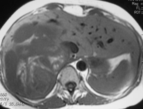
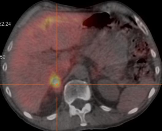
6 CASE 1 62 year old woman with hypertension. Lesion in the right adrenal detected on renal MRA?diagnosis?what further confirmatory test/tests would you perform Phaeochromocytoma

 Benign or malignant Aortic sympathetic paraganglia Visceral autonomic")
7 PHEOCHROMOCYTOMA ADRENAL PARAGANGLIOMA Carotid bodies Aortic bodies Thoracic sympathetic paraganglia Adrenal medullae Most frequent medullary tumour arising from chromaffin cells Catecholamine-secreting neoplasm Rare, but important: surgically curable form of hypertension Sporadic: ~24% have germline mutations, including mutations of RET, VHL, SDH-B, and SDH- D genes Familial (MEN, VHL, NF1, SDH, Carney s Triad) Benign or malignant Aortic sympathetic paraganglia Visceral autonomic paraganglia


8 IMAGING FEATURES: THE IMITATOR CT Pre C : >10HU Hyper vascular: Washout less than 60%* homogenous if small and heterogeneous if large MRI: No CSI signal loss

9 17% are Cystic 6HU

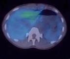
10 MIBG, FDG PET avid


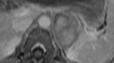
11 VHL, MEN 1, SDHB associated small lesions, usually screen detected Higher rate of malignancy than sporadic lesions


12 Malignant Phaeochromocytoma 1. Presence of metastases only definitive criteria 2. Loco regional visceral and vascular invasion 3. Benign & malignant tumors are histologically identical; the only absolute criterion for malignancy is metastasis. 4. Ki 67 increases with increasing risk of malignancy. Ki 67>30% likely malignant

13 CASE 2: 26 year old, abdominal pain and weight loss NEUROBLASTOMA/GANGLIOBLASTOMA Most frequent malignant tumour of the medulla= Small round blue cell tumor of childhood 40% occur in the adrenal, Large, heterogeneous masses, locally invasive, and lifts the aorta/ivc, extends across mid line. Coarse amorphous calcifications in 85%

14 CASE 2 COMPANION CASE: GANGLIOBLASTOMA

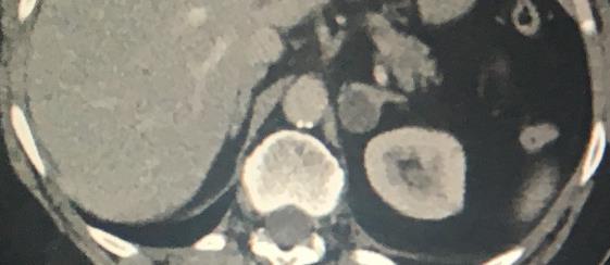
15 CASE 3 16 year old female. Generally unwell and coughing. Normal CXR US abdomen showed a left solid suprarenal mass What would you do next? (imaging and clinical)


16 5HU What is your diagnosis and why? Would you do any further imaging? What? Benign (<10HU) but not characterized *wrong age for adenoma *wrong enhancement pattern for adenoma and not a cyst on CT or US MRI Endocrine profile very important here! 15HU 25HU


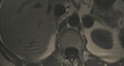
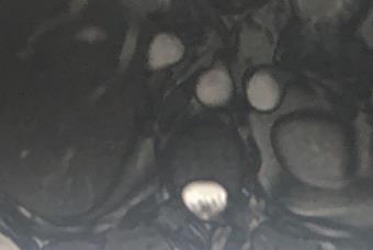
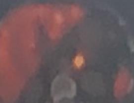
17 T2 T1 in phase T2 out of phase T1 fat saturated T1 fat saturated post contrast MIBG
18 What is your diagnosis and why? Would you do any further imaging? What? MIBG negative 30-40% are negative in phaeochromocytomas and paragangliomas MRI Features in keeping with phaeo: high T2, solid and enhancing Features not in keeping with phaeo: very low grade enhancement, low CT pre attenuation Biochemically inert: All catecholamines and adrenal cortex hormones normal What next? Biopsy: Ganglioneuroma
19 GANGLIONEUROMA Well Differentiated neoplasm of neural crest origin Benign Occurs in older paediatric to adult age group Pathology: Gross: Encapsulated, white, firm Micro: Ganglion cells & Schwann cells On Imaging : Associated with a negative CT contrast washout = slow, low grade progressive enhancement. Therefore delayed images have a higher attenuation than at 60s


20 8HU 22HU 45HU Resected Ganglioneuroma with low grade function Washout criteria: Absolute washout : 22-45/22-8 = -164%
21 TUMOURS: ARISING FROM NEUROBLASTIC /MATURE GANGLION CELLS Neuroblastoma Malignant Infants and children VMA and HVA Nor adrenalin VIP and other GI peptides Hypertension Palpitations Fever diarrhoea Ganglioneuroblastoma Ganglioblastoma Ganglioneuroma Neuroma Decreasing function Benign Incidental in older children and adults

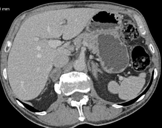
22 CASE 4 32 year old woman being investigated for mild hypertension and palpitations What are the CT findings? What information do you need?


23 What are the CT findings? -bilateral adrenal enlargement including the adrenal limbs What information do you need? Biochemical changes, syndrome? -?cortisol/aldo?catecholamines Elevated cortisol/aldo Cushing s/conns Elevated catecholamines Medullary Hyperplasia Pseudopheaochromocytoma
24 MEDULLARY HYPERPLASIA: MIMIC PHAEOCHROMOCYTOMAS Defined as expansion and extension of the adrenal medulla into limbs where medullary tissue is not normally present Diffuse or nodular/symmetric or asymmetric Familial hyperplasia Associated with MEN II, Beckwith-Wiedemann syndrome Sporadic hyperplasia also exists as a distinct entity in paediatric age group, with cystic fibrosis Imaging features not distinguishable from cortical hyperplasia on CT and MRI Increased MIBG, Ga and FDG uptake on PET which may be diffuse of focal. -focal uptake mimics a phaeo
25 MEN II : HYPERPLASIA

26 CASE 5 65 year old woman with endometrial cancer Post operatively, unable to wean off ventilation and hypotensive CT indication?abdominal bleed?collection What does the CT demonstrate? Working diagnosis? How would you confirm this?


27 What does the CT demonstrate? -bilateral adrenal enlargement -irregular contour of left adrenal? Working diagnosis? -adrenal haemorrhage? How would you confirm this? -pre operative staging scan


28 Haemorrhage and Infarction Waterhouse Frederickson syndrome Post surgical ischaemia and haemorrhage Trauma History usually leads to the diagnosis Although the adrenal cortex is more susceptible to infarction, the adrenal medulla is also affected and contributes to the hypotension. ADRENAL HAEMORRHAGE
29 SUMMARY The adrenal medulla is a sympathetic ganglion made up of neural cells Diseases affecting the peripheral nervous system and the sympathetic ganglia elsewhere, also affect the adrenal medulla Phaeochromocytomas are the best known and best described lesion but other benign and malignant lesions occur in adults Of these malignant neuroblastomas and benign ganglioneuromas have specific imaging features Syndromic associations with MEN, VHL, SDH etc may be helpful in the clinical workup Patients age has a strong influence of the likelihood of pathology
30 Thank You
The Adrenal Glands. I. Normal adrenal gland A. Gross & microscopic B. Hormone synthesis, regulation & measurement. II.
 The Adrenal Glands Thomas Jacobs, M.D. Diane Hamele-Bena, M.D. I. Normal adrenal gland A. Gross & microscopic B. Hormone synthesis, regulation & measurement II. Hypoadrenalism III. Hyperadrenalism; Adrenal
ADRENAL LESIONS 10/09/2012. Adrenal + lesion. Introduction. Common causes. Anatomy. Financial disclosure. Dr. Boraiah Sreeharsha. Nothing to declare
 ADRENAL LESIONS Financial disclosure Nothing to declare Dr. Boraiah Sreeharsha MBBS;FRCR;FRCPSC Introduction Adrenal + lesion Adrenal lesions are common 9% of the population Increase in the detection rate
Retroperitoneal Ganglioneuroma Encasing the Celiac and Superior Mesenteric Arteries
 Case Study TheScientificWorldJOURNAL (2004) 4, 974 977 ISSN 1537-744X; DOI 10.1100/tsw.2004.198 Retroperitoneal Ganglioneuroma Encasing the Celiac and Superior Mesenteric Arteries Justin K. Nelms, Eric
Adrenal masses in infancy and childhood: A clinical and radiological overview M. Mearadji
 Adrenal masses in infancy and childhood: A clinical and radiological overview M. Mearadji International Foundation for Pediatric Imaging Aid Introduction Neoplastic adrenal masses usually originate from
THE HIGHS AND LOWS OF ADRENAL GLAND PATHOLOGY
 THE HIGHS AND LOWS OF ADRENAL GLAND PATHOLOGY Symptoms of Adrenal Gland Disorders 2 Depends on whether it is making too much or too little hormone And on what you Google! Symptoms include obesity, skin
ADRENAL INCIDENTALOMA. Jamii St. Julien
 ADRENAL INCIDENTALOMA Jamii St. Julien Outline Definition Differential Evaluation Treatment Follow up Questions Case Definition The phenomenon of detecting an otherwise unsuspected adrenal mass on radiologic
SELF-ASSESSMENT MODULE REFERENCE SPR 2018 Oncologic Imaging Course Adrenal Tumors November 10, :00 12:10 p.m.
 SELF-ASSESSMENT MODULE REFERENCE SPR 2018 Oncologic Imaging Course Adrenal Tumors November 10, 2018 10:00 12:10 p.m. Staging Susan E. Sharp, MD 1. In the International Neuroblastoma Risk Group Staging
ID data. Sex: female Age: 46y/o Birthday: 1955/10/13
 ID data Sex: female Age: 46y/o Birthday: 1955/10/13 Chief Complain Right upper quadrate abdominal tenderness for one month. Present illness (1) This 46 years old female patient was in a healthy condition
Wilms Tumor and Neuroblastoma
 Wilms Tumor and Neuroblastoma Wilm s Tumor AKA: Nephroblastoma the most common intra-abdominal cancer in children. peak incidence is 2 to 3 years of age Biology somatic mutations restricted to tumor tissue
Dr.Dafalla Ahmed Babiker Jazan University
 Dr.Dafalla Ahmed Babiker Jazan University Brain tumors are the second commonest malignancy in children Infratentorial tumors are more common As a general rule they do not metastasize out of the CNS, but
Read the following article and answer the questions that follow. Refer to the Keys section to check your answers.
 ENGLISH 183 READING PRACTICE - Pheochromocytoma Read the following article and answer the questions that follow. Refer to the Keys section to check your answers. Pheochromocytoma is a tumor on the medulla
Neuroblastoma Joseph Junewick, MD FACR
 Neuroblastoma Joseph Junewick, MD FACR 03/18/2011 History 15 month old with anemia. Diagnosis Neuroblastoma Discussion Neuroblastic tumors derive from primordial neural crest cells destined for sympathetic
Daniela Faivovich K., MS VII Universidad de Chile Gillian Lieberman, MD Harvard Medical School
 Daniela Faivovich K., MS VII Universidad de Chile Gillian Lieberman, MD Harvard Medical School May 21st, 2010 56 year old male patient History of hypertension, hyperlipidemia and insulin-resistance 2009:
Evaluation of Thyroid Nodules
 Evaluation of Thyroid Nodules Stephan Kowalyk, MD January 25 28, 2018 1 Primary goal Exclude malignancy Incidental thyroid nodules If found on CT, MRI, PET scan, carotid Doppler ULTRASOUND!! January 25
Case Based Urology Learning Program
 Case Based Urology Learning Program Resident s Corner: UROLOGY Case Number 4 CBULP 2010 004 Case Based Urology Learning Program Editor: Associate Editors: Manager: Case Contributors: Steven C. Campbell,
Pediatric Retroperitoneal Masses Radiologic-Pathologic Correlation
 Acta Radiológica Portuguesa, Vol.XVIII, nº 70, pág. 61-70, Abr.-Jun., 2006 Pediatric Retroperitoneal Masses Radiologic-Pathologic Correlation Marilyn J. Siegel Mallinckrodt Institute of Radiology, Washington
The Management of adrenal incidentaloma
 The Management of adrenal incidentaloma Dimitrios Linos, MD Director of Surgery, Hygeia Hospital, Athens, Greece Consultant in Surgery, Massachusetts General Hospital, Boston, USA 8 th Postgraduate Course
PDF created with pdffactory Pro trial version
 Neuroblastoma Tumor derived from neural crest cell that form the sympathetic ganglia&adrenal medulla. Causes *unknown. *familial neuroblastoma has been reported but is rare. * The incidence is 1:100,000
Adrenal incidentaloma
 Adrenal incidentaloma Prevalence 5% post-mortem series 4% CT series 6-20% CT series in patients with Hx extra-adrenal malignancy Commoner with increasing age Associated with adrenal hyperfunction in 15%
Recommendations for cross-sectional imaging in cancer management, Second edition
 www.rcr.ac.uk Recommendations for cross-sectional imaging in cancer management, Second edition Renal and adrenal tumours Faculty of Clinical Radiology www.rcr.ac.uk Contents Renal cell carcinoma 3 Clinical
The Work-up and Treatment of Adrenal Nodules
 The Work-up and Treatment of Adrenal Nodules Lawrence Andrew Drew Shirley, MD, MS, FACS Assistant Professor of Surgical-Clinical Department of Surgery Division of Surgical Oncology The Ohio State University
Personal data. Age : 63 Gender : male
 Personal data Age : 63 Gender : male Chief complain No specific symptom or discomfort A hepatic mass, found by abdominal sonography of routine health exam on 88-12-08 Past history 1984-3-3 Old CVA with
THE FACTS YOU NEED TO KNOW
 PHEOCHROMOCYTOMA THE FACTS YOU NEED TO KNOW Pheochromocytoma is a part of the pheochromocytoma and paraganglioma group of syndromes. A pheochromocytoma is a tumor arising in the adrenal gland medulla.
Characterization of adrenal lesions on CT and MRI: all that a radiologist must know
 Characterization of adrenal lesions on CT and MRI: all that a radiologist must know Poster No.: C-2476 Congress: ECR 2013 Type: Educational Exhibit Authors: N. Benzina, S. MAJDOUB, C. H. ZARRAD, H. Zaghouani,
Evaluation and Management of Thyroid Nodules. Nick Vernetti, MD, FACE Palm Medical Group Las Vegas, Nevada
 Evaluation and Management of Thyroid Nodules Nick Vernetti, MD, FACE Palm Medical Group Las Vegas, Nevada Disclosure Consulting Amgen Speaking Amgen Objectives Understand the significance of incidental
Indications for Surgical Removal of Adrenal Glands
 The adrenal glands are orange-colored endocrine glands which are located on the top of both kidneys. The adrenal glands are triangular shaped and measure about one-half inch in height and 3 inches in length.
Presacral Neuroblastoma Joseph Junewick, MD FACR
 Presacral Neuroblastoma Joseph Junewick, MD FACR 01/12/2010 History 16 month old male with irritability. Diagnosis Presacral Neuroblastoma Additional Clinical Initial US to evaluate for intussusception
Adrenal incidentaloma guideline for Northern Endocrine Network
 Adrenal incidentaloma guideline for Northern Endocrine Network Definition of adrenal incidentaloma Adrenal mass detected on an imaging study done for indications that are not related to an adrenal problem
RECURRENT ADRENAL DISEASE. Megan Applewhite Endorama 2/19/2015 SR , SC
 RECURRENT ADRENAL DISEASE Megan Applewhite Endorama 2/19/2015 SR 2412318, SC 3421561 Category: Adrenal Attendings: Angelos & Grogan PATIENT #1 36yo woman with a hx of Cushing s Syndrome and right adrenalectomy
objectives Pitfalls and Pearls in PET/CT imaging Kevin Robinson, DO Assistant Professor Department of Radiology Michigan State University
 objectives Pitfalls and Pearls in PET/CT imaging Kevin Robinson, DO Assistant Professor Department of Radiology Michigan State University To determine the regions of physiologic activity To understand
COPYRIGHTED MATERIAL. Adrenal Imaging. 1.1 Introduction. Khaled M. Elsayes 1, Isaac R. Francis 1, Melvyn Korobkin 1 and Gerard M.
 1 Adrenal Imaging Khaled M. Elsayes 1, Isaac R. Francis 1, Melvyn Korobkin 1 and Gerard M. Doherty 2 1 Department of Radiology, University of Michigan 2 Department of Radiology and Surgery, University
Sporadic Pheochromocytoma. Bertil Hamberger Professor of Surgery Karolinska Institutet, Stockholm, Sweden
 Sporadic Pheochromocytoma Bertil Hamberger Professor of Surgery Karolinska Institutet, Stockholm, Sweden 1 Pheochromocytoma Anatomy, physiology and pathology Symptoms and diagnosis Plasma metanephrines
Endocrine system pathology
 Endocrine system pathology Central endocrine system peripheral endocrine system: thyroid gland parathyroid gland pancreas adrenal glands Thyroid gland. the weight of normal thyroid gland is about 15 grams.
PedsCases Podcast Scripts
 PedsCases Podcast Scripts This is a text version of a podcast from Pedscases.com on Approach to Abdominal Mass Part 2. These podcasts are designed to give medical students an overview of key topics in
ESUR 2018, Sept. 13 th.-16 th., 2018 Barcelona, Spain
 ESUR 2018, Sept. 13 th.-16 th., 2018 Barcelona, Spain OUR APPROACH Incidental adrenal nodule/mass Isaac R Francis, M.B;B.S University of Michigan, Ann Arbor, Michigan Disclosures None (in memory) M Korobkin,
Endocrine MR. Jan 30, 2015 Michael LaFata, MD
 Endocrine MR Jan 30, 2015 Michael LaFata, MD Brief case 55-year-old female in ED PMH: HTN, DM2, HLD, GERD CC: Epigastric/LUQ abdominal pain, N/V x2 days AF, HR 103, BP 155/85, room air CMP: Na 133, K 3.6,
Pheochromocytoma AMERICAN ASSOCIATION OF CLINICAL ENDOCRINOLOGY ILLINOIS CHAPTER OCTOBER 13, 2018
 Pheochromocytoma AMERICAN ASSOCIATION OF CLINICAL ENDOCRINOLOGY ILLINOIS CHAPTER OCTOBER 13, 2018 Steven A. De Jong, M.D., FACS, FACE Professor and Vice Chair of Surgery Chief, Division of General Surgery
Mineralocorticoids: aldosterone Angiotensin II/renin regulation by sympathetic tone; High potassium will stimulate and ACTH Increase in aldosterone
 Disease of the Adrenals 1 Zona Glomerulosa Mineralocorticoids: aldosterone Angiotensin II/renin regulation by sympathetic tone; High potassium will stimulate and ACTH Increase in aldosterone leads to salt
Endocrinology and VHL: The adrenal and the pancreas
 Overview Endocrinology and VHL: The adrenal and the pancreas LAUREN FISHBEIN MD, PHD UNIVERSITY OF COLORADO SCHOOL OF MEDICINE DIVISION OF ENDOCRINOLOGY, METABOLISM AND DIABETES DIVISION OF BIOMEDICAL
Management of adrenal incidentalomas
 31 Management of adrenal incidentalomas KEVIN MURTAGH, NANA MUHAMMAD AND MAREK MILLER The return of a scan result with reference to an incidental finding of an adrenal mass is a common scenario. 1 The
A Century of observations
 PARAGANGLIOMAS OF THE HEAD & NECK: AN OVERVIEW Michelle D. Williams, MD Associate Professor Dept. of Pathology Head & Neck Section UT MD Anderson Cancer Center Disclosure of Relevant Financial Relationships
Traumatic and Non Traumatic Adrenal Emergencies
 Traumatic and Non Traumatic Adrenal Emergencies Michael N. Patlas, MD, FRCPC (1), Christine O. Menias, MD (2), Douglas S. Katz, MD, FACR (3), Ania Z. Kielar, MD, FRCPC (4), Alla M. Rozenblit, MD (5), Jorge
ظظظ/ Omar Sami. Hussam Twaissi. Mousa Abbadi
 ظظظ/ 5 Omar Sami Hussam Twaissi Mousa Abbadi The doctor started this lecture by revising what we have taken in lecture number four, I won t re-write these stuff as it becomes boring so often. This sheet
Lecture 02. The Anomalies of Thyroid Gland & Gross Features of Suprarenal Gland. By:
 Lecture 02 The Anomalies of Thyroid Gland & Gross Features of Suprarenal Gland By: A. Prof. Dr Farooq A. Khan PMC Date: 16 th March. 2018 Thyroid Gland Enlargement Conditions which is characterized by
ORIGINAL ARTICLE A HISTOMORPHOLOGICAL STUDY OF PAEDIATRIC ADRENAL TUMOURS
 A HISTOMORPHOLOGICAL STUDY OF PAEDIATRIC ADRENAL TUMOURS M.Ramani 1, O.H.Radhika Krishna 2, K.Geetha 3, K.Ramesh Reddy 4, P.Sreenivas Reddy 5,Chandu Revathi 6, Ibraheem Javeed 7, Puja Deshmukh 8. 1. Professor,
REVIEW. Distinguishing benign from malignant adrenal masses
 Cancer Imaging (2003) 3, 102 110 DOI: 10.1102/1470-7330.2003.0006 CI REVIEW Distinguishing benign from malignant adrenal masses Isaac R Francis Professor of Radiology, Department of Radiology, University
Fig. 59 Malignant phaeochromocytoma, hepatic metastasis.
 Fig. 59 Malignant phaeochromocytoma, hepatic metastasis. X 120 Hyperte nsion Fig. 60 Malignant sympathetic paraganglioma, lymph node metastasis Primary in bladder. x 1 20 Hypertension Fig. 61 Malignant
Adrenal Mass. Cynthia Kwong SUNY Downstate Medical Center Grand Rounds October 13, 2016
 Adrenal Mass Cynthia Kwong SUNY Downstate Medical Center Grand Rounds October 13, 2016 Case Presentation 65F found to have a 4cm left adrenal mass in 2012 now presents with 6.7cm left adrenal mass PMHx:
Endocrine. Endocrine as it relates to the kidney. Sarah Elfering, MD University of Minnesota
 Endocrine Sarah Elfering, MD University of Minnesota Endocrine as it relates to the kidney Parathyroid gland Vitamin D Endocrine causes of HTN Adrenal adenoma PTH Bone Kidney Intestine 1, 25 OH Vitamin
Familial paraganglioma: A novel presentation of a case and response to therapy with radiolabelled MIBG
 HORMONES 2004, 3(2):127- Case report Familial paraganglioma: A novel presentation of a case and response to therapy with radiolabelled MIBG Justin K. Lawrence 1, Eamonn R. Maher 2, Richard Sheaves 3, Ashley
Incidental adrenal pheochromocytoma a report on three cases
 Incidental adrenal pheochromocytoma a report on three cases Incidental adrenal pheochromocytoma a report on three cases M S A Cooray 1, Uditha Bulugahapitiya 2, I S H Liyanage 3, Anuruddha M Abeygunasekera
STATE OF THE ART MANAGEMENT of PARAGANGLIOMA. IFOS, Lima, 2018
 STATE OF THE ART MANAGEMENT of PARAGANGLIOMA IFOS, Lima, 2018 VINCENT C COUSINS ENT-Otoneurology Unit, The Alfred Hospital & Department of Surgery, Monash University MELBOURNE, AUSTRALIA PARAGANGLIOMAS
Role of imaging in RCC. Ultrasonography. Solid lesion. Cystic RCC. Solid RCC 31/08/60. From Diagnosis to Treatment: the Radiologist Perspective
 Role of imaging in RCC From Diagnosis to Treatment: the Radiologist Perspective Diagnosis Staging Follow up Imaging modalities Limitations and pitfalls Duangkamon Prapruttam, MD Department of Therapeutic
Index. radiologic.theclinics.com. Note: Page numbers of article titles are in boldface type.
 Index Note: Page numbers of article titles are in boldface type. A ACC. See Adrenal cortical carcinoma. Acromegaly and the pituitary gland, 551 Acute suppurative thyroiditis, 405, 406 Addison, Thomas and
Recommendations for Reporting of Tumors of the Adrenal Cortex and Medulla
 A J C P / REPORTING OF TUMORS OF THE ADRENAL CORTEX AND MEDULLA Recommendations for Reporting of Tumors of the Adrenal Cortex and Medulla Association of Directors of Anatomic and Surgical Pathology" Key
Imaging in breast cancer. Mammography and Ultrasound Donya Farrokh.MD Radiologist Mashhad University of Medical Since
 Imaging in breast cancer Mammography and Ultrasound Donya Farrokh.MD Radiologist Mashhad University of Medical Since A mammogram report is a key component of the breast cancer diagnostic process. A mammogram
Adrenal Gland. Zona Glomerulosa (mineralcorticoids) Zona Fasciculata (glucocorticoids) Zona Reticularis (sexual hormones) Adrenaline and noradrenaline
 Zona Fasciculata (glucocorticoids) Zona Reticularis (sexual hormones) Adrenaline and noradrenaline") n Cortex Zona Glomerulosa (mineralcorticoids) Zona Fasciculata (glucocorticoids) Zona Reticularis (sexual hormones) n Medulla Adrenaline and noradrenaline n Adrenocortical Hyperfunction Hyperaldosteronism
n Cortex Zona Glomerulosa (mineralcorticoids) Zona Fasciculata (glucocorticoids) Zona Reticularis (sexual hormones) n Medulla Adrenaline and noradrenaline n Adrenocortical Hyperfunction Hyperaldosteronism
Evaluation of Liver Mass Lesions. American College of Gastroenterology 2013 Regional Postgraduate Course
 Evaluation of Liver Mass Lesions American College of Gastroenterology 2013 Regional Postgraduate Course Lewis R. Roberts, MB ChB, PhD Division of Gastroenterology and Hepatology Mayo Clinic College of
Dr Claire Smith, Consultant Radiologist St James University Hospital Leeds
 Dr Claire Smith, Consultant Radiologist St James University Hospital Leeds Imaging in jaundice and 2ww pathway Image protocol Staging Limitations Pancreatic cancer 1.2.4 Refer people using a suspected
Adrenal gland Incidentaloma
 Adrenal gland Incidentaloma Topic review 17 sep 2008 Anatomy 1 Anatomical consideration Blood supply Artery: small branches from Inf. phrenic, renal artery and aorta Vein: Rt : medial aspect to IVC Lt
Systemic Hypertension
 BCS Theme Session Cardiovascular Block Pathology of Hypertension Department of Pathology University of Sydney Systemic Hypertension Definition of Systemic hypertension: consistent blood pressure elevation
Adrenal Incidentalomas. G Stephen DeCherney, MD, MPH Clinical Professor of Medicine Division of Endocrinology UNC School of Medicine
 Adrenal Incidentalomas G Stephen DeCherney, MD, MPH Clinical Professor of Medicine Division of Endocrinology UNC School of Medicine Disclosures No financial, investment, or consulting relationship with
Neoplasia part I. Dr. Mohsen Dashti. Clinical Medicine & Pathology nd Lecture
 Neoplasia part I By Dr. Mohsen Dashti Clinical Medicine & Pathology 316 2 nd Lecture Lecture outline Review of structure & function. Basic definitions. Classification of neoplasms. Morphologic features.
SPECT- CT and PET- CT in Endocrine tumours. Prof John Buscombe
 SPECT- CT and PET- CT in Endocrine tumours Prof John Buscombe Introduc:on Parathyroid adenoma Hyperinsulinoma Adrenal imaging Pituitary imaging Parathyroid Tumours Can be seen in MEN1 Nuclear Medicine
Pathology of the endocrine system
 Pathology of the endocrine system Endocrine system A. Endocrine organs 1. Pituitary gland 2. Pineal gland 3. Thyroid gland 4. Adrenal glands 5. Parathyroid glands B. Organs with partial endocrine functions
Year 2004 Paper two: Questions supplied by Megan 1
 Year 2004 Paper two: Questions supplied by Megan 1 QUESTION 96 A 32yo woman if found to have high blood pressure (180/105mmHg) at an insurance medical examination. She is asymptomatic. Clinical examination
Neuro-endocrine and pancreatic non-adenocarcinomas. Marc Engelbrecht, AMC, Amsterdam
 Neuro-endocrine and pancreatic non-adenocarcinomas Marc Engelbrecht, AMC, Amsterdam Pancreatic Tumors q Epithelial Exocrine q Mesenchymal Ductal Adenocarcinoma (85-95%) Metastasis Lymfoma Acinar Cell Carcinoma
Malignant Focal Liver Lesions
 Malignant Focal Liver Lesions Other Than HCC Pablo R. Ros, MD, MPH, PhD Departments of Radiology and Pathology University Hospitals Cleveland Medical Center Case Western Reserve University Pablo.Ros@UHhospitals.org
Conferencia III: Dilemas en el tratamiento de Feocromocitomas y Paragangliomas. Dilemmas in Management of Pheochromocytoma and Paraganglioma
 Conferencia III: Dilemas en el tratamiento de Feocromocitomas y Paragangliomas Dilemmas in Management of Pheochromocytoma and Paraganglioma William F. Young, Jr., MD, MSc Mayo Clinic Rochester, MN, USA
Pediatric Abdominal Masses. Andrew Phelps MD Assistant Professor of Pediatric Radiology UCSF Benioff Children's Hospital
 Pediatric Abdominal Masses Andrew Phelps MD Assistant Professor of Pediatric Radiology UCSF Benioff Children's Hospital No Disclosures Take Home Message All you need to remember are the 5 common masses
Renal tumors of adults
 Renal tumors of adults Urinary Tract Tumors 2%-3% of all cancers in adults. The most common malignant tumor of the kidney is renal cell carcinoma. Tumors of the lower urinary tract are twice as common
Endocrine System. Organs and Tissues: Pituitary Adrenals Pancreas Thyroid Parathyroids
 Endocrine System Organs and Tissues: Pituitary Adrenals Pancreas Thyroid Parathyroids Bruce A. Fenderson, Ph.D. Pathology, Anatomy & Cell Biology Sidney Kimmel Medical College Bruce.Fenderson@Jefferson.edu
Odise Cenaj, Harvard Medical School Year III. Gillian Lieberman, MD
 February 2012 Radiologic evaluation of adrenal masses and an atypical radiologic presentation of adrenocortical carcinoma in a patient with primary aldosteronism Odise Cenaj, Harvard Medical School Year
the urinary system pathology Dr. Fairoz A Eltorgman
 the urinary system pathology Dr. Fairoz A Eltorgman Tumors of the renal pelvis & kidney Benign tumors of the renal pelvis: Hemangioma Leiomyoma Malignant tumors: Transitional cell carcinoma Squamous cell
Newcastle HPB MDM updated radiology imaging protocol recommendations. Author Dr John Scott. Consultant Radiologist Freeman Hospital
 Newcastle HPB MDM updated radiology imaging protocol recommendations Author Dr John Scott. Consultant Radiologist Freeman Hospital This document is intended as a guide to aid radiologists and clinicians
42 yr old male with h/o Graves disease and prior I 131 treatment presents with hyperthyroidism and undetectable TSH. 2 hr uptake 20%, 24 hr uptake 50%
 Pinhole images of the neck are acquired in multiple projections, 24hrs after the oral administration of approximately 200 µci of I123. Usually, 24hr uptake value if also calculated (normal 24 hr uptake
Nuclear medicine in endocrinology
 Nuclear medicine in endocrinology Thyroid gland: anatomy, function, inflammation, Nuclear medicine in endocrinology tumor dignitiy Parathyroid gland: localisation Adrenal cortex: function Adrenal medulla:
Mediastinal Tumors: Imaging
 Mediastinal Tumors: Imaging References Imaging in Oncology, Husband and Reznek Computed Tomography and Magnetic Resonance of the thorax, Naidich, Zerhouni, Siegelman, Mediastinal compartments Anterior:
Kidney Case 1 SURGICAL PATHOLOGY REPORT
 Kidney Case 1 Surgical Pathology Report February 9, 2007 Clinical History: This 45 year old woman was found to have a left renal mass. CT urography with reconstruction revealed a 2 cm medial mass which
A case of micturition syncope
 A case of micturition syncope Kimberly Bundick, PA-S S L I D E 1 Agenda Purpose Utilize case to illustrate classic finding of an interesting pathology Agenda Case study Epidemiology, etiology of disease
Sex: 女 Age: 51 Occupation: 無 Admission date:92/07/22
 Sex: 女 Age: 51 Occupation: 無 Admission date:92/07/22 Chief complaint Unknown fever for one month Hand tremor and left huge renal tumor was noted Present illness Suffered from fever for one month, hand
- RET/PTC rearrangement: 20% papillary thyroid cancer - RET: medullary thyroid cancer
 Thyroid Cancer UpToDate: Introduction: Risk Factors: Biology: Symptoms: Diagnosis: 1. Lenvina is the first line therapy with powerful durable response and superior PFS in pts with RAI-refractory disease.
Imaging in gastric cancer
 Imaging in gastric cancer Gastric cancer remains a deadly disease because of late diagnosis. Adenocarcinoma represents 90% of malignant tumors. Diagnosis is based on endoscopic examination with biopsies.
adrenal and parathyroid glands Done by jehad abdel aziz
 15-11-09 prof. muhammed khammash adrenal and parathyroid glands Done by jehad abdel aziz The adrenal glands:- Anatomy:- The adrenal glands are flattened, yellowish structures that weigh less than 10g in
G3.02 The malignant potential of the neoplasm should be recorded. CG3.02a
 G3.02 The malignant potential of the neoplasm should be recorded. CG3.02a Conventional adrenocortical neoplasm. Each of the below parameters is scored 0 when absent and 1 when present. 3 or more of these
XVII. Tumours of the adrenal gland and paraganglia
 XVII. Tumours of the adrenal gland and paraganglia E. C. APPLEBY 1 This classification is arranged in two parts in order to take into account the different origins, structures, and functions of the cortex
SCOPE TODAYS SESSION. Case 1: Case 2. Basic Theory Stuff: Heavy Stuff. Basic Questions. Basic Questions
 MONDAY TEACHING SCOPE TODAYS SESSION Case 1: Basic Questions Case 2 Basic Questions Basic Theory Stuff: AJCC TNM + Stage Group for Carcinoid of the Appendix Management of Carcinoid of the Appendix (NCCN)
Patient Information. Age: 8 y/o Sex: Female. Date of Admission: Date of Discharge:
 Patient Information Age: 8 y/o Sex: Female Date of Admission: 92-10-08 Date of Discharge: 92-10-18 Chief Complaint Severe admominal pain and vomiting with dysuria since last afternoon Present Illness Lower
Recent Advances in the Management of
 Recent Advances in the Management of Pheochromocytoma 6 : 4 Nalini S. Shah, Vijaya Sarathi, Reshma Pandit, Mumbai The 2004 WHO classification of endocrine tumors restricts the term Pheochromocytoma (PHEO)
Morbidity & Mortality. Mark H. Tseng MD SUNY Downstate Medical Center Lutheran Medical Center December 16, 2005
 Morbidity & Mortality Mark H. Tseng MD SUNY Downstate Medical Center Lutheran Medical Center December 16, 2005 Case presentation Pt is a xx year old Asian woman who present to the ED with cc of epigastric
Adrenal Ganglioneuroma Presenting With Adrenal Insufficiency After Unilateral Adrenalectomy
 ISPUB.COM The Internet Journal of Urology Volume 9 Number 1 Adrenal Ganglioneuroma Presenting With Adrenal Insufficiency After Unilateral Adrenalectomy S Bontha, N Sanalkumar, M Istarabadi, G Lepsien,
Renal Masses in Patients with Known Extrarenal Primary Primary Cancer Primary Primary n Met Mets s RCC Beni L mphoma Lung Breast Others
 The Importance of Stuart G. Silverman, MD, FACR Professor of Radiology Harvard ard Medical School Director, Abdominal Imaging and Intervention Brigham and Women s Hospital Boston, MA The Importance of
Contrast Enhanced Ultrasound of Parenchymal Masses in Children
 Contrast Enhanced Ultrasound of Parenchymal Masses in Children Sue C Kaste, DO On behalf of Beth McCarville, MD St. Jude Children s Research Hospital Memphis, TN Overview Share St. Jude experience with
Pathophysiology of the th E d n ocr i ne S S t ys em B. Marinov, MD, PhD Endocrine system Central: Hypothalamus
 Pathophysiology of the Endocrine System B. Marinov, MD, PhD Pathophysiology Department Medical University of Plovdiv Endocrine system Central: Hypothalamus Pituitary Pineal Peripheral Thymus Thyroid Parathyroid
Paraspinal Venous Malformation Joseph Junewick, MD FACR
 Paraspinal Venous Malformation Joseph Junewick, MD FACR 06/04/2010 History 2 year old with history of fall. Rule out spinal injury. Diagnosis Paraspinal Venous Malformation Additional Clinical CT of the
1/25/13 Right partial nephrectomy followed by completion right radical nephrectomy.
 History and Physical Case Scenario 1 45 year old white male presents with complaints of nausea, weight loss, and back pain. A CT of the chest, abdomen and pelvis was done on 12/8/12 that revealed a 12
Leonard M. Glassman MD
 BI-RADS The New BI-RADS Leonard M. Glassman MD FACR Former Chief of Breast Imaging American Institute for Radiologic Pathology Washington Radiology Associates, PC Breast Imaging Reporting and Data System
ADRENAL MR: PEARLS AND PITFALLS
 ADRENAL MR: PEARLS AND PITFALLS Frank Miller, M.D. Lee F. Rogers MD Professor of Medical Education Chief, Body Imaging Section and Fellowship Medical Director, MR Imaging Professor of Radiology Northwestern
BONE METASTASIS OF MEDIASTINAL PARAGANGLIOMA: CASE REPORT
 BONE METASTASIS OF MEDIASTINAL PARAGANGLIOMA: CASE REPORT Dr. Abhinandan Gupta 1*, Kong Long 1, Prof. Huang Jing Bai 1, Dr. Deepikal Dhakal 1, Dr. Sunil Shrestha 1, Dr. Roshan Kumar Yadav 2 and Dr. Shashi
2 to 3% of All New Visceral Cancers Peak Incidence is 6th Decade M:F = 2:1 Grossly is a Bright Yellow, Necrotic Mass with a Pseudocapsule
 GENITOURINARY PATHOLOGY Kathleen M. O Toole, M.D. Renal Cell Carcinoma 2 to 3% of All New Visceral Cancers Peak Incidence is 6th Decade M:F = 2:1 Grossly is a Bright Yellow Necrotic Mass Grossly is a Bright