Title. Author(s) Takahashi, Haruo. Issue Date Right.
|
|
|
- Georgiana Lucas
- 5 years ago
- Views:
Transcription

1 NAOSITE: Nagasaki University's Ac Title Author(s) Citation A case with posterior fossa epiderm symptoms caused by insufficiency of usefulness of free DICOM image view Takasaki, Kenji; Kumagami, Hidetaka Takahashi, Haruo Acta oto-laryngologica. Supplementu Issue Date URL Right This document is downloaded
2 Takasaki K et al. 1 A Case with Posterior-fossa Epidermoid Cyst Showing Audiovestibular Symptoms Caused by Insufficiency of Anterior Inferior Cerebellar Artery -Usefulness of Free DICOM Image Viewing and Processing Software- Kenji Takasaki, M. D. 1), Hidetaka Kumagami, M. D. 1) Akiko Baba, M. D. 1), Daisuke Fujiyama, M. D. 1), Haruo Takahashi, M. D. 1) 1) Department of Otolaryngology - Head and Neck Surgery, Nagasaki University Graduate School of Biomedical Sciences Address: Department of Otolaryngology - Head and Neck Surgery, Nagasaki University Graduate School of Biomedical Sciences 1-7-1, Sakamoto, Nagasaki , Japan Correspondence / Reprint requests: Kenji Takasaki, M. D. Department of Otolaryngology, Head and Neck Surgery, Nagasaki University Graduate School of Biomedical Sciences 1-7-1, Sakamoto, Nagasaki , Japan Phone: , Fax: ktakasa@nagasaki-u.ac.jp
3 Takasaki K et al. 2 Abstract A 58-year-old Japanese man suddenly suffered from vertigo. On physical examination, left-beating horizontal torsional spontaneous nystagmus was observed, of which the direction did not change with gaze. Other neurotological examinations revealed findings within normal limit except the left-side sensorineural hearing loss of approximately 32 db on an average. Diffusion weighted magnetic resonance imaging revealed no infarction in the brain, but demonstrated an epidermoid cyst in the left cerebello-pontine cistern region. Using free digital imaging and communications in medicine (DICOM) image viewing and processing software, it was found that the epidermoid cyst clearly compressed the left anterior inferior cerebellar artery (AICA). Therefore, we speculated that insufficiency of the left AICA caused his audiovestibular symptoms. This new technique used in the present study was considered useful when the responsible site of vertigo is suspected in the cerebello-pontine angle, where anatomical relationships between the nerves and the vessels are complicated.
4 Takasaki K et al. 3 Keywords: AICA insufficiency, Cerebello-pontine angle, DICOM image, Epidermoid cyst, Vertigo, Viewing and processing software
5 Takasaki K et al. 4 Introduction Vertigo is a common presenting symptom in patients with transient vertebro-basilar distribution ischemia (1). The anterior inferior cerebellar artery (AICA) supplies the anterolateral regions of the inferior pons, inner ear, and middle cerebellum including the flocculus (2). AICA infarction is known to be associated with hearing loss (HL), vertigo, facial weakness, ataxia, anesthesia, and Horner s syndrome (3). To date, using magnetic resonance imaging (MRI), some authors reported AICA infarction showing only HL and vertigo as clinical signs without other symptoms (4, 5); however, there has been no report that AICA insufficiency, especially with the compression by a mass lesion, caused only HL or vertigo. Recently, as digital imaging and communications in medicine (DICOM) of computed tomography (CT) and MRI, and viewing and processing software have advanced (6), it has become possible to observe and measure structures seen in the CT and MRI three-dimensionally in a way that meets our needs (7, 8). Using free DICOM image viewing and processing software, we reported a case whose left-side AICA was compressed by an epidermoid cyst at the left cerebello-pontine
6 Takasaki K et al. 5 cistern region and we speculated that the compression by the tumor caused AICA insufficiency, thus impairing the inner ear function and resulting in audiovestibular symptoms. Case Report A 58-year-old Japanese man with a long-standing history of hypertension presented with sudden onset of vertigo. He also complained of unsteady feeling. On physical examination, left-beating horizontal torsional spontaneous nystagmus was observed, of which the direction did not change with gaze. The eardrums were normal. He had not ophthalmoplegia, dysarthria, facial paralysis, facial numbness, Horner s syndrome, nor cerebellar dysmetria. Pure tone audiograms showed a sensorineural HL of approximately 32 db on average in the left ear (Figure 1). Caloric tests using 2 ml of ice water did not reveal a canal paralysis. Vestibular evoked myogenic potentials and eye tracking test were normal. Other neurotological and routine laboratory findings were all insignificant.
7 Takasaki K et al. 6 T2-weighted, T1-weighted gadolinium-enhanced, and diffusion weighted imaging (DWI) of MRI of the brain revealed no infarction. However, by three-dimensional constructive interference in steady-state (CISS) imaging, the mass was clearly demonstrated as low intensity in the left cerebello-pontine cistern region (Figure 2). The mass was not identified in conventional T1- and T2-weighted images because of the similarity in the signal intensity of the mass to that of cerebrospinal fluid (CSF), but was shown clearly by its increased signal intensity using DWI (Figure 3). From the absence of enhancement by gadolinium and similar signal intensity to the CSF, an epidermoid cyst was considered as the most possible diagnosis of the mass (9). Additionally, we used 0.5-mm-thick gapless MRI DICOM data, and an image viewing software (Virtual Place Liberty, Office Azemoto Ltd., Japan) on a personal computer to clarify the spatial anatomical relationships of this epidermoid cyst, and the vestibular nerve around the left internal auditory canal. The reconstructed CISS image demonstrated that the epidermoid cyst apparently compressed AICA at the left cerebello-pontine cistern region (Figure 4) and that it also had contact with the facial and trigeminal nerves. However, we could not recognize direct contact of the
8 Takasaki K et al. 7 cyst either to the vestibular nerve or cochlear nerve in any image (Figure 5). His vertigo disappeared over a week showing no paralytic nystagmus. We followed the patient at every six months with neurotologic, and MRI examinations. Discussion In the process of the neurological diagnosis on a patient with vertigo, we usually use selective diagnostic procedures including CT, MRI, DWI of MR, MR angiography, and angiography (10). Acoustic schwannoma, meningioma, and epidermoid cyst are the most common mass lesions around cerebellopontine angle region, where an MRI usually provides more information than a CT. On T1-weighted gadolinium-enhanced MRI, acoustic schwannoma demonstrates uniform enhancement with 10% cystic necrosis. Meningioma also shows uniform enhancement with susceptibility from calcified internal matrix and dual tail sigh on T2-weighted image. Epidermoid cyst shows variable signal intensities. Typical MRI findings of epidermoid cyst include similar or slightly increased signal intensity relative to that of CSF on both T1- and T2-weighted images. DWI, which demonstrates differences in
9 Takasaki K et al. 8 mobility of water, can differentiate solid components from fluid, therefore epidermoid cyst shows increased signal intensity (9). Since the images obtained from those examinations are two-dimensional, it is difficult to grasp three-dimensional relationship among the organs related with the pathogenesis of vertigo, especially the relationship between structures not running parallel each other, for example an artery, and a nerve. Recently, most CT and MRI are in the DICOM and viewing and processing software has been developed as a new imaging technique in the field of Radiology (6). With this technique, any arbitrarily reconstructed image desired can be obtained by changing the angle of the plane and by changing the target location. We previously reported new findings that we obtained regarding the spatial anatomy and dimensions of the eustachian tube and sphenoid sinus by using these techniques (7, 8). This is why we applied these techniques to clarify the pathogenesis of the vertigo of the present case by analyzing the spatial relationship among the tumor, nerves, and vessels. In the present case, in spite of the left-beating horizontal torsional spontaneous nystagmus, and a sensorineural HL on the left ear, there was no
10 Takasaki K et al. 9 neurotological or radiological findings indicating a brain infarction causing central vestibular dysfunction. We, therefore, suspected that the irritant nystagmus to the left was the left labyrinthine origin. Also, on CISS images, we found that the epidermoid cyst compressed the left-side AICA. The inner ear and the vestibulocochlear nerve are supplied exclusively from the labyrinthine artery, which stems from the AICA with few exceptions (2). Therefore, we speculated that the AICA inefficiency caused the irritant nystagmus originated from the left labyrinth (11). AICA infarction is known to manifest audiovestibular symptoms caused by the inner-ear dysfunction as well as brainstem signs such as crossed sensory loss, lateral gaze palsy, facial palsy, or Horner s syndrome (3). There were a few reports of AICA infarction presenting only auditovestibular symptoms without other central signs (4, 5), but there has been no report that AICA insufficiency caused vertigo and HL of the inner ear origin. We might have diagnosed that the audiovestibular symptoms in the present case were caused by brain stem compression, or neurovascular compression of the eighth cranial nerves by the tumor, if we had not used new DICOM image-viewing and processing software. Therefore, these new useful techniques
11 Takasaki K et al. 10 should be utilized fully when investigating the pathogenesis, and pathophysiology in cases with vertigo of unknown cause.
12 Takasaki K et al. 11 References 1. Williams D, Wilson TG. The diagnosis of the major and minor syndromes of basilar insufficiency. Brain. 1962;85: Kim HN, Kim YH, Park IY, Kim GR, Chung IH. Variability of the surgical anatomy of the neurovascular complex of the cerebellopontine angle. Ann Otol Rhinol Laryngol. 1990;99: Adams RD. Occlusion of the anterior inferior cerebellarartery. Arch Neurol Psychiatry. 1943;49: Son EJ, Bang JH, Kang JG. Anterior inferior cerebellar artery infarction presenting with sudden hearing loss and vertigo. Laryngoscope. 2007;117: Lee H, Ahn BH, Baloh RW. Sudden deafness with vertigo as a sole manifestation of anterior inferior cerebellar artery infarction. J Neurol Sci. 2004;222: Escott EJ, Rubinstein D. Free DICOM image viewing and processing software for your desktop computer: what's available and what it can do for you. Radiographics. 2003;23: Takasaki K, Takahashi H, Miyamoto I, Yoshida H, Yamamoto-Fukuda T, Enatsu K,
13 Takasaki K et al. 12 Kumagami H. Measurement of angle and length of the eustachian tube on computed tomography using the multiplanar reconstruction technique. Laryngoscope. 2007;117: Enatsu K, Takasaki K, Jinnouchi S, Kase K, Kumagami H, Nakamura T, Takahashi H. Surgical anatomy of the sphenoid sinus on the CT using the multiplanar reconstruction technique. Otolaryngol Head Neck Surg. in press. 9. Murakami N, Matsushima T, Kuba H, Ikezaki K, Morioka T, Mihara F, Inamura T, Fukui M. Combining steady-state constructive interference and diffusion-weighted magnetic resonance imaging in the surgical treatment of epidermoid tumors. Neurosurg Rev. 1999;22: Bruzzone MG, Grisoli M, De Simone T, Regna-Gladin C. Neuroradiological features of vertigo. Neurol Sci. 2004;25 Suppl 1:S Oas JG, Baloh RW. Vertigo and the anterior inferior cerebellar artery syndrome. Neurology. 1992;42:
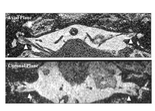
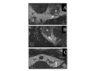
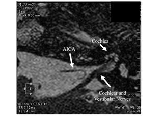
14 Takasaki K et al. 13 Legend Figure 1. Initial pure-tone audiogram showed slight sensorineural hearing loss on the left ear. Figure 2. On the three-dimensional constructive interference in steady-state (CISS) magnetic resonance imaging, the mass (*) was clearly demonstrated as low intensity in the left cerebello-pontine cistern lesion. Arrowheads indicate internal auditory canals. Figure 3. Diffusion-weighted axial magnetic resonance imaging showed high intensity in the left cerebello-pontine cistern region (*). Figure 4. Using free DICOM image viewing and processing software, reconstructed CISS images of the left-side anterior inferior cerebellar artery (AICA) in the present case are demonstrated. Plane A is close to the coronal plane perpendicular to Plane B and Plane C. Plane B is close to the sagittal plane perpendicular to Plane A and Plane C. Plane C is close to the axial plane at the level of AICA, including the whole course of the presence of AICA loop. Arrows indicate the course of AICA loop, and asterisks indicate an epidermoid cyst compressing the AICA. Figure 5. The cochlear and vestibular nerves demonstrated from the brainstem to the
15 Takasaki K et al. 14 cochlea and vestibule, AICA and cochlea are shown in the reconstructed CISS image of the left-side internal auditory canal. The epidermoid cyst is not identified in this image.

16 Figure 1 Figure 2

17 Figure 3 Figure 4
18 Figure 5
Infarction in the Territory of Anterior Inferior Cerebellar Artery Spectrum of Audiovestibular Loss
 Infarction in the Territory of Anterior Inferior Cerebellar Artery Spectrum of Audiovestibular Loss Hyung Lee, MD; Ji Soo Kim, MD; Eun-Ji Chung, MD; Hyon-Ah Yi, MD; In-Sung Chung, MD; Seong-Ryong Lee,
A n acute auditory symptom without associated neurological
 1644 PAPER Auditory disturbance as a prodrome of anterior inferior cerebellar artery infarction H Lee, Y-W Cho... See end of article for authors affiliations... Correspondence to: Dr Hyung Lee, Department
Recent Advances in Understanding Audiovestibular Loss of a Vascular Cause
 Journal of Stroke 2017;19(1):61-66 Review Recent Advances in Understanding Audiovestibular Loss of a Vascular Cause Hyun-Ah Kim, a,b Hyung Lee a,b a Department of Neurology, Keimyung University School
External carotid blood supply to acoustic neurinomas
 External carotid blood supply to acoustic neurinomas Report of two cases HARVEY L. LEVINE, M.D., ERNEST J. FERmS, M.D., AND EDWARD L. SPATZ, M.D. Departments of Radiology, Neurology, and Neurosurgery,
Vertebrobasilar Insufficiency
 Equilibrium Res Vol. (3) Vertebrobasilar Insufficiency Toshiaki Yamanaka Department of Otolaryngology-Head and Neck Surgery, Nara Medical University School of Medicine Vertebrobasilar insufficiency (VBI)
Case Report An Unusual Case of Bilateral Agenesis of the Cochlear Nerves
 Case Reports in Neurological Medicine Volume 2012, rticle D 581920, 4 pages doi:10.1155/2012/581920 Case Report n Unusual Case of Bilateral genesis of the Cochlear Nerves Vítor Yamashiro Rocha oares, 1
Imaging of Hearing Loss
 Contemporary Imaging of Sensorineural Hearing Loss Imaging of Hearing Loss Discussion Outline (SNHL) Imaging Approaches Anatomic Relationships Lesions: SNHL KL Salzman, MD University of Utah School of
A case of normal pressure hydrocephalus with peripheral vestibular disorder-like findings.
 A case of normal pressure hydrocephalus with peripheral vestibular disorder-like findings. Yuzuru Sainoo 1)2), Hidetaka Kumagami 3), Haruo Takahashi 1) 1) Goto Chuoh Hospital, Otorhinolaryngology 2) Department
MR imaging at 3.0 tesla of glossopharyngeal neuralgia by neurovascular compression
 MR imaging at 3.0 tesla of glossopharyngeal neuralgia by neurovascular compression Poster No.: C-1281 Congress: ECR 2011 Type: Scientific Exhibit Authors: M. Nishihara 1, T. Noguchi 1, H. Irie 1, K. Sasaguri
Neurovascular elements of the PCF
 Level I: Neurovascular elements of the PCF Trigeminal and Abducent Superior cerebellar artery and vein Dandy s vein Level II: Facial and Cochleovestibular AICA and internal auditory artery, veins Level
Dr. T. Venkat Kishan Asst. Prof Department of Radiodiagnosis
 Dr. T. Venkat Kishan Asst. Prof Department of Radiodiagnosis Schwannomas (also called neurinomas or neurilemmomas) constitute the most common primary cranial nerve tumors. They are benign slow-growing
SPECIAL PAPER IN CELEBRATION OF PROF. YANG'S 50 YEARS CAREER IN MEDICINE
 JOURNAL OF OTOLOGY SPECIAL PAPER IN CELEBRATION OF PROF. YANG'S 50 YEARS CAREER IN MEDICINE ADVANCES IN SURGICAL TREATMENT OF ACOUSTIC NEUROMA HAN Dongyi,CAI Chaochan Acoustic Neuroma (AN) arises from
Year 2003 Paper two: Questions supplied by Tricia
 question 43 A 42-year-old man presents with a two-year history of increasing right facial numbness. He has a history of intermittent unsteadiness, mild hearing loss and vertigo but has otherwise been well.
Case Studies in CPA/IAC
 Outline Case Studies in CPA/IAC Atul K Mallik MD PhD Department of Radiology and Imaging Sciences University of Utah Health Sciences Center Salt Lake City, Utah, USA Case based review of cerebellopontine
Acute ischemic stroke in the distribution of the anterior
 Sudden Deafness and Anterior Inferior Cerebellar Artery Infarction Hyung Lee, MD, PhD; Sung-Il Sohn, MD; Doo-Kyo Jung, MD; Yong-Won Cho, MD, PhD; Jeong-Geung Lim, MD, PhD; Sang-Doe Yi, MD, PhD; Seong-Ryong
Abnormal direction of internal auditory canal and vestibulocochlear nerve
 Medicine Otorhinolaryngology fields Okayama University Year 2004 Abnormal direction of internal auditory canal and vestibulocochlear nerve Shin Kariya kazunori Nishizaki Hirofumi Akagi Michael M. Paparella
High Signal from the Otic Labyrinth on Onenhanced Magnetic Resonance Imaging
 High Signal from the Otic Labyrinth on Onenhanced Magnetic Resonance Imaging JaneL. Weissman, 1 Hugh D. Curtin, 1 3 Barry E. Hirsch, 2 and William L. Hirsch, Jr. 1 Summary: High signal from the otic labyrinth
Meniere s disease and Sudden Sensorineural Hearing Loss
 Meniere s disease and Sudden Sensorineural Hearing Loss Tsutomu Nakashima 1,2 1 Ichinomiya Medical Treatment & Habilitation Center, Ichinomiya, Japan 2 Department of Otorhinolaryngology, Nagoya University,
SMRT Student Scope Submission
 SMRT Student Scope Submission Title & Author Title: MRI in the Detection and Diagnosis of Acoustic Neuroma Author: Manya Lovesky Andersen, student Massachusetts College of Pharmacy and Health Sciences,
Successful Treatment of Sudden Bilateral Sensorineural Hearing Loss due to Atherosclerotic Vertebral Artery Occlusion: A Case Report
 DOI: 10.5797/jnet.cr.2017-0133 Successful Treatment of Sudden Bilateral Sensorineural Hearing Loss due to Atherosclerotic Vertebral Artery Occlusion: A Case Report Takaaki Yamazaki, Hiroshi Moriwaki, Yasuyuki
Cochlear Implant Failure: Imaging Evaluation of the Electrode Course
 Clinical Radiology (2003) 58: 288 293 doi:10.1016/s0009-9260(02)00523-8, available online at www.sciencedirect.com Pictorial Review Cochlear Implant Failure: Imaging Evaluation of the Electrode Course
The Mediterranean Journal of Otology CASE REPORT
 CASE REPORT Dolichoectatic and Tortuous Vertebrobasillary Arterial System Causing Progressive Left-Sided Hearing Loss in a Patient with Previous Right-Sided Deafness Hilmi Alper fienkal, Soner Özkan, Kader
A Case of Vestibular and Oculomotor. Pathology from Bilateral AICA Watershed. Infarcts Treated with Basilar Artery Stenting
 Bilateral AICA Strokes. Kattah J, et al 1 A Case of Vestibular and Oculomotor Pathology from Bilateral AICA Watershed Infarcts Treated with Basilar Artery Stenting Jorge C Kattah, M.D * Deepak Nair M.D,
Stroke School for Internists Part 1
 Stroke School for Internists Part 1 November 4, 2017 Dr. Albert Jin Dr. Gurpreet Jaswal Disclosures I receive a stipend for my role as Medical Director of the Stroke Network of SEO I have no commercial
Kumagami, Hidetaka; Takahashi, Haru. 6489&volume=129&issue=1&spage=25.
 NAOSITE: Nagasaki University's Ac Title Author(s) Citation Amplitude and area ratios of summat in Meniere's disease. Baba, Akiko; Takasaki, Kenji; Tanak Kumagami, Hidetaka; Takahashi, Haru Acta oto-laryngologica,
Capt. Nazim ATA Aerospace Medicine Specialist Turkish Air Force AAMIMO 2013
 F-15 Pilot with ACOUSTIC NEUROMA Capt. Nazim ATA Aerospace Medicine Specialist Turkish Air Force AAMIMO 2013 Disclosure Information 84 th Annual AsMA Scientific Meeting Nazim ATA I have no financial relationships
Diagnostic criteria for vestibular neuritis
 Equilibrium Res Vol. (4) Bárány Society Diagnostic criteria for vestibular neuritis Toshihisa Murofushi Department of Otolaryngology Teikyo University School of Medicine Mizonokuchi Hospital The authors
RADIOLOGY TEACHING CONFERENCE
 RADIOLOGY TEACHING CONFERENCE John Athas, MD Monica Tadros, MD Columbia University, College of Physicians & Surgeons Department of Otolaryngology- Head & Neck Surgery September 27, 2007 CT SCAN IMAGING
Key Clinical Concepts
 Cerebrovascular Review and General Vascular Syndromes, Including Those That Impact Dizziness Key Clinical Concepts Basic Review of Cerebrovascular Circulation Circulation to the brain is divided into anterior
Gerard J. Gianoli, MD, FACS The Ear and Balance Institute Baton Rouge, Louisiana
 Gerard J. Gianoli, MD, FACS The Ear and Balance Institute Baton Rouge, Louisiana SSCD is defined anatomically as the absence of bone between the SSC and the middle fossa dura PSCD is a defect of the PSC
Major Anatomic Components of the Orbit
 Major Anatomic Components of the Orbit 1. Osseous Framework 2. Globe 3. Optic nerve and sheath 4. Extraocular muscles Bony Orbit Seven Bones Frontal bone Zygomatic bone Maxillary bone Ethmoid bone Sphenoid
Radiologic Evaluation of Petrous Apex Masses. Pavan Kavali, MS-IV Morehouse School of Medicine November 16, 2009
 Radiologic Evaluation of Petrous Apex Masses Pavan Kavali, MS-IV Morehouse School of Medicine November 16, 2009 Roadmap Petrous Apex Anatomy Patient D.S.: Clinical Presentation Differential diagnosis of
Cranial Nerve VIII (The Vestibulo-Cochlear Nerve)
") Cranial Nerve VIII (The Vestibulo-Cochlear Nerve) Please view our Editing File before studying this lecture to check for any changes. Color Code Important Doctors Notes Notes/Extra explanation Objectives
Cranial Nerve VIII (The Vestibulo-Cochlear Nerve) Please view our Editing File before studying this lecture to check for any changes. Color Code Important Doctors Notes Notes/Extra explanation Objectives
Spatial Relationship between Vestibular Schwannoma and Facial Nerve on Three-dimensional T2-weighted Fast Spin-echo MR Images
 AJNR Am J Neuroradiol 21:810 816, May 2000 Spatial Relationship between Vestibular Schwannoma and Facial Nerve on Three-dimensional T2-weighted Fast Spin-echo MR Images Sabine Sartoretti-Schefer, Spyros
Delayed Endolymphatic Hydrops: Episodic Vertigo of Delayed Onset after Profound Inner Ear Hearing Loss
 Delayed Endolymphatic Hydrops: Episodic Vertigo of Delayed Onset after Profound Inner Ear Hearing Loss Tamio Kamei 1, MD, PhD and Kenji Watanabe 2, MD 1 Professor emeritus at Gunma University, Japan 2
LA CLINICA E LA DIAGNOSI DELLA VERTIGINE VASCOLARE
 LA CLINICA E LA DIAGNOSI DELLA VERTIGINE VASCOLARE M. Mandalà Azienda Ospedaliera Universitaria Senese WHY ARE WE SCARED? NEED TO BETTER UNDERSTAND PATHOPHYSIOLOGY WHAT IS KNOWN WHAT IS EFFECTIVE and SIMPLE
CLINICAL PRESENTATION AND RADIOLOGY QUIZ QUESTION
 Donald L. Renfrew, MD Radiology Associates of the Fox Valley, 333 N. Commercial Street, Suite 100, Neenah, WI 54956 04/26/2014 Radiology Quiz of the Week # 108 Page 1 CLINICAL PRESENTATION AND RADIOLOGY
Hemorrhagic vestibular schwannoma: an unusual clinical entity Case report
 Neurosurg Focus 5 (3):Article 9, 1998 Hemorrhagic vestibular schwannoma: an unusual clinical entity Case report Dean Chou, M.D., Prakash Sampath, M.D., and Henry Brem, M.D. Departments of Neurological
Role of 3D T2 weighted imaging at 3T in evaluation of cranial nerve pathologies - An overview
 Role of 3D T2 weighted imaging at 3T in evaluation of cranial nerve pathologies - An overview Poster No.: C-1153 Congress: ECR 2013 Type: Educational Exhibit Authors: V. Kasi Arunachalam 1, R. Renganathan
Air CT Cisternography in the Diagnosis of Vascular Loop Causing Vestibular Nerve Dysfunction
 1045 Air CT Cisternography in the Diagnosis of Vascular Loop Causing Vestibular Nerve Dysfunction Fraydoon Esfahani 1 2 Kenneth D. Dolan 1 Considerable interest has developed in otology concerning the
the face department, Geneva University Hospitals and University of Geneva, Rue Micheli-du-Crest
 Final article published in Journal of Neurology 2009 Jun;256(6):1017-8. http://dx.doi.org/10.1007/s00415-009-5041-6. Sixth cranial nerve palsy and contralateral hemiparesis (Raymond s syndrome) sparing
Hyperbaric Oxygen for Idiopathic Sudden Sensorineural Hearing Loss
 Hyperbaric Oxygen for Idiopathic Sudden Sensorineural Hearing Loss Leonardo Profenna, MD, MPH Medical Director of Wound Care and Hyperbaric Medicine Connally Memorial Medical Center Introduction - ISSHL
Cranial Nerve VII & VIII
 Cranial Nerve VII & VIII Lecture Objectives Follow up the course of facial nerve from its point of central connections, exit and down to its target areas. Follow up the central connections of the facial
Acute Dizziness: Is It a Stroke? Gordon Kelley MD November 2017
 Acute Dizziness: Is It a Stroke? Gordon Kelley MD November 2017 No Disclosures Dizziness Occurs in nearly ¾ of cerebellar strokes 4 categories in classic teaching*: Vertigo Presyncope Imbalance Non-specific
Spartan Medical Research Journal
 Spartan Medical Research Journal Research at Michigan State University College of Osteopathic Medicine Volume 3 Number 2 Fall, 2018 Pages 113-122 Title: Correlation of Clinical Factors and Audiometric
Contaminated Wound: Report of a Cas
 NAOSITE: Nagasaki University's Ac Title Author(s) Citation Endovascular Treatment of a Carotid Contaminated Wound: Report of a Cas Yamaguchi, Nimpei; Kaneko, Kenichi; Takahashi, Haruo Acta medica Nagasakiensia,
Temporal Lobe Cystic Collection and Associated Oedema: A Rare Complication of Translabyrinthine Resection of Vestibular Schwannoma
 Open Access Case Report DOI: 10.7759/cureus.2217 Temporal Lobe Cystic Collection and Associated Oedema: A Rare Complication of Translabyrinthine Resection of Vestibular Schwannoma Abdurrahman Raeiq 1 1.
Essentials of Clinical MR, 2 nd edition. 14. Ischemia and Infarction II
 14. Ischemia and Infarction II Lacunar infarcts are small deep parenchymal lesions involving the basal ganglia, internal capsule, thalamus, and brainstem. The vascular supply of these areas includes the
Neuroradiology MR Protocols
 Neuroradiology MR Protocols Brain protocols N 1: Brain MRI without contrast N 2: Pre- and post-contrast brain MRI N 3 is deleted N 4: Brain MRI without or pre-/post-contrast (seizure protocol) N 5: Pre-
The New England Journal of Medicine. Review Article
 Review Article Current Concepts ACUTE VESTIBULAR SYNDROME JOHN R. HOTSON, M.D., AND ROBERT W. BALOH, M.D. RAPID, unilateral injury to either peripheral or central vestibular structures produces the acute
Magnetic Resonance Imaging. Basics of MRI in practice. Generation of MR signal. Generation of MR signal. Spin echo imaging. Generation of MR signal
 Magnetic Resonance Imaging Protons aligned with B0 magnetic filed Longitudinal magnetization - T1 relaxation Transverse magnetization - T2 relaxation Signal measured in the transverse plane Basics of MRI
T he etiology of sudden deafness can remain
 Rev Bras Otorrinolaringol. V.71, n.4, 422-6, jul./aug. 2005 ARTIGO ORIGINAL ORIGINAL ARTICLE Magnetic resonance imaging in sudden deafness Hugo Valter Lisboa Ramos 1, Flávia Alencar Barros 2, Hélio Yamashita
Computerized Tomography Demonstration of Purely Intracanalicular Acoustic Neuromas
 Atilla: CT of 1ntracanalicular Acoustic Neuromas Computerized Tomography Demonstration of Purely Intracanalicular Acoustic Neuromas SERHAN ATILLA, SEDAT ISIK. ERHAN T. ILGIT. MEHMET ARAÇ, HAKAN ÖZDEMIR
A Case of Acute Bilateral Retrocochlear Hearing Loss as an Initial Symptom of Unilateral Thalamic Hemorrhage
 online ML Comm CASE REPORT Korean J Audiol 214;18(2):-84 pissn 292-9862 / eissn 293-3797 http://dx.doi.org/1.7874/kja.214.18.2. A Case of Acute Bilateral Retrocochlear Hearing Loss as an Initial Symptom
ORIGINAL ARTICLE. Temporal Lobe Injury in Temporal Bone Fractures. imaging (MRI) to evaluate lesions of the temporal
 to evaluate lesions of the temporal") ORIGINAL ARTICLE Temporal Lobe Injury in Temporal Bone Fractures Richard M. Jones, MD; Michael I. Rothman, MD; William C. Gray, MD; Gregg H. Zoarski, MD; Douglas E. Mattox, MD Objective: To determine the
ORIGINAL ARTICLE Temporal Lobe Injury in Temporal Bone Fractures Richard M. Jones, MD; Michael I. Rothman, MD; William C. Gray, MD; Gregg H. Zoarski, MD; Douglas E. Mattox, MD Objective: To determine the
Perforating branches from ovending arteries in hemifacial spasm: anatomical correlation with vertebrobasilar configuration
 J Neurol Neurosurg Psychiatry 1999;67:73 77 73 Department of Neurosurgery, Nagoya University School of Medicine, Nagoya, Japan T Nagatani S Inao Y Suzuki J Yoshida Correspondence to: Dr T Nagatani, Department
Appearance of Normal Cranial Nerves on Steady-State Free Precession
 Note: This copy is for your personal non-commercial use only. To order presentation-ready copies for distribution to your colleagues or clients, contact us at www.rsna.org/rsnarights. EDUCATION EXHIBIT
Visual Suppression is Impaired in Spinocerebellar Ataxia Type 6 but Preserved in Benign Paroxysmal Positional Vertigo
 Diagnostics 2012, 2, 52-56; doi:10.3390/diagnostics2040052 Communication OPEN ACCESS diagnostics ISSN 2075-4418 www.mdpi.com/journal/diagnostics/ Visual Suppression is Impaired in Spinocerebellar Ataxia
The Usefulness of MR Imaging of the Temporal Bone in the Evaluation of Patients with Facial and Audiovestibular Dysfunction
 The Usefulness of MR Imaging of the Temporal Bone in the Evaluation of Patients with Facial and Audiovestibular Dysfunction Sang Uk Park, MD 1 Hyung-Jin Kim, MD 1 Young Kuk Cho, MD 1 Myung Kwan Lim, MD
Laith Sorour. Facial nerve (vii):
:") Laith Sorour Cranial nerves 7 & 8 Hello, there are edited slides please go back to them to see pictures, they are not that much important in this lecture but still, and yes slides are included :p Let s
Laith Sorour Cranial nerves 7 & 8 Hello, there are edited slides please go back to them to see pictures, they are not that much important in this lecture but still, and yes slides are included :p Let s
Neuro Vascular Relationship between Superior Cerebellar Artery and Trigeminal Nerve
 Neuro Vascular Relationship between Superior Cerebellar Artery and Trigeminal Nerve Pages with reference to book, From 140 To 143 Nawab Mohammad Khan, Mohammad Afzal Khan, Fazal Karim Aasi ( Department
Cerebellopontine Angle Masses June 2004
 TITLE: Cerebellopontine Angle Masses SOURCE: Grand Rounds Presentation, UTMB, Dept. of Otolaryngology DATE: June 2, 2004 RESIDENT PHYSICIAN: Alan L. Cowan, MD FACULTY ADVISOR: Arun Gadre, MD SERIES EDITORS:
Clinically applicable objective diagnosis of Ménière's disease by MR: How "to do" it
 Clinically applicable objective diagnosis of Ménière's disease by MR: How "to do" it Poster No.: C-0488 Congress: ECR 2013 Type: Authors: Keywords: DOI: Educational Exhibit S. Naganawa, M. Yamazaki, H.
Case Report Intracranial Capillary Hemangioma in the Posterior Fossa of an Adult Male
 Case Reports in Radiology Volume 2016, Article ID 6434623, 4 pages http://dx.doi.org/10.1155/2016/6434623 Case Report Intracranial Capillary Hemangioma in the Posterior Fossa of an Adult Male Jordan Nepute,
Patient with vertigo, dizziness and depression
 Clinical Case - Test Yourself Neuro/Head and Neck Radiology Patient with vertigo, dizziness and depression Michael Mantatzis, Paraskevi Argyropoulou, Panos Prassopoulos Radiology Department, Democritus
CASE REPORTS. Surgical Treatment of Cerebellopontine Angle Trigeminal Schwannoma Via a Retrosigmoid Intradural Approach: A Case Report
 CASE REPORTS Surgical Treatment of Cerebellopontine Angle Trigeminal Schwannoma Via a Retrosigmoid Intradural Approach: A Case Report Cédric Porret MD.,Christian Dubreuil MD. From the Otoneurosurgery Department,
Hideyuki Mitsuoka, Akira Tsunoda, Osamu Okuda, Kiyoshi Sato, and Junichi Makita
 AJNR Am J Neuroradiol 19:1823 1829, November 1998 Delineation of Small Nerves and Blood Vessels with Three-dimensional Fast Spin-Echo MR Imaging: Comparison of Presurgical and Surgical Findings in Patients
DIRECT SURGERY FOR INTRA-AXIAL
 Kitakanto Med. J. (S1) : 23 `28, 1998 23 DIRECT SURGERY FOR INTRA-AXIAL BRAINSTEM LESIONS Kazuhiko Kyoshima, Susumu Oikawa, Shigeaki Kobayashi Department of Neurosurgery, Shinshu University School of Medicine,
Vestibular-Evoked Myogenic Potentials as a Test of Otolith Function
 Original Paper Med Principles Pract 2002;11:136 140 Received: April 10, 2001 Accepted: March 17, 2002 Vestibular-Evoked Myogenic Potentials as a Test of Otolith Function Khalid Al-Sebeih a Anthony Zeitouni
15 Marzo 2014 Aspetti radiologici dei disordini vestibolari: approccio multidisciplinare
 15 Marzo 2014 Aspetti radiologici dei disordini vestibolari: approccio multidisciplinare MR Imaging of inner ear endo-perilymphatic spaces at 3T after intratympanic contrast agent administration in Definite
Acoustic Neuroma (vestibular schwannoma) basic level
 basic level") Acoustic Neuroma (vestibular schwannoma) basic level Overview An acoustic neuroma is a tumor that grows from the nerves responsible for balance and hearing. More accurately called vestibular schwannoma,
Acoustic Neuroma (vestibular schwannoma) basic level Overview An acoustic neuroma is a tumor that grows from the nerves responsible for balance and hearing. More accurately called vestibular schwannoma,
Attenuation value in HU From -500 To HU From -10 To HU From 60 To 90 HU. From 200 HU and above
 Brain Imaging Common CT attenuation values Structure Air Fat Water Brain tissue Recent hematoma Calcifications Bone Brain edema and infarction Normal liver parenchyma Attenuation value in HU From -500
Imaging Findings in Schwannomas of the Jugular Foramen
 AJNR Am J Neuroradiol 21:1139 1144, June/July 2000 Imaging Findings in Schwannomas of the Jugular Foramen O. Petter Eldevik, Trygve O. Gabrielsen, and Eva A. Jacobsen BACKGROUND AND PURPOSE: Tumors of
Meningioma in the Middle Ear: An Unusual Case of Hearing Loss
 Danezza Mae D. Lim, MD Nathaniel W. Yang, MD Department of Otorhinolaryngology Head and Neck Surgery The Medical City Meningioma in the Middle Ear: An Unusual Case of Hearing Loss When evaluating patients
Monitoring of Caloric Response and Outcome in Patients With Benign Paroxysmal Positional Vertigo
 Otology & Neurotology 28:798Y800 Ó 2007, Otology & Neurotology, Inc. Monitoring of Caloric Response and Outcome in Patients With Benign Paroxysmal Positional Vertigo *Maria I. Molina, *Jose A. López-Escámez,
Rebecca J. Clark-Bash, R. EEG\EP T., CNIMeKnowledgePlus.net Page 1
 Navigating the Auditory Pathway: Technical & Physiological Impact on IOM Rebecca Clark-Bash, R. EEG\EP T, CLTM, CNIM, F.ASET, FASNM Faculty Rebecca Clark-Bash R. EEG\EP T., CLTM, CNIM, F.ASNM, F.ASET ASNM
Pure Intracavernous Sinus Epidermoid Cyst: Diffusion-Weighted (DW) and Constructive Interference in Steady State (CISS) Images 1
 and Constructive Interference in Steady State (CISS) Images 1") Pure Intracavernous Sinus Epidermoid Cyst: Diffusion-Weighted (DW) and Constructive Interference in Steady State (CISS) Images 1 Suk Jin Park, M.D., In Kyu Yu, M.D., Min Sun Kim, M.D., Hyeon Mi Yoo, M.D.,
Pure Intracavernous Sinus Epidermoid Cyst: Diffusion-Weighted (DW) and Constructive Interference in Steady State (CISS) Images 1 Suk Jin Park, M.D., In Kyu Yu, M.D., Min Sun Kim, M.D., Hyeon Mi Yoo, M.D.,
Acoustic Neuroma (vestibular schwannoma)
") 1 2 Acoustic Neuroma (vestibular schwannoma) Overview An acoustic neuroma is a tumor that grows from the nerves responsible for balance and hearing. These tumors grow from the sheath covering the vestibulocochlear
1 2 Acoustic Neuroma (vestibular schwannoma) Overview An acoustic neuroma is a tumor that grows from the nerves responsible for balance and hearing. These tumors grow from the sheath covering the vestibulocochlear
THE NEW ZEALAND MEDICAL JOURNAL
 THE NEW ZEALAND MEDICAL JOURNAL Journal of the New Zealand Medical Association Bilateral superior canal dehiscence syndrome Jeremy Hornibrook, David O Neill-Kerr, Latham Berry, Grant Carroll Superior canal
Disclosures. Posterior Fossa Masses. I m from the Government. and I here to help! Differential Diagnosis
 Posterior Fossa Masses Differential Diagnosis James G. Smirniotopoulos, M.D. Radiology, Neurology, Biomedical Informatics Uniformed Services University Bethesda, Maryland http://rad.usuhs.edu http://medpix.usuhs.edu
Title. manometry system. Author(s) Takahashi, Haruo. Auris Nasus Larynx, 37(5), pp Issue Date
 Takahashi, Haruo. Auris Nasus Larynx, 37(5), pp Issue Date") NAOSITE: Nagasaki University's Ac Title Author(s) Citation Evaluation of swallowing pressure i sclerosis before and after cricopha manometry system. Takasaki, Kenji; Umeki, Hiroshi; En Takahashi, Haruo
NAOSITE: Nagasaki University's Ac Title Author(s) Citation Evaluation of swallowing pressure i sclerosis before and after cricopha manometry system. Takasaki, Kenji; Umeki, Hiroshi; En Takahashi, Haruo
1. Axial view, left temporal bone. Plane through the upper antrum (A), superior semicircular canal (SSC) and IAC.
, superior semicircular canal (SSC) and IAC.") PA IAC SSC A 1. Axial view, left temporal bone. Plane through the upper antrum (A), superior semicircular canal (SSC) and IAC. IAC VII M I LSC Plane through the IAC, malleus head and incus and the lateral
PA IAC SSC A 1. Axial view, left temporal bone. Plane through the upper antrum (A), superior semicircular canal (SSC) and IAC. IAC VII M I LSC Plane through the IAC, malleus head and incus and the lateral
VASCULAR LOOP AS A CAUSE OF. Brian F. McCabe, M.D. and Bruce J. Gantz, M.D.
 VASCULAR LOOP AS A CAUSE OF INCAPACITATING DIZZINESS Brian F. McCabe, M.D. and Bruce J. Gantz, M.D. Vascular compression of cranial nerves has been accepted as the cause of tic doloreaux, hemifacial spasm
Acute Vestibular Syndrome (VS or Stroke?) Three-step H.I.N.T.S. eye examination
 Three-step H.I.N.T.S. eye examination") Acute Vestibular Syndrome (VS or Stroke?) Three-step H.I.N.T.S. eye examination Head Impulse (right- and leftward) Nystagmus type Test of Skew (cover test for skew deviation) Stroke findings: I.N.F.A.R.C.T.
Acute Vestibular Syndrome (VS or Stroke?) Three-step H.I.N.T.S. eye examination Head Impulse (right- and leftward) Nystagmus type Test of Skew (cover test for skew deviation) Stroke findings: I.N.F.A.R.C.T.
A Case of Auditory Neuropathy Caused by Pontine Hemorrhage in an Adult
 CASE REPORT J Audiol Otol 217;21(2):17-111 pissn 2384-1621 / eissn 2384-171 https://doi.org/1.7874/jao.217.21.2.17 A Case of Auditory Neuropathy Caused by Pontine Hemorrhage in an Adult Seung-Hyun Chung,
OTOLOGY. 1. BRIEF DESCRIPTION OF OTOLOGIC TRAINING Rotations that include otologic training are a component of each of the four years of training.
 OTOLOGY 1. BRIEF DESCRIPTION OF OTOLOGIC TRAINING Rotations that include otologic training are a component of each of the four years of training. Longwood Rotation PGY-2 through PGY-5 years o Clinic experience
Acute Vestibular Syndrome (AVS) 12/5/2017
 12/5/2017") Sharon Hartman Polensek, MD, PhD Dept of Neurology, Emory University Atlanta VA Medical Center DIAGNOSTIC GROUPS FOR PATIENTS PRESENTING WITH DIZZINESS TO EMERGENCY DEPARTMENTS Infectious 2.9% Genitourinary
Sharon Hartman Polensek, MD, PhD Dept of Neurology, Emory University Atlanta VA Medical Center DIAGNOSTIC GROUPS FOR PATIENTS PRESENTING WITH DIZZINESS TO EMERGENCY DEPARTMENTS Infectious 2.9% Genitourinary
Internal Auditory Canal Involvement of Acoustic Neuromas: Surgical Correlates to Magnetic Resonance Imaging Findings
 Otology & Neurotology 22:92 96 200, Otology & Neurotology, Inc. Internal Auditory Canal Involvement of Acoustic Neuromas: Surgical Correlates to Magnetic Resonance Imaging Findings * Samuel H. Selesnick,
Saccades. Assess volitional horizontal saccades with special attention to. Dysfunction indicative of central involvement (pons or cerebellum)
") Saccades Assess volitional horizontal saccades with special attention to Amplitude? Duration? Synchrony? Dysfunction indicative of central involvement (pons or cerebellum) Dynamic Visual Acuity Compare
Saccades Assess volitional horizontal saccades with special attention to Amplitude? Duration? Synchrony? Dysfunction indicative of central involvement (pons or cerebellum) Dynamic Visual Acuity Compare
General Sensory Pathways of the Face Area, Taste Pathways and Hearing Pathways
 General Sensory Pathways of the Face Area, Taste Pathways and Hearing Pathways Lecture Objectives Describe pathways for general sensations (pain, temperature, touch and proprioception) from the face area.
Nystagmus-Based Approach to Vertebrobasilar Stroke Presenting as Vertigo without Initial Neurologic Signs
 Original Paper Received: April 10, 2013 Accepted: May 26, 2013 Published online: October 12, 2013 Nystagmus-Based Approach to Vertebrobasilar Stroke Presenting as Vertigo without Initial Neurologic Signs
What could be reffered to as dizziness by the patient?
 What could be reffered to as dizziness by the patient? Rotational vertigo Sense of instability Ataxia of gait Disturbance of vision Loss of contact with surroundings Nausea Loss of memory Loss of confidence
The Clinical Differentiation of Cerebellar Infarction from Common Vertigo Syndromes
 REVIEW ARTICLE The Clinical Differentiation of from Common Vertigo Syndromes James A. Nelson, MD* Erik Viirre MD, PhD * University of California at San Diego, Department of Emergency Medicine University
List the tumours that may arise in CPA:
 List the tumours that may arise in CPA: 1. Vestibular schwannoma: 75-90% 2. Meningioma: 5-10% 3. Epidermoid 5% 4. Cholesteatoma: 5% 5. Other schwannomas 2-5%: trigeminal is the most common (0.3% of intracranial
Clinical Significance of Vestibular Evoked Myogenic Potentials in Benign Paroxysmal Positional Vertigo
 Otology & Neurotology 29:1162Y1166 Ó 2008, Otology & Neurotology, Inc. Clinical Significance of Vestibular Evoked Myogenic Potentials in Benign Paroxysmal Positional Vertigo *Won Sun Yang, Sung Huhn Kim,
Sasan Dabiri, MD, Assistant Professor
 Sasan Dabiri, MD, Assistant Professor Department of Otorhinolaryngology Head & Neck Surgery Amir A lam hospital Tehran University of Medical Sciences October 2015 Outlines Anatomy of Vestibular System
ORIGINAL ARTICLE. Radiographic Classification of Temporal Bone Fractures
 ORIGINAL ARTICLE Radiographic Classification of Temporal Bone Fractures Clinical Predictability Using a New System Stewart C. Little, MD; Bradley W. Kesser, MD Objective: To compare the traditional system
Clinical Predictors of Abnormal Magnetic Resonance Imaging Findings in Patients With Asymmetric Sensorineural Hearing Loss
 Research Original Investigation Clinical Predictors of Abnormal Magnetic Resonance Imaging Findings in Patients With Asymmetric Sensorineural Hearing Loss Syed F. Ahsan, MD; Robert Standring, MD; Daniel
Auditory and Vestibular Systems
 Auditory and Vestibular Systems Objective To learn the functional organization of the auditory and vestibular systems To understand how one can use changes in auditory function following injury to localize
A Case of Intralabyrinthine Schwannoma with Extension into the Tympanic Cavity
 Int. Adv. Otol. 2012; 8:(3) 487-491 CASE REPORT A Case of Intralabyrinthine Schwannoma with Extension into the Tympanic Cavity Ryoji Hirai, Minoru Ikeda, Shuntaro Shigihara, Yasuyuki Nomura, Fusako Iikuni,