Inagaki M, Watanabe K, Yoshikawa D, Suzuki S, Ishizaki A, Matsumoto K, Haneda M, Tokusashi Y, Miyokawa N, Sato S, Kasai S.
|
|
|
- Erika Moore
- 5 years ago
- Views:
Transcription
1 Journal of Hepato-Biliary-Pancreatic Surgery (2007) 14(3): A malignant nonfunctioning pancreatic endocrine tumor with a unique pattern of intraductal growth. Inagaki M, Watanabe K, Yoshikawa D, Suzuki S, Ishizaki A, Matsumoto K, Haneda M, Tokusashi Y, Miyokawa N, Sato S, Kasai S.
2 1 Case report A malignant nonfunctioning pancreatic endocrine tumor with a unique pattern of intraductal growth Mitsuhiro Inagaki 1, Kazunori Watanabe 1, Daitaro Yoshikawa 1, Shigeki Suzuki 1, Akira Ishizaki 1, Kakuya Matsumoto 2, Mashakazu Haneda 2, Yoshihiko Tokusashi 3, Naoyuki Miyokawa 3, Sotaro Sato 4, and Shinichi Kasai 1 1 Second Department of Surgery, Asahikawa Medical College, Midorigaoka-Higashi , Asahikawa, Japan 2 Second Department of Medicine, Asahikawa Medical College, Asahikawa, Japan 3 Depaartment of Surgical Pathology, Asahikawa Medical College, Asahikawa, Japan 4 Department of Medicine, JA Hokkaido Kouseiren Obihiro Kose Hospital, Nishi 6-8-1, Obihiro, Japan Corresponding address to Mitsuhiro Inagaki, Second Department of Surgery, Asahikawa Medical College, Midorigaoka-Higashi , Asahikawa, Japan Tel: , Fax: , inagaki@asahikawa-med.ac.jp A short title: a nonfunctioning endocrine tumor Key words: pancreas, nonfunctioning endocrine tumor, PPPD, FDG-PET/CT
3 2 Abstract The intraductal growth of nonfunctioning pancreatic endocrine tumors (NFPTs) is been considered to be rare, and in our survey of the English literature only three cases have been described previously. We herein report the case of a 36-year-old male with a malignant NFPT that uniquely grew within the lumen of the main pancreatic duct (MPD) and completely obstructed the MPD by endoscopic retrograde pancreatography (ERP). Endoscopic ultrasonography clearly detected the tumor with intraductal growth. In addition, positron emission tomography using 18F-fluorodeoxyglucose (FDG) and CT by the same scanner (FDG-PET/CT) showed an enhanced uptake of FDG in the tumor. A pylorus-preserving pancreaticoduodenectomy and regional lymphadenectomy were performed under a preoperative diagnosis of a NFPT. Microscopically, positive immunoreactions for synaptophisin and vasoactive intestinal peptide indicated neuroendocrine differentiation of the tumor while in addition metastasis to a lymph node along the common hepatic artery was also observed. The patient has survived for six months after surgery without any evidence of recurrence and metastasis. Both ERP and FDG-PET/CT were thus found to be useful for predicting the malignant potential of NFPTs in the preoperative diagnosis.
4 3 Introduction Endocrine tumors of the pancreas are relatively rare, and the majority of clinically relevant pancreatic endocrine tumors are functional (1). Nonfunctioning pancreatic endocrine tumors (NFPTs) are found in 15-35% of all surgical cases, and they are usually associated with signs of an expanding mass (1, 2). NFPTs are thus generally detected at more advanced stages with an invasion of the surrounding structures or metastases to the liver and lymph nodes. Recent advances in diagnostic imaging modalities including endoscopic ultrasonography (EUS) and endoscopic retrograde pancreatography (ERP) contributed for the diagnosis of pancreatic disorders. During ERP, stenosis and a complete obstruction of the main pancreatic duct (MPD) are occasionally observed, which are generally suggestive of pancreatic malignancy. Although these findings are often demonstrated in pancreatic cancer, they are uncommon in pancreatic endocrine tumors (3-13). In addition, the intraductal growth of NFPTs may also rarely occur and our survey of the English literature showed only three such cases to have been described (10-12). We herein present a patient demonstrating a malignant NFPT with a unique pattern of intraductal growth in the head of the pancreas and an obstruction the main pancreatic duct.
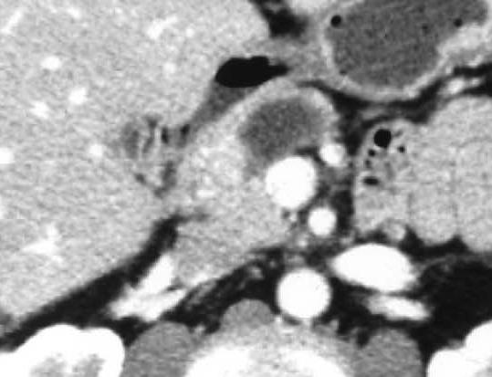
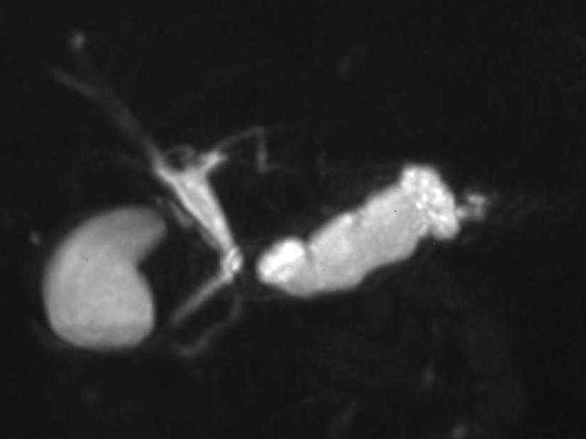
5 4 Case report A 36-year-old man was referred to our hospital for the evaluation and treatment of a pancreatic mass. He had epigastric pain while dilatation in the body and tail of MPD was also detected by ultrasonography (US). He had no past history of any pancreatic disorders. A laboratory examination on admission showed normal serum levels of amylase, lipase and pancreatic hormones including gastrin, insulin, and glucagons. In addition, the carcinoembryonic antigen, carbohydrate antigen 19-9 and elastase-i levels showed normal ranges while DUPAN-II (220 U/ml) was slightly elevated. Abdominal CT demonstrated an enhanced mass in the head of the pancreas with a dilatation of the MPD in the body and tail (Fig. 1A). On positron emission tomography using 18F-fluorodeoxyglucose (FDG) and CT with the same scanner (FDG-PET/CT), this pancreatic lesion showed an enhanced uptake of FDG, SUV: 4.67 (Fig. 1B). EUS revealed a well-defined isoechoic mass measuring about 15 mm in size within the lumen of the MPD (Fig. 1C). On ERP, an interruption of the MPD in the head of the MPD was noted (Fig. 1D). We could not detect any malignant cells in the pancreatic juice. Magnetic resonance cholangiopancreatography (MRCP) shows a dilation of the main pancreatic duct in the body and tail (Fig. 2). On MR imaging, the lesion demonstrated a low signal intensity on the T1-weighted image and a high signal intensity on the T2-weighted image with a good contrast enhancment (Fig. 3). No tumor in the parathyroid glands or the pituitary gland was detected. Although acinar cell tumors or combined tumors could not be ruled out, a pylorus-preserving pancreaticoduodenectomy (PPPD) with a pancreaticogastrostomy and a regional lymphadenectomy were performed under a preoperative diagnosis of a NFPT. Neither extra pancreatic invasion nor metastases to the lymph nodes was observed
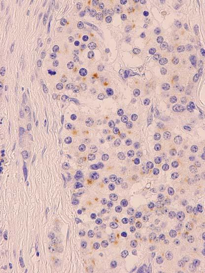
6 5 during the operation. The resected specimen of the pancreas confirmed the presence of an intraductal yellow mass which measured 16 X 15 mm in size (Fig. 4A and 4B). Microscopically, the tumor consisted of small nests and cords of uniform cuboidal cells arranged in a trabecular pattern (Fig. 4C). Immunohistochemically, the tumor was negative for insulin, gastrin, glucagons, somatostatin, and pancreatic peptide. Positive immunoreactions for synaptophisin (Fig. 4D) and vasoactive intestinal peptide (Fig. 4E) indicated the neuroendocrine differentiation of the tumor. In addition, metastasis to a lymph node along the common hepatic artery was detected. As a result, we finally diagnosed the tumor to be a malignant NFPT. The postoperative course was uneventful and the patient has survived for six months without any evidence of recurrence or metastasis.
7 6 Discussion We herein described a patient with a malignant NFPT that uniquely grew within the lumen of the MPD and obstructed the MPD. In most NFPTs, the size of the tumor correlates well with its malignant potential, and tumors measuring less than 2 cm in size are considered to be benign (14,15). On the other hand, a rare malignant NFPT (8 mm in size) has been previously reported with no abnormalities in the pancreatic ductal system (16). To the best of our knowledge only 20 cases of NFPTs have described with ERP that were accompanied by a complete obstruction of the MPD (Table 1) (3-13). Regarding clinical symptoms, pancreatitis due to a complete obstruction of the MPD has been observed 6 cases. In addition, seventeen of 20 cases (85%) were diagnosed to be malignant. A complete obstruction of the MPD caused by NFPTs indicated a malignant behavior of the tumor rather than a mass effect. We should consider the possibility of malignancy based on preoperative imaging including ERP, even if the tumor is small. Recently, FDG-PET/CT represents a useful tool for identifying tumor malignancy and also for accurately determing the preoperative staging (17). The finding of FDG-PET/CT suggested this lesion to have a malignant potential. The intraductal growth of NFPTs is thought to be very rare. There have only been four cases of NFPTs, including our case, which showed an intraductal growth pattern within the lumen of the MPD (Table 2). The fusion of the dorsal and ventral anlagen may thus have induced the MPD, while the multipotent stem cells of the pancreatic duct thereafter differentiated into endocrine cells. The origin of pancreatic endocrine tumors from hypothetical multipotent ductular stem cells has been suggested (18-21). Another possibility is that the tumor may originate from islet cells adjacent to the MPD.
8 7 Unlike typical pancreatic ductal carcinoma, the above described tumor grew slowly within the lumen of the MPD without invading the pancreatic duct epithelium. On ERP, interruption of the MPD with an intraductal filling defect in the head of the MPD was noted (Fig. 1D). On FDG-PET/CT, this pancreatic lesion showed an enhancement of FDG (Fig. 1B). Based on these findings, even though the tumor measured less than 2 cm in size, we still performed a PPPD to completely resect the tumor with sufficient margins while also performing a regional lymphadenectomy. According to the histological findings, it was finally diagnosed to be a malignant NFPT due to detect the detection of metastasis to a lymph node along the common hepatic artery. The presence of metastases does not rule out the possibility of a prolonged survival, as the overall survival rate for patients undergoing a pancreaticoduodenectomy is 81% and 70% at 5 and 10 years, respectively (22). In conclusion, we herein reported a rare case of a patient with a NFPT that uniquely grew within the lumen of the MPD without ductal involvement, while also completely obstructing the MPD. EUS and ERP were helpful for accurately delineating the intraductal growth of the tumor while also determining the resection line of the pancreas. Based on our avove findings, a malignant NFPT should therefore be included in the differential diagnosis when a complete obstruction of the MPD is demonstrated on ERP.
9 8 Figure legends Figure 1. (A) Abdominal computed tomography demonstrated an enhanced mass (arrows) in the head of the pancreas with a dilatation of the main pancreatic duct in the body and the tail of the pancreas. (B) On FDG-PET/CT, this pancreatic lesion (arrows) showed an enhanced uptake of FDG. (C) Endoscopic ultrasonography revealed a well-defined homogenous isoechoic intraductal mass (arrows) within the lumen of the main pancreatic duct. (D) Endoscopic retrograde pancreatography showed an interruption of the main pancreatic duct in the head of the pancreas. Figure 2. MRCP shows a dilation of the main pancreatic duct in the body and tail. Figure 3. On MR imaging, the lesion (arrows) demonstrated low signal intensity on T1-weighted image (A) and high signal intensity on T2-weighted image (B). (C-E) Dynamic MRI showed early enhanced tumor in the head of the pancreas. Figure 4. (A) The macroscopic findings showed a resected specimen of the pancreas to confirm the presence of an intraductal yellow mass (arrows) in the main pancreatic duct. An Arrow in Figure 4 (B) showed the position of the main pancreatic duct. (C) The microscopic findings showed the tumor to consist of small nests and cords of uniform cuboidal cells arranged in a trabecular pattern. The tumor showed positive immunoreactions for synaptophisin (D) and vasoactive intestinal peptide (E).
10 9 References 1. Kloppel G, Heitz PU. Pancreatic endocrine tumors in man. In: Polak JM, ed. Diagnostic histopathology of neuroendocrine tumors. Edinburg: Churchill Livingstone, 1993: Solcia E, Sessa F, Rindi G, Bonato M, Capella C. Pancreatic endocrine tumors: non-functioning tumors and tumors with uncommon function. In: Dayal Y, ed. Endocrine pathology of the gut and pancreas. Boca Raton: CRC Press, 1991: Sarles H, Cambon P, Choux R, Payan MJ, Odaira S, Laugier R, et al. Chronic obstructive pancreatitis due to tiny (0.6 to 8 mm) benign tumors obstructing pancreatic ducts: report of three cases. Pancreas 1988; 3: Simpson WF, Adam DB, Metcalf JS, Anderson MC. Nonfunctioning pancreatic neuroendocrine tumors presenting as pancreatitis: report of four cases. Pancreas 1988; 3: Ogawa Y, Tanaka M, Matsumoto S, Tamaguchi S, Ikeda S, Yoshimoto H. Islet cell tumors of the pancreas: the diagnostic value of endoscopic retrograde pancreatography. Int J Pancreatol 1990; 6: Mao C, Howard JM, Pancreatitis associated with neuroendocrine (islet cell) tumors of the pancreas. Am J Surg 1996; 171: Obara T, Shudo R, Fujii T, Tanno S, Muzukami Y, Izawa T, et al. Gastrointest Endosc 2000;51: Kitami C, Simizu T, Sato O, Kurosaki I, Mori S, Yamagisawa Y, et al. Malignant islet cell tumor projecting into the main pancreatic duct. J Hepatobiliary Pancreat Surg 2000; 7: Sugiyama M, Abe N, Izumisato Y, Yamaguchi Y, Yamato T, Tokuhara M, et al.
11 0 Differential diagnosis of benign versus malignant nonfunctioning islet cell tumors of the pancreas: the roles of EUS and ERCP. Gastrointest Endosc 2002; 55: Amano H, Hachimine T, Miyazaki S, Omori K, Kashihabara H, Yokoyama K. A case of non-functioning endocrine tumor of the pancreas presenting a bizarre appearance of the ampulla of Vater (in Japanese) Tan-to-sui (J Biliary Tract Pancreas) 1994; 15: Shimizu K, Shiratori K, Toki F, Suzuki M, Imaizumi T, Takasaki K, et al. Nonfunctioning islet cell tumor with a unique pattern of tumor growth. Dig Dis Sci 1999; 44: Akatsu T, Wakabayashi G, Aiura K, Suganuma K, Takigawa Y, Wada M, et al. Intraductal growth of a nonfunctioning endocrine tumor of the pancreas. J Gastroenterol 2004; 39: Terada R, Ito S, Akama F, Kashima K, Kidogawa H, Ooe H. Small nonfunctioning islet cell tumor in the body of the pancreas: report of a case. Surg Today 2004; 34: Capella C, Heitz PU, Hoefler H, Solcia E, Kloppel G. Revised classification of neuroendocrine tumors of the lung, pancreas and gut. Virchows Archiv 1995; 425: La Rosa S, Sessa F, Capella C, Riva C, Eugenio Leone B, et al. Prognostic criteria in nonfunctioning pancreatic endocrine tumors. Virchows Archiv 1996; 429: Ikenaga N, Yamaguchi K, Konomi H, Fujii K, Sugitani A, Tanaka M. A minute nonfunctioning islet tumor demonstrating malignant features. J Hepatobiliary Pancreat Surg 2005; 12:84-7.
12 1 17. Stefan H, Goerres GW, Schafer M, Sagmeister M, Bauerfeind P, Pestalozzi BC, et al. Positron emission tomography/computed tomography influences on the management of respectable pancreatic cancer and its cost-effectiveness. Ann Surg 2005; 242: Like AA, Orci L. Embryogenesis of the human pancreatic islets: A light and electron microscopic study. Diabetes 1972; 21: Heitz PU. Pancreatic endocrine tumors. In: Kloppel G, Heitz PU, eds. Pancreatic pathology. Edinburgh: Churchill-Livingstone, 1984: Heitz PU, Kasper M, Polak JM, Kloppel G. Pancreatic endocrine tunors. Immunocytochemical analysis of 125 tumors. Hum Pathol 1982; 26: Mehta S, Gittes GK. Pancreatic differentiation. J Hepatobiliary Pancreat Surg 2005;12: Sariento JM, Fanell MB, Que FG, Nagorney DM. Pancreaticoduodenectomy for islet cell tumors of the head of the pancreas: long-term survival analysis. World J Surg 2002; 26:


13 A C B D
14


15 A B C D E


16 A C B D E
17 Table 1 Reported cases of nonfunctioning pancreatic endocrine tumors that showed complete obstruction of the main pancreatic duct on endoscopic retrograde cholangiopancreatography Patient no. Age (yr)/sex Author (yr) Symptom Site Size (mm) Diagnosis 1 50/M Simpson (1988) Pancreatitis Head ND Malignant 2 63/F Simpson (1988) Pancreatitis Head 40 X 50 ND 3 51/M Ogawa (1990) ND Body ND Malignant 4 69/M Gondo (1992) ND Head-body 40 X 40 X 40 Malignant 5 62//F Gondo (1992) ND Head 47 X 45 X 35 Malignant 6 ND/F Mao (1996) Pancreatitis Tail 40 Malignant 7 ND/F Mao (1996) Pancreatitis Tail ND Malignant 8 46/M Obara (2000) Epigastric pain Body 45 Malignant 9 41/M Obara (2000) Epigastric pain Body 20 Malignant 10 57/M Kitami (2000) Pancreatitis Tail 15 X 15 Malignant 11 62/F Seki (2001) ND Head 47 Malignant 12 ND Seki (2001) Epigastric pain Head 45 Malignant 13 ND Seki (2001) Epigastric pain Head 33 Malignant 14 ND Seki (2001) Back pain Head 30 Malignant 15 ND Seki (2001) Back pain Body 13 Malignant 16 48/M Seki (2001) ND Head 8 Borderline malignant 17 75/F Sugiyama (2002) Pancreatitis Body 28 Malignant 18 43/F Akatsu (2004) None Body 25 X 8 X 12 Malignant 19 64/F Terada (2004) None Body 6 X 5 ND 20 36/M present case Epigastric pain Head 16 X 15 Malignant yr, year; ND, not described
18 Table 2 Reported cases of intraductal growth of nonfunctioning pancreatic endocrine tumors Patient no. Age (yr)/sex Author (yr) Symptom Site Size (mm) Diagnosis 1 53/F Amano (1994) Itching Head-tail 70 X 50 Malignant 2 44/F Shimizu (1998) Steatorrhea and Head-tail ND Low-grade malignant epigastric pain 3 43/M Akatsu (2004) None Body 25 X 8 X 12 Malignant 4 36/M present case None Head 16 X 15 Malignant yr, year; ND, not described
Citation American Journal of Surgery, 196(5)
") NAOSITE: Nagasaki University's Ac Title Author(s) Multifocal branch-duct pancreatic i neoplasms Tajima, Yoshitsugu; Kuroki, Tamotsu Amane; Adachi, Tomohiko; Mishima, T Kanematsu, Takashi Citation American
NAOSITE: Nagasaki University's Ac Title Author(s) Multifocal branch-duct pancreatic i neoplasms Tajima, Yoshitsugu; Kuroki, Tamotsu Amane; Adachi, Tomohiko; Mishima, T Kanematsu, Takashi Citation American
Inagaki M, Obara M, Kino S, Goto J, Suzuki S, Ishizaki A, Tanno S, Kohgo Y, Tokusashi Y, Miyokawa N, Kasai S.
 Journal of Hepato-Biliary-Pancreatic Surgery (2007) 14(3):264-269. Pylorus-preserving total pancreatectomy for an intraductal papillarymucinous neoplasm of the pancreas. Inagaki M, Obara M, Kino S, Goto
Greater Manchester and Cheshire HPB Unit Guidelines for the Assessment & Management of Hepatobiliary and Pancreatic Disease Chapter 14
 Greater Manchester and Cheshire HPB Unit Guidelines for the Assessment & Management of Hepatobiliary and Pancreatic Disease Chapter 14 Contents 14. Neuroendocrine Tumours 161 14.1. Diagnostic algorithm
A large mural nodule in branch duct intraductal papillary mucinous adenoma of the pancreas: a case report
 Haruki et al. Surgical Case Reports (2015) 1:20 DOI 10.1186/s40792-014-0009-x CASE REPORT Open Access A large mural nodule in branch duct intraductal papillary mucinous adenoma of the pancreas: a case
The Use of Pancreatoscopy in the Diagnosis of Intraductal Papillary Mucinous Tumor Lesions of the Pancreas
 CLINICAL GASTROENTEROLOGY AND HEPATOLOGY 2005;3:S53 S57 The Use of Pancreatoscopy in the Diagnosis of Intraductal Papillary Mucinous Tumor Lesions of the Pancreas KENJIRO YASUDA, MUNEHIRO SAKATA, MOOSE
Congenital dilatation of the common bile duct and pancreaticobiliary maljunction clinical implications
 Langenbecks Arch Surg (2009) 394:209 213 DOI 10.1007/s00423-008-0330-6 CURRENT CONCEPT IN CLINICAL SURGERY Congenital dilatation of the common bile duct and pancreaticobiliary maljunction clinical implications
Citation Hepato-Gastroenterology, 55(86-87),
,") NAOSITE: Nagasaki University's Ac Title Author(s) Combined pancreatic resection and p multiple lesions of the pancreas: i of the pancreas concomitant with du Kuroki, Tamotsu; Tajima, Yoshitsugu Tomohiko;
NAOSITE: Nagasaki University's Ac Title Author(s) Combined pancreatic resection and p multiple lesions of the pancreas: i of the pancreas concomitant with du Kuroki, Tamotsu; Tajima, Yoshitsugu Tomohiko;
Case Scenario 1. Discharge Summary
 Case Scenario 1 Discharge Summary A 69-year-old woman was on vacation and noted that she was becoming jaundiced. Two months prior to leaving on that trip, she had had a workup that included an abdominal
CASE REPORT. Abstract. Introduction. Case Report
 CASE REPORT Branch Duct Intraductal Papillary Mucinous Neoplasms of the Pancreas Involving Type 1 Localized Autoimmune Pancreatitis with Normal Serum IgG4 Levels Successfully Diagnosed by Endoscopic Ultrasound-guided
Management of Pancreatic Islet Cell Tumors
 Management of Pancreatic Islet Cell Tumors Ravi Dhanisetty, MD November 5, 2009 Morbidity and Mortality Conference Case Presentation 42 yr female with chronic abdominal pain. PMHx: Uterine fibroids Medications:
FDG-PET Findings of Intraductal Oncocytic Papillary Neoplasms of the Pancreas: Two Case Reports
 This is an Open Access article licensed under the terms of the Creative Commons Attribution-NonCommercial-NoDerivs 3.0 License (www.karger.com/oa-license), applicable to the online version of the article
This page explains some of the medical words that you may hear when you are finding out about pancreatic cancer and how it is treated.
 A-Z of medical words This page explains some of the medical words that you may hear when you are finding out about pancreatic cancer and how it is treated. Absorption: once your food has been broken down,
Chronic pancreatitis mimicking intraductal papillary mucinous neoplasm of the pancreas; Report of tow cases
 Jichi Medical University Journal Chronic pancreatitis mimicking intraductal papillary mucinous neoplasm of the pancreas; Report of tow cases Noritoshi Mizuta, Hiroshi Noda, Nao Kakizawa, Nobuyuki Toyama,
Yoshitsugu; Kanematsu, Takashi; Kur
 NAOSITE: Nagasaki University's Ac Title Author(s) Citation Laparoscopic Middle Pancreatectomy Surgery Kitasato, Amane; Adachi, Tomohiko; Yoshitsugu; Kanematsu, Takashi; Kur Hepato-Gastroenterology, 59(120),
A Multicentric Development Of Intraductal Papillary Mucinous Neoplasm Treated By Repeated Pancreatectomy
 ISPUB.COM The Internet Journal of Surgery Volume 7 Number 2 A Multicentric Development Of Intraductal Papillary Mucinous Neoplasm Treated By Repeated T Matsumoto, K Iwaki, H Uchida, K Yada, K Shibata,
Endoscopic Ultrasonography Assessment for Ampullary and Bile Duct Malignancy
 Diagnostic and Therapeutic Endoscopy, Vol. 3, pp. 35-40 Reprints available directly from the publisher Photocopying permitted by license only (C) 1996 OPA (Overseas Publishers Association) Amsterdam B.V.
According to the international consensus guidelines for
 ORIGINAL ARTICLE Natural History of Branch Duct Intraductal Papillary Mucinous Neoplasm With Mural Nodules A Japan Pancreas Society Multicenter Study Go Kobayashi, MD, PhD,* Naotaka Fujita, MD, PhD,* Hiroyuki
Acute Pancreatitis Associated with Neuroendocrine Tumor of the Pancreas
 CASE REPORT Acute Pancreatitis Associated with Neuroendocrine Tumor of the Pancreas José Jukemura 1, André Luis Montagnini 1, Marcos Vinícius Perini 2, José Luiz Jesus de Almeida 1, Joaquim José Gama Rodrigues
Endoscopic Resection of Ampullary Neuroendocrine Tumor
 CASE REPORT Endoscopic Resection of Ampullary Neuroendocrine Tumor Hiroyuki Fukasawa, Shigetaka Tounou, Masashi Nabetani and Tomoki Michida Abstract We report the case of a 57-year-old man with a 1.0-cm
An Intraductal Papillary Neoplasm of the Bile Duct at the Duodenal Papilla
 Published online: July 2, 2014 1662 6575/14/0072 0417$39.50/0 This is an Open Access article licensed under the terms of the Creative Commons Attribution- NonCommercial 3.0 Unported license (CC BY-NC)
EUS FNA NEUROENDOCRINE TUMORS. A. Ginès Endocopy Unit Hospital Cínic. Barcelona (Spain)
") EUS FNA NEUROENDOCRINE TUMORS A. Ginès Endocopy Unit Hospital Cínic. Barcelona (Spain) GI NEUROENDOCRINE TUMORS GENERAL CONCEPTS Rare neoplasms arising from the neuroendocrine cells of the GI tract Include:
EUS FNA NEUROENDOCRINE TUMORS A. Ginès Endocopy Unit Hospital Cínic. Barcelona (Spain) GI NEUROENDOCRINE TUMORS GENERAL CONCEPTS Rare neoplasms arising from the neuroendocrine cells of the GI tract Include:
Pancreatic Cancer. What is pancreatic cancer?
 Scan for mobile link. Pancreatic Cancer Pancreatic cancer is a tumor of the pancreas, an organ that is located behind the stomach in the abdomen. Pancreatic cancer does not always cause symptoms until
Imaging in gastric cancer
 Imaging in gastric cancer Gastric cancer remains a deadly disease because of late diagnosis. Adenocarcinoma represents 90% of malignant tumors. Diagnosis is based on endoscopic examination with biopsies.
Branch duct intraductal papillary mucinous neoplasm of the pancreas: single-center experience with 324 patients who underwent surgical resection
 Korean J Hepatobiliary Pancreat Surg 2015;19:113-120 http://dx.doi.org/10.14701/kjhbps.2015.19.3.113 Original Article Branch duct intraductal papillary mucinous neoplasm of the pancreas: single-center
Synchronous double primary squamous cell carcinoma and adenocarcinoma of the extrahepatic bile duct: a case report
 Yoo and Mun Journal of Medical Case Reports (2015) 9:116 DOI 10.1186/s13256-015-0600-1 CASE REPORT JOURNAL OF MEDICAL CASE REPORTS Open Access Synchronous double primary squamous cell carcinoma and adenocarcinoma
Index. Surg Oncol Clin N Am 16 (2007) Note: Page numbers of article titles are in boldface type.
 Note: Page numbers of article titles are in boldface type.") Surg Oncol Clin N Am 16 (2007) 465 469 Index Note: Page numbers of article titles are in boldface type. A Adjuvant therapy, preoperative for gastric cancer, staging and, 339 B Breast cancer, metabolic
Surg Oncol Clin N Am 16 (2007) 465 469 Index Note: Page numbers of article titles are in boldface type. A Adjuvant therapy, preoperative for gastric cancer, staging and, 339 B Breast cancer, metabolic
International Surgery Synchronous double cancer of the common bile duct and the ampulla of Vater without pancreaticobiliary maljunction: A case report
 International Surgery Synchronous double cancer of the common bile duct and the ampulla of Vater without pancreaticobiliary maljunction: A case report --Manuscript Draft-- Manuscript Number: Full Title:
Pancreatic Cysts. Darius C. Desai, MD FACS St. Luke s University Health Network
 Pancreatic Cysts Darius C. Desai, MD FACS St. Luke s University Health Network None Disclosures Incidence Widespread use of cross sectional imaging Seen in over 2% of patients having abdominal imaging
MULTIMEDIA ARTICLE - Clinical Imaging. Brian KP Goh 1, Yaw-Fui Alexander Chung 1,4, David CE Ng 2, Sathiyamoorthy Selvarajan 3, Khee-Chee Soo 1,4
 MULTIMEDIA ARTICLE - Clinical Imaging Positron Emission Tomography with 2-Deoxy-2-[ 18 F] Fluoro-D- Glucose in the Detection of Malignancy in Intraductal Papillary Mucinous Neoplasms of the Pancreas Brian
Multiple Primary Quiz
 Multiple Primary Quiz Case 1 A 72 year old man was found to have a 12 mm solid lesion in the pancreatic tail by computed tomography carried out during a routine follow up study of this patient with adult
ORIGINAL ARTICLE. Fate of the Pancreatic Remnant After Resection for an Intraductal Papillary Mucinous Neoplasm
 ONLINE FIRST ORIGINAL ARTICLE Fate of the Pancreatic Remnant After Resection for an Intraductal Papillary Mucinous Neoplasm A Longitudinal Level II Cohort Study Toshiyuki Moriya, MD, PhD; L. William Traverso,
Case report Serous cystadenocarcinoma of the mesentery in a man: case report and review of literature
 Gastroenterology Report 2 (2014) 306 310, doi:10.1093/gastro/gou019 Advance access publication 7 April 2014 Case report Serous cyst of the mesentery in a man: case report and review of literature Toru
Middle segment-preserving pancreatectomy for metachronous intraductal papillary mucinous neoplasm after pancreatoduodenectomy: a case report
 Yamada et al. Surgical Case Reports (2017) 3:28 DOI 10.1186/s40792-017-0306-2 CASE REPORT Middle segment-preserving pancreatectomy for metachronous intraductal papillary mucinous neoplasm after pancreatoduodenectomy:
Dr Claire Smith, Consultant Radiologist St James University Hospital Leeds
 Dr Claire Smith, Consultant Radiologist St James University Hospital Leeds Imaging in jaundice and 2ww pathway Image protocol Staging Limitations Pancreatic cancer 1.2.4 Refer people using a suspected
Predictive factors for invasive intraductal papillary mucinous neoplasm of the pancreas
 Korean J Hepatobiliary Pancreat Surg 2011;15:27-22 Original Article Predictive factors for invasive intraductal papillary mucinous neoplasm of the pancreas Dae Young Jun 1, Hyung Jun Kwon 2, Sang Geol
Disclosure of Relevant Financial Relationships
 Disclosure of Relevant Financial Relationships USCAP requires that all faculty in a position to influence or control the content of CME disclose any relevant financial relationship WITH COMMERCIAL INTERESTS
Surgical outcomes of multifocal branch duct intraductal papillary mucinous neoplasms of pancreas
 Korean J Hepatobiliary Pancreat Surg 2014;18:152-158 http://dx.doi.org/10.14701/kjhbps.2014.18.4.152 Original Article Surgical outcomes of multifocal branch duct intraductal papillary mucinous neoplasms
Esophageal Cancer Initially Thought to be Accompanied by a Solitary Metastasis to an Intrathoracic Paraaortic Lymph Node
 2012 66 5 417 421 Esophageal Cancer Initially Thought to be Accompanied by a Solitary Metastasis to an Intrathoracic Paraaortic Lymph Node a b* a a a a a a a b ʼ 418 Horio et al. Acta Med. Okayama Vol.
췌장의단일종괴형태로재발해원발성췌장암으로오인된재발성폐암
 Case Report The Korean Journal of Pancreas and Biliary Tract 2018;23:172-176 https://doi.org/10.15279/kpba.2018.23.4.172 pissn 1976-3573 eissn 2288-0941 췌장의단일종괴형태로재발해원발성췌장암으로오인된재발성폐암 대구가톨릭대학교의과대학내과학교실
Intraductal Papillary-Mucinous Neoplasm of the Pancreas Penetrating to the Stomach and the Common Bile Duct
 CASE REPORT Intraductal Papillary-Mucinous Neoplasm of the Pancreas Penetrating to the Stomach and the Common Bile Duct Norihiro Goto 1, Masahiro Yoshioka 1, Motohito Hayashi 1, Toshinao Itani 1, Jun Mimura
Matthew McCollough, M.D. April 9, 2009 University of Louisville
 Matthew McCollough, M.D. April 9, 2009 University of Louisville List the differential diagnosis for pancreatic cysts Review the epidemiology Illustrate the types of cysts through case discussions Discuss
ACUTE CHOLANGITIS AS a result of an occluded
 Digestive Endoscopy 2017; 29 (Suppl. 2): 88 93 doi: 10.1111/den.12836 Current status of biliary drainage strategy for acute cholangitis Endoscopic treatment for acute cholangitis with common bile duct
PersPeCTIves. Controversies in the management of pancreatic ipmn. Masao Tanaka
 PersPeCTIves OpiniOn Controversies in the management of pancreatic ipmn Masao Tanaka Abstract Although considerable progress has been made in our understanding of intraductal papillary mucinous neoplasm
An Unexpected Cause of Hypoglycemia
 An Unexpected Cause of Hypoglycemia Stacey A. Milan, MD FACS Surgical Oncology Nothing to disclose Disclosures Objectives Identify indications for workup of hypoglycemia Define work up for hypoglycemic
Diagnosis of tumor extension in biliary carcinoma has. Differential Diagnosis and Treatment of Biliary Strictures
 CLINICAL GASTROENTEROLOGY AND HEPATOLOGY 2009;7:S79 S83 Differential Diagnosis and Treatment of Biliary Strictures KAZUO INUI, JUNJI YOSHINO, and HIRONAO MIYOSHI Department of Internal Medicine, Second
Intraductal papillary mucinous neoplasm (IPMN) is a distinct
 is a distinct") CLINICAL GASTROENTEROLOGY AND HEPATOLOGY 2008;6:815 819 Evaluation of the Guidelines for Management of Pancreatic Branch-Duct Intraductal Papillary Mucinous Neoplasm RAYMOND S. TANG,* BENJAMIN WEINBERG,
CLINICAL GASTROENTEROLOGY AND HEPATOLOGY 2008;6:815 819 Evaluation of the Guidelines for Management of Pancreatic Branch-Duct Intraductal Papillary Mucinous Neoplasm RAYMOND S. TANG,* BENJAMIN WEINBERG,
The Pancreas. Basic Anatomy. Endocrine pancreas. Exocrine pancreas. Pancreas vasculature. Islets of Langerhans. Acinar cells Ductal System
 SGNA: Back to Basics Rogelio G. Silva, MD Assistant Clinical Professor of Medicine University of Illinois at Chicago Department of Medicine Division of Gastroenterology Advocate Christ Medical Center GI
2004 SNM Mid-Winter Educational Symposium
 1 2 20 Numeric values 15 10 5 0 SUV corr hs 18FDG Slope corr hs SUV corr hs Slope corr hs 11C-methionin 3 11C-methionine Grading and delineation of brain tumors Differentiation of malignant from benign
Magnetic resonance cholangiopancreatography (MRCP) is an imaging. technique that is able to non-invasively assess bile and pancreatic ducts,
 is an imaging. technique that is able to non-invasively assess bile and pancreatic ducts,") SECRETIN AUGMENTED MRCP Riccardo MANFREDI, MD, MBA, FESGAR Magnetic resonance cholangiopancreatography (MRCP) is an imaging technique that is able to non-invasively assess bile and pancreatic ducts, in
SECRETIN AUGMENTED MRCP Riccardo MANFREDI, MD, MBA, FESGAR Magnetic resonance cholangiopancreatography (MRCP) is an imaging technique that is able to non-invasively assess bile and pancreatic ducts, in
PANCREAS DUCTAL ADENOCARCINOMA PDAC
 CONTENTS PANCREAS DUCTAL ADENOCARCINOMA PDAC I. What is the pancreas? II. III. IV. What is pancreas cancer? What is the epidemiology of Pancreatic Ductal Adenocarcinoma (PDAC)? What are the risk factors
Synchronous double primary cancers associated with a choledochal cyst and anomalous pancreaticobiliary ductal union
 J Korean Surg Soc 2011;81:281-286 http://dx.doi.org/10.4174/jkss.2011.81.4.281 CASE REPORT JKSS Journal of the Korean Surgical Society pissn 2233-7903 ㆍ eissn 2093-0488 Synchronous double primary cancers
Kentaro Tominaga, Kenya Kamimura, Junji Yokoyama and Shuji Terai
 doi: 10.2169/internalmedicine.1700-18 http://internmed.jp CASE REPORT Usefulness of Capsule Endoscopy and Double-balloon Enteroscopy for the Diagnosis of Multiple Carcinoid Tumors in the Small Intestine:
Biliary tree dilation - and now what?
 Biliary tree dilation - and now what? Poster No.: C-1767 Congress: ECR 2012 Type: Educational Exhibit Authors: I. Ferreira, A. B. Ramos, S. Magalhães, M. Certo; Porto/PT Keywords: Pathology, Diagnostic
Topics: Staging and treatment for pancreatic cancer. Staging systems for pancreatic cancer: Differences between the Japanese and UICC systems
 M. J Hep Kobari Bil Pancr and S. Surg Matsuno: (1998) Staging 5:121 127 system for pancreatic cancer 121 Topics: Staging and treatment for pancreatic cancer Staging systems for pancreatic cancer: Differences
Preoperative Diagnosis of Adult Intussusception Caused by Small Bowel Lipoma
 377 Preoperative Diagnosis of Adult Intussusception Caused by Small Bowel Lipoma Hiroaki Shiba a Yoshinobu Mitsuyama a Ken Hanyu a Kenji Ikeuchi b Hirotaka Hayashi c Katsuhiko Yanaga a a Department of
IgG4-Negative Autoimmune Pancreatitis with Sclerosing Cholangitis and Colitis: Possible Association with Primary Sclerosing Cholangitis?
 CASE REPORT IgG4-Negative Autoimmune Pancreatitis with Sclerosing Cholangitis and Colitis: Possible Association with Primary Sclerosing Cholangitis? Keita Saeki 1, Shigenari Hozawa 1, Naoteru Miyata 1,
Diagnostic Algorithm for Autoimmune Pancreatitis in Korea
 Review Article The Korean Journal of Pancreas and Biliary Tract 2014;19(1):7-12 pissn 1976-3573 eissn 2288-0941 한국에서자가면역췌장염의진단전략 성균관대학교의과대학삼성서울병원내과학교실 이종균 Diagnostic Algorithm for Autoimmune Pancreatitis
Title. region. Author(s) Citation Surgery, 145(3), pp ; Issue Date
 Citation Surgery, 145(3), pp ; Issue Date") NAOSITE: Nagasaki University's Ac Title Author(s) Huge pancreatic pseudocyst migratin region. Tajima, Yoshitsugu; Mishima, Takehi Taiichiro; Adachi, Tomohiko; Tsuneo Citation Surgery, 145(3), pp.341-342;
NAOSITE: Nagasaki University's Ac Title Author(s) Huge pancreatic pseudocyst migratin region. Tajima, Yoshitsugu; Mishima, Takehi Taiichiro; Adachi, Tomohiko; Tsuneo Citation Surgery, 145(3), pp.341-342;
Imaging Pancreatic Neuroendocrine Tumors (PNETs): CT, MRI, EUS, Nuclear
: CT, MRI, EUS, Nuclear") Imaging Pancreatic Neuroendocrine Tumors (PNETs): CT, MRI, EUS, Nuclear Eric Tamm, M.D. Department of Diagnostic Radiology Division of Diagnostic Imaging MD Anderson Cancer Center Houston, TX Disclosure
Imaging Pancreatic Neuroendocrine Tumors (PNETs): CT, MRI, EUS, Nuclear Eric Tamm, M.D. Department of Diagnostic Radiology Division of Diagnostic Imaging MD Anderson Cancer Center Houston, TX Disclosure
Introduction of GB polyp
 Management of Gallbladder Polyp as Physician's View Sang Hyub Lee, MD, PhD Seoul National University College of Medicine Seoul National University Bundang Hospital Department of Internal Medicine Division
Management of the Mucin Filled Bile Duct. A Complication of Intraductal Papillary Mucinous Tumor of the Pancreas
 CASE REPORT Management of the Mucin Filled Bile Duct. A Complication of Intraductal Papillary Mucinous Tumor of the Pancreas Anand Patel, Louis Lambiase, Antonio Decarli, Ali Fazel Division of Gastroenterology
ITO, Yasuhiro ; FUJII, Mizue ; SHIBUYA, Takashi ; UEHARA, Jiro ; SATO, Katsuhiko ; IIZUKA, Hajime
 Journal of Dermatology (2011) 38(5):515-517. Granulocyte colony stimulating factor-producing squamous cell carcinoma of the skin ITO, Yasuhiro ; FUJII, Mizue ; SHIBUYA, Takashi ; UEHARA, Jiro ; SATO, Katsuhiko
ACG Clinical Guideline: Diagnosis and Management of Pancreatic Cysts
 ACG Clinical Guideline: Diagnosis and Management of Pancreatic Cysts Grace H. Elta, MD, FACG 1, Brintha K. Enestvedt, MD, MBA 2, Bryan G. Sauer, MD, MSc, FACG (GRADE Methodologist) 3 and Anne Marie Lennon,
A resected case of recurrent ITPN in the remnant pancreas after pancreatoduodenectomy
 Ko et al. Surgical Case Reports (2019) 5:33 https://doi.org/10.1186/s40792-019-0590-0 CASE REPORT A resected case of recurrent ITPN in the remnant pancreas after pancreatoduodenectomy Open Access Kenju
Case Report Five-Year Survival after Surgery for Invasive Micropapillary Carcinoma of the Stomach
 Case Reports in Surgery Volume 2013, Article ID 560712, 4 pages http://dx.doi.org/10.1155/2013/560712 Case Report Five-Year Survival after Surgery for Invasive Micropapillary Carcinoma of the Stomach Shigeo
Two Consecutive Cases of Ampulla of Vater Cancer Combined with Annular Pancreas and Unusual Anatomic Variation
 Korean Journal of HBP Surgery Vol. 14, No. 3, September 2010 증례 Two Consecutive Cases of Ampulla of Vater Cancer Combined with Annular Pancreas and Unusual Anatomic Variation Annular pancreas is a rare
Recommendations for the Reporting of Pancreatic Specimens Containing Malignant Tumors
 AJCP / REPORTING RECOMMENDATIONS FOR PANCREATIC SPECIMENS CONTAINING MALIGNANT TUMORS Recommendations for the Reporting of Pancreatic Specimens Containing Malignant Tumors Jorge AlboresSaavedra, David
A Case of Pancreatic Carcinoma with Bilateral Hilar
 Shinshu Med J, 66⑵:151~155, 2018 A Case of Pancreatic Carcinoma with Bilateral Hilar 18 F-FDG and 67 Ga Hyperaccumulation Satoshi Kawakami 1 )*, Yasunari Fujinaga 1), Shin Yanagisawa 1) Masumi Kadoya 1),
Common and unusual CT and MRI manifestations of pancreatic adenocarcinoma: a pictorial review
 Review Article Common and unusual CT and MRI manifestations of pancreatic adenocarcinoma: a pictorial review Min-Jie Yang, Su Li, Yong-Guang Liu, Na Jiao, Jing-Shan Gong Department of Radiology, Shenzhen
Case report Solid pseudopapillary tumor: a rare neoplasm of the pancreas
 Gastroenterology Report 2 (2014) 145 149, doi:10.1093/gastro/gou006 Advance access publication 28 February 2014 Case report Solid pseudopapillary tumor: a rare neoplasm of the pancreas Asim Shuja 1, *
doi: /j.anl
 doi: 10.1016/j.anl.2006.07.001 Synchronous unilateral parotid gland neoplasms of three different histological types Shuho Tanaka 1, Keiji Tabuchi 1, Keiko Oikawa 1, Rika Kohanawa 1, Hideki Okubo 1, Dai
Pancreas Case Scenario #1
 Pancreas Case Scenario #1 An 85 year old white female presented to her primary care physician with increasing abdominal pain. On 8/19 she had a CT scan of the abdomen and pelvis. This showed a 4.6 cm mass
Primary Pulmonary Colloid Adenocarcinoma: How Can We Obtain a Precise Diagnosis?
 doi: 10.2169/internalmedicine.1153-18 Intern Med 57: 3637-3641, 2018 http://internmed.jp CASE REPORT Primary Pulmonary Colloid Adenocarcinoma: How Can We Obtain a Precise Diagnosis? Shinsuke Ogusu 1, Koichiro
Lymphoepithelial cyst with sebaceous glands of the pancreas: a case report
 Maehira et al. Surgical Case Reports (2016) 2:98 DOI 10.1186/s40792-016-0228-4 CASE REPORT Open Access Lymphoepithelial cyst with sebaceous glands of the pancreas: a case report Hiromitsu Maehira 1*, Hisanori
CASE REPORT. Masakuni Fujii 1*, Masao Yoshioka 1, Takefumi Niguma 2, Hiroaki Saito 1,ToruKojima 2, Soichiro Nose 3 and Junji Shiode 1
 Fujii et al. Journal of Medical Case Reports 2014, 8:243 JOURNAL OF MEDICAL CASE REPORTS CASE REPORT Open Access A solid pseudopapillary neoplasm without cysts that occurred in a patient diagnosed by endoscopic
Case Scenario 1. The patient has now completed his neoadjuvant chemoradiation and has been cleared for surgery.
 Case Scenario 1 July 10, 2010 A 67-year-old male with squamous cell carcinoma of the mid thoracic esophagus presents for surgical resection. The patient has completed preoperative chemoradiation. This
Primary Pancreatic Malignant Lymphoma Diagnosed from Endoscopic Ultrasound-guided Fine-needle Aspiration Findings
 CASE REPORT Primary Pancreatic Malignant Lymphoma Diagnosed from Endoscopic Ultrasound-guided Fine-needle Aspiration Findings Nobuhiko Fukuba 1, Ichiro Moriyama 1,2, Shunji Ishihara 1, Hiroki Sonoyama
Immunoglobulin G4-Related Disease with Several Inflammatory Foci
 CASE REPORT Immunoglobulin G4-Related Disease with Several Inflammatory Foci Akira Sakamaki 1, Kenya Kamimura 1, Kazuhiko Shioji 1, Junko Sakurada 2, Takeshi Nakatsue 3, Yoko Wada 3, Michitaka Imai 1,
Autoimmune Pancreatitis: A Great Imitator
 Massachusetts General Hospital Harvard Medical School Autoimmune Pancreatitis: A Great Imitator Dushyant V Sahani MD dsahani@partners.org Autoimmune Pancreatitis: Learning Objectives Clinical manifestations
Dual-time-point FDG-PET/CT Imaging of Temporal Bone Chondroblastoma: A Report of Two Cases
 Dual-time-point FDG-PET/CT Imaging of Temporal Bone Chondroblastoma: A Report of Two Cases Akira Toriihara 1 *, Atsunobu Tsunoda 2, Akira Takemoto 3, Kazunori Kubota 1, Youichi Machida 1, Ukihide Tateishi
Intraductal papillary neoplasms in the bile ducts
 Intraductal papillary neoplasms in the bile ducts Seok Hwa Youn Myunghee Yoon Dong Hoon Shin Kosin University Gospel Hospital Department of general surgery Hepato-biliary-pancreatic division Introduction
Metachronous anterior urethral metastasis of prostatic ductal adenocarcinoma
 http://dx.doi.org/10.7180/kmj.2016.31.1.66 KMJ Case Report Metachronous anterior urethral metastasis of prostatic ductal adenocarcinoma Jeong Hyun Oh 1, Taek Sang Kim 1, Hyun Yul Rhew 1, Bong Kwon Chun
HEPATIC METASTASES. We can state 3 types of metastases depending on their treatment options:
 HEPATIC METASTASES 1. Definition Metastasis means the spread of cancer. Cancerous cells can separate from the primary tumor and enter the bloodstream or the lymphatic system (the one that produces, stores,
Ampullary Carcinoma Associated with an Annular Pancreas
 CASE REPORT Ampullary Carcinoma Associated with an Annular Pancreas Fung J Foo 1, Upkar Gill 1, Caroline S Verbeke 2, James A Guthrie 3, Krishna V Menon 1 Departments of 1 Hepatobiliary Surgery, 2 Histopathology,
Intraductal Papillary Mucinous Neoplasm of the Pancreas. Masao Tanaka Editor
 Intraductal Papillary Mucinous Neoplasm of the Pancreas Masao Tanaka Editor Intraductal Papillary Mucinous Neoplasm of the Pancreas Masao Tanaka Editor Intraductal Papillary Mucinous Neoplasm of the Pancreas
A Proposed Strategy for Treatment of Superficial Carcinoma. in the Thoracic Esophagus Based on an Analysis. of Lymph Node Metastasis
 Kitakanto Med J 2002 ; 52 : 189-193 189 A Proposed Strategy for Treatment of Superficial Carcinoma in the Thoracic Esophagus Based on an Analysis of Lymph Node Metastasis Susumu Kawate,' Susumu Ohwada,'
Ampullary neuroendocrine tumor diagnosed by endoscopic papillectomy in previously confirmed ampullary adenoma
 Case Report 2 Ampullary neuroendocrine tumor diagnosed by endoscopic papillectomy in previously confirmed ampullary adenoma Authors: Tae Hoon Lee 1, Si-Hyong Jang 2 Affiliation: Division of Gastroenterology
COLORECTAL CARCINOMA
 QUICK REFERENCE FOR HEALTHCARE PROVIDERS MANAGEMENT OF COLORECTAL CARCINOMA Ministry of Health Malaysia Malaysian Society of Colorectal Surgeons Malaysian Society of Gastroenterology & Hepatology Malaysian
Long-term Outcome of Autoimmune Pancreatitis after Oral Prednisolone Therapy
 ORIGINAL ARTICLE Long-term Outcome of Autoimmune Pancreatitis after Oral Prednisolone Therapy Takayoshi Nishino 1, Fumitake Toki 2,HiroyasuOyama 3, Kyoko Shimizu 1 and Keiko Shiratori 1 Abstract Objective
Title: The endoscopic ultrasound-assisted Rendez-Vous technique for treatment of recurrent pancreatitis due to pancreas divisum and ansa pancreatica
 Title: The endoscopic ultrasound-assisted Rendez-Vous technique for treatment of recurrent pancreatitis due to pancreas divisum and ansa pancreatica Authors: Sergio López-Durán, Celia Zaera, Juan Ángel
Diagnostic Imaging of the Liver
 S. Sakuma T. Ishigaki T. Takeuchi Diagnostic Imaging of the Liver Biliary Tract and Pancreas Data Analysis and Diagnostic Procedures With 312 Figures in 932 Separate Illustrations Springer-Verlag Berlin
Primary acinar cell carcinoma of the ampulla of Vate. The original publication is available at Instructions for use
 Title Primary acinar cell carcinoma of the ampulla of Vate Kawakami, Hiroshi; Kuwatani, Masaki; Onodera, Manabu Author(s) Itoh, Tomoo; Asaka, Masahiro CitationJournal of gastroenterology, 42(8): 694-697
MANAGEMENT OF INCIDENTALLY DETECTED GALLBLADDER CANCER
 MANAGEMENT OF INCIDENTALLY DETECTED GALLBLADDER CANCER Orlando Jorge M. Torres Full Professor and Chairman Department of Gastrointestinal Surgery Hepatopancreatobiliary Unit Federal University of Maranhão
E ndoscopic retrograde cholangiopancreatography (ERCP)
") 240 BILIARY DISEASE Endoscopic transpapillary biopsies and intraductal ultrasonography in the diagnostics of bile duct strictures: a prospective study D Domagk, C Poremba, K-H Dietl, N Senninger, A Heinecke,
240 BILIARY DISEASE Endoscopic transpapillary biopsies and intraductal ultrasonography in the diagnostics of bile duct strictures: a prospective study D Domagk, C Poremba, K-H Dietl, N Senninger, A Heinecke,
Hiroyuki Tanishima 1*, Masamichi Kimura 1, Toshiji Tominaga 1, Shinji Iwakura 1, Yoshihiko Hoshida 2 and Tetsuya Horiuchi 1
 Tanishima et al. Surgical Case Reports (2017) 3:93 DOI 10.1186/s40792-017-0366-3 CASE REPORT Open Access Lateral lymph node metastasis in a patient with T1 upper rectal cancer treated by lateral lymph
5/17/2013. Pancreatic Cancer. Postgraduate Course in General Surgery CASE 1: CASE 1: Overview. Case presentation. Differential diagnosis
 Overview Case presentation Postgraduate Course in General Surgery Differential diagnosis Diagnosis and therapy Eric K. Nakakura Koloa, HI March 26, 2013 Outcomes CASE 1: CASE 1: A 78-year-old man developed
Successful resection of pancreatic metastasis from oesophageal squamous cell carcinoma: a case report and review of the literature
 Koizumi et al. BMC Cancer (2019) 19:320 https://doi.org/10.1186/s12885-019-5549-9 CASE REPORT Open Access Successful resection of pancreatic metastasis from oesophageal squamous cell carcinoma: a case
Anatomical and Functional MRI of the Pancreas
 Anatomical and Functional MRI of the Pancreas MA Bali, MD, T Metens, PhD Erasme Hospital Free University of Brussels Belgium mbali@ulb.ac.be Introduction The use of MRI to investigate the pancreas has