BRACHYTHERAPY IN HORSES
|
|
|
- Bryan Boone
- 5 years ago
- Views:
Transcription
1 Vet Times The website for the veterinary profession BRACHYTHERAPY IN HORSES Author : DAVID DONALDSON Categories : Vets Date : June 16, 2014 DAVID DONALDSON BVSc(Hons), DipECVO, MRCVS considers the use of low-dose rate and high-dose rate brachytherapy, especially development of computerised planning to allow more precise delivery. ALTHOUGH the diagnosis of neoplasia is relatively uncommon in horses, the periocular region is commonly involved, with sarcoid and squamous cell carcinoma being the most common diagnoses. Management is commonly multimodal and integrates combinations of surgery, chemotherapy, immunotherapy, cryosurgery and radiation therapy (RT). Surgical debulking is commonly performed and improves the success rate of adjunctive therapies such as RT. Attempts at complete surgical excision followed by reconstructive blepharoplasty procedures are limited in horses due to the need to preserve a functional eyelid and the lack of skin that can be mobilised around the eye. The equine skin is firmly attached to the underlying connective tissue and has poor superficial blood supply, and, therefore, the mobilisation of adequate skin to be used in blepharoplasty procedures is limited (Gilger and Stoppini, 2005). Brachytherapy involving various radionuclides has been described in horses and, given the limitations of surgical interventions, represents an important adjunctive treatment for cases of periocular neoplasia. Brachytherapy is a form of RT where the radiation source is in direct contact with the patient. The two main types of brachytherapy are interstitial, in which the radioactive implant is placed directly 1 / 9
2 inside the tumour volume, and contact or plesiobrachytherapy, where the source is placed close to the tumour. Contact brachytherapy may be further divided into intracavitary (such as nasopharynx), intraluminal (such as cervix), endovascular and surface (for example, skin) methods. Due to the rapid fall-off in dose around radioactive sources, brachytherapy is characterised by strong dose gradients, with the highest levels of radiation being evident near the source. This allows high doses of radiation to be delivered to a tumour (greater local tumour control) while minimising the exposure of surrounding normal tissue (fewer side effects). In contrast to external beam RT in which deep-seated tumours can be irradiated, brachytherapy requires the placement of catheters directly into the tumour. Given this, brachytherapy is particularly suited to accessible tumours with well-defined margins. Brachytherapy with radium (Ra-226) was first used in 1901, soon after the discovery of radium and its radioactivity. The dangers of radiation exposure were not appreciated in the early 1900s and, as a result, radium, with its mysterious properties such as luminescence when mixed with phosphor, led to its use in many consumer products such as the very popular glow in the dark watch and clock faces. Radium remained the isotope of choice for brachytherapy until the 1950s to 1960s, when it was replaced by reactor-based radionuclides such as cobalt-60, caesium-137, iridium-192, gold-198 and iodine-125. These radium substitutes have a long history of use in low-dose rate (LDR) brachytherapy implants for treatment of human head and neck, gynaecological, breast and prostate cancers. The radiation dose used in LDR brachytherapy is less than two gray (Gy)/hr (typically 40cGy/hr to 80cGy/hr). The radioactive sources are usually coated in platinum or stainless steel and supplied in many forms, including wires, needles, tubes, pellets or seeds. Some examples of LDR brachytherapy treatments in people include permanent implantation of iodine-125 or gold-198 seeds for prostate cancer, and temporary implants of iridium-192 or caesium-137 for oropharyngeal and cervical cancer. In horses, LDR brachytherapy has been successfully used to treat periocular sarcoid with a 98 per cent success rate at more than three years (Knottenbelt and Kelly, 2000), and is considered by some to be the gold standard for tumours of this nature at this site (Byam-Cook et al, 2006; Knottenbelt and Kelly, 2000). In human radiation oncology the radiation safety concerns related to the manual handling of radiation sources have resulted in a decline in the use of LDR brachytherapy in favour of high-dose rate (HDR) brachytherapy (more than 12Gy/hr). In HDR brachytherapy a remote afterloading technique is used in which catheters are pre-placed in the tissue to be treated and the radioactive source is later loaded into these catheters by a computer-controlled delivery system after the patient is isolated in an appropriately shielded treatment room. This provides complete radiation 2 / 9
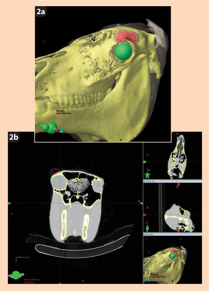
3 protection to the medical personnel delivering the treatment. Modern HDR brachytherapy is possible due to availability of high specific activity Iridium-192 sources and developments in computer-controlled afterloading technology in the 1980s and 1990s. Similar concerns regarding radiation exposure exist for LDR brachytherapy in veterinary medicine and, as a result, LDR interstitial brachytherapy in horses is not permitted in some states and facilities in the US. In the UK, interstitial LDR brachytherapy is still used in a few facilities. The Animal Health Trust (AHT) is commissioning the Varian GammaMedplus ix afterloader ( Figure 1 ), which will allow HDR brachytherapy to be performed in veterinary patients. HDR brachytherapy will be performed in the radiation protection bunker of The Kennel Club Cancer Centre recently opened at the AHT. Equine stocks have been incorporated into the bunker so HDR brachytherapy can also be performed in horses under sedation. Cross-sectional imaging The most recent development in the field of brachytherapy has been the integration of crosssectional imaging into 3D treatment planning. Sophisticated, 3D cross-sectional image-based treatment planning utilising ultrasound, CT or MRI became established as a standard practice for human external beam radiation therapy in the 1980s and 1990s. Such computerised planning is now being used in HDR brachytherapy and allows more precise delivery of the radiation dose. The afterloader uses brachytherapy treatment planning software showing the exact location of the pre-placed treatment delivery catheters. It may provide crosssectional images for 3D reconstruction or 2D radiographic film. Using various contouring tools the gross tumour volume (GTV) is delineated ( Figure 2 ). The GTV represents the tumour, which can be grossly visualised, palpated or seen using imaging. The region to be treated is referred to as the planning target volume (PTV; Figure 2 ), which encloses the GTV with a margin (usually 5mm to 10mm in brachytherapy) to account for set-up errors, as well as physiological motion and fluctuations in the size and shape of organs. The contouring tools are also used to define adjacent normal structures and organs at risk (OAR; Figure 2 ). The OARs are those more sensitive to radiation and, in the case of periocular tumours, this includes the lens, cornea, lacrimal gland, retina and optic nerve. The location of the treatment catheters is then incorporated into the treatment plan so the treatment may be simulated ( Figure 3 ). The isodose lines, which show the distribution of the radiation dose around the source, are calculated by the treatment planning software and this may be viewed either in 2D or 3D ( Figure 3 ). After the computer completes the initial calculations, the clinician (dosimetrist) can customise the radiation doses to conform to the target volume (PTV) while minimising the doses to the nearby normal tissues (particularly the OARs). Once the treatment plan has been approved by the physician, the computer transfers the treatment 3 / 9
4 plan instructions to the afterloader. The programmed instructions tell the afterloader where to direct the radioactive source, which is on the end of a guide wire, and how long it will stay (dwell time) in each position (dwell positions). The resultant distribution of radiation is shown by the isodose lines ( Figure 3 ). For equine HDR interstitial brachytherapy at the AHT the horse will be sedated and placed in stocks in the radiation bunker. The treatment catheters, which have been pre-placed through the tumour, are then connected to the HDR afterloader. All staff leave the bunker and the horse is continually monitored through closed circuit TV monitors. The treatment takes five to 10 minutes depending on the size and complexity of the implant and the activity of the source. At the end of treatment the radiation source is retracted back into the HDR afterloader. Due to financial constraints it is not feasible to perform advanced imaging such as CT on all horses with periocular neoplasms. A study is under way to compare the accuracy of using 2D radiographic film against 3D planning. This will potentially provide information to improve the efficacy of treatment using 2D planning. Other applications of HDR brachytherapy include equine skin tumours at non-periocular locations and a spectrum of neoplastic conditions in small animals, including injection site sarcomas in cats, anal gland adenocarcinomas in dogs, and nasal tumours in dogs and cats. References Byam-Cook K L, Henson F M D and Slater J D (2006). Treatment of periocular and nonocular sarcoids in 18 horses by interstitial brachytherapy with iridium-192, The Veterinary Record 159(11): Gilger B C and Stoppini R (2005). Diseases of the eyelids, conjunctiva, and nasolacrimal system. In Gilger B C (ed), Equine Ophthalmology, Elsevier Saunders, St Louis, Missouri: Knottenbelt D C and Kelly D F (2000). The diagnosis and treatment of periorbital sarcoid in the horse: 445 cases from , Veterinary Ophthalmology 3(2): / 9
 in the radiation")
5 Figure 1. The Varian GammaMedplus ix afterloader (HDR brachytherapy) in the radiation protection bunker of The Kennel Club Cancer Centre at the Animal Health Trust. 5 / 9
6 6 / 9
7 7 / 9
 is delineated in red and the planning target volume (PTV) is shown to surround the GTV in a translucent pink.")
8 Figure 2a. A 3D colour-enhanced reconstruction. Figure 2b. A reconstructed 2D transverse slice reconstruction of a horse skull in the brachytherapy treatment planning software. Using various contouring tools, the gross tumour volume (GTV) is delineated in red and the planning target volume (PTV) is shown to surround the GTV in a translucent pink. The GTV represents the tumour, which can be grossly visualised, palpated or seen using imaging, while the region to be treated encloses the GTV with a margin (usually 5mm to 10mm in brachytherapy) to account for set-up errors as well as physiological motion and fluctuations in the size and shape of organs. The bone is shown in yellow, while the eye, which is labelled as an organ at risk (OAR) due to its increased sensitivity to radiation, has been highlighted in green. Figure 3. The locations of the treatment catheters are entered into the planning software, which allows the treatment to be planned and simulated. 3a. 2D sagittal reconstruction shows the two 8 / 9
9 Powered by TCPDF ( treatment catheters placed through the tumour, in green. The GTV is shown in red, the surrounding PTV in pink and the eye (an OAR) in green. The PTV is surrounded by isodose lines, which show the periphery of the regions receiving 7Gy (yellow), 6.3Gy (light blue) and 3.5Gy (dark blue). The treatment catheters are shown to have distended segments that correspond to the dwell points at which the radioactive source will stop (dwell times). The radioactive source, which is on the end of a guide wire, will be pushed out along each catheter and stop at each dwell point for the dwell time determined by the software. The resultant distribution of radiation is shown by the isodose lines. 3b. 3D reconstruction shows the two treatment catheters (green), GTV (red), PTV (pink) and the eye (green). Only one isodose is shown (90 per cent isodose) in translucent blue. The dose may be displayed either in grays (Gy) or a percentage of the dose delivered with respect to a reference point. In this case the 90 per cent isodose is the same as the 6.3Gy isodose line. The 3D images can be rotated and viewed in any plane and provide a means of rapidly reviewing the distribution of radiation doses to the GTV, PTV and OARs. 9 / 9
Brachytherapy. What is brachytherapy and how is it used?
 Scan for mobile link. Brachytherapy Brachytherapy places radioactive sources inside the patient on a temporary or permanent basis to damage cancer cells DNA and destroy their ability to divide and grow.
Brachytherapy an Overview
 Brachytherapy an Overview Yakov Pipman, D Sc North Shore LIJ Health System Monterrey, Nov30-Dec1, 2007 Brachytherapy A procedure in therapeutic radiology that involves the irradiation of a target with
Brachytherapy The use of radioactive sources in close proximity to the target area for radiotherapy
 Brachytherapy The use of radioactive sources in close proximity to the target area for radiotherapy Interstitial Seven 192-Ir wires Interstitial implant for breast radiotherapy Intracavitary Three 137-Cs
How ICD-10 Affects Radiation Oncology. Presented by, Lashelle Bolton CPC, COC, CPC-I, CPMA
 How ICD-10 Affects Radiation Oncology Presented by, Lashelle Bolton CPC, COC, CPC-I, CPMA ICD-10 ICD-10-CM has added new challenges to the radiation oncology specialty. Approximately 220 ICD-9-CM codes
BRACHYTHERAPY IN HEAD & NECK CANCERS DR. GIRI G.V.
 BRACHYTHERAPY IN HEAD & NECK CANCERS DR. GIRI G.V. BASICS High dose to tumor = local control Spare normal structures i.e. salivary gland, mandible and muscles of mastication. Seed 100 Relative dose 10
Definitions. Brachytherapy in treatment of cancer. Implantation Techniques and Methods of Dose Specifications. Importance of Brachytherapy in GYN
 Implantation Techniques and Methods of Dose Specifications Brachytherapy Course Lecture V Krishna Reddy, MD, PhD Assistant Professor, Radiation Oncology Brachytherapy in treatment of cancer GYN Cervical
20 Prostate Cancer Dan Ash
 20 Prostate Cancer Dan Ash 1 Introduction Prostate cancer is a disease of ageing men for which the aetiology remains unknown. The incidence rises up to 30 to 40% in men over 80. The symptoms of localised
The objective of this lecture is to integrate our knowledge of the differences between 2D and 3D planning and apply the same to various clinical
 The objective of this lecture is to integrate our knowledge of the differences between 2D and 3D planning and apply the same to various clinical sites. The final aim will be to be able to make out these
Radiotherapy and tumours in veterinary practice: part one
 Vet Times The website for the veterinary profession https://www.vettimes.co.uk Radiotherapy and tumours in veterinary practice: part one Author : Aleksandra Marcinowska, Jane Dobson Categories : Companion
2015 Radiology Coding Survival Guide
 2015 Radiology Coding Survival Guide Chapter 31: Clinical Brachytherapy (77750-77799) Clinical brachytherapy involves applying radioelements into or around a treatment field. CPT guidelines clarify that
INTRODUCTION TO BRACHYTHERAPY, HISTORY AND INDICATIONS. Christine Haie-Meder Brachytherapy Unit
 INTRODUCTION TO BRACHYTHERAPY, HISTORY AND INDICATIONS Christine Haie-Meder Brachytherapy Unit HISTORY OF BRACHYTHERAPY 8/11/1895 Röentgen : X-ray discovery 22/11/1895 Mrs Röentgen s hand X-ray 1896 Becquerel
Regulatory Guidelines and Computational Methods for Safe Release of Radioactive Patients II. Brachytherapy
 Regulatory Guidelines and Computational Methods for Safe Release of Radioactive Patients II. Brachytherapy Firas Mourtada, Ph.D., DABR Chief of Clinical Physics Helen F. Graham Cancer Center Christiana
MultiSource. for a higher standard in HDR brachytherapy.
 MultiSource for a higher standard in HDR brachytherapy www.ibt-bebig.eu MultiSource HDR afterloader Multiple use: The MultiSource radiation sources have two things in common a small size and a favourable
DOSIMETRIC OPTIONS AND POSSIBILITIES OF PROSTATE LDR BRACHYTHERAPY WITH PERMANENT I-125 IMPLANTS
 DOSIMETRIC OPTIONS AND POSSIBILITIES OF PROSTATE LDR BRACHYTHERAPY WITH PERMANENT I-125 IMPLANTS Andrius IVANAUSKAS*, Eduardas ALEKNAVIČIUS*, Arvydas BURNECKIS*, Albert MILLER *Institute of Oncology Vilnius
3D ANATOMY-BASED PLANNING OPTIMIZATION FOR HDR BRACHYTHERAPY OF CERVIX CANCER
 SAUDI JOURNAL OF OBSTETRICS AND GYNECOLOGY VOLUME 11 NO. 2 1430 H - 2009 G 3D ANATOMY-BASED PLANNING OPTIMIZATION FOR HDR BRACHYTHERAPY OF CERVIX CANCER DR YASIR BAHADUR 1, DR CAMELIA CONSTANTINESCU 2,
MEDICAL POLICY. SUBJECT: BRACHYTHERAPY OR RADIOACTIVE SEED IMPLANTATION FOR PROSTATE CANCER POLICY NUMBER: CATEGORY: Technology Assessment
 MEDICAL POLICY SUBJECT: BRACHYTHERAPY OR PAGE: 1 OF: 5 If the member's subscriber contract excludes coverage for a specific service it is not covered under that contract. In such cases, medical policy
I. Equipments for external beam radiotherapy
 I. Equipments for external beam radiotherapy 5 linear accelerators (LINACs): Varian TrueBeam 6, 10 & 18 MV photons, 6-18 MeV electrons, image-guided (IGRT) and intensity modulated radiotherapy (IMRT),
Defining Target Volumes and Organs at Risk: a common language
 Defining Target Volumes and Organs at Risk: a common language Eduardo Rosenblatt Section Head Applied Radiation Biology and Radiotherapy (ARBR) Section Division of Human Health IAEA Objective: To introduce
Varian Acuity BrachyTherapy Suite One Room Integrated Image-Guided Brachytherapy
 Varian Acuity BrachyTherapy Suite One Room Integrated Image-Guided Brachytherapy The Acuity BrachyTherapy Suite Integrating Imaging, Planning, and Treatment in a Single Room Each component draws on the
REVISITING ICRU VOLUME DEFINITIONS. Eduardo Rosenblatt Vienna, Austria
 REVISITING ICRU VOLUME DEFINITIONS Eduardo Rosenblatt Vienna, Austria Objective: To introduce target volumes and organ at risk concepts as defined by ICRU. 3D-CRT is the standard There was a need for a
GYNECOLOGIC CANCER and RADIATION THERAPY. Jon Anders M.D. Radiation Oncology
 GYNECOLOGIC CANCER and RADIATION THERAPY Jon Anders M.D. Radiation Oncology Brachytherapy Comes from the Greek brakhus meaning short Brachytherapy is treatment at short distance Intracavitary vs interstitial
A Comparison of IMRT and VMAT Technique for the Treatment of Rectal Cancer
 A Comparison of IMRT and VMAT Technique for the Treatment of Rectal Cancer Tony Kin Ming Lam Radiation Planner Dr Patricia Lindsay, Radiation Physicist Dr John Kim, Radiation Oncologist Dr Kim Ann Ung,
3D CONFORMATIONAL INTERSTITIAL BRACHYTHERAPY PLANNING FOR SOFT TISSUE SARCOMA
 3D CONFORMATIONAL INTERSTITIAL BRACHYTHERAPY PLANNING FOR SOFT TISSUE SARCOMA Alina TĂNASE 1,3, M. DUMITRACHE 2,3, O. FLOREA 1 1 Emergency Central Military Hospital Dr. Carol Davila Bucharest, Romania,
Brachytherapy Planning and Quality Assurance w Classical implant systems and modern computerized dosimetry w Most common clinical applications w
 Brachytherapy Planning and Quality Assurance w Classical implant systems and modern computerized dosimetry w Most common clinical applications w Quality assurance Classical implant systems w Manchester
Brachytherapy Planning and Quality Assurance
 Brachytherapy Planning and Quality Assurance Classical implant systems Most common clinical applications and modern dosimetry methods Quality assurance Classical implant systems Manchester (Paterson-Parker)
MEDICAL MANAGEMENT POLICY
 PAGE: 1 of 8 This medical policy is not a guarantee of benefits or coverage, nor should it be deemed as medical advice. In the event of any conflict concerning benefit coverage, the employer/member summary
MEDICAL POLICY SUBJECT: BRACHYTHERAPY OR RADIOACTIVE SEED IMPLANTATION FOR PROSTATE CANCER
 MEDICAL POLICY SUBJECT: BRACHYTHERAPY OR PAGE: 1 OF: 6 If the member's subscriber contract excludes coverage for a specific service it is not covered under that contract. In such cases, medical policy
PROSTATE CANCER BRACHYTHERAPY. Kazi S. Manir MD,DNB,PDCR RMO cum Clinical Tutor Department of Radiotherapy R. G. Kar Medical College
 PROSTATE CANCER BRACHYTHERAPY Kazi S. Manir MD,DNB,PDCR RMO cum Clinical Tutor Department of Radiotherapy R. G. Kar Medical College Risk categorization Very Low Risk Low Risk Intermediate Risk High Risk
NIA MAGELLAN HEALTH RADIATION ONCOLOGY CODING STANDARD. Dosimetry Planning
 NIA MAGELLAN HEALTH RADIATION ONCOLOGY CODING STANDARD Dosimetry Planning CPT Codes: 77295, 77300, 77301, 77306, 77307, 77321, 77316, 77317, 77318, 77331, 77399 Original Date: April, 2011 Last Reviewed
FEE RULES RADIATION ONCOLOGY FEE SCHEDULE CONTENTS
 Tel: +27-21-9494060 Fax: +27-21-9494112 E-mail: leon.gouws@cancercare.co.za FEE RULES RADIATION ONCOLOGY FEE SCHEDULE CONTENTS 1. EXTERNAL BEAM RADIATION... 2 2. PLANNING OF TREATMENT... 2 3. DELIVERY
Comprehensive and Practical Brachytherapy March 04-8 March 2018, Ljubljana, Slovenia Day 1 Sunday 4 March 2018
 Day 1 Sunday 4 March 2018 Welcome and Summary of the course 13:00-13:20 15 introduction 13:20-13:50 30 Radioactivity Radioactivity: What we need to know Characteristics of LDR-PDR-HDR, radiobiological
JOURNAL OF APPLIED CLINICAL MEDICAL PHYSICS, VOLUME 6, NUMBER 2, SPRING 2005
 JOURNAL OF APPLIED CLINICAL MEDICAL PHYSICS, VOLUME 6, NUMBER 2, SPRING 2005 Advantages of inflatable multichannel endorectal applicator in the neo-adjuvant treatment of patients with locally advanced
Changing Paradigms in Radiotherapy
 Changing Paradigms in Radiotherapy Marco van Vulpen, MD, PhD Mouldroomdag-2015 Towards the elimination of invasion 1 NIH opinion on the future of oncology Twenty-five years from now,i hope that we won
Radiotherapy physics & Equipments
 Radiotherapy physics & Equipments RAD 481 Lecture s Title: An Overview of Radiation Therapy for Health Care Professionals Dr. Mohammed Emam Vision :IMC aspires to be a leader in applied medical sciences,
Page 1. Helical (Spiral) Tomotherapy. UW Helical Tomotherapy Unit. Helical (Spiral) Tomotherapy. MVCT of an Anesthetized Dog with a Sinus Tumor
 Tomotherapy. UW Helical Tomotherapy Unit. Helical (Spiral) Tomotherapy. MVCT of an Anesthetized Dog with a Sinus Tumor") Helical (Spiral) Tomotherapy Novel Clinical Applications of IMRT Linac Ring Gantry CT Detector X-Ray Fan Beam Binary Multileaf Collimator Binary MLC Leaves James S Welsh, MS, MD Department of Human Oncology
Helical (Spiral) Tomotherapy Novel Clinical Applications of IMRT Linac Ring Gantry CT Detector X-Ray Fan Beam Binary Multileaf Collimator Binary MLC Leaves James S Welsh, MS, MD Department of Human Oncology
Basics of Cervix Brachytherapy. William Small, Jr., MD Professor and Chairman Loyola University Chicago
 Gynecologic Cancer InterGroup Cervix Cancer Research Network Basics of Cervix Brachytherapy William Small, Jr., MD Professor and Chairman Loyola University Chicago Cervix Cancer Education Symposium, January
Radiotherapy in feline and canine head and neck cancer
 Bettina Kandel Like surgery radiotherapy is usually a localized type of treatment. Today it is more readily available for the treatment of cancer in companion animals and many clients are well informed
Goals and Objectives: Head and Neck Cancer Service Department of Radiation Oncology
 Goals and Objectives: Head and Neck Cancer Service Department of Radiation Oncology The head and neck cancer service provides training in the diagnosis, management, treatment, and follow-up care of head
Trina Lynd, M.S. Medical Physicist Lifefirst Imaging & Oncology Cullman, AL Tri-State Alabama, Louisiana and Mississippi Spring 2016 Meeting April
 Trina Lynd, M.S. Medical Physicist Lifefirst Imaging & Oncology Cullman, AL Tri-State Alabama, Louisiana and Mississippi Spring 2016 Meeting April 17, 2016 Discuss permanent prostate brachytherapy and
Regions Hospital Delineation of Privileges Radiation Oncology
 Regions Hospital Delineation of Privileges Radiation Oncology Applicant s Last First M. Instructions: Place a check-mark where indicated for each core group you are requesting. Review education and basic
IMRT - the physician s eye-view. Cinzia Iotti Department of Radiation Oncology S.Maria Nuova Hospital Reggio Emilia
 IMRT - the physician s eye-view Cinzia Iotti Department of Radiation Oncology S.Maria Nuova Hospital Reggio Emilia The goals of cancer therapy Local control Survival Functional status Quality of life Causes
The Evolution of RT Techniques for Gynaecological Cancers in a developing country context
 The Evolution of RT Techniques for Gynaecological Cancers in a developing country context Hannah Simonds Stellenbosch University/ Tygerberg Academic Hospital ESMO Africa 2017 I have no disclosures External
 https://patient.varian.com/sit es/default/files/videos/origin al/imrt.mp4 brachy- from Greek brakhys "short" Historically LDR has been used. Cs-137 at 0.4-0.8 Gy/h With optimally placed device, dose
NCCN GUIDELINES ON PROTON THERAPY (AS OF 4/23/18) BONE (Version , 03/28/18)
 BONE (Version , 03/28/18)") BONE (Version 2.2018, 03/28/18) NCCN GUIDELINES ON PROTON THERAPY (AS OF 4/23/18) Radiation Therapy Specialized techniques such as intensity-modulated RT (IMRT); particle beam RT with protons, carbon ions,
BONE (Version 2.2018, 03/28/18) NCCN GUIDELINES ON PROTON THERAPY (AS OF 4/23/18) Radiation Therapy Specialized techniques such as intensity-modulated RT (IMRT); particle beam RT with protons, carbon ions,
Real-time brachytherapy for prostate cancer implant analysis
 Rep Pract Oncol Radiother, 2008; 13(1): 9-14 Original Paper Received: 2007.04.17 Accepted: 2008.02.07 Published: 2008.02.29 Authors Contribution: A Study Design B Data Collection C Statistical Analysis
Medical Use of Radioisotopes
 Medical Use of Radioisotopes Therapy Radioisotopes prove to be useful in the application of brachytherapy, the procedure for using temporary irradiation close to the area of disease (i.e. cancer) 10% Medical
ADVANCED TECHNOLOGY CONSORTIUM (ATC) CREDENTIALING PROCEDURES FOR LUNG BRACHYTHERAPY IMPLANT PROTOCOLS
 CREDENTIALING PROCEDURES FOR LUNG BRACHYTHERAPY IMPLANT PROTOCOLS") ACOSOG-RTOG Lung Brachytherapy QA Page 1 of 8 ADVANCED TECHNOLOGY CONSORTIUM (ATC) CREDENTIALING PROCEDURES FOR LUNG BRACHYTHERAPY IMPLANT PROTOCOLS FACILITY QUESTIONNAIRE Institutions wishing to enter
ACOSOG-RTOG Lung Brachytherapy QA Page 1 of 8 ADVANCED TECHNOLOGY CONSORTIUM (ATC) CREDENTIALING PROCEDURES FOR LUNG BRACHYTHERAPY IMPLANT PROTOCOLS FACILITY QUESTIONNAIRE Institutions wishing to enter
Radiotherapy Of Prostate Cancer
 Radiotherapy Of Prostate Cancer 1 / 6 2 / 6 3 / 6 Radiotherapy Of Prostate Cancer We compared active monitoring, radical prostatectomy, and external-beam radiotherapy for the treatment of clinically localized
Interim Report. Prostate Cancer Fight Foundation Motorcycle Ride for Dad. February 7, Supporting Kingston s university hospitals:
 Interim Report Prostate Cancer Fight Foundation Motorcycle Ride for Dad February 7, 2014 Supporting Kingston s university hospitals: Thank you for your continued support of prostate cancer research in
Therapeutic Medical Physics. Stephen J. Amadon Jr., Ph.D., DABR
 Therapeutic Medical Physics Stephen J. Amadon Jr., Ph.D., DABR Outline 1. Why physicists are needed in medicine 2. Branches of medical physics 3. Physics in Radiation Oncology 4. Treatment types and Treatment
Klinikleitung: Dr. Kessler Dr. Kosfeld Dr. Tassani-Prell Dr. Bessmann. Radiotherapy in feline and canine head and neck cancer.
 Radiotherapy in feline and canine head and neck cancer Bettina Kandel Like surgery radiotherapy is usually a localized type of treatment. Today it is more readily available for the treatment of cancer
Rola brachyterapii w leczeniu wznów nowotworów języka i dna jamy ustnej. The role of brachytherapy in recurrent. oral cavity
 Rola brachyterapii w leczeniu wznów nowotworów języka i dna jamy ustnej The role of brachytherapy in recurrent tumours of the tongue and fundus of the oral cavity Janusz Skowronek, MD, PhD, Ass. Prof.
Brachytherapy, Noncoronary
 Brachytherapy, Noncoronary Policy Number: Original Effective Date: MM.05.004 05/10/2005 Line(s) of Business: Current Effective Date: HMO; PPO; QUEST Integration 06/01/2017 Section: Radiology Place(s) of
National System for Incident Reporting in Radiation Therapy (NSIR-RT) Taxonomy
 Taxonomy") Canadian Partnership for Quality Radiotherapy (CPQR) National System for Incident Reporting in Radiation Therapy (NSIR-RT) National System for Incident Reporting in Radiation Therapy (NSIR-RT) Taxonomy
Canadian Partnership for Quality Radiotherapy (CPQR) National System for Incident Reporting in Radiation Therapy (NSIR-RT) National System for Incident Reporting in Radiation Therapy (NSIR-RT) Taxonomy
TRANSRECTAL ULTRASOUND-GUIDED PROSTATE BRACHYTHERAPY
 TRANSRECTAL ULTRASOUND-GUIDED PROSTATE BRACHYTHERAPY 1 TRANSRECTAL ULTRASOUND-GUIDED PROSTATE BRACHYTHERAPY BRENDAN CAREY, MD TRANSRECTAL ULTRASOUND-GUIDED PROSTATE BRACHYTHERAPY 2 TRANSRECTAL ULTRASOUND-GUIDED
Dosimetric Analysis of 3DCRT or IMRT with Vaginal-cuff Brachytherapy (VCB) for Gynaecological Cancer
 for Gynaecological Cancer") Dosimetric Analysis of 3DCRT or IMRT with Vaginal-cuff Brachytherapy (VCB) for Gynaecological Cancer Tan Chek Wee 15 06 2016 National University Cancer Institute, Singapore Clinical Care Education Research
Dosimetric Analysis of 3DCRT or IMRT with Vaginal-cuff Brachytherapy (VCB) for Gynaecological Cancer Tan Chek Wee 15 06 2016 National University Cancer Institute, Singapore Clinical Care Education Research
Chapters from Clinical Oncology
 Chapters from Clinical Oncology Lecture notes University of Szeged Faculty of Medicine Department of Oncotherapy 2012. 1 RADIOTHERAPY Technical aspects Dr. Elemér Szil Introduction There are three possibilities
Understanding Radiation Therapy. For Patients and the Public
 Understanding Radiation Therapy For Patients and the Public Introduction to Radiation Oncology Radiation has been an effective tool for treating cancer for more than 100 years. Radiation oncologists are
Quality Assurance of Ultrasound Imaging in Radiation Therapy. Zuofeng Li, D.Sc. Murty S. Goddu, Ph.D. Washington University St.
 Quality Assurance of Ultrasound Imaging in Radiation Therapy Zuofeng Li, D.Sc. Murty S. Goddu, Ph.D. Washington University St. Louis, Missouri Typical Applications of Ultrasound Imaging in Radiation Therapy
Brachytherapy, Noncoronary
 Brachytherapy, Noncoronary Policy Number: Original Effective Date: MM.05.004 05/10/2005 Line(s) of Business: Current Effective Date: HMO; PPO; QUEST Integration 03/01/2016 Section: Radiology Place(s) of
PROGRESS IN RADIATION ONCOLOGY. Fox Chase Cancer Center. Jean Holland RN MSN AOCN Department of Radiation Oncology April 2017
 PROGRESS IN RADIATION ONCOLOGY Fox Chase Cancer Center Jean Holland RN MSN AOCN Department of Radiation Oncology April 2017 Radiation Therapy Therapeutic use of electromagnetic energy from a machine or
Initial experience with high dose rate brachytherapy of periorbital sarcoids in the horse
 EQUINE VETERINARY EDUCATION Equine vet. Educ. (2017) () - doi: 10.1111/eve.12782 1 Original Article Initial experience with high dose rate brachytherapy of periorbital sarcoids in the horse A. R. Hollis*
Prostate Cancer Treatment
 Scan for mobile link. Prostate Cancer Treatment Prostate cancer overview Prostate cancer is the most common form of cancer in American men, most prevalent in men over age 65 and fairly common in men 50-64
A Study on Dosimetry of Gynaecological Cancer and Quality Assurance of HDR Brachytherapy in BPKMCH, Nepal
 A Study on Dosimetry of Gynaecological Cancer and Quality Assurance of HDR Brachytherapy in BPKMCH, Nepal SB Chand, PP Chaursia, MP Adhikary, AK Jha, and S Shrestha Dept. of Radiation Oncology, BPKM Cancer
WN MEDICAL IMAGING. RADIOTHERAPY. MEDICAL PHYSICS. NUCLEAR MEDICINE. RADIOACTIVITY
 WN MEDICAL IMAGING. RADIOTHERAPY. MEDICAL PHYSICS. NUCLEAR MEDICINE. RADIOACTIVITY Definitions : (from various online encyclopaedias/dictionaries) Medical Imaging/Radiology/ Diagnostic imaging: the use
Review of brachytherapy in
 Medical Physics in the Baltic States 2017 Review of brachytherapy in Klaipėda University Hospital Romas Vilkas Rasa Dagienė Klaipėda University Hospital 1 Aims of presentation: To show progress of brachytherapy
ICRT รศ.พญ.เยาวล กษณ ชาญศ ลป
 ICRT รศ.พญ.เยาวล กษณ ชาญศ ลป Brachytherapy การร กษาด วยร งส ระยะใกล Insertion การสอดใส แร Implantation การฝ งแร Surface application การวางแร physical benefit of brachytherapy - very high dose of radiation
Corporate Medical Policy
 Corporate Medical Policy Intensity Modulated Radiation Therapy (IMRT) of Head and Neck File Name: Origination: Last CAP Review: Next CAP Review: Last Review: intensity_modulated_radiation_therapy_imrt_of_head_and_neck
HDR vs. LDR Is One Better Than The Other?
 HDR vs. LDR Is One Better Than The Other? Daniel Fernandez, MD, PhD 11/3/2017 New Frontiers in Urologic Oncology Learning Objectives Indications for prostate brachytherapy Identify pros/cons of HDR vs
Outline - MRI - CT - US. - Combinations of imaging modalities for treatment planning
 Imaging Outline - MRI - CT - US - Combinations of imaging modalities for treatment planning Imaging Part 1: MRI MRI for cervical cancer high soft tissue contrast multiplanar imaging MRI anatomy: the normal
Wireless In-Vivo Dosimetry of High Dose Rate Brachytherapy for Prostate Cancer using MOSkin Detectors
 University of Wollongong Research Online University of Wollongong Thesis Collection 2017+ University of Wollongong Thesis Collections 2018 Wireless In-Vivo Dosimetry of High Dose Rate Brachytherapy for
Evaluation of Whole-Field and Split-Field Intensity Modulation Radiation Therapy (IMRT) Techniques in Head and Neck Cancer
 Techniques in Head and Neck Cancer") 1 Charles Poole April Case Study April 30, 2012 Evaluation of Whole-Field and Split-Field Intensity Modulation Radiation Therapy (IMRT) Techniques in Head and Neck Cancer Abstract: Introduction: This study
1 Charles Poole April Case Study April 30, 2012 Evaluation of Whole-Field and Split-Field Intensity Modulation Radiation Therapy (IMRT) Techniques in Head and Neck Cancer Abstract: Introduction: This study
Recent proceedings in Brachytherapy Physics
 Recent proceedings in Brachytherapy Physics Frank-André Siebert UKSH, Campus Kiel, Germany Clinic of Radiotherapy Dept. Medical Physics Physical characteristics of brachytherapy (Courtesy Luc Beaulieu,
Evaluation of Normal Tissue Complication Probability and Risk of Second Primary Cancer in Prostate Radiotherapy
 Evaluation of Normal Tissue Complication Probability and Risk of Second Primary Cancer in Prostate Radiotherapy Rungdham Takam Thesis submitted for the degree of Doctor of Philosophy in The School of Chemistry
Chapter 13: Brachytherapy: Physical and Clinical Aspects
 Chapter 13: Brachytherapy: Physical and Clinical Aspects Set of 163 slides based on the chapter authored by N. Suntharalingam, E.B. Podgorsak, H. Tolli of the IAEA publication (ISBN 92-0-107304-6): Radiation
The Physics of Oesophageal Cancer Radiotherapy
 The Physics of Oesophageal Cancer Radiotherapy Dr. Philip Wai Radiotherapy Physics Royal Marsden Hospital 1 Contents Brief clinical introduction Imaging and Target definition Dose prescription & patient
Corporate Medical Policy
 Corporate Medical Policy Intensity Modulated Radiation Therapy (IMRT) of Abdomen and File Name: Origination: Last CAP Review: Next CAP Review: Last Review: intensity_modulated_radiation_therapy_imrt_of_abdomen_and_pelvis
Clinical Applications of Brachytherapy Radiobiology. Radiobiology is Essential
 Clinical Applications of Brachytherapy Radiobiology Dr Alexandra Stewart University of Surrey St Luke s Cancer Centre Guildford, England Radiobiology is Essential Knowledge of radiobiological principles
Quality management for Breast Brachytherapy.
 Quality management for Breast Brachytherapy. DORIN TODOR, Ph.D. Medical College of Virginia Campus Department of Radiation Oncology New England AAPM Chapter 2012 Summer Meeting, Providence, RI Quality
Intensity Modulated Radiation Therapy (IMRT)
") Intensity Modulated Radiation Therapy (IMRT) Policy Number: Original Effective Date: MM.05.006 03/09/2004 Line(s) of Business: Current Effective Date: HMO; PPO; QUEST Integration 03/01/2015 Section: Radiology
Intensity Modulated Radiation Therapy (IMRT) Policy Number: Original Effective Date: MM.05.006 03/09/2004 Line(s) of Business: Current Effective Date: HMO; PPO; QUEST Integration 03/01/2015 Section: Radiology
Implementation of advanced RT Techniques
 Implementation of advanced RT Techniques Tibor Major, PhD National Institute of Oncology Budapest, Hungary 2. Kongres radiološke tehnologije, Vukovar, 23-25. September 2016. Current RT equipments at NIO,
Abstract Purpose: Material and methods: Results: Conclusions: Key words: Purpose Address for correspondence:
 Original paper Physics Contributions The impact of activating source dwell positions outside the CTV on the dose to treated normal tissue volumes in TRUS guided 3D conformal interstitial HDR brachytherapy
Can we deliver the dose distribution we plan in HDR-Brachytherapy of Prostate Cancer?
 Can we deliver the dose distribution we plan in HDR-Brachytherapy of Prostate Cancer? Dimos Baltas Dept. of Medical Physics & Engineering, Strahlenklinik, Klinikum Offenbach GmbH 63069 Offenbach, Germany
Prostate Cancer. What is prostate cancer?
 Scan for mobile link. Prostate Cancer Prostate cancer is a tumor of the prostate gland, which is located in front of the rectum and below the bladder. Your doctor may perform a physical exam, prostate-specific
Stereotactic MR-guided adaptive radiation therapy (SMART) for locally advanced pancreatic tumors
 for locally advanced pancreatic tumors") Stereotactic MR-guided adaptive radiation therapy (SMART) for locally advanced pancreatic tumors Anna Bruynzeel, Radiation Oncologist VU University Medical Center, Amsterdam, The Netherlands Current standard
Stereotactic MR-guided adaptive radiation therapy (SMART) for locally advanced pancreatic tumors Anna Bruynzeel, Radiation Oncologist VU University Medical Center, Amsterdam, The Netherlands Current standard
THE TRANSITION FROM 2D TO 3D AND TO IMRT - RATIONALE AND CRITICAL ELEMENTS
 THE TRANSITION FROM 2D TO 3D AND TO IMRT - RATIONALE AND CRITICAL ELEMENTS ICTP SCHOOL ON MEDICAL PHYSICS FOR RADIATION THERAPY DOSIMETRY AND TREATMENT PLANNING FOR BASIC AND ADVANCED APPLICATIONS March
Corporate Medical Policy
 Corporate Medical Policy Intensity-Modulated Radiation Therapy (IMRT) of the Prostate File Name: Origination: Last CAP Review: Next CAP Review: Last Review: intensity_modulated_radiation_therapy_imrt_of_the_prostate
The Canadian National System for Incident Reporting in Radiation Treatment (NSIR-RT) Taxonomy March 11, 2015
 Taxonomy March 11, 2015") The Canadian National System for Incident Reporting in Radiation Treatment (NSIR-RT) Taxonomy March 11, 2015 Taxonomy Data Category Number Description Data Fields and Menu Choices 1. Impact 1.1 Incident
The Canadian National System for Incident Reporting in Radiation Treatment (NSIR-RT) Taxonomy March 11, 2015 Taxonomy Data Category Number Description Data Fields and Menu Choices 1. Impact 1.1 Incident
IAEA-TECDOC Implementation of microsource high dose rate (mhdr) brachytherapy in developing countries
 brachytherapy in developing countries") IAEA-TECDOC-1257 Implementation of microsource high dose rate (mhdr) brachytherapy in developing countries November 2001 The originating Section of this publication in the IAEA was: Applied Radiation Biology
IAEA-TECDOC-1257 Implementation of microsource high dose rate (mhdr) brachytherapy in developing countries November 2001 The originating Section of this publication in the IAEA was: Applied Radiation Biology
INTRODUCTION PATIENT. J. Radiat. Res., 52, (2011)
") J. Radiat. Res., 52, 54 58 (2011) Regular Paper Intracavitary Combined with CT-guided Interstitial Brachytherapy for Locally Advanced Uterine Cervical Cancer: Introduction of the Technique and a Case Presentation
J. Radiat. Res., 52, 54 58 (2011) Regular Paper Intracavitary Combined with CT-guided Interstitial Brachytherapy for Locally Advanced Uterine Cervical Cancer: Introduction of the Technique and a Case Presentation
BLADDER RADIOTHERAPY PLANNING DOCUMENT
 A 2X2 FACTORIAL RANDOMISED PHASE III STUDY COMPARING STANDARD VERSUS REDUCED VOLUME RADIOTHERAPY WITH AND WITHOUT SYNCHRONOUS CHEMOTHERAPY IN MUSCLE INVASIVE BLADDER CANCER (ISRCTN 68324339) BLADDER RADIOTHERAPY
Intensity modulated radiotherapy (IMRT) for treatment of post-operative high grade glioma in the right parietal region of brain
 for treatment of post-operative high grade glioma in the right parietal region of brain") 1 Carol Boyd March Case Study March 11, 2013 Intensity modulated radiotherapy (IMRT) for treatment of post-operative high grade glioma in the right parietal region of brain History of Present Illness:
1 Carol Boyd March Case Study March 11, 2013 Intensity modulated radiotherapy (IMRT) for treatment of post-operative high grade glioma in the right parietal region of brain History of Present Illness:
Efficient SIB-IMRT planning of head & neck patients with Pinnacle 3 -DMPO
 Investigations and research Efficient SIB-IMRT planning of head & neck patients with Pinnacle 3 -DMPO M. Kunze-Busch P. van Kollenburg Department of Radiation Oncology, Radboud University Nijmegen Medical
The future of radiation therapy. Safe and innovative options, including the CyberKnife System
 The future of radiation therapy Safe and innovative options, including the CyberKnife System Could a nonsurgical treatment be right for you? When you ve been diagnosed with a cancerous or noncancerous
First, how does radiation work?
 Hello, I am Prajnan Das, Faculty Member in the Department of Radiation Oncology at The University of Texas MD Anderson Cancer Center. We are going to talk today about some of the basic principles regarding
Brachytherapy, Noncoronary
 Brachytherapy, Noncoronary Policy Number: Original Effective Date: MM.05.004 05/10/2005 Line(s) of Business: Current Effective Date: HMO; PPO; QUEST Integration 8/29/2018 Section: Radiology Place(s) of
Image based Brachytherapy- HDR applications in Gynecological Tumors
 Image based Brachytherapy- HDR applications in Gynecological Tumors Yakov Pipman, D. Sc. North Shore LIJ Health System Sites amenable to treatment with HDR Brachytherapy GYN Breast Prostate Head and Neck
Pulsed Dose Rate for GYN Brachytherapy
 Pulsed Dose Rate for GYN Brachytherapy Firas Mourtada,, Ph.D. Department of Radiation Physics Dose equivalency to LDR Brief Introduction Radiobiology Dose Distribution: Radial dose function Source anisotropy
Quality assurance and credentialing requirements for sites using inverse planned IMRT Techniques
 TROG 08.03 RAVES Quality assurance and credentialing requirements for sites using inverse planned IMRT Techniques Introduction Commissioning and quality assurance of planning systems and treatment delivery
Patient Safety Focused QA. LDR Brachytherapy Vrinda Narayana
 Patient Safety Focused QA LDR Brachytherapy Vrinda Narayana D < 2 Gy/h Old LDR Brachytherapy? Ra-226; Cs-137; Ir-192 New Gynecological; interstitial Pd-103; I-125; Cs-131 Prostate implants Eye plaques