Analysis of MRI Volumetric Changes After Hypofractionated Stereotactic Radiotherapy for Benign Intracranial Neoplasms
|
|
|
- Anabel Lloyd
- 5 years ago
- Views:
Transcription

1 Accepted Manuscript Analysis of MRI Volumetric Changes After Hypofractionated Stereotactic Radiotherapy for Benign Intracranial Neoplasms Kathryn R. Fega, MD, PhD, Geoffrey P. Fletcher, MD, Mark R. Waddle, MD, Jennifer L. Peterson, MD, Jonathan B. Ashman, MD, PhD, David M. Barrs, MD, Bernard R. Bendok, MD, Naresh P. Patel, MD, Alyx B. Porter, MD, Sujay A. Vora, MD PII: DOI: Reference: ADRO 235 S (18) /j.adro To appear in: Advances in Radiation Oncology Received Date: 8 December 2017 Revised Date: 2 August 2018 Accepted Date: 10 August 2018 Please cite this article as: Fega KR, Fletcher GP, Waddle MR, Peterson JL, Ashman JB, Barrs DM, Bendok BR, Patel NP, Porter AB, Vora SA, Analysis of MRI Volumetric Changes After Hypofractionated Stereotactic Radiotherapy for Benign Intracranial Neoplasms, Advances in Radiation Oncology (2018), doi: /j.adro This is a PDF file of an unedited manuscript that has been accepted for publication. As a service to our customers we are providing this early version of the manuscript. The manuscript will undergo copyediting, typesetting, and review of the resulting proof before it is published in its final form. Please note that during the production process errors may be discovered which could affect the content, and all legal disclaimers that apply to the journal pertain.
2 [Category: Scientific Article] Analysis of MRI Volumetric Changes After Hypofractionated Stereotactic Radiotherapy for Benign Intracranial Neoplasms Kathryn R. Fega, MD, PhD Geoffrey P. Fletcher, MD Mark R. Waddle, MD Jennifer L. Peterson, MD Jonathan B. Ashman, MD, PhD David M. Barrs, MD Bernard R. Bendok, MD Naresh P. Patel, MD Alyx B. Porter, MD Sujay A. Vora, MD Author Affiliations: Department of Radiation Oncology (Drs Fega, Ashman, and Vora), Department of Radiology (Dr Fletcher), Emeritus Member, Department of Otolaryngology, Head and Neck Surgery (Dr Barrs), Department of Neurologic Surgery (Drs Bendok and Patel), and Department of Neurology (Dr Porter), Mayo Clinic Hospital, Phoenix, Arizona, and Department of Radiation Oncology (Drs Waddle and Peterson), Mayo Clinic, Jacksonville, Florida. Reprints: Sujay A. Vora, MD, Department of Radiation Oncology, Mayo Clinic Hospital, 5777 E Mayo Blvd, Phoenix, AZ (vora.sujay@mayo.edu). Presented at the American Society for Radiation Oncology (ASTRO) annual meeting, Boston, Massachusetts, September 25-28, 2016.
3 Role of the authors Fega: data acquisition, analysis, manuscript writing; Fletcher: revision; Waddle: revision; Peterson: revision; Ashman: revision; Barrs: revision; Bendok: revision; Patel: revision; Perter: revision; Vora: data analysis, writing. All authors have approved the final article. Conflict of interest: None. Financial Support: This research did not receive any specific grant from funding agencies in the public, commercial, or not-for-profit sectors. Text word count (includes abstract, text, references, tables, legends): 3,893 Abstract word count: 276 No. of tables: 1 No. of figures: 5 Running title: MRI Changes After SRT for Benign CNS Tumors Publisher: To expedite proof approval, send proof via to scipubs@mayo.edu Mayo Foundation for Medical Education and Research
4 -1- Analysis of MRI Volumetric Changes After Hypofractionated Stereotactic Radiotherapy for Benign Intracranial Neoplasms
5 -2- Abstract Purpose: To quantitatively assess volumetric changes after hypofractionated stereotactic radiotherapy (HFSRT) in patients treated for vestibular schwannomas and meningiomas. Methods and Materials: We retrospectively reviewed records of patients treated at our institution with HFSRT from Patients received a median dose of 25 Gy in 5 fractions. After treatment, they underwent clinical and radiologic follow-up with magnetic resonance imaging (MRI) at 3- to 12-month intervals. Gross tumor volume was outlined on each thin slice of contrast-enhanced T1 series before and on each scan after HFSRT. Volumetric changes were calculated and compared with neuroradiologist interpretations. Results: Forty-three patients underwent 182 MRI scans. Tumor types included vestibular schwannoma (n=34) and meningioma (n=9). Median follow-up time was 29 months. Median gross tumor volume was 3.1 cm 3. Local control was 81.4% for the entire cohort at the time of last follow-up. Transient volume expansion was noted in 17 patients (50%) with vestibular schwannoma and 2 (22%) with meningioma. For all patients, transient volume expansion and subsequent regression occurred at a median time of 5.5 and 12 months, respectively. Neuroradiologist agreement with regard to tumor regression, progression, or stability occurred in 155/182 total reports (85%). The largest discordance observed was a stable finding on the MRI interpretation when the measured volumetric change exceeded 20% (n=24 [13%]). Conclusion: HFSRT is associated with excellent local control and a low incidence of toxicity. With volumetric MRI measurement, transient volume
6 -3- expansion was a common finding and associated with temporary adverse effects. Although the neuroradiologist s interpretation generally agreed with the volumetric MRI measurement, the overall 15% discordance rate emphasizes the potential benefit of considering volumetric measurements, which may help clinicians correlate posttreatment symptoms with MRI findings. Keywords: benign CNS neoplasms; hypofractionated radiation therapy; MRI changes
7 -4- Abbreviations 3D, 3-dimensional CNS, central nervous system CT, computed tomography, computed tomographic HFSRT, hypofractionated stereotactic radiotherapy GTV, gross tumor volume MRI, magnetic resonance imaging PTV, planning target volume RECIST, Response Evaluation Criteria in Solid Tumors STROBE, STrengthening the Reporting of Observational Studies in Epidemiology TVE, transient volume expansion
8 -5- Introduction The management of benign meningiomas and vestibular schwannomas can be associated with unique challenges. Because these lesions are rarely life threatening, the decision to treat is often determined by current symptoms or the potential for worsening symptoms and the associated changes in quality of life. Potential treatments include surgical resection, radiotherapy, and close observation. The optimal treatment depends on tumor- and patient-related factors plus clinician and patient preferences. Surgical management remains the first-line treatment for select patients when tumor resection would improve symptoms from mass effect 1,2 or when surgery is potentially curative with gross total resection. 3,4 However, surgery may pose challenges, depending on the intracranial location of the tumor, and includes risk of complications such as infection, bleeding, and neurologic loss. Radiotherapy is an alternative to surgery, especially for patients with benign tumors that are not amenable to surgical resection or recur after surgery. 5-8 Radiotherapy options include conventionally fractionated radiotherapy, stereotactic single fraction radiosurgery, and hypofractionated stereotactic radiotherapy (HFSRT). Prior studies of conventional radiotherapy for benign central nervous system (CNS) neoplasms have shown excellent local control (>80%), but patients must undergo 5 to 6 weeks of radiotherapy Single-fraction stereotactic radiosurgery also shows excellent local control for smaller benign intracranial neoplasms, but depending on tumor-related factors (eg, location, size), they can be associated with increased risk of complications such as tumor-related
9 -6- swelling, hearing loss, and cranial neuropathies. 10,12-16 HFSRT has the ability of balancing the therapeutic ratio through fractionation and a short treatment course However, the posttreatment changes of these tumors have not been well characterized radiographically, particularly after HFSRT. Magnetic resonance imaging (MRI) is the most common method of assessing response to radiotherapy. Nevertheless, differentiating between tumor progression and radiotherapy effects can be difficult. 21 Often, the clinician relies on the radiologist s interpretation of postradiotherapy changes, but changes such as tumor-related swelling, transient volume expansion (TVE), and central necrosis can challenge the radiologist. 22 These radiotherapy effects may be more pronounced in shorter, high-dose treatments Historically, MRI-based measurements of tumor expansion or shrinkage have been interpolated by using the maximal tumor dimensions for height, width, and anterior-posterior diameter. When assessing tumor growth or shrinkage, 1- or 2-dimensional analysis may be insufficient for tumors that are irregularly shaped or multilobulated and may be subject to interobserver differences. Therefore, volumetric measurements potentially are advantageous for analyzing tumor response to radiotherapy over time. The aim of our study was to quantitatively assess volumetric changes, toxicities, and clinical outcomes after HFSRT in patients with vestibular schwannomas or meningiomas. Methods and Materials Study Design and Patient Population This study was approved by our institutional review board. The reporting of this study is in compliance with the STROBE (STrengthening the Reporting of Observational Studies in Epidemiology) statement. 26 We
10 -7- retrospectively identified all patients with vestibular schwannomas or meningiomas, who were treated at our institution (an academic tertiary medical center) with linear accelerator based HFSRT from January 1, 2002, through December 31, To meet the inclusion criteria, patients had to be at least 18 years old, with a radiographic diagnosis of a vestibular schwannoma or meningioma, and could not have received prior radiotherapy to the tumor site. Patients were required to have received HFSRT (ie, 3-5 treatments). Patients were excluded from analysis if they did not receive follow-up clinical evaluation at our institution or if they had only computed tomographic (CT) imaging. HFSRT Technique All patients were treated with a linear accelerator (Varian Medical Systems). Patient immobilization was achieved with a commercial head mask fixation system. Patients underwent a planning CT scan of the brain, and image fusion was performed with dedicated pretreatment MRI thin-cut sequences. The gross tumor volume (GTV) was delineated as a contrastenhancing tumor demonstrated on T1 postgadolinium MRI imaging. The clinical target volume was equal to the GTV. The planning target volume (PTV) was generated by the geometric expansion of GTV plus a margin (median, 2 mm; range, 0-3 mm). Patients were treated daily with image guidance. The median radiation dose was 25 Gy in 5 fractions (range, 18 Gy [3 fractions] to 35 Gy [5 fractions]). The prescribed dose was delivered to the 77% to 100% isodose line (mean, 89%) and normalized to the maximum dose to ensure coverage of at least 95% of the PTV with the prescription dose.
11 -8- Clinical and Radiologic Follow-Up After treatment, patients underwent clinical and radiologic MRI follow-up at 3- to 12-month intervals. All patients included in this study had pretreatment MRI imaging with at least 2 posttreatment MRI scans. A comprehensive clinical evaluation and neurologic examination was performed before treatment and at each clinical follow-up visit. MRI Volumetric Assessment and Response Criteria All posttreatment MRI imaging was imported into the radiation planning system and contoured by one individual (initials withheld for anonymous review).volumetric measurements were generated retrospectively by outlining the gross tumor volume, including cystic changes, on each thin-cut slice of contrast-enhanced T1 series before and on each scan after HFSRT. The thinnest cuts available (slice thickness, mm) were used for contouring. Volumetric changes were calculated for each available MRI scan and compared with the baseline tumor volume before radiotherapy. Percentage changes in size were reported as an increase, decrease, or no change compared with the original tumor volume. Modified RECIST (Response Evaluation Criteria in Solid Tumors) criteria 27 were used to define disease progression and stability. While RECIST criteria defined progression as an increase of 20% in the sum of the linear measurements, we used a volumetric approach in which 20% change in volume was considered progressive disease. Stable disease was defined as no change or a volume change <30%. Partial response to treatment was defined as at least a 30% decrease in volume. Local control was defined as either stable disease or a partial response to treatment. TVE was defined as an increase of more than 20% from baseline with a
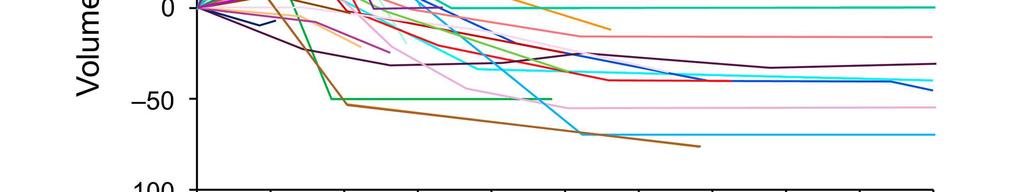
12 -9- subsequent decrease to within 20% of baseline. TVE patients were not defined as progression but categorized based on their final volume size relative to baseline. All diagnostic MRIs were read and interpreted by a member of the neuroradiology department (initials withheld for anonymous review). Tumor size was characterized as increased, decreased, or no change, based on the diameter change of the tumor measured from the coronal, sagittal, and axial cross-sections. If the neuroradiologist interpretation of a linear change agreed with our interpretation of a volumetric change (increase, decrease, or stable disease), this was considered concordant. If the report specified a change in size but the volumetric change was stable (within 20% of baseline), this was considered discordant. If a report specified no change when a volumetric change exceeding 20% was measured, this was also considered discordant. Results We identified 43 patients with 182 MRI scans who were treated with HFSRT during the study period. Baseline characteristics are shown in Table 1. The median (range) duration of follow-up was 29 (6-142) months. Figure 1 shows the final change in volume for patients with vestibular schwannoma; compared with baseline, 12 patients (35%) had a partial response to treatment, 14 (41%) remained stable, and 8 (24%) had more than 20% volumetric growth. The largest expansion was observed in patient 34, who had a very small acoustic neuroma at presentation. This lesion progressed to a volumetric change of 800%. The lesion ultimately was resected. Post-HFSRT changes in vestibular schwannoma volume over time are shown in Figure 2.
13 -10- Figure 3 shows the final change in volume for patients with meningioma; compared with baseline, 6 patients (67%) had a partial response to treatment, 3 (33%) remained stable, and no patients had a progressive expansion. Post-HFSRT changes in volume over time are shown in Figure 4. TVE was observed in 17 patients with vestibular schwannoma (50%). Median time of TVE was 6 months (range, 3-13 months), with a return to pretreatment size or decrease in size observed within a median time of 13 months (range, 6-59 months). Two patients with meningioma (22%) had TVE at a median time of 2.5 months (range, 2-3 months), with a return to pretreatment size or a decrease in size observed within a median time of 9.5 months (range, 7-12 months). TVE was associated with increased toxicity in 11 patients (65%) with vestibular schwannoma. Symptoms included decreased hearing (n=5), imbalance (n=3), dizziness (n=3), facial paresthesias (n=2), tinnitus (n=2), headache (n=2), and vertigo (n=2), with some patients experiencing multiple symptoms. Hearing loss persisted in all patients, and the other symptoms resolved for 6 patients (55%) within a median time of 6.5 months (range, 1-15 months). A total of 6 patients (30%), 5 patients with vestibular schwannoma and 1 patient with meningioma, had grade 2 toxicity requiring corticosteroids. Toxicity that included speech difficulty and weakness was seen in 1 of the 2 meningioma patients, with symptoms resolving within 12 months after radiation. Tumor volumetric assessment for each follow-up MRI was compared with the neuroradiologist s interpretation of size change for each report. The assessment was concordant with the volumetric change (in terms
14 -11- of tumor regression, stability, or progression) for 155 reports (85%). For 24 reports (13%), the report specified no change in size but the volumetric change (increase or decrease) exceeded 20%. Furthermore, for 3 reports (2%), a report specified a change in size but no volumetric change was noted. Local control was observed in 35 patients (81.4%) at the time of last follow-up. Progression or TVE was observed in 8 patients (18.6%). Progression did not occur in any patients with meningioma. A weak correlation was seen between the changes in volume as a function of time after completion of radiotherapy (Figure 5). Discussion Our findings show that subtle posttreatment MRI changes after HFSRT are common and difficult to interpret. This study provides more accurate 3D-MRI volumetric data, with changes observed over time. We believe that this information supplements the neuroradiologist s report and helps the clinician appropriately counsel patients. Although we generally observed good correlation between volumetric measurements and neuroradiologist s interpretation using RECIST criteria, we noted a 15% discordance rate. Overall, HFSRT delivered high local control rates with low observed toxicity. TVE was fairly common, although generally self-limited. Several cases were associated with short-term toxicity. Our study adds evidence to the literature that this fractionation regimen is a reasonable option. The natural disease course of vestibular schwannomas and grade 1 meningiomas is a relatively slow potential doubling time, growth rate, and
15 -12- mitotic rate. Because treatment response rates differ based on the activity of the cells in question, slow-growing cells will likely also respond more slowly to radiotherapy. Furthermore, reabsorption of killed tumor cell populations may not necessarily occur, leaving the lesion size unchanged. The opposite effect can occur as well, with lesions first increasing in size as a secondary effect of radiation and then regressing; TVE potentially causes symptoms and can be a therapeutic complication. 22,28,29 Thus, one of the main challenges for the clinician is interpreting postradiotherapy MRI scans and reports. Differentiating between partial response, stable change, TVE, and clear tumor progression from a single scan is difficult. In general, multiple scans over months to years are needed to draw a more accurate conclusion. The purpose of this study was to provide more objective data on how these tumors behave after treatment by using more accurate MRI volumetric measurements. Although RECIST criteria are useful for the interpretation of these radiologic changes after radiotherapy for malignant disease, 27 they are not ideal for benign intracranial neoplasms because of the small changes typically seen after treatment. The RECIST criteria require a relative change of more than 20% and an absolute change of more than 5 mm from baseline. They also considered lesions smaller than 10 mm to be nonmeasurable. In our series, the volumetric measurement was concordant with the neuroradiologist interpretation for 155 (85%) of reports. For 24 reports (13%), discordance was attributed to a volumetric change exceeding 20% and the neuroradiologist reporting no change. Interestingly, many of these patients had reported toxicities, which added to the clinical dilemma when
16 -13- the MRI was interpreted as showing no change. By using MRI volumes to define progression, small differences in linear measurements would be mitigated because each change on a thin-slice MRI would have a smaller effect on the overall volume. In our study, we were able to achieve a high local control rate of 83% with the use of HFSRT; this rate is likely an underestimation due to TVE changes and date of last follow-up. TVE was seen in 19 of 43 lesions (44%). This high rate was mostly attributed to treatment-related central necrosis, which caused a temporary increase in overall volume. For patients with vestibular schwannoma, the temporary size increase caused symptoms in 61%; at last follow-up, symptoms had resolved in 55% of patients. However, hearing loss persisted in all affected patients with vestibular schwannoma. Few studies have investigated the role of volumetric MRI measurements in patients with benign CNS neoplasms who have been treated with single-fraction radiosurgery, conventional fractionated stereotactic radiotherapy, or HFSRT. 17,19,20 All report good local control rates with low toxicity. However, the differences in definitions for local control and progression makes direct comparisons difficult. Matsuo et al 30 evaluated patients with vestibular schwannoma treated with single-fraction radiosurgery and used a volume change of 20% to define shrinkage or growth. They noted a transient enlargement pattern in 54.5%, with a 27% shrinkage rate and an 11% progression rate. Van de Langenberg et al 31 examined patients with vestibular schwannoma treated with single-fraction radiosurgery or conventional fractionated stereotactic radiotherapy and used a volume change of 19.7% to define shrinkage or growth. They noted that
17 -14-65% of patients had shrinkage, 13% had progression, and 54% had transient enlargement. Harrison et al 32 examined patients receiving single-fraction radiosurgery for meningiomas, defining progression as >15% volume change and regression as 15% volume change. They found regression in 67% and progression in 7%. Allowing for differences in definition, these results are similar to our findings. In terms of HFSRT, Gorman et al 20 evaluated the role of HFSRT in skull-based meningiomas and defined a partial response as a 50% decrease in maximum tumor diameter and stable disease as being between a <25% increase and <50% decrease in maximum tumor diameter. Mahadevan et al 17 evaluated clinical outcomes after HFSRT for benign skull-based tumors and reported local control as size-stability and nonprogression in the MRI scan. Kapoor et al 33 defined radiographic progression for any tumor greater than the baseline volume and significant radiographic progression when the tumor volume was more than double the treatment volume. They found that patients with tumor volumes <1 cm 3 were more likely to have significant radiologic progression compared with patients with largervolume tumors. Our study is the first to compare volumetric measurements with the interpretation of the neuroradiologist. Limitations of this study include the differences in slice thickness obtained during the MRI scan, which can markedly affect volumetric measurements (particularly for very small lesions), intraobserver differences in volumetric measurements and radiologist interpretation, and the impact of TVE on final volume calculations. In conclusion, volumetric MRI measurements provide additional information that may be used to correlate changes in tumor volume with
18 -15- symptoms or toxicities, if they arise. Clinicians should be aware of the possible discordance between radiologic reports and volume measurements, particularly in managing patients with symptoms. Continued follow-up and medical management of symptoms is important before determining true disease progression. HFSRT remains a good option for excellent local control and minimal toxicity in patients with vestibular schwannomas and meningiomas.
19 -16- References 1. Kallio M, Sankila R, Hakulinen T, Jaaskelainen J. Factors affecting operative and excess long-term mortality in 935 patients with intracranial meningioma. Neurosurgery. 1992;31(1): Samii M, Matthies C. Management of 1000 vestibular schwannomas (acoustic neuromas): the facial nerve--preservation and restitution of function. Neurosurgery. 1997;40(4): ; discussion Mendenhall WM, Friedman WA, Amdur RJ, Foote KD. Management of benign skull base meningiomas: a review. Skull Base. 2004;14(1):53-60; discussion Piepmeier J, Baehring JM. Surgical resection for patients with benign primary brain tumors and low grade gliomas. J Neurooncol. 2004;69(1-3): Kollova A, Liscak R, Novotny J, Jr., Vladyka V, Simonova G, Janouskova L. Gamma Knife surgery for benign meningioma. J Neurosurg. 2007;107(2): Kondziolka D, Lunsford LD, McLaughlin MR, Flickinger JC. Longterm outcomes after radiosurgery for acoustic neuromas. N Engl J Med. 1998;339(20): Kondziolka D, Mousavi SH, Kano H, Flickinger JC, Lunsford LD. The newly diagnosed vestibular schwannoma: radiosurgery, resection, or observation? Neurosurg Focus. 2012;33(3):E8. 8. Pollock BE, Stafford SL, Utter A, Giannini C, Schreiner SA. Stereotactic radiosurgery provides equivalent tumor control to Simpson Grade 1 resection for patients with small- to medium-size meningiomas. Int J Radiat Oncol Biol Phys. 2003;55(4):
20 Combs SE, Adeberg S, Dittmar JO, et al. Skull base meningiomas: Long-term results and patient self-reported outcome in 507 patients treated with fractionated stereotactic radiotherapy (FSRT) or intensity modulated radiotherapy (IMRT). Radiother Oncol. 2013;106(2): Minniti G, Amichetti M, Enrici RM. Radiotherapy and radiosurgery for benign skull base meningiomas. Radiat Oncol. 2009;4(42): Aoyama H, Onodera S, Takeichi N, et al. Symptomatic outcomes in relation to tumor expansion after fractionated stereotactic radiation therapy for vestibular schwannomas: single-institutional long-term experience. Int J Radiat Oncol Biol Phys. 2013;85(2): Chihara Y, Ito K, Sugasawa K, Shin M. Neurological complications after acoustic neurinoma radiosurgery: revised risk factors based on long-term follow-up. Acta Otolaryngol Suppl. 2007;559: Ebinu JO, Lwu S, Monsalves E, et al. Gamma knife radiosurgery for the treatment of cystic cerebral metastases. Int J Radiat Oncol Biol Phys. 2013;85(3): Kim YH, Kim DG, Han JH, et al. Hearing outcomes after stereotactic radiosurgery for unilateral intracanalicular vestibular schwannomas: implication of transient volume expansion. Int J Radiat Oncol Biol Phys. 2013;85(1): Kondziolka D, Mathieu D, Lunsford LD, et al. Radiosurgery as definitive management of intracranial meningiomas. Neurosurgery. 2008;62(1):53-58.
21 Sheehan JP, Lee CC, Xu Z, Przybylowski CJ, Melmer PD, Schlesinger D. Edema following Gamma Knife radiosurgery for parasagittal and parafalcine meningiomas. J Neurosurg. 2015;26: Mahadevan A, Floyd S, Wong E, Chen C, Kasper E. Clinical outcome after hypofractionated stereotactic radiotherapy (HSRT) for benign skull base tumors. Comput Aided Surg. 2011;16(3): Sakanaka K, Mizowaki T, Arakawa Y, et al. Hypofractionated stereotactic radiotherapy for acoustic neuromas: safety and effectiveness over 8 years of experience. Int J Clin Oncol. 2011;16(1): Haghighi N, Seely A, Paul E, Dally M. Hypofractionated stereotactic radiotherapy for benign intracranial tumours of the cavernous sinus. J Clin Neurosci. 2015;22(9): Gorman L, Ruben J, Myers R, Dally M. Role of hypofractionated stereotactic radiotherapy in treatment of skull base meningiomas. J Clin Neurosci. 2008;15(8): Walker AJ, Ruzevick J, Malayeri AA, et al. Postradiation imaging changes in the CNS: how can we differentiate between treatment effect and disease progression? Future Oncol. 2014;10(7): Tran DK, Jensen RL. Treatment-related brain tumor imaging changes: So-called "pseudoprogression" vs. tumor progression: Review and future research opportunities. Surg Neurol Int. 2013;4(Suppl 3):S Alomari A, Rauch PJ, Orsaria M, Minja FJ, Chiang VL, Vortmeyer AO. Radiologic and histologic consequences of radiosurgery for brain tumors. J Neurooncol. 2014;117(1):33-42.
22 Hayhurst C, Zadeh G. Tumor pseudoprogression following radiosurgery for vestibular schwannoma. Neuro Oncol. 2012;14(1): Pollock BE. Management of vestibular schwannomas that enlarge after stereotactic radiosurgery: treatment recommendations based on a 15 year experience. Neurosurgery. 2006;58(2): ; discussion von Elm E, Altman DG, Egger M, et al. The Strengthening the Reporting of Observational Studies in Epidemiology (STROBE) statement: guidelines for reporting observational studies. J Clin Epidemiol. 2008;61(4): Eisenhauer EA, Therasse P, Bogaerts J, et al. New response evaluation criteria in solid tumours: revised RECIST guideline (version 1.1). Eur J Cancer. 2009;45(2): Brandes AA, Franceschi E, Tosoni A, et al. MGMT promoter methylation status can predict the incidence and outcome of pseudoprogression after concomitant radiochemotherapy in newly diagnosed glioblastoma patients. J Clin Oncol. 2008;26(13): Ruzevick J, Kleinberg L, Rigamonti D. Imaging changes following stereotactic radiosurgery for metastatic intracranial tumors: differentiating pseudoprogression from tumor progression and its effect on clinical practice. Neurosurg Rev. 2014;37(2): Matsuo T, Okunaga T, Kamada K, Izumo T, Hayashi N, Nagata I. Long-term follow-up results of linear accelerator-based radiosurgery
23 -20- for vestibular schwannoma using serial three-dimensional spoiled gradient-echo MRI. J Clin Neurosci. 2015;22(2): van de Langenberg R, Dohmen AJC, de Bondt BJ, Nelemans PJ, Baumert BG, Stokroos RJ. Volume Changes After Stereotactic LINAC Radiotherapy in Vestibular Schwannoma: Control Rate and Growth Patterns. Int J Radiat Oncol Biol Phys.84(2): Harrison G, Kano H, Lunsford LD, Flickinger JC, Kondziolka D. Quantitative tumor volumetric responses after Gamma Knife radiosurgery for meningiomas. J Neurosurg. 2016;124(1): Kapoor S, Batra S, Carson K, et al. Long-term outcomes of vestibular schwannomas treated with fractionated stereotactic radiotherapy: an institutional experience. Int J Radiat Oncol Biol Phys. 2011;81(3):
24 -21- Legends Figure 1. Final change in volume for patients with vestibular schwannoma. Numbers represent individual patients. Figure 2. Changes in vestibular schwannoma volume after hypofractionated stereotactic radiotherapy (n=34). One patient had a maximum volume change of 800% (change at last follow-up). Figure 3. Final change in volume for patients with meningioma. Numbers represent individual patients. Figure 4. Changes in meningioma volume after hypofractionated stereotactic radiotherapy (n=9). Figure 5. Volume change in tumor size from baseline over time, stratified by tumor type.
25 Table 1. Baseline Characteristics (N=43) Characteristic Value Male sex, No. (%) 20 (47) Age, median (range), y 68 (33-87) Primary intracranial tumor, No. (%) Vestibular schwannoma Meningioma 34 (79) 9 (21) Gross tumor volume, median, cm Radiation dose and fractionation, No. (%) 35 Gy, 5 treatments 27.5 Gy, 5 treatments 25 Gy, 5 treatments 21 Gy, 3 treatments 20 Gy, 5 treatments 20 Gy, 4 treatments 2 (5) 1 (2) 23 (53) 2 (5) 12 (28) 1 (2) 18 Gy, 3 treatments 2 (5) MRI scans per patient after radiotherapy, median (range) Vestibular schwannoma Meningioma Abbreviation: MRI, magnetic resonance imaging. 3 (2-11) 4 (2-7)
26
27


28
29

30
31 Summary Interpreting MRI changes after hypofractionated stereotactic radiotherapy (HFSRT) for benign CNS neoplasms can be challenging for the clinician. This retrospective study quantitates the subtle changes with volumetric MRI measurements and analyzes the changes over time. Temporal changes in tumor size, toxicity, and correlation with neuroradiologist interpretation are reported. Local control was excellent and toxicity was low. We noted a mild discordance between the radiology report and MRI measurements, suggesting a clinical use for volumetric measurements.
Otolaryngologist s Perspective of Stereotactic Radiosurgery
 Otolaryngologist s Perspective of Stereotactic Radiosurgery Douglas E. Mattox, M.D. 25 th Alexandria International Combined ORL Conference April 18-20, 2007 Acoustic Neuroma Benign tumor of the schwann
Vestibular schwannoma (VS), also known as acoustic neuroma
, also known as acoustic neuroma") ORIGINAL RESEARCH O.W.M. Meijer E.J. Weijmans D.L. Knol B.J. Slotman F. Barkhof W.P. Vandertop J.A. Castelijns Tumor-Volume Changes after Radiosurgery for Vestibular Schwannoma: Implications for Follow-
ORIGINAL RESEARCH O.W.M. Meijer E.J. Weijmans D.L. Knol B.J. Slotman F. Barkhof W.P. Vandertop J.A. Castelijns Tumor-Volume Changes after Radiosurgery for Vestibular Schwannoma: Implications for Follow-
PRINCESS MARGARET CANCER CENTRE CLINICAL PRACTICE GUIDELINES
 PRINCESS MARGARET CANCER CENTRE CLINICAL PRACTICE GUIDELINES CENTRAL NERVOUS SYSTEM MENINGIOMA CNS Site Group Meningioma Author: Dr. Norm Laperriere Date: February 20, 2018 1. INTRODUCTION 3 2. PREVENTION
LONG-TERM FOLLOW-UP OF ACOUSTIC SCHWANNOMA RADIOSURGERY WITH MARGINAL TUMOR DOSES OF 12 TO 13 Gy
 doi:10.1016/j.ijrobp.2007.01.001 Int. J. Radiation Oncology Biol. Phys., Vol. 68, No. 3, pp. 845 851, 2007 Copyright 2007 Elsevier Inc. Printed in the USA. All rights reserved 0360-3016/07/$ see front
Stereotactic Radiosurgery and Stereotactic Body Radiation Therapy
 Stereotactic Radiosurgery and Stereotactic Body Radiation Therapy Policy Number: Original Effective Date: MM.05.008 05/12/1999 Line(s) of Business: Current Effective Date: HMO; PPO; QUEST Integration 04/01/2015
The New England Journal of Medicine LONG-TERM OUTCOMES AFTER RADIOSURGERY FOR ACOUSTIC NEUROMAS
 LONG-TERM OUTCOMES AFTER RADIOSURGERY FOR ACOUSTIC NEUROMAS DOUGLAS KONDZIOLKA, M.D., L. DADE LUNSFORD, M.D., MARK R. MCLAUGHLIN, M.D., AND JOHN C. FLICKINGER, M.D. ABSTRACT Background Stereotactic radiosurgery
Serial Follow-up MR Imaging after Gamma Knife Radiosurgery for Vestibular Schwannoma
 AJNR Am J Neuroradiol 21:1540 1546, September 2000 Serial Follow-up MR Imaging after Gamma Knife Radiosurgery for Vestibular Schwannoma Hiroyuki Nakamura, Hidefumi Jokura, Kou Takahashi, Nagatoshi Boku,
Results of acoustic neuroma radiosurgery: an analysis of 5 years experience using current methods
 See the Letter to the Editor and the Response in this issue in Neurosurgical Forum, pp 141 142. J Neurosurg 94:1 6, 2001 Results of acoustic neuroma radiosurgery: an analysis of 5 years experience using
Paraganglioma of the Skull Base. Ross Zeitlin, MD Medical College of Wisconsin Milwaukee, WI
 Paraganglioma of the Skull Base Ross Zeitlin, MD Medical College of Wisconsin Milwaukee, WI Case Presentation 63-year-old female presents with right-sided progressive conductive hearing loss for several
FRACTIONATED STEREOTACTIC RADIOTHERAPY FOR ACOUSTIC NEUROMAS
 PII S0360-3016(02)02763-3 Int. J. Radiation Oncology Biol. Phys., Vol. 54, No. 2, pp. 500 504, 2002 Copyright 2002 Elsevier Science Inc. Printed in the USA. All rights reserved 0360-3016/02/$ see front
Stereotactic Radiosurgery/Fractionated Stereotactic Radiotherapy for Acoustic Neuroma (Vestibular Schwannomas)
") Strategic Commissioning Group West Midlands Commissioning Policy (WM/38) Stereotactic Radiosurgery/Fractionated Stereotactic Radiotherapy for Acoustic Neuroma (Vestibular Schwannomas) Version 1 July 2010
Strategic Commissioning Group West Midlands Commissioning Policy (WM/38) Stereotactic Radiosurgery/Fractionated Stereotactic Radiotherapy for Acoustic Neuroma (Vestibular Schwannomas) Version 1 July 2010
Hemorrhagic vestibular schwannoma: an unusual clinical entity Case report
 Neurosurg Focus 5 (3):Article 9, 1998 Hemorrhagic vestibular schwannoma: an unusual clinical entity Case report Dean Chou, M.D., Prakash Sampath, M.D., and Henry Brem, M.D. Departments of Neurological
Selected radiosurgery cases from the Rotating Gamma Institute Debrecen, Hungary
 Selected radiosurgery cases from the Rotating Gamma Institute Debrecen, Hungary László Bognár M.D., Ph.D., József G. Dobai M.D., Gábor Csiky and Imre Fedorcsák M.D., Ph.D. Department of Neurosurgery, Medical
Stereotactic Radiosurgery and Stereotactic Body Radiation Therapy
 Stereotactic Radiosurgery and Stereotactic Body Radiation Therapy Policy Number: Original Effective Date: MM.05.008 05/12/1999 Line(s) of Business: Current Effective Date: HMO; PPO; QUEST 03/01/2013 Section:
Forward treatment planning techniques to reduce the normalization effect in Gamma Knife radiosurgery
 Received: 7 November 2016 Revised: 9 August 2017 Accepted: 21 August 2017 DOI: 10.1002/acm2.12193 RADIATION ONCOLOGY PHYSICS Forward treatment planning techniques to reduce the normalization effect in
Stereotactic Radiosurgery and Stereotactic Body Radiation Therapy
 Stereotactic Radiosurgery and Stereotactic Body Radiation Therapy Policy Number: Original Effective Date: MM.05.008 05/12/1999 Line(s) of Business: Current Effective Date: HMO; PPO; QUEST Integration 04/01/2017
Overview of MLC-based Linac Radiosurgery
 SRT I: Comparison of SRT Techniques 1 Overview of MLC-based Linac Radiosurgery Grace Gwe-Ya Kim, Ph.D. DABR 2 MLC based Linac SRS Better conformity for irregular target Improved dose homogeneity inside
Sponsored by: Congress of Neurological Surgeons (CNS) and the Section on Tumors
 and the Section on Tumors") 1 2 3 4 5 6 7 8 CONGRESS OF NEUROLOGICAL SURGEONS SYSTEMATIC REVIEW AND EVIDENCE-BASED GUIDELINE ON THE ROLE OF RADIOSURGERY AND RADIATION THERAPY IN THE MANAGEMENT OF PATIENTS WITH VESTIBULAR SCHWANNOMAS
1 2 3 4 5 6 7 8 CONGRESS OF NEUROLOGICAL SURGEONS SYSTEMATIC REVIEW AND EVIDENCE-BASED GUIDELINE ON THE ROLE OF RADIOSURGERY AND RADIATION THERAPY IN THE MANAGEMENT OF PATIENTS WITH VESTIBULAR SCHWANNOMAS
Cyberknife Radiotherapy for Vestibular Schwannoma
 Cyberknife Radiotherapy for Vestibular Schwannoma GordonT. Sakamoto, MD a, *, Nikolas Blevins, MD b, Iris C. Gibbs, MD c KEYWORDS Stereotactic radiosurgery Vestibular schwannomas Cyberknife Fractionation
S tereotactic radiosurgery, whether delivered by a gamma
 1536 PAPER Gamma knife stereotactic radiosurgery for unilateral acoustic neuromas J G Rowe, M W R Radatz, L Walton, A Hampshire, S Seaman, A A Kemeny... See end of article for authors affiliations... Correspondence
Stereotactic Radiosurgery and Stereotactic Body Radiation Therapy
 Stereotactic Radiosurgery and Stereotactic Body Radiation Therapy Policy Number: Original Effective Date: MM.05.008 05/12/1999 Line(s) of Business: Current Effective Date: HMO; PPO; QUEST Integration 11/20/2015
SUCCESSFUL TREATMENT OF METASTATIC BRAIN TUMOR BY CYBERKNIFE: A CASE REPORT
 SUCCESSFUL TREATMENT OF METASTATIC BRAIN TUMOR BY CYBERKNIFE: A CASE REPORT Cheng-Ta Hsieh, 1 Cheng-Fu Chang, 1 Ming-Ying Liu, 1 Li-Ping Chang, 2 Dueng-Yuan Hueng, 3 Steven D. Chang, 4 and Da-Tong Ju 1
Hypofractionated radiosurgery for meningiomas a safer alternative for large tumors?
 Original Article Hypofractionated radiosurgery for meningiomas a safer alternative for large tumors? Damon E. Smith 1, Sanjay Ghosh 2, Michael O Leary 2, Colin Chu 1, David Brody 2 1 Genesis Healthcare
Brain Tumor Treatment
 Scan for mobile link. Brain Tumor Treatment Brain Tumors Overview A brain tumor is a group of abnormal cells that grows in or around the brain. Tumors can directly destroy healthy brain cells. They can
Dosimetry, see MAGIC; Polymer gel dosimetry. Fiducial tracking, see CyberKnife radiosurgery
 Subject Index Acoustic neuroma, neurofibromatosis type 2 complications 103, 105 hearing outcomes 103, 105 outcome measures 101 patient selection 105 study design 101 tumor control 101 105 treatment options
THE EFFECTIVE OF BRAIN CANCER AND XAY BETWEEN THEORY AND IMPLEMENTATION. Mustafa Rashid Issa
 THE EFFECTIVE OF BRAIN CANCER AND XAY BETWEEN THEORY AND IMPLEMENTATION Mustafa Rashid Issa ABSTRACT: Illustrate malignant tumors that form either in the brain or in the nerves originating in the brain.
Utility of 18 F-FDG PET/CT in metabolic response assessment after CyberKnife radiosurgery for early stage non-small cell lung cancer
 Utility of F-FDG PET/CT in metabolic response assessment after CyberKnife radiosurgery for early stage non-small cell lung cancer Ngoc Ha Le 1*, Hong Son Mai 1, Van Nguyen Le 2, Quang Bieu Bui 2 1 Department
PRINCESS MARGARET CANCER CENTRE CLINICAL PRACTICE GUIDELINES
 PRINCESS MARGARET CANCER CENTRE CLINICAL PRACTICE GUIDELINES CENTRAL NERVOUS SYSTEM BRAIN METASTASES CNS Site Group Brain Metastases Author: Dr. Norm Laperriere Date: February 20, 2018 1. INTRODUCTION
NON MALIGNANT BRAIN TUMOURS Facilitator. Ros Taylor Advanced Neurosurgical Nurse Practitioner Southmead Hospital Bristol
 NON MALIGNANT BRAIN TUMOURS Facilitator Ros Taylor Advanced Neurosurgical Nurse Practitioner Southmead Hospital Bristol Neurosurgery What will be covered? Meningioma Vestibular schwannoma (acoustic neuroma)
Gamma knife radiosurgery for Koos grade 4 vestibular schwannomas
 Gamma knife radiosurgery for Koos grade 4 vestibular schwannomas David Mathieu MD FRCSC, Christian Iorio-Morin MD PhD, Fahd Al Subaie MD MSc FRCSC Division of neurosurgery, Université de Sherbrooke, Centre
Image Fusion, Contouring, and Margins in SRS
 Image Fusion, Contouring, and Margins in SRS Sarah Geneser, Ph.D. Department of Radiation Oncology University of California, San Francisco Overview Review SRS uncertainties due to: image registration contouring
Stereotactic Radiosurgery of World Health Organization Grade II and III Intracranial Meningiomas
 Stereotactic Radiosurgery of World Health Organization Grade II and III Intracranial Meningiomas Treatment Results on the Basis of a 22-Year Experience Bruce E. Pollock, MD 1,2 ; Scott L. Stafford, MD
Neurological Change after Gamma Knife Radiosurgery for Brain Metastases Involving the Motor Cortex
 ORIGINAL ARTICLE Brain Tumor Res Treat 2016;4(2):111-115 / pissn 2288-2405 / eissn 2288-2413 http://dx.doi.org/10.14791/btrt.2016.4.2.111 Neurological Change after Gamma Knife Radiosurgery for Brain Metastases
Dr. T. Venkat Kishan Asst. Prof Department of Radiodiagnosis
 Dr. T. Venkat Kishan Asst. Prof Department of Radiodiagnosis Schwannomas (also called neurinomas or neurilemmomas) constitute the most common primary cranial nerve tumors. They are benign slow-growing
Comparison of RECIST version 1.0 and 1.1 in assessment of tumor response by computed tomography in advanced gastric cancer
 Original Article Comparison of RECIST version 1.0 and 1.1 in assessment of tumor response by computed tomography in advanced gastric cancer Gil-Su Jang 1 *, Min-Jeong Kim 2 *, Hong-Il Ha 2, Jung Han Kim
WHOLE-BRAIN RADIOTHERAPY WITH SIMULTANEOUS INTEGRATED BOOST TO MULTIPLE BRAIN METASTASES USING VOLUMETRIC MODULATED ARC THERAPY
 doi:10.1016/j.ijrobp.2009.03.029 Int. J. Radiation Oncology Biol. Phys., Vol. 75, No. 1, pp. 253 259, 2009 Copyright Ó 2009 Elsevier Inc. Printed in the USA. All rights reserved 0360-3016/09/$ see front
Collection of Recorded Radiotherapy Seminars
 IAEA Human Health Campus Collection of Recorded Radiotherapy Seminars http://humanhealth.iaea.org The Role of Radiosurgery in the Treatment of Gliomas Luis Souhami, MD Professor Department of Radiation
Postoperative LINAC-Based Stereotactic Radiotherapy for Grade I Intracranial Meningioma in Subtype Classification
 Postoperative LINAC-Based Stereotactic Radiotherapy for Grade I Intracranial Meningioma in Subtype Classification Peerapong Lueangapapong MD*, Mantana Dhanachai MD**, Ake Hansasuta MD* * Division of Neurosurgery,
Specialised Services Policy: CP22. Stereotactic Radiosurgery
 Specialised Services Policy: CP22 Document Author: Assistant Director of Planning Executive Lead: Director of Planning ad Performance Approved by: Management Group Issue Date: 01 July 2015 Review Date:
Stereotactic Radiosurgery and Stereotactic Body Radiation Therapy
 Stereotactic Radiosurgery and Stereotactic Body Radiation Therapy Policy Number: Original Effective Date: MM.05.008 05/12/1999 Line(s) of Business: Current Effective Date: HMO; PPO; QUEST 04/01/2014 Section:
ANALYSIS OF TREATMENT OUTCOMES WITH LINAC BASED STEREOTACTIC RADIOSURGERY IN INTRACRANIAL ARTERIOVENOUS MALFORMATIONS
 ANALYSIS OF TREATMENT OUTCOMES WITH LINAC BASED STEREOTACTIC RADIOSURGERY IN INTRACRANIAL ARTERIOVENOUS MALFORMATIONS Dr. Maitri P Gandhi 1, Dr. Chandni P Shah 2 1 Junior resident, Gujarat Cancer & Research
Fractionated Stereotactic Radiotherapy. Rationale, indications, & treatment techniques
 Fractionated Stereotactic Radiotherapy Rationale, indications, & treatment techniques Radiobiological principles The BED (Gy) = D(1 + d/α/β) Assume BED 1 = BED 2 for tissue of an unknown α/β: Optic
Brain Tumors. What is a brain tumor?
 Scan for mobile link. Brain Tumors A brain tumor is a collection of abnormal cells that grows in or around the brain. It poses a risk to the healthy brain by either invading or destroying normal brain
Stereotactic Diffusion Tensor Tractography For Gamma Knife Stereotactic Radiosurgery
 Disclosures The authors of this study declare that they have no commercial or other interests in the presentation of this study. This study does not contain any use of offlabel devices or treatments. Stereotactic
Estimating the Risks of Adverse Radiation Effects After Gamma Knife Radiosurgery for Arteriovenous Malformations
 Estimating the Risks of Adverse Radiation Effects After Gamma Knife Radiosurgery for Arteriovenous Malformations Hideyuki Kano, MD, PhD; John C. Flickinger, MD; Daniel Tonetti, MD; Alan Hsu, MD; Huai-che
Chapter 5 Section 3.1
 Radiology Chapter 5 Section 3.1 Issue Date: March 27, 1991 Authority: 32 CFR 199.4(b)(2), (b)(2)(x), (c)(2)(viii), and (g)(15) 1.0 CPT 1 PROCEDURE CODES 37243, 61793, 61795, 77261-77421, 77427-77799, 0073T
and Strength of Recommendations
 ASTRO with ASCO Qualifying Statements in Bold Italics s patients with T1-2, N0 non-small cell lung cancer who are medically operable? 1A: Patients with stage I NSCLC should be evaluated by a thoracic surgeon,
Cover Page. The handle holds various files of this Leiden University dissertation
 Cover Page The handle http://hdl.handle.net/1887/36461 holds various files of this Leiden University dissertation Author: Wiggenraad, Ruud Title: Stereotactic radiotherapy of intracranial tumors : optimizing
Survival and Intracranial Control of Patients With 5 or More Brain Metastases Treated With Gamma Knife Stereotactic Radiosurgery
 ORIGINAL ARTICLE Survival and Intracranial Control of Patients With 5 or More Brain Metastases Treated With Gamma Knife Stereotactic Radiosurgery Ann C. Raldow, BS,* Veronica L. Chiang, MD,w Jonathan P.
Results of Surgery of Cerebellopontine angle Tumors
 Original Article Iranian Journal of Otorhinolaryngology, Vol. 27(1), Serial No.78, Jan 2015 Abstract Results of Surgery of Cerebellopontine angle Tumors Faramarz Memari 1, * Fatemeh Hassannia 1, Seyed
Stereotactic Radiosurgery. Extracranial Stereotactic Radiosurgery. Linear accelerators. Basic technique. Indications of SRS
 Stereotactic Radiosurgery Extracranial Stereotactic Radiosurgery Annette Quinn, MSN, RN Program Manager, University of Pittsburgh Medical Center Using stereotactic techniques, give a lethal dose of ionizing
Radioterapia degli adenomi ipofisari
 Radioterapia degli adenomi ipofisari G Minniti Radiation Oncology, Sant Andrea Hospital, University of Rome Sapienza, and IRCCS Neuromed, Pozzilli (IS) Roma 6-9 Novembre 14 ! Outline " Radiation techniques
Extracranial doses in stereotactic and conventional radiotherapy for pituitary adenomas
 JOURNAL OF APPLIED CLINICAL MEDICAL PHYSICS, VOLUME 7, NUMBER 2, SPRING 2006 Extracranial doses in stereotactic and conventional radiotherapy for pituitary adenomas Thomas Samuel Ram, a Paul B. Ravindran,
Information for patients. Acoustic Neuroma. Neurosurgery: Neurosciences. Supported by
 Information for patients Acoustic Neuroma Neurosurgery: Neurosciences Supported by What is an Acoustic Neuroma You have been diagnosed as having an acoustic neuroma. An acoustic neuroma also known as a
Defining Target Volumes and Organs at Risk: a common language
 Defining Target Volumes and Organs at Risk: a common language Eduardo Rosenblatt Section Head Applied Radiation Biology and Radiotherapy (ARBR) Section Division of Human Health IAEA Objective: To introduce
PRINCESS MARGARET CANCER CENTRE CLINICAL PRACTICE GUIDELINES
 PRINCESS MARGARET CANCER CENTRE CLINICAL PRACTICE GUIDELINES CENTRAL NERVOUS SYSTEM ANAPLASTIC GLIOMAS CNS Site Group Anaplastic Gliomas Author: Dr. Norm Laperriere Date: February 20, 2018 1. INTRODUCTION
Ac o u s t i c neuromas, also known as vestibular. Predictors of hearing preservation after stereotactic radiosurgery for acoustic neuroma
 J Neurosurg 111:863 873, 2009 Predictors of hearing preservation after stereotactic radiosurgery for acoustic neuroma Clinical article Hi d e y u k i Ka n o, M.D., Ph.D., 1,3 Do u g l a s Ko n d z i o
Thierry M. Muanza, MSc, MD, FRCPC,, McGill University Segal Cancer Centre, Jewish General Hospital Montreal, QC, Canada
 Thierry M. Muanza, MSc, MD, FRCPC,, McGill University Segal Cancer Centre, Jewish General Hospital Montreal, QC, Canada Déclarations Aucun conflit d intérêt Objectifs d apprentissage Évolution de la radiothérapie
11/27/2017. Modern Treatment of Meningiomas. Disclosures. Modern is Better? No disclosures relevant to this presentation
 Modern Treatment of Meningiomas Michael A. Vogelbaum MD, PhD Professor of Neurosurgery Cleveland Clinic Disclosures No disclosures relevant to this presentation IP and royalties related to drug and device
Gamma Knife radiosurgery with CT image-based dose calculation
 JOURNAL OF APPLIED CLINICAL MEDICAL PHYSICS, VOLUME 16, NUMBER 6, 2015 Gamma Knife radiosurgery with CT image-based dose calculation Andy (Yuanguang) Xu, 1a Jagdish Bhatnagar, 1 Greg Bednarz, 1 Ajay Niranjan,
PRINCESS MARGARET CANCER CENTRE CLINICAL PRACTICE GUIDELINES
 PRINCESS MARGARET CANCER CENTRE CLINICAL PRACTICE GUIDELINES CENTRAL NERVOUS SYSTEM LOW GRADE GLIOMAS CNS Site Group Low Grade Gliomas Author: Dr. Norm Laperriere 1. INTRODUCTION 3 2. PREVENTION 3 3. SCREENING
ORIGINAL ARTICLE GAMMA KNIFE STEREOTACTIC RADIOSURGERY FOR SALIVARY GLAND NEOPLASMS WITH BASE OF SKULL INVASION FOLLOWING NEUTRON RADIOTHERAPY
 ORIGINAL ARTICLE GAMMA KNIFE STEREOTACTIC RADIOSURGERY FOR SALIVARY GLAND NEOPLASMS WITH BASE OF SKULL INVASION FOLLOWING NEUTRON RADIOTHERAPY James G. Douglas, MD, MS, 1,2 Robert Goodkin, MD, 1,2 George
Herlev radiation oncology team explains what MRI can bring
 Publication for the Philips MRI Community Issue 46 2012/2 Herlev radiation oncology team explains what MRI can bring The radiotherapy unit at Herlev University Hospital investigates use of MRI for radiotherapy
Year 2003 Paper two: Questions supplied by Tricia
 question 43 A 42-year-old man presents with a two-year history of increasing right facial numbness. He has a history of intermittent unsteadiness, mild hearing loss and vertigo but has otherwise been well.
Let s speak the same language: Standardization in Terminology
 Faculty Disclosure Let s speak the same language: Standardization in Terminology Michael Torrens Gamma Knife Department, Hygeia Hospital, Athens, Greece Why is standardization necessary? Neurosurgical
LINAC Radiosurgery and Radiotherapy Treatment of Acoustic Neuromas
 Otolaryngol Clin N Am 40 (2007) 541 570 LINAC Radiosurgery and Radiotherapy Treatment of Acoustic Neuromas Ilya Likhterov, BA a, Robert M. Allbright, MD b, Samuel H. Selesnick, MD c,d,e, * a Weill Cornell
Long-term follow-up reveals low toxicity of radiosurgery for vestibular schwannoma q
 Radiotherapy and Oncology 82 (2007) 83 89 www.thegreenjournal.com Vestibular schwannoma Long-term follow-up reveals low toxicity of radiosurgery for vestibular schwannoma q Isabelle Rutten a, *, Brigitta
Dr Eddie Mee. Neurosurgeon Auckland City Hospital, Ascot Integrated Hospital, MercyAscot Hospitals, Auckland
 Dr Eddie Mee Neurosurgeon Auckland City Hospital, Ascot Integrated Hospital, MercyAscot Hospitals, Auckland 16:30-17:25 WS #48: Current Management of Brain Bleeds and Tumours 17:35-18:30 WS #58: Current
STEREOTACTIC RADIATION THERAPY. Monique Blanchard ANUM Radiation Oncology Epworth HealthCare
 STEREOTACTIC RADIATION THERAPY Monique Blanchard ANUM Radiation Oncology Epworth HealthCare Overview Stereotactic radiation therapy at Epworth Healthcare What is stereotactic radiation therapy? Delivery
Intensity modulated radiotherapy (IMRT) for treatment of post-operative high grade glioma in the right parietal region of brain
 for treatment of post-operative high grade glioma in the right parietal region of brain") 1 Carol Boyd March Case Study March 11, 2013 Intensity modulated radiotherapy (IMRT) for treatment of post-operative high grade glioma in the right parietal region of brain History of Present Illness:
1 Carol Boyd March Case Study March 11, 2013 Intensity modulated radiotherapy (IMRT) for treatment of post-operative high grade glioma in the right parietal region of brain History of Present Illness:
8/2/2018. Acknowlegements: TCP SPINE. Disclosures
 A Presentation for the AAPM Annual meeting, Aug 2, 2018 Nashville, TN Stereotactic Radiosurgery for Spinal Metastases: Tumor Control Probability Analyses and Recommended Reporting Standards for Future
Cerebellopontine angle (CPA) meningiomas are a. Stereotactic radiosurgery for cerebellopontine angle meningiomas. Clinical article
 meningiomas are a. Stereotactic radiosurgery for cerebellopontine angle meningiomas. Clinical article") J Neurosurg 120:708 715, 2014 AANS, 2014 Stereotactic radiosurgery for cerebellopontine angle meningiomas Clinical article Seong-Hyun Park, M.D., Ph.D., 1,3,4 Hideyuki Kano, M.D., Ph.D., 1,3 Ajay Niranjan,
J Neurosurg 120:708 715, 2014 AANS, 2014 Stereotactic radiosurgery for cerebellopontine angle meningiomas Clinical article Seong-Hyun Park, M.D., Ph.D., 1,3,4 Hideyuki Kano, M.D., Ph.D., 1,3 Ajay Niranjan,
ORIGINAL ARTICLE. Hearing Loss and Changes in Transient Evoked Otoacoustic Emissions After Gamma Knife Radiosurgery for Acoustic Neurinomas
 ORIGINAL ARTICLE Hearing Loss and Changes in Transient Evoked Otoacoustic Emissions After Gamma Knife Radiosurgery for Acoustic Neurinomas Francesco Ottaviani, MD; Cesare Bartolomeo Neglia, MD; Laura Ventrella,
Acoustic Neuroma. Presenting Signs and Symptoms of an Acoustic Neuroma:
 Acoustic Neuroma An acoustic neuroma is a benign tumor which arises from the nerves behind the inner ear and which may affect hearing and balance. The incidence of symptomatic acoustic neuroma is estimated
Hiroyuki Hanakawa, Nobuya Monden, Kaori Hashimoto, Aiko Oka, Isao Nozaki, Norihiro Teramoto, Susumu Kawamura
 Accepted Manuscript Radiation-induced laryngeal angiosarcoma: Case report Hiroyuki Hanakawa, Nobuya Monden, Kaori Hashimoto, Aiko Oka, Isao Nozaki, Norihiro Teramoto, Susumu Kawamura PII: S2468-5488(18)30005-5
Injury to the facial nerve is a common complication. Efficacy of facial nerve sparing approach in patients with vestibular schwannomas
 See the corresponding editorial in this issue, pp 915 916. J Neurosurg 115:917 923, 2011 Efficacy of facial nerve sparing approach in patients with vestibular schwannomas Clinical article Raqeeb Haque,
REVISITING ICRU VOLUME DEFINITIONS. Eduardo Rosenblatt Vienna, Austria
 REVISITING ICRU VOLUME DEFINITIONS Eduardo Rosenblatt Vienna, Austria Objective: To introduce target volumes and organ at risk concepts as defined by ICRU. 3D-CRT is the standard There was a need for a
Stereotactic radiosurgery in the management of acoustic neuromas associated with neurofibromatosis Type 2
 Stereotactic radiosurgery in the management of acoustic neuromas associated with neurofibromatosis Type 2 Brian R. Subach, M.D., Douglas Kondziolka, M.D., M. Sc., F.R.C.S.(C), L. Dade Lunsford, M.D., F.A.C.S.,
MEASUREMENT OF EFFECT SOLID TUMOR EXAMPLES
 MEASUREMENT OF EFFECT SOLID TUMOR EXAMPLES Although response is not the primary endpoint of this trial, subjects with measurable disease will be assessed by standard criteria. For the purposes of this
Proton Stereotactic Radiotherapy: Clinical Overview. Brian Winey, Ph.D. Physicist, MGH Assistant Professor, HMS
 Proton Stereotactic Radiotherapy: Clinical Overview Brian Winey, Ph.D. Physicist, MGH Assistant Professor, HMS Acknowledgements Radiation Oncologists and Physicists at various institutions (MGH, MDACC,
PII S (01) CLINICAL INVESTIGATION
 CLINICAL INVESTIGATION") PII S0360-3016(01)01559-0 Int. J. Radiation Oncology Biol. Phys., Vol. 50, No. 5, pp. 1265 1278, 2001 Copyright 2001 Elsevier Science Inc. Printed in the USA. All rights reserved 0360-3016/01/$ see front
PII S0360-3016(01)01559-0 Int. J. Radiation Oncology Biol. Phys., Vol. 50, No. 5, pp. 1265 1278, 2001 Copyright 2001 Elsevier Science Inc. Printed in the USA. All rights reserved 0360-3016/01/$ see front
SBRT in early stage NSCLC
 SBRT in early stage NSCLC Optimal technique and tumor dose Frank Zimmermann Clinic of Radiotherapy and Radiation Oncology University Hospital Basel Petersgraben 4 CH 4031 Basel radioonkologiebasel.ch Techniques
A lthough more than 90% of intracranial meningiomas are
 226 PAPER Complications after gamma knife radiosurgery for benign meningiomas J H Chang, J W Chang, J Y Choi, Y G Park, S S Chung... See end of article for authors affiliations... Correspondence to: Professor
Disclosures. Neurological Manifestations of Von Hippel Lindau Syndrome. Objectives. Overview. None No conflicts of interest
 Neurological Manifestations of Von Hippel Lindau Syndrome ARNOLD B. ETAME MD, PhD NEURO-ONCOLOGY/NEUROSURGERY Moffitt Cancer Center Disclosures None No conflicts of interest VHL Alliance Annual Family
Evaluation of Whole-Field and Split-Field Intensity Modulation Radiation Therapy (IMRT) Techniques in Head and Neck Cancer
 Techniques in Head and Neck Cancer") 1 Charles Poole April Case Study April 30, 2012 Evaluation of Whole-Field and Split-Field Intensity Modulation Radiation Therapy (IMRT) Techniques in Head and Neck Cancer Abstract: Introduction: This study
1 Charles Poole April Case Study April 30, 2012 Evaluation of Whole-Field and Split-Field Intensity Modulation Radiation Therapy (IMRT) Techniques in Head and Neck Cancer Abstract: Introduction: This study
Acoustic neuromas (vestibular schwannomas) are generally
 are generally") CHAPTER 6 Navigating Change and the Acoustic Neuroma Story: Methods, Outcomes, and Myths L. Dade Lunsford, M.D., F.A.C.S., Ajay Niranjan, M.B.B.S., M.Ch., John C. Flickinger, M.D., and Douglas Kondziolka,
CHAPTER 6 Navigating Change and the Acoustic Neuroma Story: Methods, Outcomes, and Myths L. Dade Lunsford, M.D., F.A.C.S., Ajay Niranjan, M.B.B.S., M.Ch., John C. Flickinger, M.D., and Douglas Kondziolka,
Gamma Knife Radiosurgery A tool for treating intracranial conditions. CNSA Annual Congress 2016 Radiation Oncology Pre-congress Workshop
 Gamma Knife Radiosurgery A tool for treating intracranial conditions CNSA Annual Congress 2016 Radiation Oncology Pre-congress Workshop ANGELA McBEAN Gamma Knife CNC State-wide Care Coordinator Gamma Knife
Linac or Non-Linac Demystifying And Decoding The Physics Of SBRT/SABR
 Linac or Non-Linac Demystifying And Decoding The Physics Of SBRT/SABR PhD, FAAPM, FACR, FASTRO Department of Radiation Oncology Indiana University School of Medicine Indianapolis, IN, USA Indra J. Das,
Evaluation of Monaco treatment planning system for hypofractionated stereotactic volumetric arc radiotherapy of multiple brain metastases
 Evaluation of Monaco treatment planning system for hypofractionated stereotactic volumetric arc radiotherapy of multiple brain metastases CASE STUDY Institution: Odette Cancer Centre Location: Sunnybrook
Leksell Gamma Knife Icon. Treatment information
 Leksell Gamma Knife Icon Treatment information You may be feeling frightened or overwhelmed by your recent diagnosis. It can be confusing trying to process a diagnosis, understand a new and challenging
Fractionated stereotactic radiation therapy improves cranial neuropathies in patients with skull base meningiomas: a retrospective cohort study.
 Thomas Jefferson University Jefferson Digital Commons Department of Radiation Oncology Faculty Papers Department of Radiation Oncology 1-1-2012 Fractionated stereotactic radiation therapy improves cranial
The Effect of Sub-Pixel MRI shifts on Radiosurgical Dosimetry for Vestibular Schwannomas. Departments of Neurosurgery 1 and Radiation Oncology 2,
 The Effect of Sub-Pixel MRI shifts on Radiosurgical Dosimetry for Vestibular Schwannomas. Jonathan A. Borden, M.D. 1, Jen-San Tsai, Ph.D. 2 and Anita Mahajan, M.D. 2 Departments of Neurosurgery 1 and Radiation
Evaluation of linear accelerator-based stereotactic radiosurgery in the management of meningiomas: a single center experience
 JBUON 2013; 18(3): 717-722 ISSN: 1107-02, online ISSN: 2241-293 www.jbuon.com E-mail: editorial_office@jbuon.com ORIGINAL ARTICLE Evaluation of linear accelerator-based stereotactic radiosurgery in the
Radiosurgical Treatment of Vestibular Schwannomas in Patients With Neurofibromatosis Type 2
 Radiosurgical Treatment of Vestibular Schwannomas in Patients With Neurofibromatosis Type 2 Tumor Control and Hearing Preservation Ji Hoon Phi, MD 1, Dong Gyu Kim, MD, PhD 1, Hyun-Tai Chung, PhD 1, Joongyub
CNS SESSION 3/8/ th Multidisciplinary Management of Cancers: A Case based Approach
 CNS SESSION Chair: Ruben Fragoso, MD/PhD UC Davis Fellow: Michael Cardenas, MD UC Davis Panel: Gordon Li, MD Stanford Seema Nagpal, MD Stanford Jennie Taylor, MD UCSF HPI: 46 yo right handed woman who
Alessandra Gorgulho, MD, MSc
 Manejo do Meningioma que compromete o seio cavernoso: quando eu irradio Alessandra Gorgulho, MD, MSc Chefe Clínico-Científica Centro HCor de Neurociências Professora Visitante, Departamento de Neurocirurgia,
biij Initial experience in treating lung cancer with helical tomotherapy
 Available online at http://www.biij.org/2007/1/e2 doi: 10.2349/biij.3.1.e2 biij Biomedical Imaging and Intervention Journal CASE REPORT Initial experience in treating lung cancer with helical tomotherapy
Original Date: April 2016 Page 1 of 7 FOR CMS (MEDICARE) MEMBERS ONLY
 MEMBERS ONLY") National Imaging Associates, Inc. Clinical guidelines STEREOTACTIC RADIATION THERAPY: STEREO RADIOSURGERY (SRS) AND STEREOTACTIC BODY RADIATION THERAPY (SBRT) CPT4 Codes: Please refer to pages 5-6 LCD
National Imaging Associates, Inc. Clinical guidelines STEREOTACTIC RADIATION THERAPY: STEREO RADIOSURGERY (SRS) AND STEREOTACTIC BODY RADIATION THERAPY (SBRT) CPT4 Codes: Please refer to pages 5-6 LCD
IMAGE-GUIDED RADIOSURGERY USING THE GAMMA KNIFE
 IMAGE-GUIDED RADIOSURGERY USING THE GAMMA KNIFE L. D. LUNSFORD INTRODUCTION Image guided brain surgery became a reality in the mid-1970s after the introduction of the first methods to obtain axial imaging
Stereotactic radiotherapy
 Stereotactic radiotherapy Influence of patient positioning and fixation on treatment planning - clinical results Frank Zimmermann Institut für Radioonkologie Universitätsspital Basel Petersgraben 4 CH