Paraganglioma of the Skull Base. Ross Zeitlin, MD Medical College of Wisconsin Milwaukee, WI
|
|
|
- Aron Allison
- 5 years ago
- Views:
Transcription
1 Paraganglioma of the Skull Base Ross Zeitlin, MD Medical College of Wisconsin Milwaukee, WI
2 Case Presentation 63-year-old female presents with right-sided progressive conductive hearing loss for several years Mild pulsatile tinnitus No other neurologic complaints Physical exam: Red-purple mass located behind right tympanic membrane, no cranial nerve (CN) deficits
3 Case Presentation CT temporal bone and MRI internal auditory canal: 4 mm soft tissue mass along the right cochlear promontory consistent with a glomus tympanicum Underwent right tympanoplasty with tumor resection, with pathology demonstrating paraganglioma Lost to follow-up until 4 years later, with progressive disequilibrium, right sensorineural hearing loss, and right pulsatile tinnitus
: Evidence of recurrence in right")
 Pre-operative CT 2013 Repeat CT")
4 Case Presentation Repeat CT temporal bone (right; orange arrow denotes tumor) in comparison to initial pre-operative CT (left): Evidence of recurrence in right middle ear cavity in the hypotympanum (red arrow) with new moth-eaten osseous destruction of the temporal bone (yellow arrow) Pre-operative CT 2013 Repeat CT 2017
 along the medial margin of the right internal jugular")

5 Case Presentation MRI brain with contrast: Enhancing tumor (red arrow) along the medial margin of the right internal jugular vein, centered at the right jugular foramen with slight extension below the foramen. T1+Contrast: Axial Plane T1+Contrast: Coronal Plane
6 Background Affects approximately 1 case per 1.3 million patients per year Most common tumor of the middle ear Female predominance Most occur in patients aged Mostly benign, but <5% can metastasize
7 Terminology Jugulotympanic paraganglioma are also termed: Glomus jugulare tumors Arise from Jacobson nerve (branch of CN IX) or Arnold nerve (branch of CN X) within the jugular foramen Glomus tympanicum tumors Arise from the Jacobson nerve in the middle ear/cochlear promontory
8 Pathophysiology Neuroendocrine tumors arising from autonomic paraganglia (small organs of neuroendocrine cells derived from the embryonic neural crest) Most parasympathetic paragangliomas are nonsecreting, distributed along vascular or neural structures of the skull base and neck Histologically, they are comprised of clusters of chief cells in a highly vascular stroma Can be locally invasive within the temporal bone/skull base and adjacent structures
9 Classification Fisch Classifications: Type A: Limited to middle ear cleft/arise along tympanic plexus Type B: Invasion into hypotympanum/limited to tympanomastoid area with no infralabyrinthine compartment involvement Type C: Involves the infralabyrinthine compartment of the temporal bone, extending to the petrous apex Type D: Intracranial extension
10 Classification Glasscock-Jackson: Type 1: Involves jugular bulb, middle ear, mastoid process Type 2: Extends under internal auditory canal Type 3: Extends into petrous apex Type 4: Extends beyond petrous apex into clivus or infratemporal fossa Note: Types 2-4 may have intracranial extension
11 Clinical Presentation Gradual onset of symptoms Middle ear involvement: Conductive hearing loss, ear fullness, pulsatile tinnitus, otorrhea; otalgia is uncommon Involvement of inner ear: Vertigo, sensorineural hearing loss CN IX-XI involvement: Dysphonia, dysphagia, loss of gag reflex Intracranial involvement: Headache, nausea
12 Work-Up Thorough neurologic and otoscopic exam Audiogram CT temporal bone with contrast and with thin slicing Delineates extent of osseous involvement MR brain with contrast Salt and pepper appearance of intermixed highintensity signals and signal voids: represents fast flowing blood MR angiogram may be helpful for further tumor delineation
13 Treatment options Observation Asymptomatic, tumor size <2-3 cm Surgery Early stage tumors: Tympanoplastic resection More advanced tumors or those with jugular involvement: Resection using infratemporal approach Radiotherapy Often used when resection would require extensive sacrifice of critical vascular or neural structures as well as for recurrent tumor after prior surgery May utilize either fractionated external beam radiation therapy (EBRT) or stereotactic radiosurgery (SRS) approaches
14 Surgical Treatment Tympanoplastic surgery Low risk of damage to cranial nerves Resection using infratemporal More extensive One systematic review of retrospective series reports high risk of post-operative cranial neuropathies
15 Radiotherapy: Principles Goal: Achieve durable radiographic and clinical stability However, tumors often to not regress in size Locally symptomatic lesions should be considered for surgery when anatomically feasible
16 Systematic Review: RT vs Surgery Suarez et al: Systematic study examining efficacy and safety of surgery (n=715 in 41 studies), fractionated RT (n=461 in 20 studies), and SRS (n=254 in 14 studies) for jugular paragangliomas (JPGs) Mean duration of follow up: 65.6 months Surgery vs RT in JPGs: Tumor control: 78.2% vs 91.5% (SS) Major complications: 28.2% vs 11.4% (SS) CN palsies after treatment (per patient): 0.9 vs 0.08 (SS) Conventional EBRT vs SRS in JPGs: Tumor control: 89.1% vs 93.7% (NS) Major complications: 10.4% vs 6.5% (NS) CN palsies after treatment (per patient): 0.15 vs (NS) Conclusions: EBRT and SRS offer similar chance of tumor control with lower risks of morbidity compared to surgery in patients with JPGs.
17 Retrospective Series: Fractionated RT Dupin et al: Retrospective series examining survival and toxicity outcomes for head and neck paraganglioma patients (n=66) receiving fractionated RT (mean dose 45 Gy in 25 fractions) Median follow up: 4.1 years Outcomes: Local control: 100% at 5 years, 98.7% at 10 years Cause-specific death: 2 patients within 6 months following RT Acute toxicity: 9 patients hospitalized for weight loss, nausea, mucositis, or ophthalmic zoster Late toxicity: 2 patients with vascular complications (middle cerebral artery and carotid stenosis) and 2 patients with RT-related meningiomas 15 and 18 years post-treatment Conclusion: Conventional fractionated EBRT is effective and safe, and achieves excellent local control.
18 Systematic Review: SRS Guss et al: Systematic review and meta-analysis of data on management of jugular paraganglioma tumors using SRS (n=335 patients in 19 studies) with either Gamma Knife-, CyberKnife-, or linear accelerator-based technologies. Clinical control of 95% and tumor control of 96% at mean or median follow up time of > 36 months
19 Retrospective Series: SRS Sheehan et al reports a multicenter retrospective analysis examining outcomes after SRS in 132 patients undergoing 134 procedures. Median dose 15 Gy; median follow up 50.5 months Outcomes: Overall tumor control: 93% at 5 years Pulsatile tinnitus improved in 49% of patients New or progressive CN deficits noted in 15% of patients Improvement in preexisting CN deficits noted in 11% of patients Conclusions: Gamma knife SRS was well tolerated, provides high rate of local control, and improves symptomatic tinnitus in approximately ½ of patients. Overall neurologic status and CN function were preserved or improved in the majority of patients after SRS.
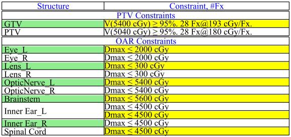
20 Radiotherapy: Treatment Planning Fractionated EBRT: Dose: Gy at Gy/fraction to the PTV SRS: Dose: Gy in single fraction to ~50% isodose line Choice of approach depends on tumor size, normal tissue constraints, and tumor delineation.
21 Radiotherapy: Treatment Planning Target Definition: GTV: Grossly visible disease as defined by contrast-enhanced CT and/or MRI CTV: Typically none is used unless the disease is poorly defined PTV: 1-5 mm depending on image-guidance and immobilization
22 Case Presentation: Fractionated RT Given the concern for tumor delineation on MR for SRS planning, the patient underwent fractionated RT to a dose of 50.4 Gy in 28 fractions at 1.8 Gy per fraction.
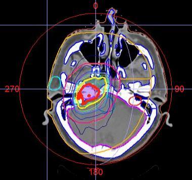
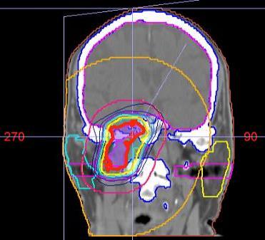
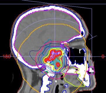
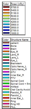
23 Case Presentation: Treatment Planning
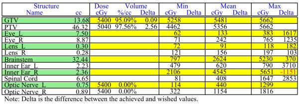
24 Case Presentation: Treatment Planning DVH Summary:
25 Toxicities Acute: Fatigue, skin reactions, transient mucositis, ear congestion, middle ear effusion, xerostomia Long-term: Decreased hearing, hypopituitarisim, xerostomia; more rarely, osteomyelitis, bone necrosis, brain necrosis, vascular compromise due to stenosis
26 Follow-Up Extrapolated from NCCN, clinical and radiographic follow up every 6-12 months for the first 3 years, then annually thereafter for 10 years, as recurrence can take several years to present
27 References Dupin, C. et al (2014) Treatment of head and neck paragangliomas with external beam radiation therapy. Int J Radiat Oncol Biol Phys 89(2): Fisch, U. Mattox, D. (1988) Microsurgery of the skull base. Thieme, Stuttgart-New York, page 149. Guss, ZD. et al (2011) Radiosurgery of glomus jugulare tumors: a meta-analysis. Int J Radiat Oncol Biol Phys 81(4):e Jackson, CG. et al (1982) Glomus tumors. Diagnosis, classification, and management of large lesions. Arch Otolaryngol 108(7): Sheehan, JP. et al (2012) Gamma Knife surgery for the management of glomus tumors: a multicenter study. J Neurosurg. 117(2): Suárez, C. et al (2013) Jugular and vagal paragangliomas: Systematic study of management with surgery and radiotherapy. Head Neck 35(8):
Otolaryngologist s Perspective of Stereotactic Radiosurgery
 Otolaryngologist s Perspective of Stereotactic Radiosurgery Douglas E. Mattox, M.D. 25 th Alexandria International Combined ORL Conference April 18-20, 2007 Acoustic Neuroma Benign tumor of the schwann
STATE OF THE ART MANAGEMENT of PARAGANGLIOMA. IFOS, Lima, 2018
 STATE OF THE ART MANAGEMENT of PARAGANGLIOMA IFOS, Lima, 2018 VINCENT C COUSINS ENT-Otoneurology Unit, The Alfred Hospital & Department of Surgery, Monash University MELBOURNE, AUSTRALIA PARAGANGLIOMAS
Solitary paraganglioma of the hypoglossal nerve: A case report with magnetic resonance imaging findings
 ISSN 1507-6164 DOI: 10.12659/AJCR.889509 Received: 2013.06.30 Accepted: 2013.07.16 Published: 2013.10.18 : A case report with magnetic resonance imaging findings Authors Contribution: Study Design A Data
Refresher Course EAR TUMOR. Sasikarn Chamchod, MD Chulabhorn Hospital
 Refresher Course EAR TUMOR Sasikarn Chamchod, MD Chulabhorn Hospital Reference: Perez and Brady s Principles and Practice of radiation oncology sixth edition Outlines Anatomy Epidemiology Clinical presentations
Case Report Recurrent paraganglioma of occipito-temporal bone masquerading as temporal bone malignancy
 Bangladesh J Otorhinolaryngol 2013; 19(1): 58-64 Case Report Recurrent paraganglioma of occipito-temporal bone masquerading as temporal bone malignancy Sushil Kumar Aggarwal 1, Rajkumar 2 Abstract: A 33-year-old
Glomus Jugulare. Paragangliomas, Chemodectoma, Ganglia tympanica, vascular tumors of middle ear.
 Glomus Jugulare SYNONYMS Paragangliomas, Chemodectoma, Ganglia tympanica, vascular tumors of middle ear. DEFINITION Glomus tumors are benign slow growing vascular tumors arising from GLOMUS BODIES present
Robotic Stereotactic Radiosurgery in Patients with Unresectable Glomus Jugulare Tumors
 Technology in Cancer Research and Treatment ISSN 1533-0346 Volume 12, Number 2, April 2013 Adenine Press (2013) Robotic Stereotactic Radiosurgery in Patients with Unresectable Glomus Jugulare Tumors www.tcrt.org
GUIDELINES FOR THE MANAGEMENT OF SKULL BASE TUMOURS. Version: 1 AngCN-SSG-BC6
 GUIDELINES FOR THE MANAGEMENT OF SKULL BASE TUMOURS Version: 1 Ref: AngCN-SSG-BC6 CONTENTS Page No 1 Introduction... 4 2 Imaging... 4 2.1 Other investigations... 4 3 Referral... 4 3.1 Accepting referrals...
The Ear The ear consists of : 1-THE EXTERNAL EAR 2-THE MIDDLE EAR, OR TYMPANIC CAVITY 3-THE INTERNAL EAR, OR LABYRINTH 1-THE EXTERNAL EAR.
 The Ear The ear consists of : 1-THE EXTERNAL EAR 2-THE MIDDLE EAR, OR TYMPANIC CAVITY 3-THE INTERNAL EAR, OR LABYRINTH 1-THE EXTERNAL EAR Made of A-AURICLE B-EXTERNAL AUDITORY MEATUS A-AURICLE It consists
Primary Jugular Foramen Meningioma: Imaging Appearance and Differentiating Features
 ndré J. Macdonald 1 Karen L. Salzman 1 H. Ric Harnsberger 1 Erik Gilbert 2 lough Shelton 2 Received May 16, 2003; accepted after revision ugust 12, 2003. 1 Department of Diagnostic Radiology, University
PRINCESS MARGARET CANCER CENTRE CLINICAL PRACTICE GUIDELINES
 PRINCESS MARGARET CANCER CENTRE CLINICAL PRACTICE GUIDELINES CENTRAL NERVOUS SYSTEM BRAIN METASTASES CNS Site Group Brain Metastases Author: Dr. Norm Laperriere Date: February 20, 2018 1. INTRODUCTION
Radiologic Evaluation of Petrous Apex Masses. Pavan Kavali, MS-IV Morehouse School of Medicine November 16, 2009
 Radiologic Evaluation of Petrous Apex Masses Pavan Kavali, MS-IV Morehouse School of Medicine November 16, 2009 Roadmap Petrous Apex Anatomy Patient D.S.: Clinical Presentation Differential diagnosis of
Impact of Gamma Knife Radiosurgery on the neurosurgical management of skull-base lesions: The Combined Approach
 Radiosurgery as part of the neurosurgical armamentarium: Educational Symposium November 24 th 2011 Impact of Gamma Knife Radiosurgery on the neurosurgical management of skull-base lesions: The Combined
The Temporal Bone Anatomy & Pathology
 Department of Radiology University of California San Diego The Temporal Bone Anatomy & Pathology John R. Hesselink, M.D. Temporal Bone Axial View Temporal Bone Coronal View Longitudinal Fracture The Temporal
ORIGINAL ARTICLE GAMMA KNIFE STEREOTACTIC RADIOSURGERY FOR SALIVARY GLAND NEOPLASMS WITH BASE OF SKULL INVASION FOLLOWING NEUTRON RADIOTHERAPY
 ORIGINAL ARTICLE GAMMA KNIFE STEREOTACTIC RADIOSURGERY FOR SALIVARY GLAND NEOPLASMS WITH BASE OF SKULL INVASION FOLLOWING NEUTRON RADIOTHERAPY James G. Douglas, MD, MS, 1,2 Robert Goodkin, MD, 1,2 George
External carotid blood supply to acoustic neurinomas
 External carotid blood supply to acoustic neurinomas Report of two cases HARVEY L. LEVINE, M.D., ERNEST J. FERmS, M.D., AND EDWARD L. SPATZ, M.D. Departments of Radiology, Neurology, and Neurosurgery,
Paragangliomas of the Jugular Bulb and Carotid Body: MR Imaging
 83 Paragangliomas of the Jugular Bulb and Carotid Body: MR Imaging with Short Sequences and Gd-DTPA Enhancement T. Vogl 1 R. Bruning 1 H. Schedel 1 K. Kang 1 G. Grevers D. Hahn 1 J. Lissner 1 Twenty-six
Advanced Vascular Imaging: Pulsatile Tinnitus. Disclosures. Pulsatile Tinnitus: Differential Diagnosis. Pulsatile Tinnitus
 Advanced Vascular Imaging: Pulsatile Tinnitus Patrick Turski MD, Zach Clark MD, Tabby Kennedy MD The Objectives of this presentation are to: Review the differential diagnosis of pulsatile tinnitus Discuss
The treatment of glomic tumors has been controversial
 Rev Bras Otorrinolaringol. V.71, n.6, 752-7, nov./dec. 2005 ORIGINAL ARTICLE Radiation therapy for glomus tumors of the temporal bone Celso Dall Igna 1, arcelo B. Antunes 2, Daniela Pernigotti Dall Igna
Gross Anatomy of the. TEMPORAL BONE, EXTERNAL EAR, and MIDDLE EAR
 Gross Anatomy of the TEMPORAL BONE, EXTERNAL EAR, and MIDDLE EAR M1 Gross and Developmental Anatomy 9:00 AM, December 11, 2008 Dr. Milton M. Sholley Professor of Anatomy and Neurobiology Assignment: Head
PRINCESS MARGARET CANCER CENTRE CLINICAL PRACTICE GUIDELINES
 PRINCESS MARGARET CANCER CENTRE CLINICAL PRACTICE GUIDELINES CENTRAL NERVOUS SYSTEM MENINGIOMA CNS Site Group Meningioma Author: Dr. Norm Laperriere Date: February 20, 2018 1. INTRODUCTION 3 2. PREVENTION
Acoustic Neuroma. Presenting Signs and Symptoms of an Acoustic Neuroma:
 Acoustic Neuroma An acoustic neuroma is a benign tumor which arises from the nerves behind the inner ear and which may affect hearing and balance. The incidence of symptomatic acoustic neuroma is estimated
Case Report Squamous Cell Carcinoma of the External Auditory Canal: ACaseReport
 Case Reports in Otolaryngology Volume 2011, Article ID 615210, 4 pages doi:10.1155/2011/615210 Case Report Squamous Cell Carcinoma of the External Auditory Canal: ACaseReport Harry Boamah, 1 Glenn Knight,
1. Axial view, left temporal bone. Plane through the upper antrum (A), superior semicircular canal (SSC) and IAC.
, superior semicircular canal (SSC) and IAC.") PA IAC SSC A 1. Axial view, left temporal bone. Plane through the upper antrum (A), superior semicircular canal (SSC) and IAC. IAC VII M I LSC Plane through the IAC, malleus head and incus and the lateral
PA IAC SSC A 1. Axial view, left temporal bone. Plane through the upper antrum (A), superior semicircular canal (SSC) and IAC. IAC VII M I LSC Plane through the IAC, malleus head and incus and the lateral
Gross Anatomy of the. TEMPORAL BONE, EXTERNAL EAR, and MIDDLE EAR. Assignment: Head to Toe Temporomandibular Joint (TMJ)
") Gross Anatomy the TEMPORAL BONE, EXTERNAL EAR, and MIDDLE EAR M1 Gross and Developmental Anatomy 9:00 AM, December 11, 2008 Dr. Milton M. Sholley Pressor Anatomy and Neurobiology Assignment: Head to Toe
Gross Anatomy the TEMPORAL BONE, EXTERNAL EAR, and MIDDLE EAR M1 Gross and Developmental Anatomy 9:00 AM, December 11, 2008 Dr. Milton M. Sholley Pressor Anatomy and Neurobiology Assignment: Head to Toe
Temporal Bone Carcinoma: Results of Surgery for Primary and Secondary Malignancies
 ORIGINAL ARTICLE Temporal Bone Carcinoma: Results of Surgery for Primary and Secondary Malignancies Milan Stankovic, M.D. From the Department of Otorhinolaryngology, Medical Faculty Nis, Serbia. Correspondence:
STEREOTACTIC RADIATION THERAPY. Monique Blanchard ANUM Radiation Oncology Epworth HealthCare
 STEREOTACTIC RADIATION THERAPY Monique Blanchard ANUM Radiation Oncology Epworth HealthCare Overview Stereotactic radiation therapy at Epworth Healthcare What is stereotactic radiation therapy? Delivery
Case Conference: SBRT for spinal metastases D A N I E L S I M P S O N M D 3 / 2 7 / 1 2
 Case Conference: SBRT for spinal metastases D A N I E L S I M P S O N M D 3 / 2 7 / 1 2 Case 79 yo M with hx of T3N0 colon cancer diagnosed in 2008 metastatic liver disease s/p liver segmentectomy 2009
Fractionated Stereotactic Radiotherapy. Rationale, indications, & treatment techniques
 Fractionated Stereotactic Radiotherapy Rationale, indications, & treatment techniques Radiobiological principles The BED (Gy) = D(1 + d/α/β) Assume BED 1 = BED 2 for tissue of an unknown α/β: Optic
Stereotactic Radiosurgery and Stereotactic Body Radiation Therapy
 Stereotactic Radiosurgery and Stereotactic Body Radiation Therapy Policy Number: Original Effective Date: MM.05.008 05/12/1999 Line(s) of Business: Current Effective Date: HMO; PPO; QUEST Integration 11/20/2015
Selected radiosurgery cases from the Rotating Gamma Institute Debrecen, Hungary
 Selected radiosurgery cases from the Rotating Gamma Institute Debrecen, Hungary László Bognár M.D., Ph.D., József G. Dobai M.D., Gábor Csiky and Imre Fedorcsák M.D., Ph.D. Department of Neurosurgery, Medical
PRIMARY SQUAMOUS cell carcinoma
 Squamous Cell Carcinoma of the Temporal Bone A Radiographic-Pathologic Correlation ORIGINAL ARTICLE M. Boyd Gillespie, MD; Howard W. Francis, MD; Nelson Chee, MD; David W. Eisele, MD Objective: To assess
RADIOLOGY TEACHING CONFERENCE
 RADIOLOGY TEACHING CONFERENCE John Athas, MD Monica Tadros, MD Columbia University, College of Physicians & Surgeons Department of Otolaryngology- Head & Neck Surgery September 27, 2007 CT SCAN IMAGING
NCCN GUIDELINES ON PROTON THERAPY (AS OF 4/23/18) BONE (Version , 03/28/18)
 BONE (Version , 03/28/18)") BONE (Version 2.2018, 03/28/18) NCCN GUIDELINES ON PROTON THERAPY (AS OF 4/23/18) Radiation Therapy Specialized techniques such as intensity-modulated RT (IMRT); particle beam RT with protons, carbon ions,
BONE (Version 2.2018, 03/28/18) NCCN GUIDELINES ON PROTON THERAPY (AS OF 4/23/18) Radiation Therapy Specialized techniques such as intensity-modulated RT (IMRT); particle beam RT with protons, carbon ions,
OTOLOGY. 1. BRIEF DESCRIPTION OF OTOLOGIC TRAINING Rotations that include otologic training are a component of each of the four years of training.
 OTOLOGY 1. BRIEF DESCRIPTION OF OTOLOGIC TRAINING Rotations that include otologic training are a component of each of the four years of training. Longwood Rotation PGY-2 through PGY-5 years o Clinic experience
Stereotactic Radiosurgery and Stereotactic Body Radiation Therapy
 Stereotactic Radiosurgery and Stereotactic Body Radiation Therapy Policy Number: Original Effective Date: MM.05.008 05/12/1999 Line(s) of Business: Current Effective Date: HMO; PPO; QUEST Integration 04/01/2017
Case Studies in the Skull Base
 Case Studies in the Skull Base Amy C Tsai, MD Neuroradiology Fellow Department of Radiology and Imaging Sciences University of Utah Health Sciences Center Salt Lake City, Utah, USA No disclosures related
Hypofractionated radiosurgery for meningiomas a safer alternative for large tumors?
 Original Article Hypofractionated radiosurgery for meningiomas a safer alternative for large tumors? Damon E. Smith 1, Sanjay Ghosh 2, Michael O Leary 2, Colin Chu 1, David Brody 2 1 Genesis Healthcare
AUDITORY APPARATUS. Mr. P Mazengenya. Tel 72204
 AUDITORY APPARATUS Mr. P Mazengenya Tel 72204 Describe the anatomical features of the external ear Describe the tympanic membrane (ear drum) Describe the walls of the middle ear Outline the structures
Directorate Medical Operations Patients and Information Nursing Policy Commissioning Development. Clinical Reference Group Chairs
 Clinical Commissioning Policy: Stereotactic Radiosurgery/ Radiotherapy for Glomus Tumours (skull base paragangliomas, glomus jugulare tumours) September 2013 Reference: NHS ENGLAND D05/P/f England 1 NHS
ANALYSIS OF TREATMENT OUTCOMES WITH LINAC BASED STEREOTACTIC RADIOSURGERY IN INTRACRANIAL ARTERIOVENOUS MALFORMATIONS
 ANALYSIS OF TREATMENT OUTCOMES WITH LINAC BASED STEREOTACTIC RADIOSURGERY IN INTRACRANIAL ARTERIOVENOUS MALFORMATIONS Dr. Maitri P Gandhi 1, Dr. Chandni P Shah 2 1 Junior resident, Gujarat Cancer & Research
Radiosurgery for the control of Glomus Jugulare Tumours
 Radiosurgery for the control of Glomus Jugulare Tumours M. E. Bari,A.A. Kemeny,D.M.C. Forster,M.W.R.Radatz ( Department of Neurosurgery, Aga Khan University Hospital*, Karachi and Royal Hallamshire Hospital,
Stereotactic Radiosurgery. Extracranial Stereotactic Radiosurgery. Linear accelerators. Basic technique. Indications of SRS
 Stereotactic Radiosurgery Extracranial Stereotactic Radiosurgery Annette Quinn, MSN, RN Program Manager, University of Pittsburgh Medical Center Using stereotactic techniques, give a lethal dose of ionizing
Stereotactic Radiosurgery/Fractionated Stereotactic Radiotherapy for Acoustic Neuroma (Vestibular Schwannomas)
") Strategic Commissioning Group West Midlands Commissioning Policy (WM/38) Stereotactic Radiosurgery/Fractionated Stereotactic Radiotherapy for Acoustic Neuroma (Vestibular Schwannomas) Version 1 July 2010
Strategic Commissioning Group West Midlands Commissioning Policy (WM/38) Stereotactic Radiosurgery/Fractionated Stereotactic Radiotherapy for Acoustic Neuroma (Vestibular Schwannomas) Version 1 July 2010
CHONDROSARCOMAS OF THE TEMPORAL BONE PRESENTATION AND MANAGEMENT
 CHONDROSARCOMAS OF THE TEMPORAL BONE PRESENTATION AND MANAGEMENT Authors Mr Mallappa Raghu FRCS, Clinical Research Fellow Mr Ioannis Moumoulidis MRCS, Clinical Research Fellow Mr Ranit De, FRCS (ORL-HNS),
Dr. T. Venkat Kishan Asst. Prof Department of Radiodiagnosis
 Dr. T. Venkat Kishan Asst. Prof Department of Radiodiagnosis Schwannomas (also called neurinomas or neurilemmomas) constitute the most common primary cranial nerve tumors. They are benign slow-growing
Abstract. Samuel Hahn, M.D. 1 James N. Palmer, M.D. 1 Nithin D. Adappa, M.D. 1
 19 A Catecholamine-Secreting Skull Base Sinonasal Paraganglioma Presenting with Labile Hypertension in a Patient with Previously Undiagnosed Genetic Mutation Samuel Hahn, M.D. 1 James N. Palmer, M.D. 1
Feasibility of Proton Beam Therapy for Chordoma and Chondrosarcoma of the Skull Base
 ORIGINAL ARTICLE Feasibility of Proton Beam Therapy for Chordoma and Chondrosarcoma of the Skull Base Hiroshi Fuji, M.D., Ph.D., 1 Yoko Nakasu, M.D., Ph.D., 2 Yuji Ishida, M.D., Ph.D., 3 Satoshi Horiguchi,
Imaging of Petrous Apex: Anatomy and Pathology
 University of Utah Head and Neck Conference 2018 Petrous apex Imaging of Petrous Apex: Anatomy and Pathology Philip Chapman MD University of Alabama, Birmingham Good News PAs tend to be symmetric A quick
Stereotactic Radiosurgery and Stereotactic Body Radiation Therapy
 Stereotactic Radiosurgery and Stereotactic Body Radiation Therapy Policy Number: Original Effective Date: MM.05.008 05/12/1999 Line(s) of Business: Current Effective Date: HMO; PPO; QUEST Integration 04/01/2015
Update on Pediatric Brain Tumors
 Update on Pediatric Brain Tumors David I. Sandberg, M.D. Director of Pediatric Neurosurgery & Associate Professor Dr. Marnie Rose Professorship in Pediatric Neurosurgery Pre-talk Questions for Audience
Collection of Recorded Radiotherapy Seminars
 IAEA Human Health Campus Collection of Recorded Radiotherapy Seminars http://humanhealth.iaea.org The Role of Radiosurgery in the Treatment of Gliomas Luis Souhami, MD Professor Department of Radiation
Waleed F. Mourad MD, Kenneth S. Hu MD, Louis B. Harrison MD
 Waleed F. Mourad MD, Kenneth S. Hu MD, Louis B. Harrison MD Department of Radiation Oncology Beth Israel Medical Centers (BIMC) St. Luke's-Roosevelt Hospital (SLRH) Continuum Cancer Centers of New York
GLOMUS TUMORS ~ A REVIEW OF 8 CASES WITH INTRODUCTION OF A MODIFIED COMBINED INTRA-AND EXTRACRANIAL APPROACH
 Chinese Journal of Cancer Research 9(1):56-60.199Z GLOMUS TUMORS ~ A REVIEW OF 8 CASES WITH INTRODUCTION OF A MODIFIED COMBINED INTRA-AND EXTRACRANIAL APPROACH Zhao Suping ~..~ Xiao Jianyun ~ ffj~: Tao
Linac Based SBRT for Low-intermediate Risk Prostate Cancer in 5 Fractions: Preliminary Report of a Phase II Study with FFF Delivery
 Linac Based SBRT for Low-intermediate Risk Prostate Cancer in 5 Fractions: Preliminary Report of a Phase II Study with FFF Delivery FILIPPO ALONGI MD Radiation Oncology & Radiosurgery Istituto Clinico
Meningioma in the Middle Ear: An Unusual Case of Hearing Loss
 Danezza Mae D. Lim, MD Nathaniel W. Yang, MD Department of Otorhinolaryngology Head and Neck Surgery The Medical City Meningioma in the Middle Ear: An Unusual Case of Hearing Loss When evaluating patients
Dosimetry, see MAGIC; Polymer gel dosimetry. Fiducial tracking, see CyberKnife radiosurgery
 Subject Index Acoustic neuroma, neurofibromatosis type 2 complications 103, 105 hearing outcomes 103, 105 outcome measures 101 patient selection 105 study design 101 tumor control 101 105 treatment options
Extracranial doses in stereotactic and conventional radiotherapy for pituitary adenomas
 JOURNAL OF APPLIED CLINICAL MEDICAL PHYSICS, VOLUME 7, NUMBER 2, SPRING 2006 Extracranial doses in stereotactic and conventional radiotherapy for pituitary adenomas Thomas Samuel Ram, a Paul B. Ravindran,
REVISITING ICRU VOLUME DEFINITIONS. Eduardo Rosenblatt Vienna, Austria
 REVISITING ICRU VOLUME DEFINITIONS Eduardo Rosenblatt Vienna, Austria Objective: To introduce target volumes and organ at risk concepts as defined by ICRU. 3D-CRT is the standard There was a need for a
Radiation Therapy for Liver Malignancies
 Outline Radiation Therapy for Liver Malignancies Albert J. Chang, M.D., Ph.D. Department of Radiation Oncology, UCSF March 23, 2014 Rationale for developing liver directed therapies Liver directed therapies
Cyberknife Radiotherapy for Vestibular Schwannoma
 Cyberknife Radiotherapy for Vestibular Schwannoma GordonT. Sakamoto, MD a, *, Nikolas Blevins, MD b, Iris C. Gibbs, MD c KEYWORDS Stereotactic radiosurgery Vestibular schwannomas Cyberknife Fractionation
Gamma knife radiosurgery for Koos grade 4 vestibular schwannomas
 Gamma knife radiosurgery for Koos grade 4 vestibular schwannomas David Mathieu MD FRCSC, Christian Iorio-Morin MD PhD, Fahd Al Subaie MD MSc FRCSC Division of neurosurgery, Université de Sherbrooke, Centre
HBA THE BODY Head & Neck Written Examination October 23, 2014
 HBA 531 - THE BODY Head & Neck Written Examination October 23, 2014 Name: NOTE 2: When asked to trace nerve, artery, or vein pathways, do so by using arrows, e.g., structure a structure b structure c...
Advanced Technology Consortium (ATC) Credentialing Procedures for 3D Conformal Therapy Protocols 3D CRT Benchmark*
 Credentialing Procedures for 3D Conformal Therapy Protocols 3D CRT Benchmark*") Advanced Technology Consortium (ATC) Credentialing Procedures for 3D Conformal Therapy Protocols 3D CRT Benchmark* Purpose: To evaluate an institution s 3D treatment planning process and the institution
Advanced Technology Consortium (ATC) Credentialing Procedures for 3D Conformal Therapy Protocols 3D CRT Benchmark* Purpose: To evaluate an institution s 3D treatment planning process and the institution
Our Experience with Surgical Treatment of Tympanojugular Pragangliomas
 Prague Medical Report / Vol. 111 (2010) No. 1, p. 25 34 25) Our Experience with Surgical Treatment of Tympanojugular Pragangliomas Skřivan J. 1, Zvěřina E. 1, Kluh J. 1, Chovanec M. 1, Pádr R. 2 1 Charles
CLINICAL PRESENTATION AND RADIOLOGY QUIZ QUESTION
 Donald L. Renfrew, MD Radiology Associates of the Fox Valley, 333 N. Commercial Street, Suite 100, Neenah, WI 54956 04/26/2014 Radiology Quiz of the Week # 108 Page 1 CLINICAL PRESENTATION AND RADIOLOGY
Radiotherapy and Brain Metastases. Dr. K Van Beek Radiation-Oncologist BSMO annual Meeting Diegem
 Radiotherapy and Brain Metastases Dr. K Van Beek Radiation-Oncologist BSMO annual Meeting Diegem 24-02-2017 Possible strategies Watchful waiting Surgery Postop RT to resection cavity or WBRT postop SRS
NON MALIGNANT BRAIN TUMOURS Facilitator. Ros Taylor Advanced Neurosurgical Nurse Practitioner Southmead Hospital Bristol
 NON MALIGNANT BRAIN TUMOURS Facilitator Ros Taylor Advanced Neurosurgical Nurse Practitioner Southmead Hospital Bristol Neurosurgery What will be covered? Meningioma Vestibular schwannoma (acoustic neuroma)
Overview of MLC-based Linac Radiosurgery
 SRT I: Comparison of SRT Techniques 1 Overview of MLC-based Linac Radiosurgery Grace Gwe-Ya Kim, Ph.D. DABR 2 MLC based Linac SRS Better conformity for irregular target Improved dose homogeneity inside
Skull-2. Norma Basalis Interna Norma Basalis Externa. Dr. Heba Kalbouneh Associate Professor of Anatomy and Histology
 Skull-2 Norma Basalis Interna Norma Basalis Externa Dr. Heba Kalbouneh Associate Professor of Anatomy and Histology Norma basalis interna Base of the skull- superior view The interior of the base of the
SKULL BASE LESIONS THAT MAY MIMICK DISEASE
 SKULL BASE LESIONS THAT MAY MIMICK DISEASE AUTHORS: MYERS, TANDBERG, LORENZO UNIVERSITY OF NEW MEXICO DIAGNOSTIC RADIOLOGY Learning Objectives The participant will identify normal anatomic variants that
Josh Howard CMD Upendra Parvathaneni MBBS, FRANZCR
 Anatomic and Dosimetric Correlation in the Treatment of Advanced Larynx Cancer- When is the Brachial Plexus at Risk? Josh Howard CMD Upendra Parvathaneni MBBS, FRANZCR AAMD 39th Annual Meeting - Seattle
Major Anatomic Components of the Orbit
 Major Anatomic Components of the Orbit 1. Osseous Framework 2. Globe 3. Optic nerve and sheath 4. Extraocular muscles Bony Orbit Seven Bones Frontal bone Zygomatic bone Maxillary bone Ethmoid bone Sphenoid
Evaluation of Monaco treatment planning system for hypofractionated stereotactic volumetric arc radiotherapy of multiple brain metastases
 Evaluation of Monaco treatment planning system for hypofractionated stereotactic volumetric arc radiotherapy of multiple brain metastases CASE STUDY Institution: Odette Cancer Centre Location: Sunnybrook
Hands-on Course in: Lateral Skull Base Surgery Piacenza, Italy November 2016
 Piacenza, Italy Case n.01 Right PBC Age 43 years Sex Female History RT for nasopharyngeal carcinoma (1993) Right-sided tympanoplasty EAC chole (2003) Onset of right hemifacial spasm and progressive right
4D Radiotherapy in early ca Lung. Prof. Manoj Gupta Dept of Radiotherapy & oncology I.G.Medical College Shimla
 4D Radiotherapy in early ca Lung Prof. Manoj Gupta Dept of Radiotherapy & oncology I.G.Medical College Shimla Presentation focus on ---- Limitation of Conventional RT Why Interest in early lung cancer
Cerebellopontine Angle Masses June 2004
 TITLE: Cerebellopontine Angle Masses SOURCE: Grand Rounds Presentation, UTMB, Dept. of Otolaryngology DATE: June 2, 2004 RESIDENT PHYSICIAN: Alan L. Cowan, MD FACULTY ADVISOR: Arun Gadre, MD SERIES EDITORS:
ORIGINAL ARTICLE. Temporal Lobe Injury in Temporal Bone Fractures. imaging (MRI) to evaluate lesions of the temporal
 to evaluate lesions of the temporal") ORIGINAL ARTICLE Temporal Lobe Injury in Temporal Bone Fractures Richard M. Jones, MD; Michael I. Rothman, MD; William C. Gray, MD; Gregg H. Zoarski, MD; Douglas E. Mattox, MD Objective: To determine the
ORIGINAL ARTICLE Temporal Lobe Injury in Temporal Bone Fractures Richard M. Jones, MD; Michael I. Rothman, MD; William C. Gray, MD; Gregg H. Zoarski, MD; Douglas E. Mattox, MD Objective: To determine the
LONG-TERM FOLLOW-UP OF ACOUSTIC SCHWANNOMA RADIOSURGERY WITH MARGINAL TUMOR DOSES OF 12 TO 13 Gy
 doi:10.1016/j.ijrobp.2007.01.001 Int. J. Radiation Oncology Biol. Phys., Vol. 68, No. 3, pp. 845 851, 2007 Copyright 2007 Elsevier Inc. Printed in the USA. All rights reserved 0360-3016/07/$ see front
Defining Target Volumes and Organs at Risk: a common language
 Defining Target Volumes and Organs at Risk: a common language Eduardo Rosenblatt Section Head Applied Radiation Biology and Radiotherapy (ARBR) Section Division of Human Health IAEA Objective: To introduce
Sponsored by: Congress of Neurological Surgeons (CNS) and the Section on Tumors
 and the Section on Tumors") 1 2 3 4 5 6 7 8 CONGRESS OF NEUROLOGICAL SURGEONS SYSTEMATIC REVIEW AND EVIDENCE-BASED GUIDELINE ON THE ROLE OF RADIOSURGERY AND RADIATION THERAPY IN THE MANAGEMENT OF PATIENTS WITH VESTIBULAR SCHWANNOMAS
1 2 3 4 5 6 7 8 CONGRESS OF NEUROLOGICAL SURGEONS SYSTEMATIC REVIEW AND EVIDENCE-BASED GUIDELINE ON THE ROLE OF RADIOSURGERY AND RADIATION THERAPY IN THE MANAGEMENT OF PATIENTS WITH VESTIBULAR SCHWANNOMAS
Injury retrotympanic white, blue and red. Clinicalradiological
 Injury retrotympanic white, blue and red. Clinicalradiological correlation Poster No.: C-0211 Congress: ECR 2013 Type: Educational Exhibit Authors: R. Esteban Saiz, R. Castañón Martinez, M. Rebolledo Vicente,
CLASSIFICATION. 2 main systems:
 DEFINITION Sound sensation that originates in the head and is not attributable to any perceivable external sound. (popping, clicking, pulsing & pure or multiple tones) Sounds of differing quality Mild
Ashley Pyfferoen, MS, CMD. Gundersen Health Systems La Crosse, WI
 Ashley Pyfferoen, MS, CMD Gundersen Health Systems La Crosse, WI 3 Radiation Oncologists 3 Physicists 2 Dosimetrists 9 Radiation Therapists o o o o o o o o o Brachial Plexus Anatomy Brachial Plexopathy
Dr. Sami Zaqout Faculty of Medicine IUG
 Auricle External Ear External auditory meatus The Ear Middle Ear (Tympanic Cavity) Auditory ossicles Internal Ear (Labyrinth) Bony labyrinth Membranous labyrinth External Ear Auricle External auditory
Head&Neck Imaging. ssregypt.com. Parapharyngeal Spaces. Mamdouh mahfouz MD
 Head&Neck Imaging Parapharyngeal Spaces ssregypt.com Mamdouh mahfouz MD mamdouh.m5@gmail.com Definitio n Fat filled triangular space lateral the pharynx Extends from the skull base to the oropharynx Parapharyngeal
List the tumours that may arise in CPA:
 List the tumours that may arise in CPA: 1. Vestibular schwannoma: 75-90% 2. Meningioma: 5-10% 3. Epidermoid 5% 4. Cholesteatoma: 5% 5. Other schwannomas 2-5%: trigeminal is the most common (0.3% of intracranial
Chapters from Clinical Oncology
 Chapters from Clinical Oncology Lecture notes University of Szeged Faculty of Medicine Department of Oncotherapy 2012. 1 RADIOTHERAPY Technical aspects Dr. Elemér Szil Introduction There are three possibilities
Protocol of Radiotherapy for Small Cell Lung Cancer
 107 年 12 月修訂 Protocol of Radiotherapy for Small Cell Lung Cancer Indication of radiotherapy Limited stage: AJCC (8th edition) stage I-III (T any, N any, M0) that can be safely treated with definitive RT
PRINCESS MARGARET CANCER CENTRE CLINICAL PRACTICE GUIDELINES
 PRINCESS MARGARET CANCER CENTRE CLINICAL PRACTICE GUIDELINES CENTRAL NERVOUS SYSTEM EPENDYMOMA Last Revision Date July 2015 1 CNS Site Group Ependymoma Author: Dr. Norm Laperriere 1. INTRODUCTION 3 2.
Kingdom of Bahrain Arabian Gulf University College of Medicine and Medical Sciences Year 6 ENT SMC Otitis Media (Dr.
 Kingdom of Bahrain Arabian Gulf University College of Medicine and Medical Sciences Year 6 ENT SMC Otitis Media (Dr. Jalal Almarzooq) - Anatomy of the ear: The ear is divided into 3 parts: External ear.
Selecting the Optimal Treatment for Brain Metastases
 Selecting the Optimal Treatment for Brain Metastases Clinical Practice Today CME Co-provided by Learning Objectives Upon completion, participants should be able to: Understand the benefits, limitations,
Radiation Therapy for Soft Tissue Sarcomas
 Radiation Therapy for Soft Tissue Sarcomas Alexander R. Gottschalk, MD, PhD Assistant Professor, Radiation Oncology University of California, San Francisco 1/25/08 NCI: limb salvage vs. amputation 43 patients
Gamma Knife Radiosurgery A tool for treating intracranial conditions. CNSA Annual Congress 2016 Radiation Oncology Pre-congress Workshop
 Gamma Knife Radiosurgery A tool for treating intracranial conditions CNSA Annual Congress 2016 Radiation Oncology Pre-congress Workshop ANGELA McBEAN Gamma Knife CNC State-wide Care Coordinator Gamma Knife
Background Principles and Technical Development
 Contents Part I Background Principles and Technical Development 1 Introduction and the Nature of Radiosurgery... 3 Definitions of Radiosurgery... 5 Consequences of Changing Definitions of Radiosurgery...
Linac or Non-Linac Demystifying And Decoding The Physics Of SBRT/SABR
 Linac or Non-Linac Demystifying And Decoding The Physics Of SBRT/SABR PhD, FAAPM, FACR, FASTRO Department of Radiation Oncology Indiana University School of Medicine Indianapolis, IN, USA Indra J. Das,
Stereotactic Radiosurgery and Stereotactic Body Radiation Therapy
 Stereotactic Radiosurgery and Stereotactic Body Radiation Therapy Policy Number: Original Effective Date: MM.05.008 05/12/1999 Line(s) of Business: Current Effective Date: HMO; PPO; QUEST 03/01/2013 Section:
The Ear. Dr. Heba Kalbouneh Assistant Professor of Anatomy and Histology
 The Ear Dr. Heba Kalbouneh Assistant Professor of Anatomy and Histology The Ear The ear consists of the external ear; the middle ear (tympanic cavity); and the internal ear (labyrinth), which contains
Tania Kaprealian, M.D. Assistant Professor UCLA Department of Radiation Oncology August 22, 2015
 Tania Kaprealian, M.D. Assistant Professor UCLA Department of Radiation Oncology August 22, 2015 Most common brain tumor, affecting 8.5-15% of cancer patients. Treatment options: Whole brain radiation
Lung Cancer Radiotherapy
 Lung Cancer Radiotherapy Indications, Outcomes, and Impact on Survivorship Care Malcolm Mattes, MD Assistant Professor WVU Department of Radiation Oncology When people think about radiation, they think