Unusual Manifestation of Stomach Cancer; Pictorial review and review of the literature
|
|
|
- Jeffery Lane
- 5 years ago
- Views:
Transcription
1 Unusual Manifestation of Stomach Cancer; Pictorial review and review of the literature Poster No.: C-1386 Congress: ECR 2014 Type: Educational Exhibit Authors: D. H. Lee, S. K. Moon; Seoul/KR Keywords: Cancer, Staging, CT, Stomach (incl. Oesophagus), Oncology, Gastrointestinal tract DOI: /ecr2014/C-1386 Any information contained in this pdf file is automatically generated from digital material submitted to EPOS by third parties in the form of scientific presentations. References to any names, marks, products, or services of third parties or hypertext links to thirdparty sites or information are provided solely as a convenience to you and do not in any way constitute or imply ECR's endorsement, sponsorship or recommendation of the third party, information, product or service. ECR is not responsible for the content of these pages and does not make any representations regarding the content or accuracy of material in this file. As per copyright regulations, any unauthorised use of the material or parts thereof as well as commercial reproduction or multiple distribution by any traditional or electronically based reproduction/publication method ist strictly prohibited. You agree to defend, indemnify, and hold ECR harmless from and against any and all claims, damages, costs, and expenses, including attorneys' fees, arising from or related to your use of these pages. Please note: Links to movies, ppt slideshows and any other multimedia files are not available in the pdf version of presentations. Page 1 of 37
2 Learning objectives The purpose of this pictorial review is to investigate the previous literature about MDCT diagnosis and staging of stomach cancer, and to illustrate various imaging features of characteristic histology subtypes and unusual disease spread of stomach cancer. Page 2 of 37
3 Background Stomach cancer is the 4th most common cancer and the 2nd leading cause of cancerrelated death worldwide. MDCT is a recently reliable and widely used diagnostic modalities for the diagnosis and preoperative staging with other modality (i.e. gastroscopy and EUS). Accurate assessment of local tumor invasion (T staging in the TNM staging system) is the most significant element in determining appropriate treatment plans. The advances of MDCT techniques renewed the interest in using CT for T staging. Some unusual histology of stomach cancers have characteristic radiologic features on MDCT images. On the other hand, some gastric cancer can show unusual manifestation or radiologic features. Page 3 of 37
4 Findings and procedure details 1. Staging of stomach cancer (1) Clinical staging of stomach cancer (Fig. 1) (2) CT T staging of stomach cancer * T1 stomach cancer - Invisible on MDCT images (especially T1a cancer) - Abnormally enhancing intraluminal mass or wall thickening, involving the most inner layer but maintaining the smooth outer border and the outer low attenuation layer (Fig. 2) - Abnormal fold change on 3D volume images (Fig. 3) * T2 stomach cancer - Abnormally enhancing mass or wall thickening with/without ulceration, showing intramural involvement and irregular outer border of the lesion, but maintaining the thin most outer layer and clear perigastric fat plane around tumor (Fig. 4 and 5) * T3 stomach cancer - Transmural lesion involving the entire gastric wall and either maintaining clear perigastric fat plane around tumor or fine perigastric fat spiculation, but not invading beyond adjacent perigastric vessels, especially along the lesser and greater omentum (Fig. 6 and 7) * T4a stomach cancer - Transmural lesion involving the entire gastric wall with irregular/nodular outer border with perigastric fat infiltration and invading beyond the adjacent perigastric vessels (Fig. 8) * T4b stomach cancer - Transmural lesion obliterating fat plane between gastric tumor and adjacent organ or invading adjacent organ (Fig. 9) (3) Local staging of stomach cancer with MDCT * A systematic review by Kwee J Clin Oncol 2007;25: ) - Diagnostic accuracy; moderate methodological quality. Page 4 of 37
5 ; EUS %, MDCT 77.1% %, MRI 71.4% % - Sensitivity for assessing serosal involvement ; EUS 77.8% - 100%, MDCT 82.8% - 100%, MRI 89.5% % - Specificity for assessing serosal involvement ; EUS 67.9% - 100%, MDCT 80% %, MRI 91.4%-100% - Early gastric cancer (EGC) ; EUS > MDCT - Advanced gastric cancer (AGC); EUS < MDCT ; EUS has a limited depth of penetration, and is of limited usefulness in the overall assessment of more distant spread. 2. Advanced technique of MDCT in the diagnosis of stomach cancer Adequate distension of stomach with water or gas improved lesion detection rates, and the accuracy of local T staging. Its thinner collimation, and faster scanning also improved the diagnostic accuarcy by improving spatial resolution. 2D multi-planar reformatted (MPR) images and 3D volume rendering images or virtual gastroscopic images are valuable in the accurate diagnosis and staging of stomach cancer. Although it still has some limitation in staging of EGC and nodal staging (N), it currently palys the key role in the diagnosis and staging of stomach cancer. 3. Morphologic classification of stomach cancer Stomach cancer is classified according to the morphologic type (Fig. 11~16). 4. Unusual manifestations of stomach cancer In retrospective review of the cases of stomach cancer in our hospital for the recent 20 years, we recognized several unusual manifestations of stomach cancer. These may be related with following factors; - Unusual histological subtypes - Unusual configuration - Unusual spread of the disease - Unusual complication Page 5 of 37
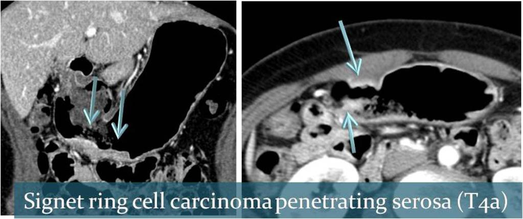
6 The cases with stomach cancer which are demonstrated from now on manifest unusual imaging features. Typical histological subtypes of stomach cancer - Tubular adenocarcinoma; well, moderately, and poorly differentiated - Papillary adenocarcinoma - Mucinous adenocarcinoma - Signet ring cell adenocarcinoma ; Poorly differentiated and signet ring cell adenocarcinoma which together are considered poorly differentiated. Poorly differentiated adenocarcinoma primarily corresponds with diffuse-type gastric cancers. Atypical histological subtypes of stomach cancer - Sarcomatoid adenocarcinoma - Squamous cell carcinoma - Hepatoid adenocarcinoma **Atypical histological subtypes (1) Mucinous adenocarcinoma WHO defines mucinous adenocarcinoma as an adenocarcinoma more than 50% of the tumor contains extracellular mucin pools. It was used to be confused with SRC because of mucus secretion. It has been characterized by layered enhancement and poorly enhanced outer or middle layer (Fig. 17 and 18). It may have calcifications (Fig. 19). (2) Signet ring cell carcinoma (SRC) SRC has been characterized by cells containing a sufficient intracytoplasmic volume of mucin to compress the nucleus against the periphery of the cell, and by its potential to diffusely infiltrate the stomach wall and to cause a marked scirrhous reaction. SRC has been known with its poor prognosis related with larger tumor size, more LN mets, deeper invasion, and more Borrmann type IV lesions than other histological subtypes. Abundant immature fibrosis of SRC may induce the high degree of enhancement on CT scan (Fig. 20). (3) Poorly differentiated adenocarcinoma Page 6 of 37
7 Poorly differentiated adenocarcinoma primarily corresponds with diffuse-type stomach cancers (linitis plastica). With SRC, this undifferentiated cancer prefer to invade stomach wall deeply and spread lymphatics (Fig. 21). (4) Sarcomatoid adenocarcinoma Sarcomatoid adenocarcinoma (carcinosarcoma) is rare malignant tumor with biphasic histology of carcinomatous and sarcomatous component. In the upper GI tract, it most frequently observed in the esophagus. About 50 cases of stomach carcinosarcoma have been reported, mostly in Japan, and predominantly in the male population, mostly over the age of 60 years. Carcinosarcoma of stomach may be polypoid, exophytic or endophytic, with generally ulcerated surfaces, and form bulky tumor masses (Fig. 22). (5) Hepatoid adenocarcinoma The term "hepatoid adenocarcinoma of the stomach" was proposed for primary gastric carcinomas characterized histololgically by hepatoid differentiation and production of large amounts of AFP. However, the diagnosis of hepatoid adenocarcinoma is not dependent on whether AFP was produced and considered that diagnosis was better based on the recognition of the characteristic histologic features (Fig. 23). There is sometimes confusion about whether hepatoid carcinoma originates from the stomach or the liver, because most patients show multiple liver metastasis preoperatively. Because of the extremely poor prognosis of hepatoid adenocarcinoma of the stomach, it is important to ensure that the diagnosis is accurate. Even if metastasis is absent preoperatively, liver metastasis can occur within a year after surgery. Thus, close observation and long-term follow-up of patients are required. Imaging features - Gastric wall thickening with eccentric contrast enhancement, ulceration, or irregular lobulation - Diffuse hepatic metastasis and lymph node metastasis - Predilection site; gastric antrum (6) Squamous cell carcinoma Squamous cell carcinoma of the stomach is uncommon, with an incidence of less than 1% worldwide. Because it is rare, the mechanism of this disease has not been wellunderstood (Fig. 24). (7) Neuroendocrine carcinoma (gastric carcinoid) * WHO classification - Well-differentiated endocrine tumor (carcinoid) Page 7 of 37
8 - Well-differentiated endocrine carcinoma (malignant carcinoid) - Poorly differentiated endocrine carcinoma Neuroendocrine tumors of GI tract usually produce bioactive subastances and show immunoreactivity to neuroendocrine markers. Stomach neuroendocrine carcinoma ; 3 types 1.Type I; associated with chronic atrophic gastritis, grow from the fundus and body, the most common type of stomach carcinoid, usually small (<1-2cm), often multiple 2.Type II; frequently associated with Zollinger-Ellison syndrome and MEN 1, also small (1-2cm) and often multiple, sometimes metastasis 3.Type III; absence of underlying gastric diseaseor hypergastrinemia, usually large (>2cm) single neoplasm located in the body and fundus, typically vascular invasion at the time of diagnosis * CT findings (Fig. 25) - Small size; well-defined subepithelial tumor and well enhancement - Large size; ulcerofungating or ulceroinfiltrative tumor, appear similar to AGC **Unusual configuration of stomach cancer (1) Exophytic stomach cancer An adenocarcinoma arising in the gastric mucosa may present as an exophytic gastric mass with a tumor center lying beyond the confines of the stomach in rare circumstances (Fig. 26). It is hard to differentiate a malignant GIST from an exophytic adenocarcinoma of the stomach. Antral location, thickening of the gastric wall adjacent to an exogastric mass, LN enlargement and discordant images between US and CT are typical findings of exophytic stomach cancer, and allow distinction with between exophytic stomach cancer and GIST. (2) Synchronous tumors The synchronous occurrence of GIST in the stomach and EGC is very uncommon in the literature. GIST may be concomitant with gastric lymphomas or with carcinoid tumors (Fig. 27~28). (3) Tumor thrombosis Page 8 of 37
9 Stomach cancer generally spreads through the lymphatic and /or portal venous system, frequently resulting in the occurrence of metastases to the regional LNs and liver. However, the formation of a tumor thrombus in the portal venous system is extremely rare in patients with stomach cancer (Fig. 29~30). Tumor thrombus can communicate with primary tumor, and it may invade vascular wall directly. On US and CT images, contiguous thrombus is strongly suggested as having tumor origin. Tumor thrombus can occur in portal vein developed from the left gastric vein, right gastroepiploic vein, and short gastric vein (Fig. 29). (4) Direct small bowel invasion Stomach cancer can invade the transverse colon directly through the mesocolon. However, direct invasion of the small bowel is uncommon (Fig. 31). (5) Ureter metastasis Ureter metastasis from stomach cancer is rare and has been diagnosed only at autopsy. * Mechanism; via perivascular lymphatic channel * CT findings (Fig. 32) - Thickened enhanced ureteral wall with periureteral infiltrations and obstructive hydronephrosis and hydroureter - Accompanying multiple LN metastasis around the aorta and peritoneal seeding (6) Cancer peritonitis vs. TB peritonitis in a AGC patient Cancer peritonitis frequently shows ascites. The discovery of new-onset ascites, thickening, nodularity, omental cake/smudge and enhancement of the peritoneum are suggestive of a malignant process. However, TB peritonitis can show similar features. Classically, tuberculous peritonitis has been described as having three imaging patterns (wet, fibrotic fixed, and dry plastic). In all three patterns, soft-tissue masses or nodules that stud the peritoneal surfaces or infiltrate the omentum and mesenteries represent caseous nodules and fibrosis (Fig. 33). (7) Stomach cancer perforation Perforated stomach cancer is rare with an incidence of %, and is not frequentlly diagnosed peroperatively. The cause of gastric perforation is difficult to identify during emergency surgery.malignant gastric perfuration is commonly regarded as a sign of a terminal disease, because it is thought to contribute to the peritoneal dissemination of cancer cells and early recurrence (Fig. 34). Thus, the cause of gastric perforation should be thoroughly evaluated on imaging study. Page 9 of 37

10 Images for this section: Fig. 1: TNM staging system of stomach cancer in the 7th edition of AJCC cancer staging system Page 10 of 37

11 Fig. 2: T1 stomach cancer in CT images Page 11 of 37

12 Fig. 3: T1 stomach cancer in a 3D volume rendering image Page 12 of 37

13 Fig. 4: T2 stomach cancer in CT images. Page 13 of 37

14 Fig. 5: T2 stomach cancer in a CT image Page 14 of 37

15 Fig. 6: T3 stomach cancer in a CT axial image Page 15 of 37

16 Fig. 7: T3 stomach cancer in a CT coronal image Page 16 of 37

17 Fig. 8: T4a stomach cancer in CT images Page 17 of 37
18 Fig. 9: T4b stomach cancer Page 18 of 37
19 Fig. 10 Page 19 of 37


20 Fig. 11: Morphologic classification of stomach cancer Bosman FT et al. (2010) Chapter 3 Tumours of the Stomach, WHO Classification of the Digestive System, Fourth Edition Fig. 12: Barium study of Borrmann type I AGC Page 20 of 37

21 Fig. 13: A CT image of Borrmann type I stomach cancer Page 21 of 37

22 Fig. 14: Barium study and a CT image of Borrmann type II AGC Page 22 of 37

23 Fig. 15: A CT image and barium study of Borrmann type III AGC Page 23 of 37

24 Fig. 16: Barium study and a CT image of Borrmann type IV AGC Page 24 of 37

25 Fig. 17: Mucinous adenocarcinoma Page 25 of 37

26 Fig. 18: Mucinous adenocarcinoma Page 26 of 37

27 Fig. 19: Mucinous adenocarcinoma Page 27 of 37


28 Fig. 20: Signet ring cell carcinoma penetrating serosa (T4a) Fig. 21: A 43-year old woman with diffuse-type stomach cancer. Submucsal tumor spread shows prominent contrast enhancement on CT scans. Repeated endoscopic biopsy only revealed chronic gastritis. But it was confirmed as poorly differentiated adenocarcinoma. Fig. 22: Sarcomatoid adenocarcinoma and multiple liver metastasis in a 69-year old woman Page 28 of 37


29 Fig. 23: A 65-year old man with pathologically proven hepatoid adenocarcinoma of the stomach. Wall thickening of gastric lower body and multiple liver metastases. Fig. 24: Squamous cell carcinoma of the stomach and liver metastasis in a 56-year old man. Bulky necrotic mass at gastric cardia with exophytic growth Page 29 of 37


30 Fig. 25: A rapidly growing subepithelial tumor of the gastric cardia in a 82 year-old man, which was confimed as a poorly differentiated neuroendocrine carcinoma Fig. 26: An exophytic mucinous adenocarcinoma at the posterior wall of the stomach body. Abnormally thickened and enhanced overlying and adjacent gastric mucosa of the exogastric mass, and mass effect to the pancreas tail. Fig. 27: A synchronous EGC and GIST in a 70-year old woman. EGC in a CT image and endoscopy. Page 30 of 37


31 Fig. 28: A synchronous EGC and GIST in a 70-year old woman. GIST in endoscopy and a CT image. Page 31 of 37

32 Fig. 29: A 78-year old man with AGC at the cardia, fundus, and high body. Direct extension of stomach cancer to coronary vein and extending to distal splenic vein, splenoportal and distal SMV. Fig. 30: A 66-year old man with AGC at the antrum (small arrows. Tumor thrombus in the main portal vein and SMV. Page 32 of 37


33 Fig. 31: Direct invasion of mid jejunum by stomach cancer at the antrum in a patient with incomplete rotation of jejunum. Fig. 32: A 69-year old man with AGC and ureter metastasis Page 33 of 37

34 Fig. 33: A 68-year old woman with AGC at the lower body (small arrow). Biopsy proven TB peritonitis and improvement after TB medication 3 months later. Fig. 34: Stomach cancer perforation Page 34 of 37
35 Conclusion MDCT gave a quantum leap in the diagnosis and staging of stomach cancer. It can demonstrate some unusual manifestation of stomach cancer, and be helpful for the accurate staging, and diagnosis of disease status. Page 35 of 37
36 References 1. A case of two primary gastric malignancies: adenocarcinoma and squamous cell carcinoma of the stomach. Gastrointest Endosc May;75(5): doi: / j.gie Epub 2011 Jul Carcinosarcoma (pure endocrine cell carcinoma with sarcoma components) of the stomach. Pathol Int Aug;53(8): Carcinosarcoma of the stomach: a case report and review of the literature. World J Gastroenterol Nov 7;13(41): Imaging in local staging of gastric cancer: a systematic review. J Clin Oncol May 20;25(15): Exophytic adenocarcinoma of the stomach: computed tomography and ultrasonography features with emphasis on differentiation from a malignant gastrointestinal stromal tumor. Clin Imaging Nov-Dec;32(6): doi: / j.clinimag Exophytic adenocarcinoma of the stomach: CT findings. AJR Am J Roentgenol Jul;163(1): Gastric hepatoid adenocarcinoma: CT findings. Abdom Imaging MayJun;32(3): Hepatoid adenocarcinoma: computed tomographic imaging findings with histopathologic correlation in 6 cases. J Comput Assist Tomogr NovDec;31(6): Hepatoid adenocarcinoma of the stomach with liver metastasis mimicking hepatocellular carcinoma: a case report. Cases J Aug 11;2:6317. doi: / Histological subtype of gastric cancer in Japan. Lancet Oncol Feb;11(2): doi: /S (09) Mucinous versus nonmucinous gastric carcinoma: differentiation with helical CT. Radiology May;223(2): Neuroendocrine and mucinous differentiation in signet ring cell carcinoma of the stomach: evidence for a common cell of origin in composite tumors.hum Pathol Oct;42(10): doi: /j.humpath Epub 2011 Mar 21. Page 36 of 37
37 13. Clinicopathological features, surgical management, and disease outcome of perforated gastric cancer. J Surg Oncol Sep 15;91(4): Neuroendocrine tumors of the stomach (gastric carcinoids) are on the rise: small tumors, small problems? Endoscopy Aug;42(8): doi: / s Epub 2010 Jul Preoperative staging of gastric cancer by endoscopic ultrasonography and multidetector-row computed tomography. J Gastroenterol Hepatol Mar;25(3): doi: /j x. 16. Dedicated multidetector CT of the stomach: spectrum of diseases. Radiographics May-Jun;23(3): Neuroendocrine neoplasms of the gastrointestinal tract: classification, pathologic basis, and imaging features. Radiographics Nov-Dec;27(6): CT of the stomach: spectrum of disease. Radiographics Sep;16(5): CT and PET in stomach cancer: preoperative staging and monitoring of response to therapy. Radiographics Jan-Feb;26(1): Scirrhous gastric carcinoma: endoscopy versus upper gastrointestinal radiography. Radiology May;231(2): Advanced gastric carcinoma with signet ring cell carcinoma versus non-signet ring cell carcinoma: differentiation with multidetector CT. J Comput Assist Tomogr NovDec;30(6): Successful surgical treatment of gastric cancer with a tumor thrombus in the portal and splenic veins: report of a case. Surg Today. 1998;28(10): Unusual gastric tumors: radiologic-pathologic correlation. Radiographics NovDec;19(6): Page 37 of 37
Unusual Manifestation of Stomach Cancer; Pictorial review and review of the literature
 Unusual Manifestation of Stomach Cancer; Pictorial review and review of the literature Poster No.: C-1386 Congress: ECR 2014 Type: Educational Exhibit Authors: D. H. Lee, S. K. Moon; Seoul/KR Keywords:
Imaging in gastric cancer
 Imaging in gastric cancer Gastric cancer remains a deadly disease because of late diagnosis. Adenocarcinoma represents 90% of malignant tumors. Diagnosis is based on endoscopic examination with biopsies.
CT evaluation of small bowel carcinoid tumors
 CT evaluation of small bowel carcinoid tumors Poster No.: C-0060 Congress: ECR 2015 Type: Educational Exhibit Authors: N. V. V. P. Costa, L. Nascimento, T. Bilhim ; Estoril/PT, PT, 1 2 3 1 2 3 Lisbon/PT
Curious case of Misty Mesentery
 Curious case of Misty Mesentery Poster No.: C-1385 Congress: ECR 2015 Type: Authors: Keywords: DOI: Educational Exhibit T. Simelane 1, H. Khosa 2, N. Ramesh 2 ; 1 Dublin/IE, 2 Portlaoise/IE Abdomen, Anatomy,
Pneumo-esophageal 64-MDCT technique for gastric cancer evaluation
 Pneumo-esophageal 64-MDCT technique for gastric cancer evaluation Poster No.: C-1627 Congress: ECR 2010 Type: Scientific Exhibit Topic: GI Tract Authors: M. Ulla, E. Gentile, E. Levy, D. Cavadas, J. Ithurralde
Local staging of colon cancer: the current role of CT
 Local staging of colon cancer: the current role of CT Poster No.: C-2699 Congress: ECR 2018 Type: Authors: Keywords: DOI: Educational Exhibit A. P. Pissarra, R. R. Domingues Madaleno, C. Sanches, L. Curvo-
Lesions of the pancreaticoduodenal groove, a pictorial review
 Lesions of the pancreaticoduodenal groove, a pictorial review Poster No.: C-2131 Congress: ECR 2013 Type: Educational Exhibit Authors: E. Ni Mhurchu, L. Lavelle, I. Murphy, S. Skehan ; IE, Dublin/ IE Keywords:
Imaging characterization of renal clear cell carcinoma
 Imaging characterization of renal clear cell carcinoma Poster No.: C-0327 Congress: ECR 2011 Type: Educational Exhibit Authors: S. Ballester 1, A. Gaser 2, M. Dotta 1, M. F. CAPPA 1, F. Hammar 1 ; 1 2
64-MDCT imaging of the pancreas: Scan protocol optimisation by different scan delay regimes
 64-MDCT imaging of the pancreas: Scan protocol optimisation by different scan delay regimes Poster No.: C-051 Congress: ECR 2009 Type: Scientific Exhibit Topic: Abdominal and Gastrointestinal Authors:
MDCT signs differentiating retroperitoneal and intraperitoneal lesions- diagnostic pearls
 MDCT signs differentiating retroperitoneal and intraperitoneal lesions- diagnostic pearls Poster No.: C-0987 Congress: ECR 2015 Type: Educational Exhibit Authors: D. V. Bhargavi, R. Avantsa, P. Kala; Bangalore/IN
Diffuse high-attenuation within mediastinal lymph nodes on non-enhanced CT scan: Usefulness in the prediction of benignancy
 Diffuse high-attenuation within mediastinal lymph nodes on non-enhanced CT scan: Usefulness in the prediction of benignancy Poster No.: C-1785 Congress: ECR 2012 Type: Authors: Keywords: DOI: Scientific
A pictorial review of normal anatomical appearences of Pericardial recesses on multislice Computed Tomography.
 A pictorial review of normal anatomical appearences of Pericardial recesses on multislice Computed Tomography. Poster No.: C-1787 Congress: ECR 2012 Type: Educational Exhibit Authors: N. Ahmed 1, G. Avery
Esophageal Cancer Staging Essentials: The New TNM Staging System (7th edition) and Clinicoradiologic Implications
 and Clinicoradiologic Implications") Esophageal Cancer Staging Essentials: The New TNM Staging System (7th edition) and Clinicoradiologic Implications Poster No.: E-0060 Congress: ESTI 2012 Type: Scientific Exhibit Authors: K. Lee, T. J.
Esophageal Cancer Staging Essentials: The New TNM Staging System (7th edition) and Clinicoradiologic Implications Poster No.: E-0060 Congress: ESTI 2012 Type: Scientific Exhibit Authors: K. Lee, T. J.
Biliary tree dilation - and now what?
 Biliary tree dilation - and now what? Poster No.: C-1767 Congress: ECR 2012 Type: Educational Exhibit Authors: I. Ferreira, A. B. Ramos, S. Magalhães, M. Certo; Porto/PT Keywords: Pathology, Diagnostic
Hyperechoic breast lesions can be malignant.
 Hyperechoic breast lesions can be malignant. Poster No.: C-0041 Congress: ECR 2015 Type: Educational Exhibit Authors: G. Babu, R. bradley; Edinburgh/UK Keywords: Breast, Ultrasound, Biopsy, Cancer DOI:
Low-dose computed tomography (CT) protocol in the screening of patients with social exposure to asbestos
 protocol in the screening of patients with social exposure to asbestos") Low-dose computed tomography (CT) protocol in the screening of patients with social exposure to asbestos Poster No.: C-3032 Congress: ECR 2010 Type: Scientific Exhibit Topic: Radiographers Authors: P.
Low-dose computed tomography (CT) protocol in the screening of patients with social exposure to asbestos Poster No.: C-3032 Congress: ECR 2010 Type: Scientific Exhibit Topic: Radiographers Authors: P.
CT findings in multifocal or diffuse non-mucinous bronchioloalveolar carcinoma (BAC)
") CT findings in multifocal or diffuse non-mucinous bronchioloalveolar carcinoma (BAC) Poster No.: C-2192 Congress: ECR 2014 Type: Educational Exhibit Authors: I. Sandu, A. R. Popita, I.-A. Brumboiu; Cluj-Napoca/RO
CT findings in multifocal or diffuse non-mucinous bronchioloalveolar carcinoma (BAC) Poster No.: C-2192 Congress: ECR 2014 Type: Educational Exhibit Authors: I. Sandu, A. R. Popita, I.-A. Brumboiu; Cluj-Napoca/RO
Purpose. Methods and Materials
 Thin-section CT findings in peripheral lung cancer of 3 cm or smaller: are there any characteristic features for predicting tumor histology or do they depend only on tumor size? Poster No.: C-1893 Congress:
The "whirl sign". Diagnostic accuracy for intestinal volvulus.
 The "whirl sign". Diagnostic accuracy for intestinal volvulus. Poster No.: C-0670 Congress: ECR 2014 Type: Scientific Exhibit Authors: M. Pire, M. Marti, A. Borobia, A. Verón; Madrid/ES Keywords: Abdomen,
Vacuum-assisted breast biopsy using computer-aided 3.0 T- MRI guidance: diagnostic performance in 173 lesions
 Vacuum-assisted breast biopsy using computer-aided 3.0 T- MRI guidance: diagnostic performance in 173 lesions Poster No.: C-2870 Congress: ECR 2017 Type: Scientific Exhibit Authors: A. Pozzetto, L. Camera,
Adenomyosis by myometrial Invasion of endometriosis: Comparison with typical adenomyosis
 Adenomyosis by myometrial Invasion of endometriosis: Comparison with typical adenomyosis Poster No.: C-1294 Congress: ECR 2010 Type: Scientific Exhibit Topic: Genitourinary Authors: S. Moon, H. K. Lim,
Triple-negative breast cancer: which typical features can we identify on conventional and MRI imaging?
 Triple-negative breast cancer: which typical features can we identify on conventional and MRI imaging? Poster No.: C-1862 Congress: ECR 2013 Type: Educational Exhibit Authors: V. Bertani 1, A. Gualano
Spectrum of findings of sclerosing adenosis at breast MRI.
 Spectrum of findings of sclerosing adenosis at breast MRI. Poster No.: C-0738 Congress: ECR 2012 Type: Scientific Exhibit Authors: F. Vasselli 1, F. Pediconi 2, M. Telesca 2, M. Luciani 2, V. Casali 2,
The Radiologic Features of Xanthogranulomatous Cholecystitis: An Important Mimic of Gallbladder Carcinoma
 The Radiologic Features of Xanthogranulomatous Cholecystitis: An Important Mimic of Gallbladder Carcinoma Poster No.: C-0691 Congress: ECR 2014 Type: Authors: Keywords: DOI: Educational Exhibit H. L. khosa
CT findings of gastric and intestinal anisakiasis as cause of acute abdominal pain
 CT findings of gastric and intestinal anisakiasis as cause of acute abdominal pain Poster No.: C-2258 Congress: ECR 2015 Type: Educational Exhibit Authors: S. Marcos 1, J. Gonzalez 1, L. Sarria Octavio
Slowly growing malignant nodules and rapidly growing benign nodules: Evaluation of the value of volume doubling time
 Slowly growing malignant nodules and rapidly growing benign nodules: Evaluation of the value of volume doubling time Poster No.: C-208 Congress: ECR 2009 Type: Educational Exhibit Topic: Chest Authors:
MRI in staging of rectal carcinoma
 MRI in staging of rectal carcinoma Poster No.: C-0152 Congress: ECR 2015 Type: Scientific Exhibit Authors: J. R. Ramos Rodriguez, M. Atencia Ballesteros, M. D. M. Muñoz Ruiz, A. J. Márquez Moreno, M. D.
Ultrasound assessment of T1 Squamous Cell Carcinomas of the Tongue.
 Ultrasound assessment of T1 Squamous Cell Carcinomas of the Tongue. Poster No.: C-2014 Congress: ECR 2015 Type: Educational Exhibit Authors: S. R. Rice, G. Price, L. Firmin, S. Morley, T. Beale; London/UK
Monophasic versus biphasic contrast application in CT of patients with head and neck tumour
 Monophasic versus biphasic contrast application in CT of patients with head and neck tumour Poster No.: C-3331 Congress: ECR 2010 Type: Topic: Authors: Keywords: DOI: Scientific Exhibit Head and Neck G.
Comparison of multidetector-row computed tomography findings of IgG4-related sclerosing cholangitis and cholangiocarcinoma
 Comparison of multidetector-row computed tomography findings of IgG4-related sclerosing cholangitis and cholangiocarcinoma Poster No.: C-0245 Congress: ECR 2014 Type: Scientific Exhibit Authors: M. Yata,
Characterisation of cervical lymph nodes by US and PET-CT
 Characterisation of cervical lymph nodes by US and PET-CT Poster No.: C-1807 Congress: ECR 2010 Type: Educational Exhibit Topic: Head and Neck Authors: J. I. Garcia Gomez; Mexico City/MX Keywords: cervical
High density thrombi of pulmonary embolism on precontrast CT scan: Is it dangerous?
 High density thrombi of pulmonary embolism on precontrast CT scan: Is it dangerous? Poster No.: C-1753 Congress: ECR 2013 Type: Authors: Keywords: DOI: Scientific Exhibit B. Y. Lee, H. R. KIM, J. I. Jung,
MR imaging of FIGO stage I uterine cervical cancer: The diagnostic impact of 3T-MRI
 MR imaging of FIGO stage I uterine cervical cancer: The diagnostic impact of 3T-MRI Poster No.: C-1191 Congress: ECR 2010 Type: Educational Exhibit Topic: Genitourinary Authors: M. Takeuchi, K. Matsuzaki,
Pattern based approach for differential diagnosis of small bowel neoplasms using MDCT
 Pattern based approach for differential diagnosis of small bowel neoplasms using MDCT Poster No.: C-1400 Congress: ECR 2014 Type: Educational Exhibit Authors: P. Bhari Thippeswamy, C. Anuradha, A. Polimood,
Role of ultrasound in the evaluation of the ileocecal valve
 Role of ultrasound in the evaluation of the ileocecal valve Poster No.: C-1581 Congress: ECR 2010 Type: Scientific Exhibit Topic: GI Tract Authors: M. Mohammed, M. Hussain, U. Momin, S. Lakhtakia, N. D.
CT Enteroclysis in the Diagnosis of Crohn's Disease (CD)
") CT Enteroclysis in the Diagnosis of Crohn's Disease (CD) Poster No.: C-2291 Congress: ECR 2012 Type: Scientific Exhibit Authors: I. Kiss, A. Rosztóczy, F. Nagy, T. Wittmann, A. Palko; Szeged/HU Keywords:
CT Enteroclysis in the Diagnosis of Crohn's Disease (CD) Poster No.: C-2291 Congress: ECR 2012 Type: Scientific Exhibit Authors: I. Kiss, A. Rosztóczy, F. Nagy, T. Wittmann, A. Palko; Szeged/HU Keywords:
Computed tomography and Modified RECIST criteria for assessment of response in malignant pleural mesothelioma
 Computed tomography and Modified RECIST criteria for assessment of response in malignant pleural mesothelioma Poster No.: C-0729 Congress: ECR 2013 Type: Scientific Exhibit Authors: A. Marin, I. Pozek,
Digital breast tomosynthesis (DBT) occult breast cancers: clinical, radiological and histopathological features.
 occult breast cancers: clinical, radiological and histopathological features.") Digital breast tomosynthesis (DBT) occult breast cancers: clinical, radiological and histopathological features. Poster No.: C-1707 Congress: ECR 2015 Type: Scientific Exhibit Authors: V. Vinci 1, A. Iqbal
Digital breast tomosynthesis (DBT) occult breast cancers: clinical, radiological and histopathological features. Poster No.: C-1707 Congress: ECR 2015 Type: Scientific Exhibit Authors: V. Vinci 1, A. Iqbal
Role of positron emission mammography (PEM) for assessment of axillary lymph node status in patients with breast cancer
 for assessment of axillary lymph node status in patients with breast cancer") Role of positron emission mammography (PEM) for assessment of axillary lymph node status in patients with breast cancer Poster No.: C-1260 Congress: ECR 2011 Type: Scientific Paper Authors: K. M. Kulkarni,
Role of positron emission mammography (PEM) for assessment of axillary lymph node status in patients with breast cancer Poster No.: C-1260 Congress: ECR 2011 Type: Scientific Paper Authors: K. M. Kulkarni,
Magnetic Resonance Imaging of Perianal Fistulas
 Magnetic Resonance Imaging of Perianal Fistulas Poster No.: C-0317 Congress: ECR 2014 Type: Authors: Keywords: DOI: Educational Exhibit A. P. Sathe, E. Soh, K. Y. Seto, B. Yeh, D. W. Y. chee, R. Quah,
Pneumatosis intestinalis, not always a surgical emergency
 Pneumatosis intestinalis, not always a surgical emergency Poster No.: C-2233 Congress: ECR 2012 Type: Educational Exhibit Authors: E. Vanhoutte, M. Lefere, R. Vanslembrouck, D. Bielen, G. De 1 1 2 1 1
Intraluminal gas in non-perforated acute appendicitis: a CT sign of gangrenous appendicitis
 Intraluminal gas in non-perforated acute appendicitis: a CT sign of gangrenous appendicitis Poster No.: C-978 Congress: ECR 202 Type: Scientific Exhibit Authors: D. Plata Ariza, E. MARTINEZ CHAMORRO, J.
Purpose. Methods and Materials. Results
 Prevalence and significance of hypoattenuating hepatic lesions deemed too small to characterise: How are we following up these lesions and what are the outcomes? Poster No.: C-014 Congress: ECR 2009 Type:
Is ascites a sensible predictive sign of peritoneal involvement in patients with ovarian carcinoma?: our experience with FDG-PET/CT
 Is ascites a sensible predictive sign of peritoneal involvement in patients with ovarian carcinoma?: our experience with FDG-PET/CT Poster No.: C-1019 Congress: ECR 2013 Type: Scientific Exhibit Authors:
Emerging Referral Patterns for Whole-Body Diffusion Weighted Imaging (WB-DWI) in an Oncology Center
 in an Oncology Center") Emerging Referral Patterns for Whole-Body Diffusion Weighted Imaging (WB-DWI) in an Oncology Center Poster No.: C-1296 Congress: ECR 2014 Type: Scientific Exhibit Authors: G. Petralia 1, G. Conte 1, S.
Emerging Referral Patterns for Whole-Body Diffusion Weighted Imaging (WB-DWI) in an Oncology Center Poster No.: C-1296 Congress: ECR 2014 Type: Scientific Exhibit Authors: G. Petralia 1, G. Conte 1, S.
Diffuse Alveolar Hemorrhage: Initial and Follow-up HRCT Features
 Diffuse Alveolar Hemorrhage: Initial and Follow-up HRCT Features Poster No.: E-0037 Congress: ESTI 2012 Type: Authors: Keywords: Scientific Exhibit M. Y. Kim; Seoul/KR Lung, CT-High Resolution, CT, Computer
Ureteropelvic Junction Obstruction (UPJO) syndrome: imaging with Multidetector CT (MDCT) prior to minimally invasive treatment
 syndrome: imaging with Multidetector CT (MDCT) prior to minimally invasive treatment") Ureteropelvic Junction Obstruction (UPJO) syndrome: imaging with Multidetector CT (MDCT) prior to minimally invasive treatment Poster No.: C-1753 Congress: ECR 2011 Type: Scientific Exhibit Authors: E.
Ureteropelvic Junction Obstruction (UPJO) syndrome: imaging with Multidetector CT (MDCT) prior to minimally invasive treatment Poster No.: C-1753 Congress: ECR 2011 Type: Scientific Exhibit Authors: E.
Cierny-Mader classification of chronic osteomyelitis: Preoperative evaluation with cross-sectional imaging
 Cierny-Mader classification of chronic osteomyelitis: Preoperative evaluation with cross-sectional imaging Poster No.: C-590 Congress: ECR 2009 Type: Topic: Educational Exhibit Musculoskeletal Authors:
Esophagus: Spectrum of pathologies on Barium Swallow
 Esophagus: Spectrum of pathologies on Barium Swallow Poster No.: C-1426 Congress: ECR 2013 Type: Authors: Keywords: DOI: Educational Exhibit E. Dhamija 1, D. Chandan 1, D. Srivastava 2 ; 1 New Delhi/IN,
Intracystic papillary carcinoma of the breast
 Intracystic papillary carcinoma of the breast Poster No.: C-1932 Congress: ECR 2011 Type: Educational Exhibit Authors: V. Dimarelos, F. TZIKOS, N. Kotziamani, G. Rodokalakis, 1 2 3 1 1 1 2 T. MALKOTSI
Audit of split-bolus CT urography for the investigation of haematuria over a 12 month period at two district general hospitals
 Audit of split-bolus CT urography for the investigation of haematuria over a 12 month period at two district general hospitals Poster No.: C-1349 Congress: ECR 2010 Type: Educational Exhibit Topic: Genitourinary
Extrapulmonary Manifestations of Tuberculosis: A Radiologic Review
 Extrapulmonary Manifestations of Tuberculosis: A Radiologic Review Poster No.: C-1958 Congress: ECR 2014 Type: Authors: Educational Exhibit J. Isern 1, S. Llaverias Borrell 1, A. Olarte 1, E. Grive 1,
Quantitative imaging of hepatic cirrhosis on abdominal CT images
 Quantitative imaging of hepatic cirrhosis on abdominal CT images Poster No.: C-0556 Congress: ECR 2013 Type: Authors: Keywords: DOI: Scientific Exhibit S. Kido, A. Nakamura, Y. Hirano; Ube/JP Cirrhosis,
Ultra-low dose CT of the acute abdomen: Spectrum of imaging findings
 Ultra-low dose CT of the acute abdomen: Spectrum of imaging findings Poster No.: C-1452 Congress: ECR 2010 Type: Educational Exhibit Topic: GI Tract Authors: P. A. Vlachou, C. Kloeters, S. Kandel, P. Hein,
Radiofrequency ablation combined with conventional radiotherapy: a treatment option for patients with medically inoperable lung cancer
 Radiofrequency ablation combined with conventional radiotherapy: a treatment option for patients with medically inoperable lung cancer Poster No.: C-0654 Congress: ECR 2011 Type: Scientific Paper Authors:
Complications of Perianal Crohn s Disease - Adenocarcinoma & Extensive Fistulization
 Complications of Perianal Crohn s Disease - Adenocarcinoma & Extensive Fistulization Poster No.: C-0711 Congress: ECR 2013 Type: Educational Exhibit Authors: P. Faria João 1, D. Penha 2, P. Cabral 1, E.
Soft tissues lymphoma, the great pretender. MRI diagnostic keys.
 Soft tissues lymphoma, the great pretender. MRI diagnostic keys. Poster No.: C-2133 Congress: ECR 2015 Type: Educational Exhibit Authors: L. Caminero, M. E. Banegas Illescas, M. L. Rozas, M. Y. Torres,
Diffusion-weighted MRI (DWI) "claw sign" is useful in differentiation of infectious from degenerative Modic I signal changes of the spine
 claw sign is useful in differentiation of infectious from degenerative Modic I signal changes of the spine") Diffusion-weighted MRI (DWI) "claw sign" is useful in differentiation of infectious from degenerative Modic I signal changes of the spine Poster No.: C-0894 Congress: ECR 2012 Type: Scientific Exhibit
Diffusion-weighted MRI (DWI) "claw sign" is useful in differentiation of infectious from degenerative Modic I signal changes of the spine Poster No.: C-0894 Congress: ECR 2012 Type: Scientific Exhibit
CT assessment of acute coalescent mastoiditis.
 CT assessment of acute coalescent mastoiditis. Poster No.: C-1794 Congress: ECR 2010 Type: Educational Exhibit Topic: Head and Neck Authors: A. Thomson, S. J. Thomas, A. Hutchings, E. Tilley; Portsmouth/UK
Acute abdominal venous thromboses- the hyperdense noncontrast CT sign
 Acute abdominal venous thromboses- the hyperdense noncontrast CT sign Poster No.: C-1095 Congress: ECR 2011 Type: Educational Exhibit Authors: M. Goldstein, K. Jhaveri; Toronto, ON/CA Keywords: Abdomen,
A Randomized Controlled Study to Compare Image Quality between Fenestrated and Non-Fenestrated Intravenous Catheters for Cardiac MDCT
 A Randomized Controlled Study to Compare Image Quality between Fenestrated and Non-Fenestrated Intravenous Catheters for Cardiac MDCT Poster No.: C-0623 Congress: ECR 2017 Type: Authors: Keywords: DOI:
Ultrasonic evaluation of superior mesenteric vein in cancer of the pancreatic head
 Ultrasonic evaluation of superior mesenteric vein in cancer of the pancreatic head Poster No.: C-1430 Congress: ECR 2012 Type: Authors: Keywords: DOI: Scientific Exhibit E. Fisenko, N. Vetsheva, E. Pershina;
Assessment of renal cell carcinoma by two PET tracer : dual-time-point C-11 methionine and F-18 fluorodeoxyglucose
 Assessment of renal cell carcinoma by two PET tracer : dual-time-point C-11 methionine and F-18 fluorodeoxyglucose Poster No.: C-0805 Congress: ECR 2015 Type: Scientific Exhibit Authors: S. Ito, K. Kato,
Emergency radiology of the large-bowel: What radiologists should know
 Emergency radiology of the large-bowel: What radiologists should know Poster No.: C-1659 Congress: ECR 2016 Type: Educational Exhibit Authors: A. Falkowski, D. Boll; Basle/CH Keywords: Colon, Emergency,
AFib is the most common cardiac arrhythmia and its prevalence and incidence increases with age (Fuster V. et al. Circulation 2006).
.") Feasibility, image quality and radiation dose of coronary CT angiography (CCTA) in patients with atrial fibrillation using a new generation 256 multi-detector CT (MDCT) Poster No.: C-2378 Congress: ECR
Feasibility, image quality and radiation dose of coronary CT angiography (CCTA) in patients with atrial fibrillation using a new generation 256 multi-detector CT (MDCT) Poster No.: C-2378 Congress: ECR
Peritoneal tuberculosis: retrospective analysis of clinical and radiologic findings in our institution from 2003 to 2013
 Peritoneal tuberculosis: retrospective analysis of clinical and radiologic findings in our institution from 2003 to 2013 Poster No.: C-2332 Congress: ECR 2015 Type: Scientific Exhibit Authors: A. Viteri,
CT EVALUATION OF GASTRIC LESIONS:
 CT EVALUATION OF GASTRIC LESIONS: Pictural essay Hasni Bouraoui I, Kahloun A, Jemni H, Elouni F, Moulahi H, Daadoucha A, Ben Ali A, Sriha B, Tlili Graies K Departments of Radiology, Gastro enterology,
Pulmonary changes induced by radiotherapy. HRCT findings
 Pulmonary changes induced by radiotherapy. HRCT findings Poster No.: C-2299 Congress: ECR 2015 Type: Educational Exhibit Authors: R. E. Correa Soto, M. Albert Antequera, K. Müller Campos, D. 1 2 4 3 1
Scientific Exhibit. Authors: D. Takenaka, Y. Ohno, Y. Onishi, K. Matsumoto, T.
 The feasibility of biphasic contrast-media-injection-protocol for chest imaging on 320-slice volume MDCT: Direct comparison of biphasic and bolus contrast-media injection protocols on 320-slice volume
Stomach Computerized Tomography indications, technique, examples. VUH SK Radiology and nuclear medicine center Radiologist Dileta Rutkauskaitė
 Stomach Computerized Tomography indications, technique, examples VUH SK Radiology and nuclear medicine center Radiologist Dileta Rutkauskaitė Stomach Computerized Tomography gastroente rologist Oncologist
Pleomorphic carcinoma of the lung: which CT findings predict poor prognosis?
 Pleomorphic carcinoma of the lung: which CT findings predict poor prognosis? Poster No.: C-1887 Congress: ECR 2015 Type: Scientific Exhibit Authors: A. Fujisaki, T. Aoki, S. Kinoshita, Y. Hayashida, Y.
Radiological features of Legionella Pneumophila Pneumonia
 Radiological features of Legionella Pneumophila Pneumonia Poster No.: E-0048 Congress: ESTI 2012 Type: Scientific Exhibit Authors: M. Vinciguerra, L. Stefanetti, E. Teti, G. Argentieri, L. G. 1 1 1 1 1
Radiologic Findings of Mucocele-like Tumors of the breast: Can we differentiate pure benign from associated with high risk lesions?
 Radiologic Findings of Mucocele-like Tumors of the breast: Can we differentiate pure benign from associated with high risk lesions? Poster No.: C-0332 Congress: ECR 2014 Type: Educational Exhibit Authors:
Small-bowel obstruction due to bezoar: CT diagnosis and characterization
 Small-bowel obstruction due to bezoar: CT diagnosis and characterization Poster No.: C-1450 Congress: ECR 2013 Type: Scientific Exhibit Authors: I. lópez blasco, S. Paz Maya, R. Dosdá Muñoz, D. Soriano
PET/CT depiction of ATS mediastinal nodal stations: What every radiologist should know - diagnostic strategies and potential pitfalls
 PET/CT depiction of ATS mediastinal nodal stations: What every radiologist should know - diagnostic strategies and potential pitfalls Poster No.: C-236 Congress: ECR 2009 Type: Educational Exhibit Topic:
Excavated pulmonary nodule: steps to diagnosis?
 Excavated pulmonary nodule: steps to diagnosis? Poster No.: C-1044 Congress: ECR 2014 Type: Authors: Keywords: DOI: Educational Exhibit W. Mnari, M. MAATOUK, A. Zrig, B. Hmida, M. GOLLI; Monastir/ TN Metastases,
PI-RADS classification: prognostic value for prostate cancer grading
 PI-RADS classification: prognostic value for prostate cancer grading Poster No.: C-1622 Congress: ECR 2014 Type: Scientific Exhibit Authors: I. Platzek, A. Borkowetz, T. Paulus, T. Brauer, M. Wirth, M.
A pictorial essay depicting CT and MR characteristic of adrenal pathologies: Indian study
 A pictorial essay depicting CT and MR characteristic of adrenal pathologies: Indian study Poster No.: C-0703 Congress: ECR 2011 Type: Educational Exhibit Authors: A. J. B. Baxi, K. L. Tourani, N. R. Thanugonda,
Evaluation of BI-RADS 3 lesions in women with a high risk of hereditary breast cancer.
 Evaluation of BI-RADS 3 lesions in women with a high risk of hereditary breast cancer. Poster No.: C-0346 Congress: ECR 2014 Type: Scientific Exhibit Authors: A. Thomas 1, R. Dominguez Oronoz 1, S. Roche
Lung cancer in patients with chronic empyema
 Lung cancer in patients with chronic empyema Poster No.: P-0025 Congress: ESTI 2015 Type: Scientific Poster Authors: Y. Lee, C.-K. Park; Guri/KR Keywords: Neoplasia, Biopsy, PET-CT, CT, Thorax, Lung DOI:
Contrast-enhanced ultrasound (CEUS) in the evaluation and characterization of complex renal cysts
 in the evaluation and characterization of complex renal cysts") Contrast-enhanced ultrasound (CEUS) in the evaluation and characterization of complex renal cysts Poster No.: C-2812 Congress: ECR 2018 Type: Educational Exhibit Authors: J. A. Torres de Abreu Macedo,
Contrast-enhanced ultrasound (CEUS) in the evaluation and characterization of complex renal cysts Poster No.: C-2812 Congress: ECR 2018 Type: Educational Exhibit Authors: J. A. Torres de Abreu Macedo,
Utility of PET-CT for detection of N2 or N3 nodal mestastases in the mediastinum in patients with non-small cell lung cancer (NSCLC)
") Utility of PET-CT for detection of N2 or N3 nodal mestastases in the mediastinum in patients with non-small cell lung cancer (NSCLC) Poster No.: C-1360 Congress: ECR 2015 Type: Scientific Exhibit Authors:
Utility of PET-CT for detection of N2 or N3 nodal mestastases in the mediastinum in patients with non-small cell lung cancer (NSCLC) Poster No.: C-1360 Congress: ECR 2015 Type: Scientific Exhibit Authors:
Gastrointestinal Angiodysplasia: CT Findings
 Gastrointestinal Angiodysplasia: CT Findings Poster No.: C-1792 Congress: ECR 2012 Type: Authors: Keywords: DOI: Educational Exhibit G. Anguita Martinez, A. Fernandez Alfonso, D. C. Olivares Morello, J.
Idiopathic dilatation of the pulmonary artery : radiographic and MDCT features in 6 cases
 Idiopathic dilatation of the pulmonary artery : radiographic and MDCT features in 6 cases Poster No.: P-0075 Congress: ESTI 2014 Type: Authors: Educational Poster J. J. Woo 1, K. Y. Lee 2, Y. Cho 1, J.
Primary epiploic appendagitis versus omental infarction : The role of MDCT
 Primary epiploic appendagitis versus omental infarction : The role of MDCT e-poster: EE-125 Congress: ESGAR 2010 Type: Educational Exhibit Topic: Diagnostic / Mesentery and Peritoneum Authors: P. Kraniotis,
Pleomorphic adenoma head and neck
 Pleomorphic adenoma head and neck Poster No.: C-1042 Congress: ECR 2015 Type: Educational Exhibit Authors: M. E. Pérez Montilla, I. Bravo Rey, E. Roldán Romero, F. BravoRodríguez; Cordoba/ES Keywords:
Radiological Investigation of Renal Colic in an Emergency Department of a Teaching Hospital
 Radiological Investigation of Renal Colic in an Emergency Department of a Teaching Hospital Poster No.: C-0892 Congress: ECR 2014 Type: Authors: Keywords: DOI: Scientific Exhibit A. Koo; Leeds, West Yorkshire/UK
Acute Pancreatitis, its Complications and Prognostic Correlation by Modified CT Severity Index
 Acute Pancreatitis, its Complications and Prognostic Correlation by Modified CT Severity Index Poster No.: R-0022 Congress: RANZCR-AOCR 2012 Type: Scientific Exhibit Authors: K. P. Bellam Premnath, K.
Breast cancer tumor size: Correlation between MRI and histopathology
 Breast cancer tumor size: Correlation between MRI and histopathology Poster No.: C-0409 Congress: ECR 2010 Type: Topic: Scientific Exhibit Breast Authors: H. Khan, M. Hoosein, M. Alattar, S. Tenant, L.
Sonographic and Mammographic Features of Phyllodes Tumours of the Breast: Correlation with Histological Grade
 Sonographic and Mammographic Features of Phyllodes Tumours of the Breast: Correlation with Histological Grade Poster No.: C-0046 Congress: ECR 2014 Type: Authors: Keywords: DOI: Scientific Exhibit C. Y.
Pelvic inflammatory disease - spectrum of tomodensitometric findings
 Pelvic inflammatory disease - spectrum of tomodensitometric findings Poster No.: C-2451 Congress: ECR 2015 Type: Educational Exhibit Authors: E. Matos, A. T. Almeida, D. Castelo; Vila Nova de Gaia/PT Keywords:
Diffusion-weighted MR imaging for Diagnosis of Uterine Leiomyomas
 Diffusion-weighted MR imaging for Diagnosis of Uterine Leiomyomas Poster No.: C-0111 Congress: ECR 2015 Type: Scientific Exhibit Authors: A. Er 1, G. Pekindil 2, M. Gök 3, A. R. Kandiloglu 2, A. G. Tamay
ARDS - a must know. Page 1 of 14
 ARDS - a must know Poster No.: C-1683 Congress: ECR 2016 Type: Authors: Keywords: DOI: Educational Exhibit M. Cristian; Turda/RO Education and training, Edema, Acute, Localisation, Education, Digital radiography,
The predicament of cancer presenting during pregnancy
 The predicament of cancer presenting during pregnancy Poster No.: C-3001 Congress: ECR 2010 Type: Educational Exhibit Topic: Radiographers Authors: D. O'Mahony, G. Murphy, G. Wilson, M. T. Keogan; Dublin/IE
Categorical Classification of Spiculated Mass on Breast MRI
 Categorical Classification of Spiculated Mass on Breast MRI Poster No.: C-1974 Congress: ECR 2013 Type: Authors: Scientific Exhibit Y. Kanda 1, S. Kanao 2, M. Kataoka 2, K. Togashi 2 ; 1 Kyoto City/JP,
Radiologic and pathologic correlation of non-mass like breast lesions on US and MRI: Benign, high risk, versus malignant
 Radiologic and pathologic correlation of non-mass like breast lesions on US and MRI: Benign, high risk, versus malignant Poster No.: C-1161 Congress: ECR 2013 Type: Educational Exhibit Authors: J. Kwak,
The solitary pulmonary nodule: Assessing the success of predicting malignancy
 The solitary pulmonary nodule: Assessing the success of predicting malignancy Poster No.: C-0829 Congress: ECR 2010 Type: Scientific Exhibit Topic: Chest Authors: R. W. K. Lindsay, J. Foster, K. McManus;
Seemingly isolated greater trochanter fractures do not exist
 Seemingly isolated greater trochanter fractures do not exist Poster No.: B-0950 Congress: ECR 2012 Type: Scientific Paper Authors: D. Dunker, J. H. Göthlin, M. Geijer ; Gothenburg/SE, Lund/SE Keywords: