Immunohistochemical classification of haematolymphoid tumours. Stephen Hamilton-Dutoit Institute of Pathology Aarhus University Hospital
|
|
|
- Francine Gardner
- 5 years ago
- Views:
Transcription
1 Immunohistochemical classification of haematolymphoid tumours Stephen Hamilton-Dutoit Institute of Pathology Aarhus University Hospital
2 Malignant lymphoproliferative diseases What are they?
3 Haematolymphoid Neoplasias: Leukaemia vs Lymphoma C L O N A L LYMPHOID M A L I G N A N C I E S


4 Haematolymphoid Neoplasias: Leukaemia vs Lymphoma C L O N A L LYMPHOID M A L I G N A N C I E S Bone marrow Blood Lymph node Extranodal site Leukaemia Lymphoma
5 Malignant lymphoproliferative diseases How common are they?
6 Malignant lymphoproliferative diseases Malignant lymphoma Leukaemia Acute lymphoblastic leukaemia Chronic lymphocytic leukaemia (CLL) Ca. 1,600 per year in DK Ca. 700,000 per year in the world (?) 8 th commonest cancer globally


7 Age & sex: Non-Hodgkins lymphoma (UK) Age specific incidence Cases per year
8 Estimated Annual Incidence 60,000 45,000 30,000 15, Year
9 Malignant lymphoproliferative diseases What causes them? Largely unkown...but involves: Changes in genes e.g. mutations, translocations inherited radiation chemicals infections sporadic Changes in the immune system immune deficiencies autoimmune diseases chronic infections


10 Malignant lymphoproliferative diseases What causes them?
11 Malignant lymphoproliferative diseases How are they classified?

")
12 Thomas Hodgkin Thomas Hodgkin dissection (Prof. Robert Carswell)

13 Thomas Hodgkin Hodgkin s original case: abdominal nodes Gordon Museum, King s College London


14 Thomas Hodgkin Hodgkin s original case: CD15 (1991) Gordon Museum, King s College London

15 WHO Classification of Tumours of Haematopoietic and Lymphoid Tissues, s 80s: Kiel classification B vs T cells: IHC!! 90s: REAL classification WHO (latest..2017) Real disease entities Clinical features Morphology Immunophenotype Molecular genetics
16 Lymphoma Hodgkins lymphoma Non-Hodgkins lymphoma HL, LP HL, NS HL, MC HL, LR HL, LD B-cell precursor peripheral 32 subtypes T/NK-cell precursor peripheral 20 subtypes

17 Updated WHO Classification 2016 (2017)


18 Updated WHO Classification 2016 (2017) It has not got any smaller! > 100 lymphoma entities
19 Why does it all have to be so complicated!? Because of Personalized Medicine - Many subtypes of lymphoma are rare - But... they require specific treatments
20 Updated WHO Classification 2017) Major immunophenotypic changes: Diffuse large B-cell lymphoma COO cell of origin analysis now required to distinguish GCB vs ABC/non-GC types either by gene expression profiling or immunohistochemistry IHC for MYC and BCL2 expression to identify double -expressors

21 Lymphoma frequencies HL DLBCL ALCL Follicular 2002 SEER database. O Connor
22 What is lymphoma? Clonal malignancy mutational events cause cells to freeze at a single stage of normal lymphocyte differentiation Morphology, immunophenotype & molecular features: mirror stages of normal lymphocyte development

23 T and B-cell differentiation: Stage-specific surface antigen expression

24 Lymphoid neoplasms: Correlation with normal T or B-cell differentiation


25 What is lymphoma? Clonal malignancy mutational events cause cells to freeze at a single stage of normal lymphocyte differentiation Morphology, immunophenotype & molecular features: mirror stages of normal lymphocyte development Resemble normal haematopoietic cells in their: morphology, immunophenotype, molecular genetics
26 Lymphoma & Leukaemia diagnosis Clinical features Morphology Immunophenotype Molecular diagnosis
27 Lymphoma differential diagnosis Assess morphology: cell size architecture But...that s not enough!
28 Lymphoma & Leukaemia diagnosis Clinical features Morphology Immunophenotype Molecular diagnosis


 BCL-2 Ki-67 CD20")
29 Lymphoma differential diagnosis Assess morphology: cell size architecture Select appropriate immune panel(s) BCL-2 Ki-67 CD20 pos CD30 pos OCT2 pos EBV LMP1

30 Enlarged lymph node Is it malignant? Emphasis on lymphoma classification Reactive vs malignant - often more challenging diagnosis Use IHC to evaluate lymphoid tissue cytology and architecture Correlate immunophenotype with disease entity
31 International recommendations for lymphoma diagnostics Danish lymphoma group See Lymfomdiagnostik UK: RCPath / BCSH
 Originally for surface antigens on leucocytes")
32 What are CD numbers? CD: clusters of differentiation Classification system for antigens (and antibodies) Originally for surface antigens on leucocytes Now includes other cells and intracellular antigens (no CD no.) 10 workshops since 1982 Currently > 350 CD antigens

33 IHC Dogma (also applies in diagnostic haematopathology) IHC complements routine staining Helps characterize cells and architecture No single antibody is disease specific Antibodies should be used in panels Interpret findings in relation to the histology
34 Diagnostic Applications of IHC 1 Reactive vs malignant Polyclonal vs monoclonal Ig Follicular hyperplasia vs follicular lymphoma Diff. diagnosis of small cell B-cell lymphomas CLL/SLL vs MALT vs FL vs Mantle cell Aggressive B-cell lymphomas DLBCL vs BL vs BL-like / grey-zone NHL DLBCL cell of origin GCB vs ABC
35 Diagnostic Applications of IHC 2 EBV LMP1 T-cell lymphoma vs B-cell lymphoma T-cell lymphoma vs T-zone hyperplasia Hodgkin lymphoma vs NHL Hodgkin lymphoma NLPHL vs classical HL Lymphoblastic vs. Myeloblastic vs. Burkitt Undifferentiated malignant tumor Lymphoma prognosis e.g. Ki-67; ALK; c-myc Targeted therapy e.g. CD20 / Rituximab; CD30 / Brentuximab; Alemtuzumab (anti-cd52)


 CD56 CD57 EMA S100 CD68 CD163 CD20 CD5 CD30")
36 Useful antigens in haematopathology CD45 B-cell specific CD19 CD20 CD79α Pax-5 OCT-2 / BOB1 Ig T-cell specific CD3 CD5 CD2 CD7 CD1a CD4 CD8 PD-1/CXCL-13 (TFH) Other CD30 CD10 Bcl-2 Bcl-6 ALK c-myc CD21 CD23 CD15 TdT Cyclin-D1 SOX-11 CD56 TIA-1, granzyme, perforin Other EBV LMP1 EBNA2 (EBER) CD56 CD57 EMA S100 CD68 CD163 CD20 CD5 CD30 PD1 EBV LMP1
37 Basic IHC panel for lymphoma diagnosis CD45 CD20 CD79α (PAX-5) kappa/lambda CD3 CD5 CD30 CD43 Bcl-2 Bcl-6 CD23 (CD21) Cyclin-D1 Ki-67
 hæmopoietic cells Not expressed on non-bm-derived cells CD45 isoforms are more lineage specific")

38 Basic stains: CD45 Membrane glycoprotein family Positive in all (?) hæmopoietic cells Not expressed on non-bm-derived cells CD45 isoforms are more lineage specific Reactive LN: CD45 In lymphomas: Most NHLs positive Often/always negative in: Precursor LB Plasma cell neoplasia Anaplastic large cell lymphoma Hodgkins lymphoma: LP: Popcorn cells positive HRS cells in classical HL are negative HL, NC: CD45

39 Basic stain: Immunoglobulin IHC-Ig first protocol for IHC in FFPE still one of the hardest to perform & evaluate! plasmacytoma monoclonal Ig-kappa


40 Basic stains: Immunoglobulin B-cell specific Normal κ:λ ratio ca. 3-4:1 Monotypic Ig restriction Suggests clonality >10:1 or < 0.2:1 = restriction Cytoplasmic Ig easily shown In lymphomas: Cy Ig: lymphoplasmacytic; myeloma; MZL; DLBCL, FL Surface Ig DLBCL: cyt-κ DLBCL: cyt-λ

")
41 Basic stains: Immunoglobulin Tonsil; sig -λ Surface Ig B-NHL clonality Requires sensitive, optimised technique Interpretation difficult (serum Ig) Tonsil; sig -λ
42 Basic stains: CD20 Many B-cell neoplasms Negative in: early precursor B-LB plasma cell neoplasms Negative in T-cell lymphomas rare cases positive Hodgkins lymphoma HL-LP: 90% positive Other types variably positive (10% - 30%; not all HRS cells) Predictive marker for Rituximab therapy Follicular lymphoma: CD20


43 Usual staining pattern of B-cell neoplasms DLBCL: λ CD5 Cyclin D1
44 Small cell B-Cell lymphomas: Differential Diagnosis SLL SLL Small lymphocytic NHL? MCL MCL Mantle cell NHL
45 Small B-Cell Lymphomas: Overall Survival Marginal Zone 0.7 Survival Small Lymphocytic (CLL) Mantle Cell 0.1 Log Rank Test: p< Years Armitage et al, 1997


46 B-cell Small Lymphocytic Lymphoma (CLL) Morphology small lymphocytes proliferation centres Immunology surface IgMD weak CD19, 20, 79a + CD5 + CD23 + CD10, CycD1 - CD23 CD5


47 Mantle Cell Lymphoma Morphology small-medium medium lymphocytes cleaved / irregular blastoid variant nodular / mantle / diffuse CD5 Immunology surface Ig + CD19, 20, 22, 79a + CD5 + CD23 - Cyclin D1 + CD10 - Cyclin D1
 Mantle cell NHL (90%) 10%+ DLBCL B-CLL B-cells dim reactive T-cells")
48 Basic stains: CD5 Modulates T & B cell signalling Pan-T cell marker 95% thymocytes 100% post-thymic T-cells expression with maturity Minor population normal B-cells: ca. 10%+ peripheral B-cells in autoimmunity Lymphomas: 90% T-cell neoplasias B-cell NHL B-CLL / SLL (90%) Mantle cell NHL (90%) 10%+ DLBCL B-CLL B-cells dim reactive T-cells strong*

49 Basic stains: Cyclin D1 cyclin family control cell cycle normal proliferating cells, e.g. basal epidermal cells positive variable clone sensitivity Bcl-1 gene product at 11q13 upregulated in cells with t(11;14) >90% MCLs positive (nuclear) 15% myelomas positive (nuclear) Cyclin-D1 Mantle cell NHL: cyclin-d1
50 Immunophenotype: Small B-Cell Lymphomas CD20 CD79A CD10 CD23 CD5 CD43 bcl-2 CyclinD1 TdT CLL FL MCL LPL / MZL / SMZ / MALT / HCL BLB - / / - + /


51 Follicular Lymphoma CD20 Morphology germinal centre cells CBs & CCs follicular CD3 Immunology surface Ig + CD19, 20, 22, 79a + BCL-2 + CD10 +/- Bcl-6 + CD5 - CD10 BCL-2
 Positive in neoplastic germinal centres Often negative in skin lymphoma Ca 10% of follicular")
52 Basic stain: bcl-2 Reactive B-cell follicle Apoptosis inhibitor Nuclear and cytoplasmic stain Normal: Mature B- and T-cells Negative in cortical thymocytes and germinal centre cells In lymphoma: Positive in most peripheral B-NHL and T-NHL Negative in BL Associated with, but not specific for t (14;18) Positive in neoplastic germinal centres Often negative in skin lymphoma Ca 10% of follicular lyphomas re bcl-2 negative Follicular lymphoma
53 BCL-2Mab#100 BCL-2Mab#c2


54 Diffuse Large B-cell Lymphoma CB IB B-ana Morphology large cells nucleoli diffuse Immunology surface Ig +/- cytoplasmic Ig -/+ CD19, 20, 22, 79a + CD30 -/+ CD38, CD138 pc CD5 10% CD10 40% bcl6 79% mum1 50% TCRBCL CD20 CD79


55 Basic stain: Ki- 67 Reactive tonsil - Ki67 Nuclear protein Expressed in all cell cycle stages except G0 In lymphomas: DLBCL - Ki67 Roughly indolent / aggressive / highly aggressive NHL Prognosis? Characteristic pattern in HRS cells in HL Testicular Burkitt - Ki67

 SLL, MCL, MZL, HCL: negative Reactive")
56 Basic stain: Bcl-6 Nuclear protooncogene product Normal: germinal centre cells In lymphomas: follicular lymphoma most BL variable DLBCL cell of origin staining in DLBCL HL-LP (not classical) SLL, MCL, MZL, HCL: negative Reactive tonsil: BCL6
57 IHC for DLBCL Add to basic panel: CD10 CD138 MUM1

58 Secondary stain: CD10 >90% precursor B-LB (membrane & paranuclear stain) ca. 25% precursor T-LB Burkitt lymphoma Follicular lymphoma Interfollicular CD10+ cells suggets lymphoma Some DLBCL Cell of origin algorithm in DLBCL GCB vs ABC Follicular lymphoma CD10 Interfollicular tumour cells
 Germinal Centre")

59 Large B-cell Lymphomas Molecular Variants Gene profiling identified 2 types of DLBCL (Cell Of Origin COO) Germinal Centre B-cell Activated B-cell Molecular profiling not applicable in routine setting IHC surrogate molecular profiling Hans cell of origin classifier
 & Activated B cell (ABC)")
60 DLBCL - the HANS Classifier: Germinal centre (GC) & Activated B cell (ABC) types
61 DLBCL - cell of origin : Competing IHC classifiers

 proportion of double-hit B-NHL (e.")
62 Immunophenotyping in Aggressive B-NHL Immunophenotyping gray zone DLBCL /BL OFTEN TRICKY!! CD20 CD3 CD10 TdT Ki-67 Bcl-2 DLBCL-like morphology BL-like immunophenotype (BCL2 neg ) proportion of double-hit B-NHL (e.g ( e.g.. c-myc c / bcl-2 rearranged)

63 IHC for c-myc and bcl-2 identifies double-hit & double-expressor B-NHL c-myc bcl-2
64 Updated WHO Classification 2017 Major immunophenotypic changes: Diffuse large B-cell lymphoma COO cell of origin analysis now required to distinguish GCB vs ABC/non-GC types either by gene expression profiling or immunohistochemistry IHC for MYC and BCL2 expression to identify double -expressors

65 Hodgkins lymphoma: differential diagnosis T-cell rich, B-cell lymphoma: CD20 Hodgkins lymphoma, LP: CD20 Classical Hodgkin lymphoma, MC: CD30
 some clones stain plasma cells (Ber-H2) Pattern: Membrane with dot-like Golgi Reactive LN: activated")
66 Basic stain: CD30 TNF-R family Ki-1 antigen Activation antigen Normal expression: activated parafollicular immunoblasts virally infected cells (EBV) some clones stain plasma cells (Ber-H2) Pattern: Membrane with dot-like Golgi Reactive LN: activated B-cells

67 CD30 in lymphoma CD30+ lymphoproliferations : Primary skin anaplastic large cell lymphoma (ALCL) Systemic ALCL Lymphomatoid papulosis Mycosis fungoides transformation Hodgkin lymphoma HRS cells in classical types Popcorn cells in HL-LP: 0% -10% Ca. 30% of other T-cell NHL Ca. 20% DLBCL Target for Brentuximab Hodgkins lymphoma: CD30 ALCL sinus pattern CD30
68 IHC for Hodgkins Lymphoma Add to basic panel: PAX-5 (ALCL?) BCL-6, CD57, BOB-1, OCT-2 (HL, LP?) ALK (ALCL?) EBV (CD15)
69 HL vs ALCL: Immunophenotype HL ALK - pos T/null - ALC ALK - neg T/null - ALC ALK EBV > 40 % - - CD CD15 ca. 90 % < 5 % - / + EMA - ca. 50 % ca. 50 % PAX5 > 80 % - - CD20 ca. 25 % - - CD3 ca. 2 % + / - + / - CD45 - ca. 50 % ca. 50 % CD43 - most + most + Granzyme/ perforin % ca. 90 % ca. 70 % TCR genes G R R Ig genes R (single cell) G G
 In lymphoma: Hodgkin lymphoma: Classical types: 25% - 50% positive in HRS cells: LMP1+ EBNA2- HL-LP: L&H/Popcorn cells negative EBV+ immundefect associated lymphomas Variable")
70 Secondary stain: EBV Most viral antigens not relevant Latent membrane protein 1 Normal primary infection (IM) Latency patterns II and III HRS-cell-like morphology EBNA2 Nuclear reaction Normal primary infection (IM) In lymphoma: Hodgkin lymphoma: Classical types: 25% - 50% positive in HRS cells: LMP1+ EBNA2- HL-LP: L&H/Popcorn cells negative EBV+ immundefect associated lymphomas Variable (diagnostically useful) latency patterns Sporadic B-NHL Ca. 5% (EBV + DLBCL, NOS) T cell lymphomas Variably positive (5% - 100% depending on type) ALCL are negative HL, MC - LMP1
71 T-cell lymphoma: immunophenotype Complex!

72 Basic stain: CD3 transmembrane molecule Ig superfamily part of T-cell receptor most specific T-cell marker pan-t cell marker thymocytes: cyt. membrane most post-thymic T-cells activated NK-celler Reactive LN: CD3 Normal tonsil

73 CD3 in lymphoma >90% peripheral TCLs Primitive precursor T-LB in cytoplasm B-cell lymphomas negative Hodgkin lymphoma negative (NK-lymfomer: cyt. expression) Precursor T-LB CD3-cyt
74 IHC for PTL Add to basic panel: CD1a CD2 CD4 CD7 CD8 CD3epsilon, TdT, CD43 T-LB? CD10, CD21, CD23, PD-1 AILD? CD56, CD57, perforin, granzyme B, TIA-1 NK/NK-like? EBV
 Negative in primary")
75 Secondary stain: Anaplastic lymphoma kinase (ALK, CD246) Normal tissues only in CNS In neoplasia: ALCL with t(2;5) or other translocation positive prognostic factor cellular localisation varies with partner gene ALK-positive ALCL ALK-ve B-cell NHL (rare) Negative in primary cutaneous ALCL
 precursor LBs negative in most peripheral TCLs some AMLs (up to")
76 Secondary stain: Terminal deoxynucleotidyl transferase (TdT) Nuclear protein involved in DNA synthesis Normal expression: early thymocytes pre-b and pre-pre-b cells In lymphomas: stem cell leukaemias most (>90%) precursor LBs negative in most peripheral TCLs some AMLs (up to 20%)
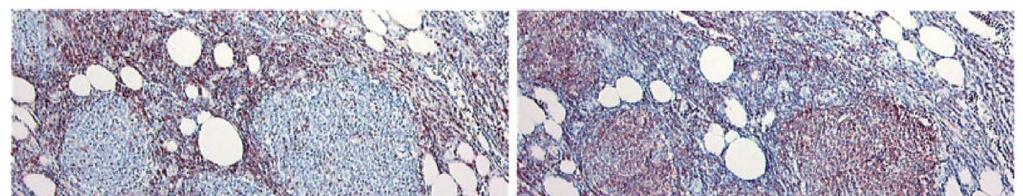
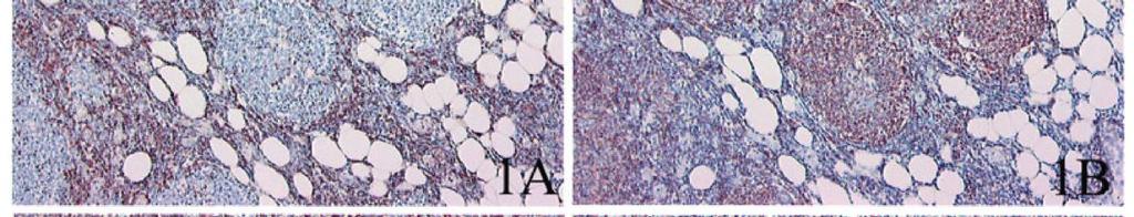
77 Basic stain: CD21 Membrane glycoprotein Normal: Mature B cells mantle zone & marginal zone B cells Lost on B-cell activation Follicular dendritic reticulum cells in GCs C3d/EBV receptor In lymphomas: most follicular lymphomas some other B-cell NHL FDC network in GC-derived tumours MCL, HL, AILD AILD-T-cell lymphoma AILD-T-cell lymphoma: CD21


78 Basic stain: CD21 Membrane glycoprotein Normal: Mature B cells mantle zone & marginal zone B cells Lost on B-cell activation Follicular dendritic reticulum cells in GCs C3d/EBV receptor In lymphomas: most follicular lymphomas some other B-cell NHL FDC network in GC-derived tumours MCL, HL, AILD AILD-T-cell lymphoma AILD-T-cell lymphoma: CD21
79 Nodal PTCL - immunophenotype
80 Oncogenes/ Tumor Suppressor Genes Evaluation by Immunohistochemistry Bcl-2: Follicular lymphoma, t(14;18) antigen expression not specific for translocation Cyclin D1: Mantle cell lymphoma, t(11;14); myelomas (15%) p53: Progression in lymphomas, high grade lymphomas Bcl-6: Germinal center origin c-myc cell of origin staining in DLBCL Prognosis in DLBCL double hit & double-expressor lymphomas (with Bcl-2) ALK-1: ALCL; NPM/ALK (t2;5) CD99: Lymphoblastic, myeloblastic c-myc bcl-2
81 IHC for lymphoma vs other Add to basic panel: panck S-100 Melan-A

 Myeloid")
82 IHC for lymphoid vs myeloid Add to basic panel Myeloperoxidase CD43 CD68 CD163 CD33 (CD14, CD15, CD34, CD61, glycophorin C) Myeloid Sarcoma CD33


83 Myeloid sarcoma: testis Myeloid sarcoma: testis CD33 Myeloperoxidase Lysozyme


 HL")

 B-CLL")
84 Targeted therapy Rituximab (anti-cd20) B-cell NHL FL: CD20 Brentuximab (anti-cd30) HL ALCL CD30+ DLBCL ALCL: CD30 Alemtuzumab (anti-cd52) B-CLL T-cell lymphoma T-cell lymphomas: CD52

85 Immune checkpoint inhibitory therapy? PD-1 AILD Hodgkin PD-L1 PD-1 AILD: PD-1 Gravelle P, et al. Oncotarget 8.27 (2017):
86 Thanks!
Classification! Immunohistochemical classification of haematolymphoid tumours. Malignant lymphoproliferative diseases
 Immunohistochemical classification of haematolymphoid tumours Haematolymphoid Neoplasias: Leukaemia vs Lymphoma C L O N A L M A L I G N A N C I E S Stephen Hamilton-Dutoit Institute of Pathology Aarhus
Immunopathology of Lymphoma
 Immunopathology of Lymphoma Noraidah Masir MBBCh, M.Med (Pathology), D.Phil. Department of Pathology Faculty of Medicine Universiti Kebangsaan Malaysia Lymphoma classification has been challenging to pathologists.
Aggressive B-Cell Lymphomas
 Aggressive B-cell Lymphomas Aggressive B-Cell Lymphomas Stephen Hamilton Dutoit Institute of Pathology Aarhus Kommunehospital B-lymphoblastic lymphoma Diffuse large cell lymphoma, NOS T-cell / histiocyte-rich;
Small B-cell (Histologically Low Grade) Lymphoma
 Lymphoma") Frequency of Lymphoid Neoplasms Small B-cell (Histologically Low Grade) Lymphoma Stephen Hamilton-Dutoit Institute of Pathology Aarhus University Hospital B-cell neoplasms 88% Diffuse large B-cell lymphoma
Frequency of Lymphoid Neoplasms Small B-cell (Histologically Low Grade) Lymphoma Stephen Hamilton-Dutoit Institute of Pathology Aarhus University Hospital B-cell neoplasms 88% Diffuse large B-cell lymphoma
Differential diagnosis of hematolymphoid tumors composed of medium-sized cells. Brian Skinnider B.C. Cancer Agency, Vancouver General Hospital
 Differential diagnosis of hematolymphoid tumors composed of medium-sized cells Brian Skinnider B.C. Cancer Agency, Vancouver General Hospital Lymphoma classification Lymphoma diagnosis starts with morphologic
Non-Hodgkin s Lymphomas Version
 NCCN Clinical Practice Guidelines in Oncology (NCCN Guidelines ) Non-Hodgkin s Lymphomas Version 2.2015 NCCN.org Continue Use of Immunophenotyping/ Genetic Testing in Differential Diagnosis of Mature B-Cell
Methods used to diagnose lymphomas
 Institut für Pathologie Institut für Pathologie Methods used to diagnose lymphomas Prof. Dr.Med. Leticia Quintanilla-Fend Molecular techniques NGS histology Cytology AS-PCR Sanger seq. MYC Immunohistochemistry
Lymphoma Update: Lymphoma Update: What s Likely to be New in the New WHO. Patrick Treseler, MD, PhD University of California San Francisco
 Lymphoma Update: What s Likely to be New in the New WHO Blood 127:2375; 2016 Patrick Treseler, MD, PhD University of California San Francisco Lymphoma Update: What IS New in the New WHO! Patrick Treseler,
7 Omar Abu Reesh. Dr. Ahmad Mansour Dr. Ahmad Mansour
 7 Omar Abu Reesh Dr. Ahmad Mansour Dr. Ahmad Mansour -Leukemia: neoplastic leukocytes circulating in the peripheral bloodstream. -Lymphoma: a neoplastic process in the lymph nodes, spleen or other lymphatic
Approach to Core Biopsy Specimens
 BDIAP 108th Symposium on Haematopathology Joint Meeting of the BDIAP and BLPG at-bristol, Anchor Road, Harbourside, Bristol BS1 5DB 15th - 17th May 2014 Approach to Core Biopsy Specimens Dr Stefan Dojcinov
Many of the hematolymphoid disorders are derived
 REVIEW ARTICLE Practical Immunohistochemistry in Hematopathology: A Review of Useful Antibodies for Diagnosis Ji Lu, MD and Karen L. Chang, MD Abstract: This review article offers some useful panels of
Hepatic Lymphoma Diagnosis An Algorithmic Approach
 Hepatic Lymphoma Diagnosis An Algorithmic Approach Ryan M. Gill, M.D., Ph.D. University of California, San Francisco PLEASE TURN OFF YOUR CELL PHONES Disclosure of Relevant Financial Relationships USCAP
FOLLICULARITY in LYMPHOMA
 FOLLICULARITY in LYMPHOMA Reactive Follicular Hyperplasia Follicular Hyperplasia irregular follicles Follicular Hyperplasia dark and light zones Light Zone Dark Zone Follicular hyperplasia MIB1 Follicular
Pearls and pitfalls in interpretation of lymphoid lesions in needle biopsies
 Pearls and pitfalls in interpretation of lymphoid lesions in needle biopsies Megan S. Lim MD PhD University of Pennsylvania October 8, 2018 Objectives To understand how the trend toward less invasive lymph
A Practical Guide To Diagnose B-Cell Lymphomas on FNAs. Nancy P. Caraway, M.D.
 A Practical Guide To Diagnose B-Cell Lymphomas on FNAs Nancy P. Caraway, M.D. Major Factors Impacting Dx Lymphomas on Small Bxs Classification systems Immunophenotyping by multiprobe flow cytometry and
High grade B-cell lymphomas (HGBL): Altered terminology in the 2016 WHO Classification (Update of the 4 th Edition) and practical issues Xiao-Qiu Li,
: Altered terminology in the 2016 WHO Classification (Update of the 4 th Edition) and practical issues Xiao-Qiu Li,") High grade B-cell lymphomas (HGBL): Altered terminology in the 2016 WHO Classification (Update of the 4 th Edition) and practical issues Xiao-Qiu Li, M.D., Ph.D. Fudan University Shanghai Cancer Center
High grade B-cell lymphomas (HGBL): Altered terminology in the 2016 WHO Classification (Update of the 4 th Edition) and practical issues Xiao-Qiu Li, M.D., Ph.D. Fudan University Shanghai Cancer Center
ISIMM Tata Conference on Immunohistochemistry. Kolkata, India, January Immunohistochemistry. A cost effective approach to lymphoma diagnosis
 ISIMM Tata Conference on Immunohistochemistry. Kolkata, India, January 2018 Immunohistochemistry A cost effective approach to lymphoma diagnosis Clive R. Taylor, M.D., Ph.D., Department of Pathology, Keck
3/23/2017. Disclosure of Relevant Financial Relationships. Pitfalls in Immunohistochemistry in Hematopathology: CD20 and CD3 Can Let Me Down?!
 Pitfalls in Immunohistochemistry in Hematopathology: CD20 and CD3 Can Let Me Down?! Judith A. Ferry Massachusetts General Hospital Disclosure of Relevant Financial Relationships USCAP requires that all
Defined lymphoma entities in the current WHO classification
 Defined lymphoma entities in the current WHO classification Luca Mazzucchelli Istituto cantonale di patologia, Locarno Bellinzona, January 29-31, 2016 Evolution of lymphoma classification Rappaport Lukes
VENTANA hematopathology solutions. Deliver diagnostic confidence
 VENTANA hematopathology solutions Deliver diagnostic confidence 2 Hematopathology diagnostic solutions Contents VENTANA hematopathology assays 3 Detecting and subtyping hematological cancers 4 The importance
Non-Hodgkin lymphomas (NHLs) Hodgkin lymphoma )HL)
 Hodgkin lymphoma )HL)") Non-Hodgkin lymphomas (NHLs) Hodgkin lymphoma )HL) Lymphoid Neoplasms: 1- non-hodgkin lymphomas (NHLs) 2- Hodgkin lymphoma 3- plasma cell neoplasms Non-Hodgkin lymphomas (NHLs) Acute Lymphoblastic Leukemia/Lymphoma
Non-Hodgkin lymphomas (NHLs) Hodgkin lymphoma )HL) Lymphoid Neoplasms: 1- non-hodgkin lymphomas (NHLs) 2- Hodgkin lymphoma 3- plasma cell neoplasms Non-Hodgkin lymphomas (NHLs) Acute Lymphoblastic Leukemia/Lymphoma
VENTANA hematopathology solutions Comprehensive aids for detecting and subtyping
 VENTANA hematopathology solutions Comprehensive aids for detecting and subtyping 1 12/4/2015 9:47:24 AM 2 Hematopathology diagnostic solutions Contents VENTANA hematopathology assays 3 Detecting and subtyping
Aggressive B-cell Lymphomas
 Neoplastic Hematopathology Update 2018 Aggressive B-cell Lymphomas Raju K. Pillai City of Hope National Medical Center I do not have any disclosures Disclosures Outline New entities and changes in WHO
DETERMINATION OF A LYMPHOID PROCESS
 Chapter 2 Applications of Touch Preparation Cytology to Intraoperative Consultations: Lymph Nodes and Extranodal Tissues for Evaluation of Hematolymphoid Disorders INTRODUCTION As discussed in Chap. 1,
Lymphoma/CLL 101: Know your Subtype. Dr. David Macdonald Hematologist, The Ottawa Hospital
 Lymphoma/CLL 101: Know your Subtype Dr. David Macdonald Hematologist, The Ottawa Hospital Function of the Lymph System Lymph Node Lymphocytes B-cells develop in the bone marrow and influence the immune
WHO UPDATE ON LYMPHOMAS. Dr Priya Mary Jacob Asst Professor, Pathology.
 WHO UPDATE ON LYMPHOMAS Dr Priya Mary Jacob Asst Professor, Pathology 3 rd 4 th 4 th revised 2001 2008 2017 The Change The Significance of the Change- Diagnostic, Prognostic The Rationale behind the change.
HODGKIN LYMPHOMA DR. ALEJANDRA ZARATE OSORNO HOSPITAL ESPAÑOL DE MEXICO
 HODGKIN LYMPHOMA DR. ALEJANDRA ZARATE OSORNO HOSPITAL ESPAÑOL DE MEXICO HODGKIN LYMPHOMA CLASSIFICATION Lukes & Butler Rye WHO-2016 Linphocytic and/or histiocytic Nodular & diffuse Nodular Sclerosis Lymphocyte
WHO Classification. B-cell chronic lymphocytic leukemia/small T-cell granular lymphocytic leukemia
 Blood Malignancies-II Prof. Dr. Herman Hariman, a Ph.D, SpPK (KH). Prof. Dr. Adikoesoema Aman, SpPK (KH) Dept. of Clinical Pathology, School of Medicine, University of North Sumatra WHO classification
Common Problem Areas. WHO Classification. Defines separate diseases (entities) with their CLINICAL AGGRESSIVENESS LOW GRADE / HIGH GRADE
 with their CLINICAL AGGRESSIVENESS LOW GRADE / HIGH GRADE") WHO Classification Defines separate diseases (entities) with their CLINICAL AGGRESSIVENESS REVIEW OF MOST COMMON LYMPHOMA ENTITIES Dr Stefan Dojcinov LOW GRADE / HIGH GRADE (June 2014) The Non-Hodgkin
WHO Classification Defines separate diseases (entities) with their CLINICAL AGGRESSIVENESS REVIEW OF MOST COMMON LYMPHOMA ENTITIES Dr Stefan Dojcinov LOW GRADE / HIGH GRADE (June 2014) The Non-Hodgkin
Burkitt lymphoma. Sporadic Endemic in Africa associated with EBV Translocations involving MYC gene on chromosome 8
 Heme 8 Burkitt lymphoma Sporadic Endemic in Africa associated with EBV Translocations involving MYC gene on chromosome 8 Most common is t(8;14) Believed to be the fastest growing tumor in humans!!!! Morphology
Aggressive B-cell Lymphoma 2013
 Aggressive B-cell Lymphoma 2013 Diffuse Large B-Cell Lymphoma Burkitt Lymphoblastic lymphoma Gray zone Intermediate DLBCL/HL Intermediate BL/DLBCL Diffuse Large B-cell lymphoma Common morphology: diffuse
Lymphoma Immunophenotyping: A New Era in Paraffin-Section Immunohistochemistry
 Advances in Anatomic Pathology Vol. 8, No. 4, pp. 218 239 2001 Lippincott Williams & Wilkins, Inc., Philadelphia Lymphoma Immunophenotyping: A New Era in Paraffin-Section Immunohistochemistry Eric D. Hsi
Contents. vii. Preface... Acknowledgments... v xiii
 Contents Preface... Acknowledgments... v xiii SECTION I 1. Introduction... 3 Knowledge-Based Diagnosis... 4 Systematic Examination of the Lymph Node... 7 Cell Type Identification... 9 Cell Size and Cellularity...
5003 Immunohistochemistry in hematopathology, what's in, what's out, what's useful
 www.ascp.org/ascp2014 5003 Immunohistochemistry in hematopathology, what's in, what's out, what's useful Kathryn Rizzo, DO, PhD VIRGINIA COMMONWEALTH UNIVERSITY Department of Pathology School of Medicine
Pathology #07. Hussein Al-Sa di. Dr. Sohaib Al-Khatib. Mature B-Cell Neoplasm. 0 P a g e
 Pathology #07 Mature B-Cell Neoplasm Hussein Al-Sa di Dr. Sohaib Al-Khatib 0 P a g e Thursday 18/2/2016 Our lecture today (with the next 2 lectures) will be about lymphoid tumors This is a little bit long
LYMPHOMAS an overview of some subtypes of NHLs
 One of the confusing aspects of the lymphoid neoplasms concerns the use of the descriptive terms "leukemia" and "lymphoma." LYMPHOMAS an overview of some subtypes of NHLs Leukemia is used for lymphoid
Lymphoma: What You Need to Know. Richard van der Jagt MD, FRCPC
 Lymphoma: What You Need to Know Richard van der Jagt MD, FRCPC Overview Concepts, classification, biology Epidemiology Clinical presentation Diagnosis Staging Three important types of lymphoma Conceptualizing
Large cell immunoblastic Diffuse histiocytic (DHL) Lymphoblastic lymphoma Diffuse lymphoblastic Small non cleaved cell Burkitt s Non- Burkitt s
 Lymphoblastic lymphoma Diffuse lymphoblastic Small non cleaved cell Burkitt s Non- Burkitt s") Non Hodgkin s Lymphoma Introduction 6th most common cause of cancer death in United States. Increasing in incidence and mortality. Since 1970, the incidence of has almost doubled. Overview The types of
Non Hodgkin s Lymphoma Introduction 6th most common cause of cancer death in United States. Increasing in incidence and mortality. Since 1970, the incidence of has almost doubled. Overview The types of
Incidence. Bimodal age incidence 15-40, >55 years Childhood form (0-14) more common in developing countries M:F=1.5:1; in all subtypes except NS
 more common in developing countries M:F=1.5:1; in all subtypes except NS") Hodgkin Lymphoma Hodgkin Lymphoma 30% of all lymphomas Absolute incidence unchanged Arise in lymph node, cervical region Neoplastic tissues usually contain a small number of tumor cells Incidence Bimodal
Hodgkin Lymphoma Hodgkin Lymphoma 30% of all lymphomas Absolute incidence unchanged Arise in lymph node, cervical region Neoplastic tissues usually contain a small number of tumor cells Incidence Bimodal
Aggressive B-cell Lymphomas Updated WHO classification Elias Campo
 Aggressive B-cell Lymphomas Updated WHO classification Elias Campo Hospital Clinic, University of Barcelona Diffuse Large B-cell Lymphoma A Heterogeneous Category Subtypes with differing: Histology and
11/2/2017. Immunodeficiencies. Joo Y. Song, MD Assistant Professor of Clinical Pathology. I have no financial disclosures.
 I have no financial disclosures Joo Y. Song, MD Assistant Professor of Clinical Pathology City of Hope National Medical Center Immunodeficiencies Transplant Autoimmunity Drugs T-cell dysfunction (Age,
Update on the Classification of Aggressive B-cell Lymphomas and Hodgkin Lymphoma
 Update on the Classification of Aggressive B-cell Lymphomas and Hodgkin Lymphoma Nancy Lee Harris, M. D. Massachusetts General Hospital Harvard Medical School Aggressive B-cell Lymphomas WHO 4 th Edition
EQA SCHEME CIRCULATION 33 EDUCATIONAL SLIDES DR GRAEME SMITH MONKLANDS DGH
 EQA SCHEME CIRCULATION 33 EDUCATIONAL SLIDES DR GRAEME SMITH MONKLANDS DGH CASE E1 M: 68 yrs Left destructive sinonasal lesion.?lymphoma?adenocarcinoma CD20 CD10 BCL6 MIB1 Answers Diffuse large B cell
PhenoPath. Diagnoses you can count on B CELL NON-HODGKIN LYMPHOMA
 PhenoPath Diagnoses you can count on B CELL NON-HODGKIN LYMPHOMA C urrent diagnosis of B cell non-hodgkin lymphoma (B-NHL) is based on the 2008 WHO Classification of Tumours of Haematopoietic and Lymphoid
T cell lymphoma diagnostics and differential diagnosis to Hodgkin lymphoma
 T cell lymphoma diagnostics and differential diagnosis to Hodgkin lymphoma Sylvia Hartmann Dr. Senckenberg Institute of Pathology Goethe University Frankfurt Overview Borderline ALCL classical HL Borderline
88-year-old Female with Lymphadenopathy. Faizi Ali, MD
 88-year-old Female with Lymphadenopathy Faizi Ali, MD Clinical History A 88-year-old caucasian female presented to our hospital with the complaints of nausea, vomiting,diarrhea, shortness of breath and
10/31/2017. Immunodeficiencies. Outline. Discuss EBV. Non-destructive Polymorphic Monomorphic Therapies Challenges
 I have no financial disclosures Joo Y. Song, MD Assistant Professor of Clinical Pathology City of Hope National Medical Center Immunodeficiencies Outline Transplant Congenital Autoimmunity T-cell/immune
Lymphoma: The Basics. Dr. Douglas Stewart
 Lymphoma: The Basics Dr. Douglas Stewart Objectives What is lymphoma? How common is it? Why does it occur? How do you diagnose it? How do you manage it? How do you follow patients after treatment? What
Molecular Pathology of Lymphoma (Part 1) Rex K.H. Au-Yeung Department of Pathology, HKU
 Rex K.H. Au-Yeung Department of Pathology, HKU") Molecular Pathology of Lymphoma (Part 1) Rex K.H. Au-Yeung Department of Pathology, HKU Lecture outline Time 10:00 11:00 11:15 12:10 12:20 13:15 Content Introduction to lymphoma Review of lymphocyte biology
Molecular Pathology of Lymphoma (Part 1) Rex K.H. Au-Yeung Department of Pathology, HKU Lecture outline Time 10:00 11:00 11:15 12:10 12:20 13:15 Content Introduction to lymphoma Review of lymphocyte biology
The next lymphoma classification Luca Mazzucchelli Istituto cantonale di patologia, Locarno
 Evolution of classification The next classification Luca Mazzucchelli Istituto cantonale di patologia, Locarno The Lymphoma Forum of Excellence, Bellinzona, January 2011 Rappaport Lukes and Collins (immunophenotype)
Flow cytometric analysis of B-cell lymphoproliferative disorders
 Flow cytometric analysis of B-cell lymphoproliferative disorders David M. Dorfman, M.D., Ph.D. Department of Pathology Brigham and Women s Hospital and Harvard Medical School Boston, MA Objectives Review
Aggressive B cell Lymphomas
 Aggressive B cell Lymphomas I have nothing to disclose. Disclosures Raju K. Pillai City of Hope National Medical Center Outline WHO 2016 Classification Large B cell Lymphomas New entities and changes in
Anaplastic Large Cell Lymphoma (of T cell lineage)
") Anaplastic Large Cell Lymphoma (of T cell lineage) Definition T-cell lymphoma comprised of large cells with abundant cytoplasm and pleomorphic, often horseshoe-shaped nuclei CD30+ Most express cytotoxic
Anaplastic Large Cell Lymphoma (of T cell lineage) Definition T-cell lymphoma comprised of large cells with abundant cytoplasm and pleomorphic, often horseshoe-shaped nuclei CD30+ Most express cytotoxic
Primary Cutaneous CD30-Positive T-cell Lymphoproliferative Disorders
 Primary Cutaneous CD30-Positive T-cell Lymphoproliferative Disorders Definition A spectrum of related conditions originating from transformed or activated CD30-positive T-lymphocytes May coexist in individual
Aggressive B cell Lymphomas
 Aggressive B cell Lymphomas Raju K. Pillai City of Hope National Medical Center I have no disclosures Outline What is new in the WHO 2016 classification Insights from genomic studies Double Hit Lymphoma
HIGH GRADE B-CELL LYMPHOMA DAVID NOLTE, MD (PGY-2) HUSSAM AL-KATEB, PHD, FACMG DEBORAH FUCHS, MD
 HUSSAM AL-KATEB, PHD, FACMG DEBORAH FUCHS, MD") HIGH GRADE B-CELL LYMPHOMA DAVID NOLTE, MD (PGY-2) HUSSAM AL-KATEB, PHD, FACMG DEBORAH FUCHS, MD OUTLINE High grade B-cell lymphoma with MYC and BCL2 and/or BCL6 rearrangements Patient presentation 2008/2016
HIGH GRADE B-CELL LYMPHOMA DAVID NOLTE, MD (PGY-2) HUSSAM AL-KATEB, PHD, FACMG DEBORAH FUCHS, MD OUTLINE High grade B-cell lymphoma with MYC and BCL2 and/or BCL6 rearrangements Patient presentation 2008/2016
Pathology of the indolent B-cell lymphomas Elias Campo
 Pathology of the indolent B-cell lymphomas Elias Campo Hospital Clinic, University of Barcelona Small B-cell lymphomas Antigen selection NAIVE -B LYMPHOCYTE MEMORY B-CELL MCL FL LPL MZL CLL Small cell
The spectrum of flow cytometry of the bone marrow
 The spectrum of flow cytometry of the bone marrow Anna Porwit Lund University Faculty of Medicine Dept. of Clinical Sciences Div. Oncology and Pathology anna.porwit@med.lu.se Disclosure of speaker s interests
Lymphoid Neoplasms. Sylvie Freeman Department of Clinical Immunology, University of Birmingham
 Lymphoid Neoplasms Sylvie Freeman Department of Clinical Immunology, University of Birmingham Incidence of Haematological Malignancies UK2001 (CRUK) Malignancy New Cases All Cancers 271,000 Leukaemia 6,760
Lymph node cytopathology : A practical approach to lymphoproliferative disorders
 Lymph node cytopathology : A practical approach to lymphoproliferative disorders Koray Ceyhan, M.D Department of Pathology Faculty of Medicine Ankara University Ankara, Turkey Diagnostic use of FNA in
Classifications of lymphomas
 Classifications of lymphomas Lukes and Collins Kiel classification Working formulation REAL classification (1994) WHO classification (2000) WHO CLASSIFICATIONF OF NEOPLASMS HAEMATOPETIC AND LYMPHOID TISSUES
Mimics of Lymphoma in Routine Biopsies. I have nothing to disclose regarding the information to be reported in this talk.
 Mimics of Lymphoma in Routine Biopsies Patrick Treseler, MD, PhD Professor of Pathology University of California San Francisco I have nothing to disclose regarding the information to be reported in this
From Morphology to Molecular Pathology: A Practical Approach for Cytopathologists Part 1-Cytomorphology. Songlin Zhang, MD, PhD LSUHSC-Shreveport
 From Morphology to Molecular Pathology: A Practical Approach for Cytopathologists Part 1-Cytomorphology Songlin Zhang, MD, PhD LSUHSC-Shreveport I have no Conflict of Interest. FNA on Lymphoproliferative
Mimics of Lymphoma in Routine Biopsies. Mixed follicular and paracortical hyperplasia. Types of Lymphoid Hyperplasia
 Mimics of Lymphoma in Routine Biopsies Patrick Treseler, MD, PhD Professor of Pathology University of California San Francisco Types of Lymphoid Hyperplasia Follicular hyperplasia (B-cells) Paracortical
NEW ENTITIES IN AGGRESSIVE B CELL LYMPHOMA. Joon Seong Park, M.D. Dept. of Hematology-Oncology Ajou University School of Medicine
 NEW ENTITIES IN AGGRESSIVE B CELL LYMPHOMA Joon Seong Park, M.D. Dept. of Hematology-Oncology Ajou University School of Medicine Historical background of Lymphoma classification Rappaport classification
3/24/2017 DENDRITIC CELL NEOPLASMS: HISTOLOGY, IMMUNOHISTOCHEMISTRY, AND MOLECULAR GENETICS. Disclosure of Relevant Financial Relationships
 DENDRITIC CELL NEOPLASMS: HISTOLOGY, IMMUNOHISTOCHEMISTRY, AND MOLECULAR GENETICS Jason L. Hornick, M.D., Ph.D. Director of Surgical Pathology and Immunohistochemistry Brigham and Women s Hospital Professor
9/28/2017. Follicular Lymphoma and Nodal Marginal Zone Lymphoma. Follicular Lymphoma Definition. Low-Grade B-Cell Lymphomas in WHO Classification
 and L. Jeffrey Medeiros, MD DISCLOSURES I do not have anything to disclose Low-Grade B-Cell Lymphomas in WHO Classification Lymphoma Type Frequency Follicular lymphoma 22.1 % Extranodal MALT-lymphoma 7.6
During past decades, because of the lack of knowledge
 Staging and Classification of Lymphoma Ping Lu, MD In 2004, new cases of non-hodgkin s in the United States were estimated at 54,370, representing 4% of all cancers and resulting 4% of all cancer deaths,
FLOW CYTOMETRY PRINCIPLES AND PRACTICE. Toby Eyre Consultant Haematologist Oxford University Hospitals NHS Foundation Trust June 2018
 FLOW CYTOMETRY PRINCIPLES AND PRACTICE Toby Eyre Consultant Haematologist Oxford University Hospitals NHS Foundation Trust June 2018 Aims and Objectives Principles of flow cytometry Preparation Steps involved
Gray Zones and Double Hits Distinguishing True Burkitt Lymphoma from Other High-Grade B-NHLs Burkitt Lymphoma Burkitt-Like Lymphoma DLBCL Patrick Tres
 Gray Zones and Double Hits Distinguishing True Burkitt Lymphoma from Other High-Grade B-NHLs Burkitt Lymphoma Burkitt-Like Lymphoma DLBCL Patrick Treseler, MD, PhD University of California San Francisco
The History of Lymphoma Classification and the 2017 Revision
 The History of Lymphoma Classification and the 2017 Revision ESMO Perceptorship on Lymphoma, Lugano 2018 German Ott Department of Clinical Pathology, Robert-Bosch-Krankenhaus and Dr. Margarete Fischer-Bosch
Case year old male with abdominal lymphadenopathy Treated with 8 cycles of R-CHOP One year later B-symptoms and progressive disease
 Codirectors Tsieh Sun, M.D., FASCP Francisco Vega, M.D., Ph.D. Department of Hematopathology UT MD Anderson Cancer Center Houston Texas There is no conflict of interest involved in the content and presentation
Change Summary - Form 2018 (R3) 1 of 12
 1 of 12") Summary - Form 2018 (R3) 1 of 12 Form Question Number (r3) Type Description New Text Previous Text Today's date was removed 2018 N/A Today's Date Removed from Key Fields 2018 N/A HCT Type 2018 N/A Product
Summary - Form 2018 (R3) 1 of 12 Form Question Number (r3) Type Description New Text Previous Text Today's date was removed 2018 N/A Today's Date Removed from Key Fields 2018 N/A HCT Type 2018 N/A Product
Exploring the Borderlands between Diffuse Large B-cell Lymphoma and Classical Hodgkin s Lymphoma
 Exploring the Borderlands between Diffuse Large B-cell Lymphoma and Classical Hodgkin s Lymphoma Elaine S. Jaffe National Cancer Institute Bethesda, MD, USA On the Pathological Changes In Hodgkin s Disease
Plasma cell myeloma (multiple myeloma)
") Plasma cell myeloma (multiple myeloma) Common lymphoid neoplasm, present at old age (70 years average) Remember: plasma cells are terminally differentiated B-lymphocytes that produces antibodies. B-cells
Plasma cell myeloma (multiple myeloma) Common lymphoid neoplasm, present at old age (70 years average) Remember: plasma cells are terminally differentiated B-lymphocytes that produces antibodies. B-cells
Corrigenda. WHO Classification of Tumours of Haematopoietic and Lymphoid Tissues (revised 4th edition): corrections made in second print run
: corrections made in second print run") Corrigenda WHO Classification of Tumours of Haematopoietic and Lymphoid Tissues (revised 4th edition): corrections made in second print run In addition to corrections of minor typographical errors, corrections
Corrigenda WHO Classification of Tumours of Haematopoietic and Lymphoid Tissues (revised 4th edition): corrections made in second print run In addition to corrections of minor typographical errors, corrections
Clinical Policy: Bendamustine (Bendeka, Treanda) Reference Number: PA.CP.PHAR.307
 Reference Number: PA.CP.PHAR.307") Clinical Policy: (Bendeka, Treanda) Reference Number: PA.CP.PHAR.307 Effective Date: 01/18 Last Review Date: 11/17 Coding Implications Revision Log Description The intent of the criteria is to ensure that
Clinical Policy: (Bendeka, Treanda) Reference Number: PA.CP.PHAR.307 Effective Date: 01/18 Last Review Date: 11/17 Coding Implications Revision Log Description The intent of the criteria is to ensure that
Methotrexate-associated Lymphoproliferative Disorders
 Methotrexate-associated Lymphoproliferative Disorders Definition A lymphoid proliferation or lymphoma in a patient immunosuppressed with methotrexate, typically for treatment of autoimmune disease (rheumatoid
Diffuse large B-cell lymphoma (DLBCL) is one of the
 is one of the") Practical Applications in Immunohistochemistry Evaluation of Diffuse Large B-Cell Lymphoma and Related Large B-Cell Lymphomas Dennis P. O Malley, MD; Aaron Auerbach, MD; Lawrence M. Weiss, MD Context.
Practical Applications in Immunohistochemistry Evaluation of Diffuse Large B-Cell Lymphoma and Related Large B-Cell Lymphomas Dennis P. O Malley, MD; Aaron Auerbach, MD; Lawrence M. Weiss, MD Context.
WHO 4th ED Classification of Mature B-cell Neoplasms
 WHO 4th ED Classification of Mature B-cell Neoplasms Chronic lymphocytic leukemia /Small lymphocytic lymphoma B-cell prolymphocytic leukaemia Splenic marginal zone lymphoma Hairy cell leukemia Splenic
Aggressive B-cell lymphomas and gene expression profiling towards individualized therapy?
 Aggressive B-cell lymphomas and gene expression profiling towards individualized therapy? Andreas Rosenwald Institute of Pathology, University of Würzburg, Germany Barcelona, June 18, 2010 NEW WHO CLASSIFICATION
Approach at lymph node pathology and ancillary techniques
 Approach at lymph node pathology and ancillary techniques Hans Konrad Müller-Hermelink Institute of Pathology, University of Würzburg Würzburg, Germany 1000 km 400 km Würzburg Germany: 80.000.000 population
Integrated Hematopathology. Morphology and FCI with IHC
 Integrated Hematopathology Morphology and FCI with IHC FrontMatter.indd i 9/6/2009 9:30:12 PM FrontMatter.indd ii 9/6/2009 9:30:18 PM Integrated Hematopathology Morphology and FCI with IHC Cherie H Dunphy,
Low-grade B-cell lymphoma
 Low-grade B-cell lymphoma Patho-Basic 11. September 2018 Stephan Dirnhofer Pathology Outline Definition LPL, MBL/CLL/SLL, MCL FL Subtypes & variants Diagnosis including Grading Transformation Summary Be
Monoclonal B-cell Lymphocytosis
 Entity Centred Approach Lymphoma Classification: WHO and Beyond Clinically meaningful categories Dr Stefan Dojcinov University Hospital of Wales, Cardiff WHO UPDATE - NEW ENTITIES Early lesions lymphoma
Pathology of aggressive lymphomas
 Institute of Pathology Pathology of aggressive lymphomas Leticia Quintanilla-Martinez Changes in the new 2016 WHO Aggressive B-cell lymphoid neoplasms Major changes that impact how cases should be evaludated
Combinations of morphology codes of haematological malignancies (HM) referring to the same tumour or to a potential transformation
 referring to the same tumour or to a potential transformation") Major subgroups according to the World Health Organisation (WHO) Classification Myeloproliferative neoplasms (MPN) Myeloid and lymphoid neoplasms with eosinophilia and abnormalities of PDGFRA, PDGFRB or
Major subgroups according to the World Health Organisation (WHO) Classification Myeloproliferative neoplasms (MPN) Myeloid and lymphoid neoplasms with eosinophilia and abnormalities of PDGFRA, PDGFRB or
Diagnostic Molecular Pathology of Lymphoid Neoplasms
 Diagnostic Molecular Pathology of Lymphoid Neoplasms (Part II) Rational use of molecular testing in lymphomas Beirut, Lebanon Friday December 2, 2011: Hematopathology Session Adam Bagg University of Pennsylvania
HENATOLYMPHOID SYSTEM THIRD YEAR MEDICAL STUDENTS- UNIVERSITY OF JORDAN AHMAD T. MANSOUR, MD. Parts 2 and 3
 HENATOLYMPHOID SYSTEM THIRD YEAR MEDICAL STUDENTS- UNIVERSITY OF JORDAN AHMAD T. MANSOUR, MD Parts 2 and 3 NEOPLASTIC LYMPHOID DISEASES Introduction o The bone marrow is the source of all cells in the
Thomas Hodgkin and Hodgkin lymphoma
 J Hematopathol (2014) 7:123 138 DOI 10.1007/s12308-014-0214-3 REVIEW ARTICLE Thomas Hodgkin and Hodgkin lymphoma Judith A. Ferry Received: 26 June 2014 /Accepted: 31 July 2014 /Published online: 12 August
Classification of Hematologic Malignancies. Patricia Aoun MD MPH
 Classification of Hematologic Malignancies Patricia Aoun MD MPH Objectives Know the basic principles of the current classification system for hematopoietic and lymphoid malignancies Understand the differences
Prevalent lymphomas in Africa
 Prevalent lymphomas in Africa Dr Zainab Mohamed Clinical Oncologist GSH/UCT Groote Schuur Hospital Disclaimer I declare that I have no conflict of interest Groote Schuur Hospital Denis Burkitt 1911-1993
Diagnosis of lymphoid neoplasms has been
 Iranian Journal of Pathology (2007)2 (1), 1-61 Review Article Mehdi Nassiri Dep. of Pathology, University of Miami Miller School of Medicine, Miami, USA Abstract Correct diagnosis and classification of
GENETIC MARKERS IN LYMPHOMA a practical overview. P. Heimann Dpt of Medical Genetics Erasme Hospital - Bordet Institute
 GENETIC MARKERS IN LYMPHOMA a practical overview P. Heimann Dpt of Medical Genetics Erasme Hospital - Bordet Institute B and T cell monoclonalities Rearrangement of immunoglobin and TCR genes may help
Lymphoma and Pseudolymphoma
 Lymphoma and Pseudolymphoma Laura B. Pincus, MD Co-Director, Cutaneous Lymphoma Clinic Associate Professor Dermatology and Pathology University of California, San Francisco I HAVE NO RELEVANT RELATIONSHIPS
New Concepts and Clinical Implications of the 2008 WHO Classification of Lymphomas Elaine S. Jaffe, M.D.
 New Concepts and Clinical Implications of the 2008 WHO Classification of Lymphomas Elaine S. Jaffe, M.D. The first step in wisdom is to know the things themselves; this notion consists in having a true
The development of clonality testing for lymphomas in the Bristol Genetics Laboratory. Dr Paula Waits Bristol Genetics Laboratory
 The development of clonality testing for lymphomas in the Bristol Genetics Laboratory Dr Paula Waits Bristol Genetics Laboratory Introduction The majority of lymphoid malignancies belong to the B cell
Low-Grade B-Cell Lymphomas in WHO Classification. Follicular Lymphoma Definition. Follicular Lymphoma Clinical Features 11/7/2017 DISCLOSURES
 Low-Grade B-Cell Lymphomas in WHO Classification DISCLOSURES I do not have anything to disclose Lymphoma Type Frequency Follicular lymphoma 22.1 % Extranodal MALT-lymphoma 7.6 % Small lymphocytic lymphoma/cll
Lymphadenopathies mimicking lymphoma and vice versa. Session 3
 Lymphadenopathies mimicking lymphoma and vice versa Session T/ NK Cell System The human and rodent T and NK cell system is characterized by many highly specialized cell suopulations and functions, that
Case 3. Ann T. Moriarty,MD
 Case 3 Ann T. Moriarty,MD Case 3 59 year old male with asymptomatic cervical lymphadenopathy. These images are from a fine needle biopsy of a left cervical lymph node. Image 1 Papanicolaou Stained smear,100x.