TIPS AND PITFALLS IN PLAIN FILM INTERPRETATION
|
|
|
- Rebecca Washington
- 5 years ago
- Views:
Transcription


1 TIPS AND PITFALLS IN PLAIN FILM INTERPRETATION Dr Philip Touska MBBS, BMedSci(Hons), MRCS, DO-HNS, FRCR Radiology Fellow Guy s & St Thomas Hospitals
2 LEARNING OBJECTIVES Where do we go wrong? Common pitfalls Brief recap of basics Technique, adequacy and anatomy Cases Interactive session

3 Errare humanum est Seneca WHERE CAN WE GO WRONG? Technical E.g. Rotation, limits of technique Perceptive Missed finding, distraction by obvious abnormalities Interpretative Misdiagnosis
4 SYSTEMATIC APPROACH AABCDEF Adequacy (technical quality) Airways Breathing (lungs) Circulation (heart and vessels & hila) Densities (bones/prostheses/surgical clips) Edges (incl. skin/breasts) Fundus of stomach (gastric bubble/under the diaphragm)
5


6 PA radiograph = outpatients and some inpatients AP radiograph = most inpatients Magnification ~22% Sources: Fundamentals of radiography. (1) (2) Young-Cheol J et al. IJST 2015.


7 Leftward rotation = slightly enlarged cardiac silhouette
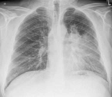
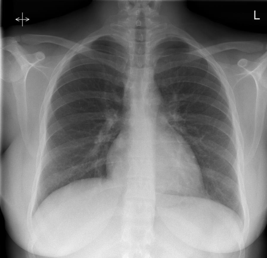
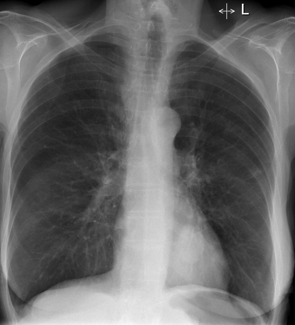
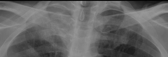
8 CHEST RADIOGRAPH
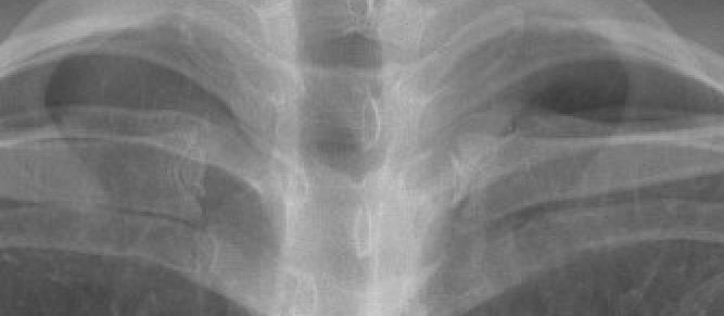
9 Airway Bronchi
10 Lungs and fissure

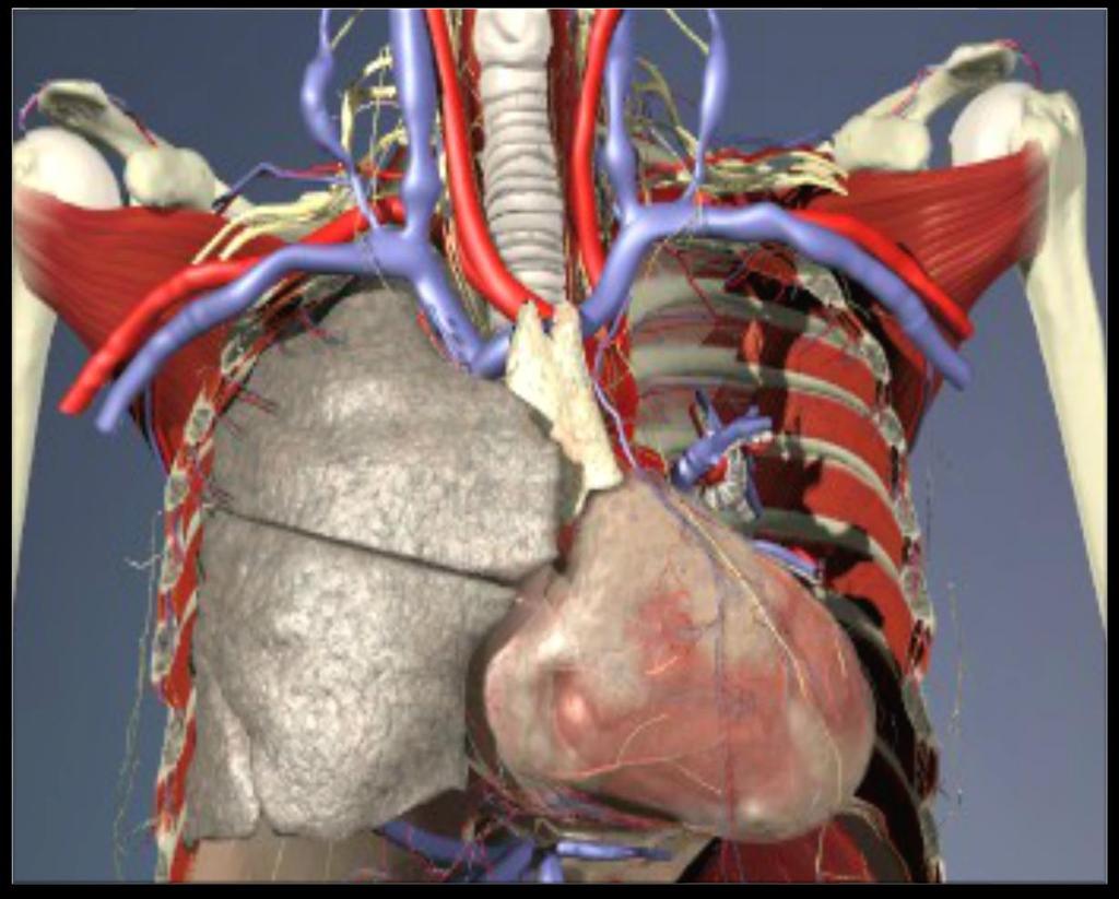
11 Left subclavian a. SVC Azygous vein Pulmonary veins Pulmonary arteries Thoracic aorta RA, LAA, LV
12 Scapula Ribs Vertebrae Gastric bubble
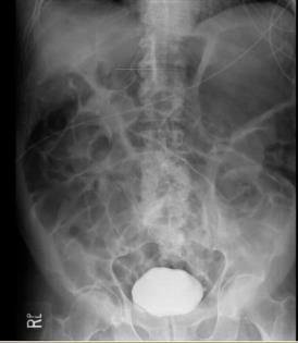
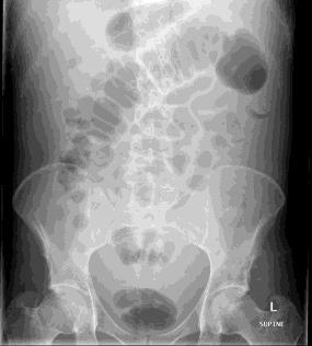
13 SYSTEMATIC APPROACH ABDO X Air (in right place) Bowel (wall thickness) Dense structures (bones/calcification) Organs external object/artefacts

14 Gastric bubble Caecum Ascending colon Transverse colon Descending colon Sigmoid colon & rectum Liver L kidney R psoas muscle
15 INTERACTIVE CASES Fingers on buzzers please

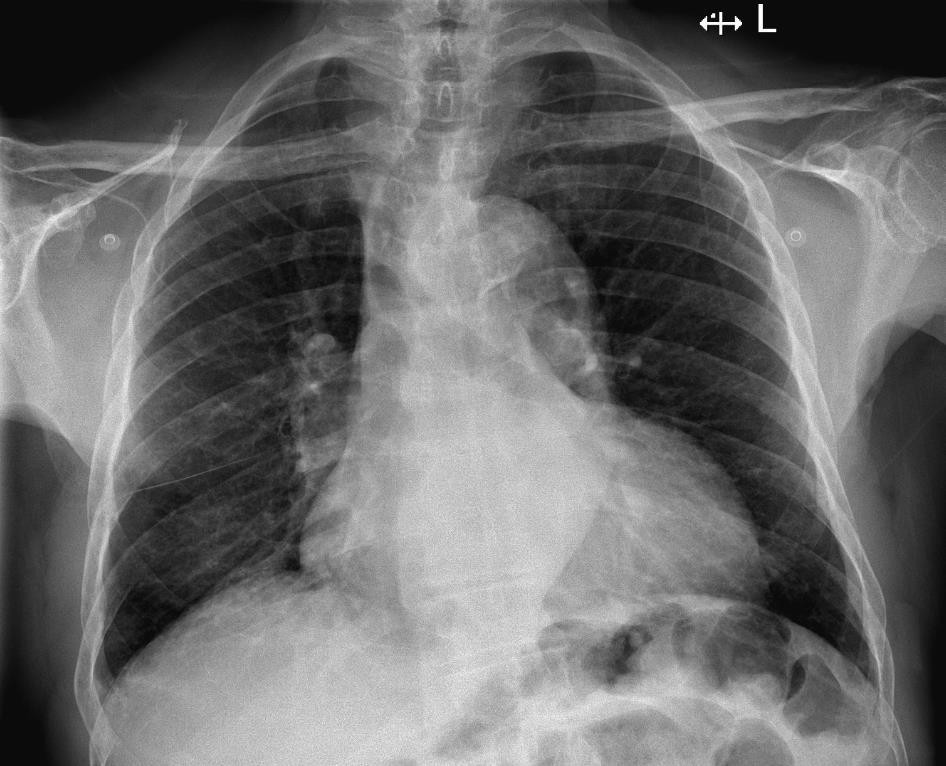
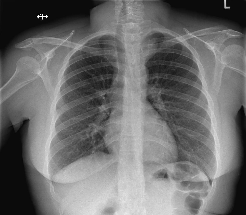
16 43 year old SOB
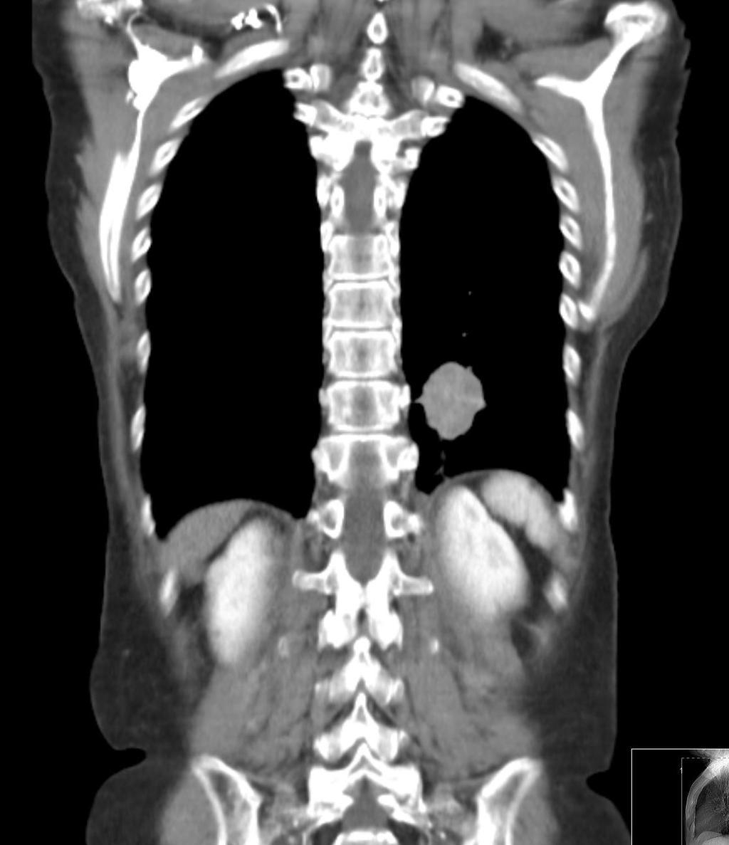
17 WHAT IS THE DIAGNOSIS? 1. Left upper lobe collapse 2. Upper mediastinal mass 3. Aortic rupture 4. Lymphadenopathy

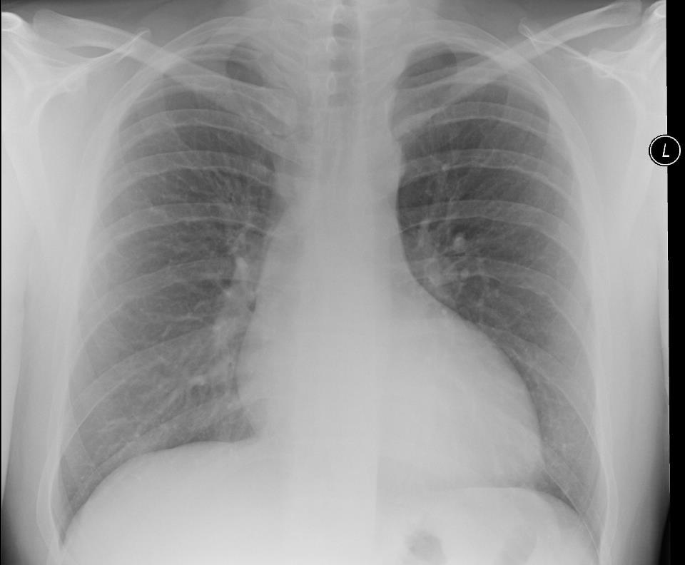
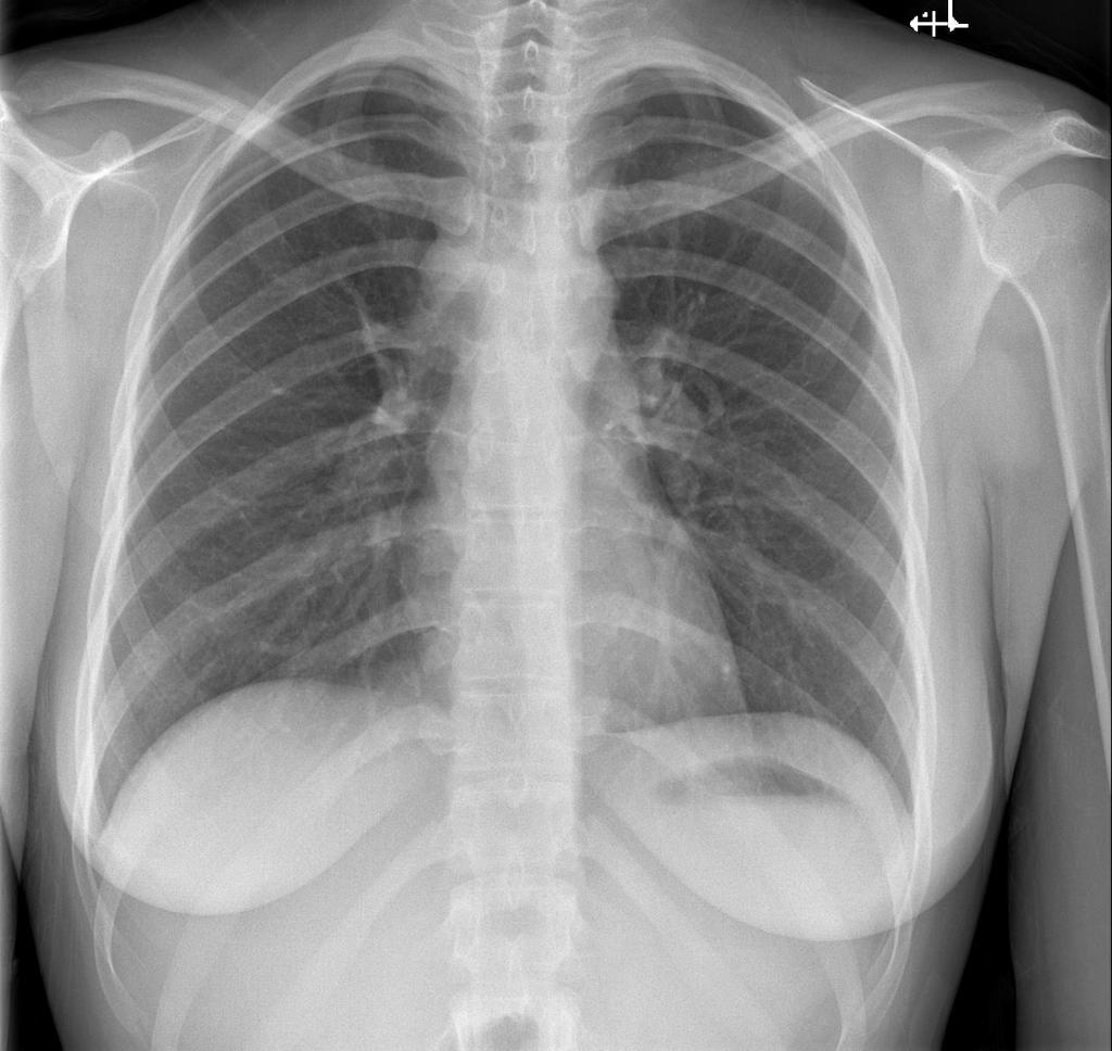
18 37 year old chest pain & pyrexia
19 WHAT IS THE MAIN DIAGNOSIS? 1. Right lower lobe consolidation 2. Middle lobe consolidation 3. Primary lung malignancy

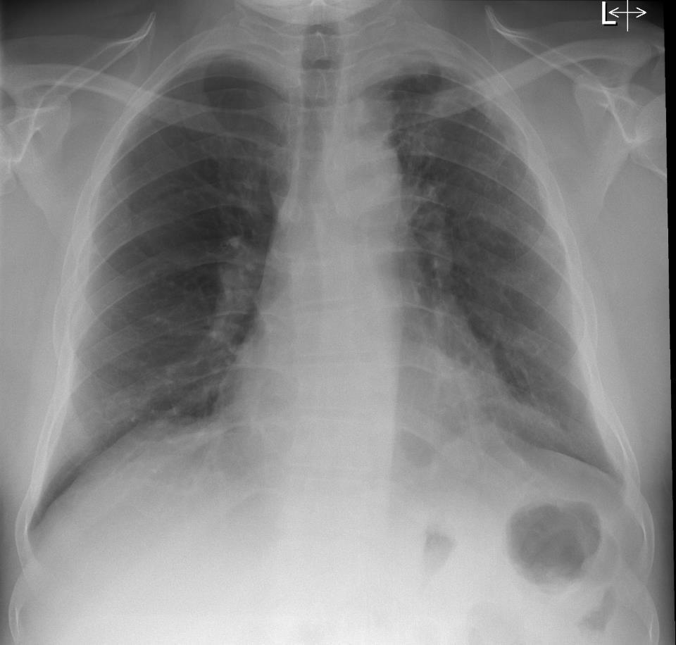
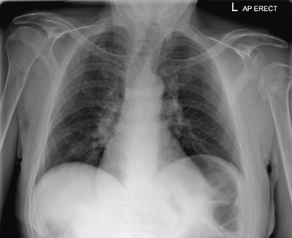
20 51 year old SOB and cough
21 WHERE IS THE MAIN ABNORMALITY? 1. Right upper zone 2. Left upper zone 3. Right middle zone 4. Left middle zone 5. Right lower zone 6. Left lower zone

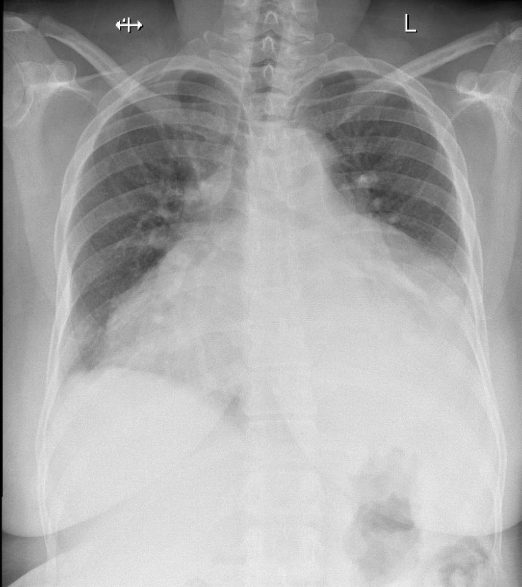
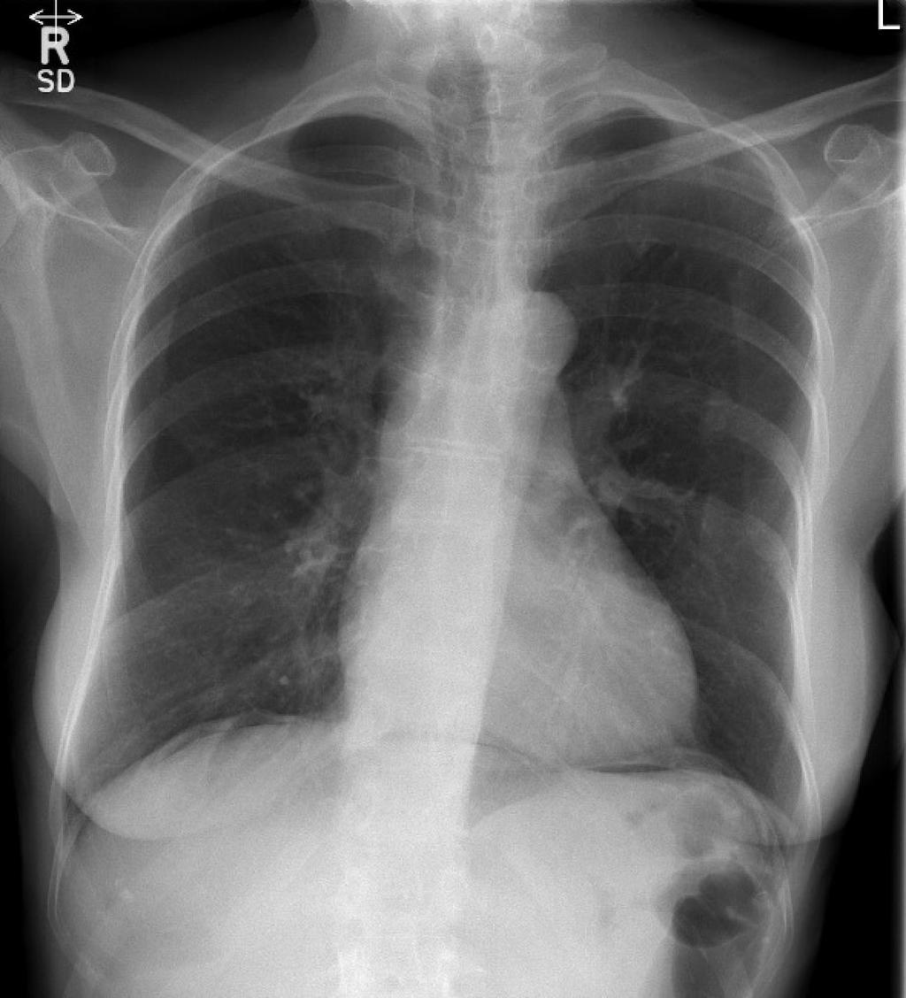
22 48 year old chest pain
23 WHERE IS THE MAIN ABNORMALITY? 1. Right upper zone 2. Left upper zone 3. Right middle zone 4. Left middle zone 5. Right lower zone 6. Left lower zone
24
25

26 65 year old SOB and cough
27 WHAT IS THE DIAGNOSIS? 1. Normal study 2. Tuberculosis 3. Primary lung cancer 4. Upper lobe collapse
28

29 53 year old SOB and cough
30 WHAT IS THE NATURE OF THE ABNORMALITY? 1. Pulmonary arterial hypertension 2. Lymphadenopathy 3. Primary lung cancer

31 38 year old acute chest pain
32 WHAT IS THE DIAGNOSIS? 1. Normal study 2. Pneumomediastinum/pneumopericardium
33
34

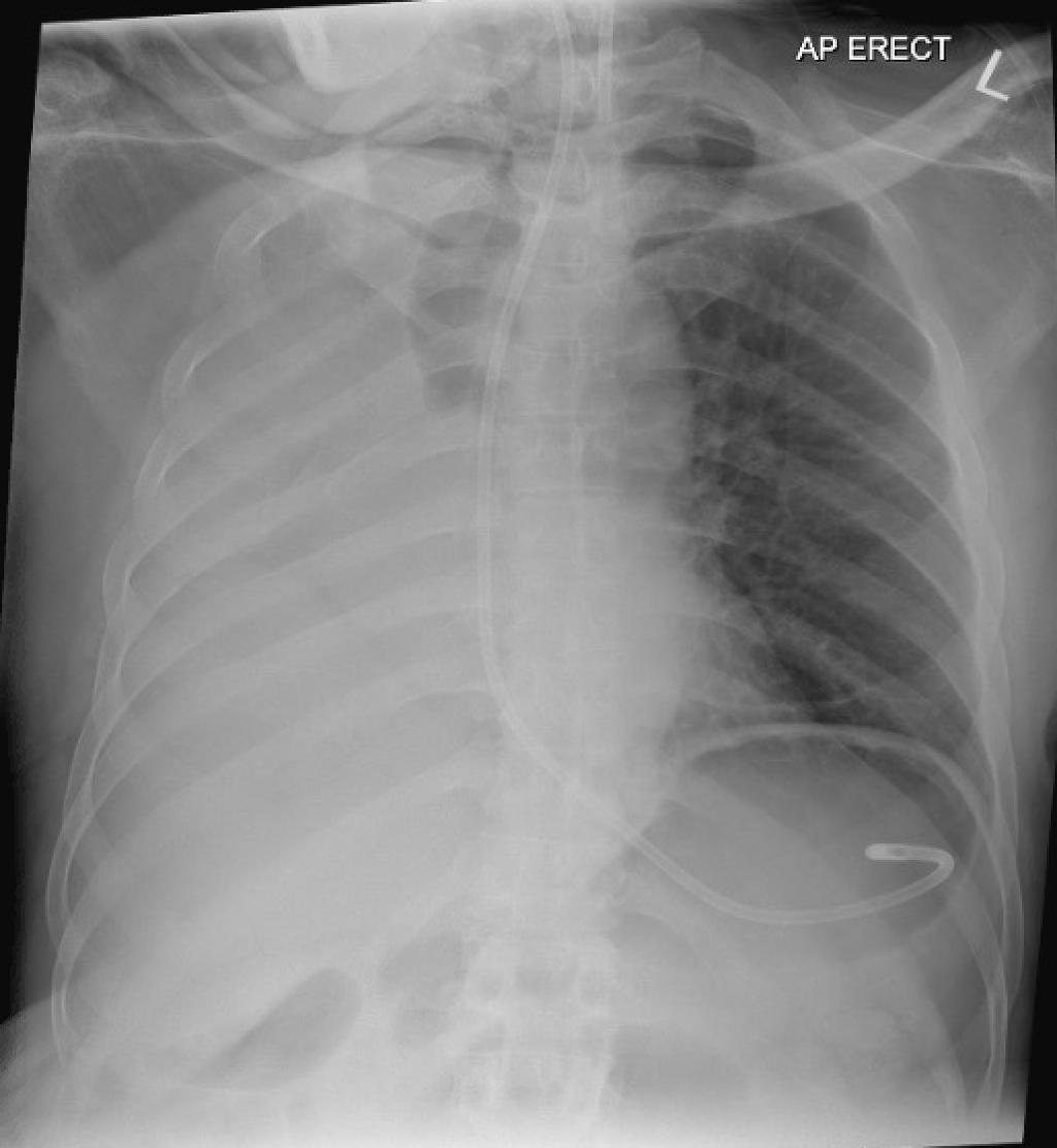
35 78 year old confused NG tube check
36 WHERE IS THE ABNORMALITY? 1. Airway 2. Lung parenchyma 3. Hila 4. Below the diaphragm

37 82 year old cough and SOB
38 WHAT IS THE DIAGNOSIS? 1. Left upper lobe collapse 2. Left lower lobe pneumonia 3. Left lower lobe collapse
39

40 56 year old - chest pain
41 HOW MANY ABNORMALITIES? 1. One 2. Two 3. Three

42 34 year old acute chest pain & cough
43 WHAT IS THE DIAGNOSIS? 1. Pneumonia 2. Acute cardiac failure 3. Sickle crisis 4. Hyperparathyroidism

44 47 year old SOB
45 WHAT IS THE DIAGNOSIS? 1. Acute pulmonary oedema 2. Incidental lung cancer 3. Combination of above
46

47 21 year old chest pain and SOB
48 WHAT IS THE DIAGNOSIS? 1. Lymphoma 2. Pneumonia 3. Tuberculosis

49 60 year old chest pain
50 WHERE IS THE ABNORMALITY? 1. Airway 2. Lungs 3. Mediastinum 4. Bones

51 19 year old chest pain and SOB
52 WHERE IS THE ABNORMALITY? 1. Right upper zone 2. Left upper zone 3. Right middle zone 4. Left middle zone

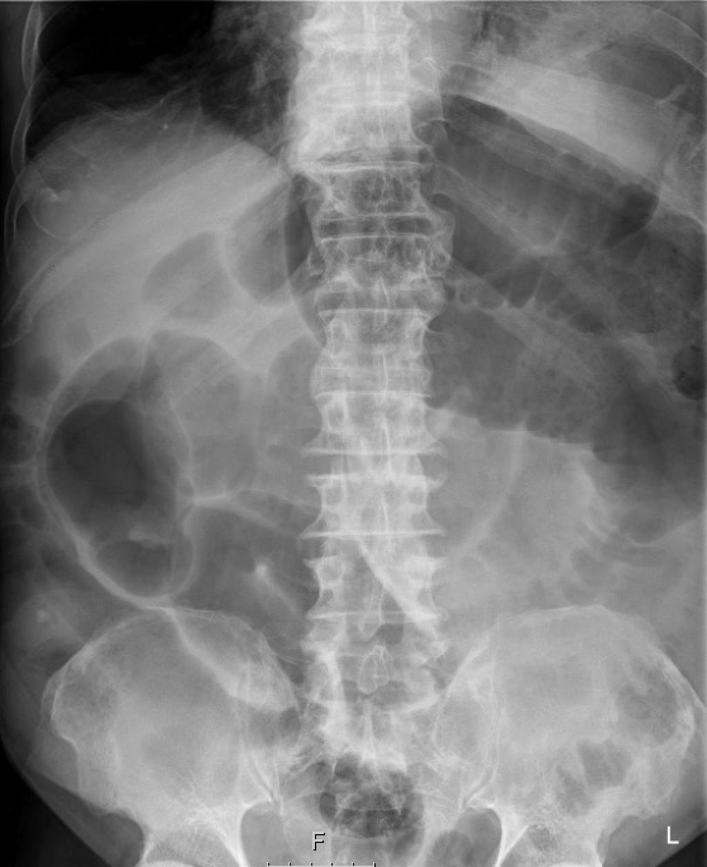
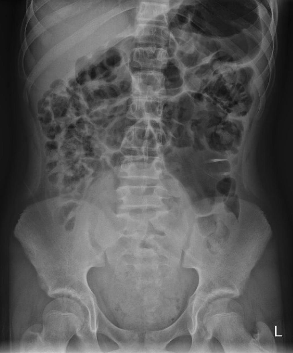
53 84 year old abdominal pain
54 IS THERE AN ACUTE ABNORMALITY ON THIS FILM? 1. Yes 2. No
55
56
57 WHAT IS THE DIAGNOSIS? 1. Small bowel obstruction 2. Sigmoid volvulus 3. Caecal volvulus 4. Perforation
58
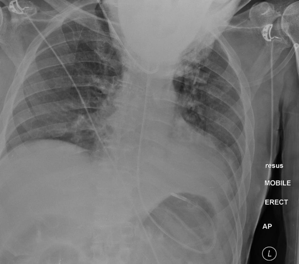
59
60

61 45 year old abdominal pain & PR bleeding
62 WHAT IS THE DIAGNOSIS? 1. Infective colitis 2. Ischaemia 3. Inflammatory bowel disease 4. Any of the above

63 56 year old abdominal pain
64

65 57 year old abdominal pain
66 SUMMARY AVOIDING PITFALLS Poor/limited technique Rotation, limited inspiration, AP projection, limitations of the technique Perceptive errors Missed finding, distraction by obvious abnormalities Interpretative errors Misdiagnosis Solution: Know the limitations of the study/technique consider other e.g. CT tekn Solution: Use a systematic approach; avoid satisfaction of search Solution: Consider alternatives, phone a friend!

67 THANK YOU

68 SILHOUETTES
69 WHAT TO LOOK FOR? 1. Bowel Is it small bowel or large bowel? Is it dilated? 3, 6, 9 cm rule Does it look normal? 2. Is there air where it shouldn t be? - Perforation 3. Densities (stones/surgical clips/foreign bodies) 4. Anything else? Bones/soft tissues/organs/masses

 Valvulae conniventes")
70 SMALL BOWEL Characteristics Often invisible under normal circumstances Position: central Size: small (max 3cm diameter) Valvulae conniventes (uninterupted lines)

 Haustra")
71 LARGE BOWEL Characteristics Usually visible under normal circumstances faecal shadowing Position: peripheral (note: sigmoid & transverse colons are floppy may be central) Size: large (max diameters: 6cm transverse colon, 9cm caecum) Haustra (interrupted lines)
72

73
74

75
76
77
78
79

80 39 year old SOB and cough
81
82
83
Undergraduate Teaching
 Prof. James F Meaney Undergraduate Teaching Chest X-Ray Understanding the normal anatomical by reference to cross sectional imaging Radiology? It s FUN! Cryptic puzzle Sudoku (Minecraft?) It s completely
Chest X-ray Interpretation
 Chest X-ray Interpretation Introduction Routinely obtained Pulmonary specialist consultation Inherent physical exam limitations Chest x-ray limitations Physical exam and chest x-ray provide compliment
X-rays. Dr Will Dooley
 X-rays Dr Will Dooley Plan Chest X-Rays Abdominal X-Rays Exam approach Presentation skills EMQ EMQ- answers Chest X-Ray - Systematic Approach D R Details RIP Image Quality +/- OBVIOUS ABNORMALITY A B C
Chest X-ray (CXR) Interpretation Brent Burbridge, MD, FRCPC
 Interpretation Brent Burbridge, MD, FRCPC") Chest X-ray (CXR) Interpretation Brent Burbridge, MD, FRCPC An approach to reviewing a chest x-ray will create a foundation that will facilitate the detection of abnormalities. You should create your own
Chest X-ray (CXR) Interpretation Brent Burbridge, MD, FRCPC An approach to reviewing a chest x-ray will create a foundation that will facilitate the detection of abnormalities. You should create your own
CHEST & ABDOMINAL X-RAYS MALIKA IBRAHIM CORE MEDICAL TRAINEE BLACKPOOL VICTORIA HOSPITAL DATA INTERPRETATION COURSE FEB 20, 2017
 CHEST & ABDOMINAL X-RAYS MALIKA IBRAHIM CORE MEDICAL TRAINEE BLACKPOOL VICTORIA HOSPITAL DATA INTERPRETATION COURSE FEB 20, 2017 1. Sample x-rays 2. Basic chest x-ray interpretation skills 3. Chest x-ray
X-Rays. Kunal D Patel Research Fellow IMM
 X-Rays Kunal D Patel Research Fellow IMM The 12-Steps } 1: Name 2: Date 3: Old films 4: What type of view(s) 5: Penetration } Pre-read 6: Inspiration 7: Rotation Quality Control 8: Angulation 9: Soft tissues
Radiological Anatomy of Thorax. Dr. Jamila Elmedany & Prof. Saeed Abuel Makarem
 Radiological Anatomy of Thorax Dr. Jamila Elmedany & Prof. Saeed Abuel Makarem Indications for Chest x - A chest x-ray may be used to diagnose and plan treatment for various conditions, including: Diseases/Fractures
ASSESSING THE PLAIN ABDOMINAL RADIOGRAPH M A A M E F O S U A A M P O F O
 ASSESSING THE PLAIN ABDOMINAL RADIOGRAPH M A A M E F O S U A A M P O F O Introduction The abdomen (less formally called the belly, stomach, is that part of the body between the thorax (chest) and pelvis,
Chest XRay interpretation INTERPRETATIONS Identifications: Name & Date Technical evaluation Basic Interpretations
 Chest XRay interpretation INTERPRETATIONS Identifications: Name & Date Technical evaluation Basic Interpretations TECHNICAL EVALUATION 1. Projection: AP/PA view To differentiate between AP & PA films,
Approach to CXR. Terminology. 1.Identification. Greg Blecher SCH Respir Fellow. Correct patient Correct date and time Correct examination
 Approach to CXR Greg Blecher SCH Respir Fellow From Rob Posteraro http://home.earthlink.net/~rhpos/cxr_interpret.txt.html ; http://home.earthlink.net/~rhpos/cxr_main.txt.html) Approach to viewing Chest
Introduction and Definitions
 Bowel obstruction Introduction and Definitions Accounts for 5% of all acute surgical admissions Patients are often extremely ill requiring prompt assessment, resuscitation and intensive monitoring Obstruction
FUNDAMENTALS OF CXR INTERPRETATION THE BASICS
 FUNDAMENTALS OF CXR INTERPRETATION THE BASICS PART I QUALITY ASSESSMENT 1 PATIENT-DEPENDENT FACTORS 3 REVIEW OF IMPORTANT ANATOMY 7 LUNGS AND PLEURA 11 DIAPHRAGMS 13 BONES AND SOFT TISSUES 14 A BRIEF LOOK
Disclosure. Clinical Chest Radiography Interpretation Part I
 Clinical Chest Radiography Interpretation Part I Anthony M. Angelow, PhD(c), MSN, ACNPC, AGACNP-BC, CEN Associate Lecturer, Fitzgerald Health Education Associates Clinical practice Division of Trauma Surgery
10/17/2016. Nuts and Bolts of Thoracic Radiology. Objectives. Techniques
 Nuts and Bolts of Thoracic Radiology October 20, 2016 Carleen Risaliti Objectives Understand the basics of chest radiograph Develop a system for interpreting chest radiographs Correctly identify thoracic
Image Interpretation and Evaluation
 Semester 2 / Commences January 20 Credits Each Course is composed of Modules & Activities. Modules: Image Interpretation Thoracic radiography Abdominal pelvic imaging Musculoskeletal Imaging Neuroimaging
Cardiac Radiography. Jared D. Christensen, M.D.
 Cardiac Radiography Jared D. Christensen, M.D. Cardiac radiography Jared D. Christensen, M.D. Overview Basic Concepts Technique Normal anatomy Cases Technique 3 Standard Views Posterior-Anterior (PA) Anterior-Posterior
Manage TB Dr. A. Chitrakumar Madras Medical College and RGGGH Institute of Thoracic Medicine, Chennai
 Manage TB Dr. A. Chitrakumar Madras Medical College and RGGGH Institute of Thoracic Medicine, Chennai Lecture 16 Radiology in diagnosis of Tuberculosis Session 01 So, welcome to the session Radiology in
Chest Radiology Interpretation: Findings of Tuberculosis
 Chest Radiology Interpretation: Findings of Tuberculosis Get out your laptops, smart phones or other devices pollev.com/chestradiology Case #1 1 Plombage Pneumonia Cancer 2 Reading the TB CXR Be systematic!
The Chest X-ray for Cardiologists
 Mayo Clinic & British Cardiovascular Society at the Royal College of Physicians, London : 21-23-October 2013 Cases-Controversies-Updates 2013 The Chest X-ray for Cardiologists Michael Rubens Royal Brompton
UERMMMC Department of Radiology. Basic Chest Radiology
 UERMMMC Department of Radiology Basic Chest Radiology PHYSICS DENSITIES BONE SOFT TISSUES WATER FAT AIR TELEROENTGENOGRAM Criteria for an Ideal Chest Radiograph 1. Upright 2. Posteroanterior View 3. Full
UNDERSTANDING X-RAYS: ABDOMINAL IMAGING THE ABDOMEN
 UNDERSTANDING X-RAYS: ABDOMINAL IMAGING THE ABDOMEN Radiology Enterprises radiologyenterprises@gmail.com www.radiologyenterprises.com STOMACH AND SMALL BOWEL STOMACH AND SMALL BOWEL Swallowed air is a
Learning Radiology: Recognizing the Basics. Text with Student Consult Online Access Code
 Learning Radiology: Recognizing the Basics. Text with Student Consult Online Access Code Herring, W ISBN-13: 9780323074445 Table of Contents 1. Recognizing Anything The "colorful" world of radiology A
Interpreting thoracic x-ray of the supine immobile patient: Syllabus
 Interpreting thoracic x-ray of the supine immobile patient: Syllabus Johannes Godt Dep. of Radiology and Nuclear Medicine Oslo University Hospital Ullevål NORDTER 2017, Helsinki Content - Why bedside chest
Lab Monitor Images Dissection of the Abdominal Vasculature + Lower Digestive System
 Lab Monitor Images Dissection of the Abdominal Vasculature + Lower Digestive System Stomach & Duodenum Frontal (AP) View Nasogastric tube 2 1 3 4 Stomach Pylorus Duodenum 1 Duodenum 2 Duodenum 3 Duodenum
Chest X rays and Case Studies. No disclosures. Outline 5/31/2018. Carlo Manalo, M.D. Department of Radiology Loma Linda University Children s Hospital
 Chest X rays and Case Studies Carlo Manalo, M.D. Department of Radiology Loma Linda University Children s Hospital No disclosures. Outline Importance of history Densities delineated on radiography An approach
Signs in Chest Radiology
 Signs in Chest Radiology Jonathan H. Chung, MD Disclosures No pertinent disclosures Jonathan H. Chung, MD Assistant Professor Institute t of fadvanced d Biomedical Imaging National Jewish Health Denver,
Plain abdomen The standard films are supine & erect AP views (alternative to erect, lateral decubitus film is used in ill patients).
.") Plain abdomen The standard films are supine & erect AP views (alternative to erect, lateral decubitus film is used in ill patients). The stomach can be readily identified by its location, gastric rugae
Plain abdomen The standard films are supine & erect AP views (alternative to erect, lateral decubitus film is used in ill patients). The stomach can be readily identified by its location, gastric rugae
Nasogastric tube. Stomach. Pylorus. Duodenum 1. Duodenum 2. Duodenum 3. Duodenum 4
 Esophagus Barium Swallow Stomach and Duodenum 4 year old Upper GI Nasogastric tube Stomach and Duodenum 4 year old Upper GI Nasogastric tube Stomach Pylorus Duodenum 1 Duodenum 2 Duodenum 3 Duodenum 4
B-I-2 CARDIAC AND VASCULAR RADIOLOGY
 (YEARS 1 3) CURRICULUM FOR RADIOLOGY 13 B-I-2 CARDIAC AND VASCULAR RADIOLOGY KNOWLEDGE To describe the normal anatomy of the heart and vessels including the lymphatic system as demonstrated by radiographs,
4/16/2017. Learning Objectives. Interpretation of the Chest Radiograph. Components. Production of the Radiograph. Density & Appearance
 Interpretation of the Arthur Jones, EdD, RRT Learning Objectives Identify technical defects in chest radiographs Identify common radiographic abnormalities This Presentation is Approved for 1 CRCE Credit
Disclosure. Clinical Chest Radiography Interpretation Part II
 Clinical Chest Radiography Interpretation Part II Anthony M. Angelow, PhD(c), MSN, ACNPC, AGACNP-BC, CEN Associate Lecturer, Fitzgerald Health Education Associates Clinical practice Division of Trauma
Lines and tubes. 1 Nasogastric tubes Endotracheal tubes Central lines Permanent pacemakers Chest drains...
 Lines and tubes 1 Nasogastric tubes... 15 2 Endotracheal tubes.... 19 3 Central lines... 21 4 Permanent pacemakers.... 25 5 Chest drains... 30 This page intentionally left blank 1 Nasogastric tubes Background
What s Your Diagnosis? Jessica Eisenbarth. Signalment: Jazz is a female intact 2 year old German Shorthaired Pointer.
 What s Your Diagnosis? Jessica Eisenbarth Signalment: Jazz is a female intact 2 year old German Shorthaired Pointer. Presenting complaint: Jazz was presented to the K-State emergency service on August
Dana Alrafaiah. - Moayyad Al-Shafei. -Mohammad H. Al-Mohtaseb. 1 P a g e
 - 6 - Dana Alrafaiah - Moayyad Al-Shafei -Mohammad H. Al-Mohtaseb 1 P a g e Quick recap: Both lungs have an apex, base, mediastinal and costal surfaces, anterior and posterior borders. The right lung,
Tests Your Pulmonologist Might Order. Center For Cardiac Fitness Pulmonary Rehab Program The Miriam Hospital
 Tests Your Pulmonologist Might Order Center For Cardiac Fitness Pulmonary Rehab Program The Miriam Hospital BASIC ANATOMY OF THE LUNGS Lobes of Lung 3 lobes on the Right lung 2 lobes on the Left Blood
Do you want to be an excellent Radiologist? - Focus on the thoracic aorta on lateral chest image!!!
 The lateral chest radiograph: Challenging area around the thoracic aorta!!! Do you want to be an excellent Radiologist? - Focus on the thoracic aorta on lateral chest image!!! Dong Yoon Han 1, So Youn
Chest and cardiovascular
 Module 1 Chest and cardiovascular A. Doss and M. J. Bull 1. Regarding the imaging modalities of the chest: High resolution computed tomography (HRCT) uses a slice thickness of 4 6 mm to identify mass lesions
Radiology. Undergraduate Radiology Sample Questions
 Radiology Undergraduate Radiology Sample Questions April 2012 The following examples are offered of questions that might be used to assess undergraduate radiology. There are 3 different styles: An OSCE
Radiology of the abdomen Lecture -1-
 Radiology of the abdomen Lecture -1- Objectives To know radiology modalities used in abdomen imaging mainly GI tract. To know advantages and disadvantages of each modality. To know indications and contraindications
Reporting SPECT-VQ. Alp Notghi
 Reporting SPECT-VQ Alp Notghi 20 year old female 24 weeks pregnant Clinical History : SOB and chest pain for past 3 days.?pe Doppler USS excluded DVT Case 4413041 Normal Case 4413041 CXR report: The heart
THE GOOFY ANATOMIST QUIZZES
 THE GOOFY ANATOMIST QUIZZES 7. LUNGS Q1. Fill in the blanks: the lung has lobes and fissures. A. Right, three, two. B. Right, two, one. C. Left, three, two. D. Left, two, three. Q2. The base of the lung
Interesting Cases. Pulmonary
 Interesting Cases Pulmonary 54M with prior history of COPD, hep B/C, and possible history of TB presented with acute on chronic dyspnea, and productive cough Hazy opacity overlying the left hemithorax
Interactive Lecture. Lecture 7 - Interactive. Radiology of cardiorespiratory disease. Editing File. Done By. Color Coding Important Notes Extra
 Lecture 7 - Interactive 436 Teams Interactive Lecture Radiology of cardiorespiratory disease Done By Team Leaders: Khalid Alshehri Hanin Bashaikh Team Members: Ghaida Alsaeed Maha Alissa Nawwaf AlHarbi
Acute Abdomen 急腹症 钱黎俊. Radiology, Renji Hospital. Shanghai Jiaotong University School of Medicine
 Acute Abdomen 急腹症 Radiology, Renji Hospital Shanghai Jiaotong University School of Medicine 钱黎俊 Learning objectives To understand the normal anatomy of the erect abdominal plain film To understand and
Interpretation of the chest radiograph Elizabeth Puddy MB ChB FCARCSI Catherine Hill MB ChB MRCP FRCR
 Interpretation of the chest radiograph Elizabeth Puddy MB ChB FCARCSI Catherine Hill MB ChB MRCP FRCR The traditional technique used in the acquisition and development of a chest radiograph uses methods
Objectives. What is a Chest X Ray? CXR Workshop. Definition (diagnostic tool/internal PE) Types. Cost
 Types. Cost") Objectives CAPA 2011 Christy Wilson, PA C Georgia Lung Associates Identify the radiographic landmarks on a chest radiograph Recognize identifiers of poor quality on the chest radiograph Outline an approach
Objectives CAPA 2011 Christy Wilson, PA C Georgia Lung Associates Identify the radiographic landmarks on a chest radiograph Recognize identifiers of poor quality on the chest radiograph Outline an approach
Introduction to Chest CT Interpretation. Objectives 8/28/2017
 Introduction to Chest CT Interpretation Deborah Stein ACNP BC, CCRN NP Education Specialist Department of Anesthesia and Critical Care Medicine August 28, 2017 Objectives Basic Principles Thoracic Anatomy
X-Rays. Prepared by Prof.Dr. Magda Hassab Allah Assist.lecturer Marwa Al Hady
 X-Rays Prepared by Prof.Dr. Magda Hassab Allah Assist.lecturer Marwa Al Hady CHEST X-RAYS Normal Chest X-ray Comments on chest X ray includes examination of 1- Bony cage (ribs,clavicles &vertebral column
Pitfalls of the Pediatric Chest and Abdomen SPR 2017
 Pitfalls of the Pediatric Chest and Abdomen SPR 2017 Richard I. Markowitz, MD, FACR Children s Hospital of Philadelphia Perelman School of Medicine University of Pennsylvania No Disclosures Cognitive Perceptual
After the Chest X-Ray:
 After the Chest X-Ray: What To Do Next Alan S. Brody Professor of Radiology and Pediatrics Chief of Thoracic Imaging Cincinnati Children s Hospital Cincinnati, Ohio USA What Should We Do Next? CT scan?
Abdominal radiology 腹部放射線學
 Abdominal radiology 腹部放射線學 台北醫學大學 - 市立萬芳醫院 留偉順 laowilson@hotmail.com The Normal Abdominal Series Chest Supine abdomen Erect abdomen Left lateral decubitus abdomen Learning objectives Understanding normal
Pulmonary Embolism. Thoracic radiologist Helena Lauri
 Pulmonary Embolism Thoracic radiologist Helena Lauri 8.5.2017 Statistics 1-2 out of 1000 adults annually are diagnosed with deep vein thrombosis (DVT) and/or pulmonary embolism (PE) About half of patients
DATA INTERPRETATION FOR MEDICAL STUDENTS
 DATA INTERPRETATION FOR MEDICAL STUDENTS CASES Second Edition Paul K Hamilton BSc(Hons), MB BCh BAO(Hons) MRCP(UK) MD Consultant Physician Belfast Health and Social Care Trust Belfast United Kingdom Ian
Sectional Anatomy Quiz - III
 Sectional Anatomy - III Rashid Hashmi * Rural Clinical School, University of New South Wales (UNSW), Wagga Wagga, NSW, Australia A R T I C L E I N F O Article type: Article history: Received: 30 Jun 2018
CT Chest. Verification of an opacity seen on the straight chest X ray
 CT Chest Indications: To assess equivocal plain x-ray findings Staging of lung neoplasm Merastatic workup of extra thoraces malignancies Diagnosis of diffuse lung diseases with HRCT Assessment of bronchietasis
Imaging of Respiratory Disorders: M2 Pathology correlated with Radiology
 Imaging of Respiratory Disorders: M2 Pathology correlated with Radiology by (c) Dr Goh Poh Sun MBBS(Melb), FRCR(UK), FAMS(Singapore), MHPE(Maastricht) Senior Consultant Radiologist and Associate Professor
Shades of Gray Interpretation of Perioperative Imaging
 Stanford Hospital and Clinics DEPARTMENT OF CARDIOTHORACIC SURGERY-THORACIC AORTIC SURGERY UNIT FALK CARDIOVASCULAR RESEARCH CENTER STANFORD, CALIFORNIA 94305-5407 MICHAEL SHEEHAN, MSN, RNFA, NPC TELEPHONE
Large veins of the thorax Brachiocephalic veins
 Large veins of the thorax Brachiocephalic veins Right brachiocephalic vein: formed at the root of the neck by the union of the right subclavian & the right internal jugular veins. Left brachiocephalic
CT Evaluation of Bowel Wall Thickening. Dr: Adel El Badrawy; M.D. Lecturer of Radio Diagnosis Faculty of Medicine Mansoura University.
 CT Evaluation of Bowel Wall Thickening By Dr: Adel El Badrawy; M.D. Lecturer of Radio Diagnosis Faculty of Medicine Mansoura University. The CT findings of bowel wall thickening includes 1 Degree of thickening.
Lecturer: Ms DS Pillay ROOM 2P24 25 February 2013
 Lecturer: Ms DS Pillay ROOM 2P24 25 February 2013 Thoracic Wall Consists of thoracic cage Muscle Fascia Thoracic Cavity 3 Compartments of the Thorax (Great Vessels) (Heart) Superior thoracic aperture
The External Anatomy of the Lungs. Prof Oluwadiya KS
 The External Anatomy of the Lungs Prof Oluwadiya KS www.oluwadiya.com Introduction The lungs are the vital organs of respiration Their main function is to oxygenate the blood by bringing inspired air into
Right lung. -fissures:
 -Right lung is shorter and wider because it is compressed by the right copula of the diaphragm by the live.. 2 fissure, 3 lobes.. hilum : 2 bronchi ( ep-arterial, hyp-arterial ), one artery mediastinal
Many of the disease processes that acutely affect the abdomen produce
 ABC of Emergency Radiology Radiographic signs oftrauma THE ABDOMEN-II D A Nicholson, P A Driscoll Many of the disease processes that acutely affect the abdomen produce radiographic signs, but most of these
10/14/2018 Dr. Shatarat
 2018 Objectives To discuss mediastina and its boundaries To discuss and explain the contents of the superior mediastinum To describe the great veins of the superior mediastinum To describe the Arch of
Abdomen and Pelvis CT (1) By the end of the lecture students should be able to:
 By the end of the lecture students should be able to:") RAD 451 Abdomen and Pelvis CT (1) By the end of the lecture students should be able to: State the common indications for Abdomen and pelvis CT exams Identify possible contra indications for Abdomen and
RAD 451 Abdomen and Pelvis CT (1) By the end of the lecture students should be able to: State the common indications for Abdomen and pelvis CT exams Identify possible contra indications for Abdomen and
OVARIES URETER FALLOPIAN TUBES BLADDER UROGENITAL OPENINGS (BOTH SEXES) PENIS VAGINA UTERUS
 PENIS VAGINA UTERUS") URETER OVARIES FALLOPIAN TUBES BLADDER UROGENITAL OPENINGS (BOTH SEXES) PENIS VAGINA UTERUS REPRODUCTIVE PRODUCE FEMALE HORMONES EXCRETORY FROM KIDNEY TO BLADDER EXCRETORY STORES URINE REPRODUCTIVE TRANSPORTS
URETER OVARIES FALLOPIAN TUBES BLADDER UROGENITAL OPENINGS (BOTH SEXES) PENIS VAGINA UTERUS REPRODUCTIVE PRODUCE FEMALE HORMONES EXCRETORY FROM KIDNEY TO BLADDER EXCRETORY STORES URINE REPRODUCTIVE TRANSPORTS
Colonoscopy Explained
 Colonoscopy Explained Your doctor has recommended that you have a medical procedure called a colonoscopy to evaluate or treat your condition. This brochure will help you understand how a colonoscopy can
Thoracic Imaging: A Case of Metastatic Adenocarcinoma of Unknown Primary
 January 28, 2009 Thoracic Imaging: A Case of Metastatic Adenocarcinoma of Unknown Primary Kristina Mirabeau-Beale, Harvard Medical School Year III Gillian Lieberman, MD Agenda Introduce Patient RS Discuss
MISSED FINDINGS IN EMERGENCY RADIOLOGY: CASE BASE SESSION 5 th Nordic Trauma Radiology Course Oslo, Norway
 MISSED FINDINGS IN EMERGENCY RADIOLOGY: CASE BASE SESSION 5 th Nordic Trauma Radiology Course Oslo, Norway K.SHANMUGANATHAN M.D. EASILY MISSED FINDINGS IN EMERGENCY RADIOLOGY OBJECTIVES Commonly missed
Medical application of transabdominal ultrasound in gastrointestinal diseases
 Medical application of transabdominal ultrasound in gastrointestinal diseases Hsiu-Po Wang Department of Emergency Medicine National Taiwan University Hospital Real-time ultrasound has become a standard
Radiology of the respiratory disease
 Radiology of the respiratory disease [ Color index: Important Notes Extra ] [ Editing file Feedback Share your notes Shared notes ] Resources: - 435 Slides - 434 Team - 435 Notes Done by: - Mai Alageel
LUNG PATTERNS IN THE DOG NORMAL AND PATHOLOGICAL
 TRADITION AND MODERNITY IN VETERINARY MEDICINE, 2018, vol. 3, No 1(4): 7 14 LUNG PATTERNS IN THE DOG NORMAL AND PATHOLOGICAL Kalin Spasov 1, Michaela Kunovska 2, Dimo Dimov 3 1 University of Forestry,
Case 1. A 35-year-old male presented with fever, cough, and purulent sputum for one week. This was his CXR (Fig. 1.1). What is the diagnosis?
. What is the diagnosis?") 1 Interpreting Chest X-Rays CASE 1 Fig. 1.1 Case 1. A 35-year-old male presented with fever, cough, and purulent sputum for one week. This was his CXR (Fig. 1.1). What is the diagnosis? CASE 1 Interpreting
1 Interpreting Chest X-Rays CASE 1 Fig. 1.1 Case 1. A 35-year-old male presented with fever, cough, and purulent sputum for one week. This was his CXR (Fig. 1.1). What is the diagnosis? CASE 1 Interpreting
Acute Aortic Syndromes
 Acute Aortic Syndromes Carole J. Dennie, MD Acute Thoracic Aortic Syndromes Background Non-Traumatic Acute Thoracic Aortic Syndromes Carole Dennie MD FRCPC Associate Professor of Radiology and Cardiology
Mediastinum It is a thick movable partition between the two pleural sacs & lungs. It contains all the structures which lie
 Dr Jamila EL medany OBJECTIVES At the end of the lecture, students should be able to: Define the Mediastinum. Differentiate between the divisions of the mediastinum. List the boundaries and contents of
Role of radiology and imaging in the daignosis of acute abdominal conditions
 Role of radiology and imaging in the daignosis of acute abdominal conditions Miah MAY Introduction In our day to day practice we have to face many of the acute abdominal conditions. As we know acute abdomen
RADIOLOGY FOR FINALS THE ULTIMATE REVISION GUIDE
 RADIOLOGY FOR FINALS THE ULTIMATE REVISION GUIDE By Lina Fazlanie (4th year medical student) & Ian Bickle (Radiology SpR) This short focused revision guide features 20 of the most popular x-rays that are
Imaging of Thoracic Trauma: Tips and Traps. Arun C. Nachiappan, MD Associate Professor of Clinical Radiology University of Pennsylvania
 Imaging of Thoracic Trauma: Tips and Traps Arun C. Nachiappan, MD Associate Professor of Clinical Radiology University of Pennsylvania None Disclosures Objectives Describe blunt and penetrating traumatic
TB Radiology for Nurses Garold O. Minns, MD
 TB Nurse Case Management Salina, Kansas March 31-April 1, 2010 TB Radiology for Nurses Garold O. Minns, MD April 1, 2010 TB Radiology for Nurses Highway Patrol Training Center Salina, KS April 1, 2010
8/14/2017. Objective: correlate radiographic findings of common lung diseases to actual lung pathologic features
 What is that lung disease? Pulmonary Patterns & Correlated Pathology Dr. Russell Tucker, DACVR Objective: correlate radiographic findings of common lung diseases to actual lung pathologic features Improved
Long Case Set 02. Dr Raviraj Uppoor. Dr Sameer Shamshuddin. Consultant Radiologist Cumberland Infirmary, Carlisle, UK
 Long Case Set 02 www.frcrtutorials.com Dr Raviraj Uppoor MBBS, DMRD, DNB, FRCR Consultant Radiologist Cumberland Infirmary, Carlisle, UK Dr Sameer Shamshuddin MBBS, DMRD, FRCR Consultant Radiologist Royal
Looking Outside the Box: Incidental Extracardiac Finding in Echo
 Looking Outside the Box: Incidental Extracardiac Finding in Echo Dr. Aijaz Shah Head of Division, Adult Echocardiography Laboratory Prince Sultan Cardiac Centre Riyadh Case 1 17 year old boy presented
Eun-Young Kang, M.D., Jae Wook Lee, M.D., Ji Yung Choo, M.D., Hwan Seok Yong, M.D., Ki Yeol Lee, M.D., Yu-Whan Oh, M.D.
 Eun-Young Kang, M.D., Jae Wook Lee, M.D., Ji Yung Choo, M.D., Hwan Seok Yong, M.D., Ki Yeol Lee, M.D., Yu-Whan Oh, M.D. Department of Radiology, Korea University Guro Hospital, College of Medicine, Korea
Shedding Light on Neonatal X-rays. Objectives. Indications for X-Rays 5/14/2018
 Shedding Light on Neonatal X-rays Barbara C. Mordue, MSN, NNP-BC Neonatal Nurse Practitioner LLUH Children s Hospital, NICU Objectives Utilize a systematic approach to neonatal x-ray interpretation Identify
Lecture 3. Inflammatory Processes
 Lecture 3 Inflammatory Processes Process: Increased vascular permeability Water and cellular infiltrations Results: Abscess, ulceration, cavitation Penetration, perforation and fistula formation Scarring,
Lung Cancer - Suspected
 Lung Cancer - Suspected Shared Decision Making Lung Cancer: http://www.enhertsccg.nhs.uk/ Patient presents with abnormal CXR Lung cancer - clinical presentation History and Examination Incidental finding
Pulmonary Patterns & Correlated Pathology
 Pulmonary Patterns & Correlated Pathology Russell Tucker, DVM, DACVR Washington State University College of Veterinary Medicine Objective: correlate radiographic findings of common lung diseases to actual
Radiology Pathology Conference
 Radiology Pathology Conference Sharlin Johnykutty,, MD, Cytopathology Fellow Sara Majewski, MD, Radiology Resident Friday, August 28, 2009 Presentation material is for education purposes only. All rights
STREAM. Human Body Project Pages Website QR Code body project/
 STREAM Human Body Project Pages 1 16 Website QR Code https://sites.google.com/a/wyckoffschools.org/human body project/ Project Checklist Did you include Head a brain that can open to show the inside as
Corso Base di Cardiologia Unisvet 2012
 Principi di Radiologia del torace Dr Luca Ferasin DVM PhD CertVC PGCert(HE) DipECVIM-CA (Cardiology) MRCVS European and RCVS Recognised Specialist in Veterinary Cardiology Introduction Thoracic radiography
Imminent Cardiac Collapse: The Catastrophe You Cannot Afford To Miss
 Imminent Cardiac Collapse: The Catastrophe You Cannot Afford To Miss Presenting Authors Ameya J Baxi, MD (baxi@uthscsa.edu) Carlos Restrepo, MD Disclaimer: We do not have any conflict of interest or financial
Abdominal Imaging: Luminal organs. Rowland Illing MA BMBCh DM FLS MRCS(Eng) FRCR
 FRCR") Abdominal Imaging: Luminal organs Rowland Illing MA BMBCh DM FLS MRCS(Eng) FRCR Aims Reference text & resources Management of a patient Imaging what and when to use What to ask and how to describe Segments
Abdominal Imaging: Luminal organs Rowland Illing MA BMBCh DM FLS MRCS(Eng) FRCR Aims Reference text & resources Management of a patient Imaging what and when to use What to ask and how to describe Segments
Introduction to Chest Radiography
 Introduction to Chest Radiography RSTH 366: DIAGNOSTIC TECHNIQUES Alan Alipoon BS, RCP, RRT Instructor Department of Cardiopulmonary Sciences 1 Introduction Discovered in 1895 by Wilhelm Roentgen Terminology
Duodenum retroperitoneal
 Duodenum retroperitoneal C shaped Initial region out of stomach into small intestine RETROperitoneal viscus Superior 1 st part duodenal cap ; moves upwards and backwards to lie on the R crura medial to
OUTLINE ANATOMY, RADIOGRAPHY,
 16 ABDOMEN R OUTLINE SUMMARY OF PROJECTIONS, 84 ANATOMY, 85 Abdominopelvic cavity, 85 SUMMARY OF ANATOMY, 86 SUMMARY OF PATHOLOGY, 86 EXPOSURE TECHNIQUE CHART, 87 ABBREVIATIONS, 87 RADIOGRAPHY, 88 Abdominal
Gastrointestinal Pathology. August 2007
 Gastrointestinal Pathology August 2007 Case 1 Dysphagia and halitosis Case 1 Dilatation of the oesophagus with a smooth narrowing of its lower end. The large volume of contained fluid indicates delayed
All You Need to Know About Situs and Looping Disorders: Embryology, Anatomy, and Echocardiography
 All You Need to Know About Situs and Looping Disorders: Embryology, Anatomy, and Echocardiography Helena Gardiner Co-Director of Fetal Cardiology, The Fetal Center, University of Texas at Houston Situs
Chest imaging (Case-based teaching) 胸腔病例教學 謝叔強 財團法人恩主公醫院主治醫師萬芳醫院放射科兼任主治醫師.
 胸腔病例教學 謝叔強 財團法人恩主公醫院主治醫師萬芳醫院放射科兼任主治醫師.") Chest imaging (Case-based teaching) 胸腔病例教學 謝叔強 財團法人恩主公醫院主治醫師萬芳醫院放射科兼任主治醫師 e510019@gmail.com Check List(1) 1. Check patient data, position, technical quality and normal anatomy. 2. Review systematically
Chest imaging (Case-based teaching) 胸腔病例教學 謝叔強 財團法人恩主公醫院主治醫師萬芳醫院放射科兼任主治醫師 e510019@gmail.com Check List(1) 1. Check patient data, position, technical quality and normal anatomy. 2. Review systematically
COLORECTAL CANCER FAISALGHANISIDDIQUI MBBS; FCPS; PGDIP-BIOETHICS; MCPS-HPE
 COLORECTAL CANCER FAISALGHANISIDDIQUI MBBS; FCPS; PGDIP-BIOETHICS; MCPS-HPE PROFESSOR OF SURGERY & DIRECTOR, PROFESSIONAL DEVELOPMENT CENTRE J I N N A H S I N D H M E D I C A L U N I V E R S I T Y faisal.siddiqui@jsmu.edu.pk
Semiotics in Radiology
 Adelino Santos Health Technology College Coimbra, Portugal Collaboration of António Agudo Student of Radiology College of Health Technology Coimbra, Portugal What are the most important points to evaluate